Báo cáo khoa học: Submembraneous microtubule cytoskeleton: regulation of microtubule assembly by heterotrimeric G proteins pptx
Bạn đang xem bản rút gọn của tài liệu. Xem và tải ngay bản đầy đủ của tài liệu tại đây (339.2 KB, 10 trang )
MINIREVIEW
Submembraneous microtubule cytoskeleton: regulation of
microtubule assembly by heterotrimeric G proteins
Sukla Roychowdhury
1
and Mark M. Rasenick
2
1 Neuroscience and Metabolic Disorder Unit, Border Biomedical Research Center and Department of Biological Sciences, University of
Texas, El Paso, TX, USA
2 Department of Physiology and Biophysics, Psychiatry, University of Illinois, Chicago, IL, USA
Microtubules constitute a crucial part of the cytoskele-
ton and are involved in cell division and differentia-
tion, cell motility, intracellular transport, and cell
morphology [1,2]. These functions of microtubules are
critically dependent upon the ability to polymerize and
depolymerize. During mitosis, the interphase network
of microtubules radiating throughout the cell changes
into a bipolar spindle that mediates the accurate
segregation of chromosomes. The half-life of micro-
tubules changes from 5 to 10 min to 30 s to 1 min
during this transition [3]. By contrast, the stability of
microtubules increases significantly during differentia-
tion [4]. The major component of microtubules is the
heterodimeric protein, tubulin. Tubulin dimer binds
Keywords
cAMP; cytoskeleton; G protein-coupled
receptor; G-protein; GTPase; microtubules;
neurite outgrowth; RGS; synaptic plasticity;
tubulin
Correspondence
M. M. Rasenick, Department of Physiology
and Biophysics, University of Illinois at
Chicago, 835 S. Wolcott m ⁄ c 901, Chicago,
IL 60612, USA
Fax: +1 312 996 1414
Tel: +1 312 996 6641
E-mail:
S. Roychowdhury, Department of Biological
Sciences, University of Texas at El Paso,
500 West University Avenue, El Paso,
TX 79968, USA
Fax: +1 915 747 5808
Tel: +1 915 747 5943
E-mail:
(Received 15 April 2008, revised 18 July
2008, accepted 30 July 2008)
doi:10.1111/j.1742-4658.2008.06614.x
Heterotrimeric G proteins participate in signal transduction by transferring
signals from cell surface receptors to intracellular effector molecules.
G proteins also interact with microtubules and participate in microtubule-
dependent centrosome ⁄ chromosome movement during cell division, as well
as neuronal differentiation. In recent years, significant progress has been
made in our understanding of the biochemical ⁄ functional interactions
between G protein subunits (a and bc) and microtubules, and the molecu-
lar details emerging from these studies suggest that a and bc subunits of
G proteins interact with tubulin ⁄ microtubules to regulate the assembly ⁄
dynamics of microtubules, providing a novel mechanism for hormone- or
neurotransmitter-induced rapid remodeling of cytoskeleton, regulation of
the mitotic spindle for centrosome ⁄ chromosome movements in cell division,
and neuronal differentiation in which structural plasticity mediated by
microtubules is important for appropriate synaptic connections and signal
transmission.
Abbreviations
AGS3, activator of G protein signaling 3; GDI, guanine nucleotide dissociation inhibitors; Gia, alpha subunit of inhibitory G protein Gi; GoLoco
motif, Gai ⁄ o-Loco interaction motif; GPCR, G protein-coupled receptors; GPR motif, G protein regulatory motif; Gbc, bc subunit of G protein;
LGN, first identified as a Gai2-interacting protein and named LGN based on the presence of N-terminal Leu-Gly-Asn repeats; Loco,
Drosophila Gia-interacting protein.
4654 FEBS Journal 275 (2008) 4654–4663 ª 2008 The Authors Journal compilation ª 2008 FEBS
2 mol of GTP per mole of tubulin. Although both
molecules of GTP are noncovalently bound, only one
is exchangeable with free GTP (the E-site in b-tubulin).
The presence of GTP enhances the polymerization pro-
cess, and hydrolysis of GTP to GDP (most likely by
an intrinsic tubulin GTPase) occurs subsequent to
microtubule polymerization [5]. GTP hydrolysis by
b-tubulin is a key element in determining the dynamic
behavior of microtubules, and this hydrolysis creates a
microtubule consisting largely of GDP–tubulin, but a
small region of GTP-liganded tubulin, called a ‘GTP
cap,’ remains at the end (Fig. 1). Loss of the cap
results in the transition from growth to shortening
(catastrophe), whereas re-acquisition of the GTP cap
results in a transition from shortening to growth (res-
cue) [6]. This characteristic dynamic behavior, termed
‘dynamic instability,’ allows a rapid remodeling of
microtubules. An important consequence of dynamic
instability is that it allows microtubules to search spe-
cific target sites within the cell more effectively [7]. A
large group of proteins known as microtubule-asso-
ciated proteins are known to promote microtubule
assembly and to stabilize microtubules both in vitro
and in vivo (Fig. 1) [8–11]. Microtubule destabilization
is achieved by a growing number of proteins, which
include stathmin ⁄ Op18 (a small heat-stable protein
that is abundant in many types of cancer cells), kata-
nin, and some kinesin-related motor proteins [12,13].
These proteins have been shown to stimulate transi-
tions from elongation to shortening of microtubules
and are referred to as catastrophe-promoters (Fig. 1).
Although much effort has been made in identifying
and characterizing the cellular factors that regulate
microtubule assembly and dynamics, the precise spatial
and temporal control of the process is not clearly
understood [14].
Heterotrimeric G proteins are comprised of a, b,
and c subunits, with the former binding and hydrolyz-
ing GTP. Activation of these G proteins follows ago-
nist binding to a G protein-coupled receptor (GPCR)
and binding of GTP to the Ga subunit. The activated
Ga and Gbc modulate membrane-associated G protein
effectors such as adenylyl cyclase, phospholipase, phos-
phodiesterase or ion channels. GPCRs are activated by
number of hormones, neurotransmitters and odorants
and are coded for by a family or almost 1000 genes in
humans. Similarly, several genes for G proteins exist
and these code for 20 a subunits, 5 b subunits and 14
c subunits. G protein a subunits, which provide the
primary determinant for ‘information flow’ from the
activated GPCR are grouped into four families: Gs
(for stimulatory), which activates adenylyl cyclase; Gi
(inhibitory), which inhibits adenylyl cyclase (Gt, the
photoreceptor G protein, transducins are also in this
family); Gq, which activates phospholipase C; and
G12 ⁄ 13, which is not discussed here. Note that there is
a great deal of ‘flexibility’ in this system and G protein
a and bc subunits are quite plastic in their activation
of downstream effectors.
Results obtained by us and others over nearly
30 years have revealed a complex between certain het-
erotrimeric G protein alpha subunits (Gsa, Gi1a and
Gqa) with a K
d
of 115–130 nm [15,16]. Tubulin has
been shown to activate or inhibit adenylyl cyclase via
the direct transfer of GTP to Gsa or Gia1 [17,18].
More relevant to this review, Gsa and Gia have been
shown to activate tubulin GTPase and, in doing so,
modulate microtubule dynamics [19]. This review
focuses on our current understanding of G protein-
regulated microtubule assembly and the cellular and
physiological aspects of this regulation.
Beyond transmembrane signaling:
the interaction of G proteins with
microtubules
Although heterotrimeric G proteins are well known for
their function in the downstream signaling of GPCRs,
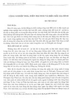
MAPs
Kinesin-related
Motor proteins
Polymerization Depolymerization
Stathmin/Op18
Nocodazole
γ
γ
-tubulin
Tubulin-GTP Tubulin-GDP
Microtubules with GTP Cap
Fig. 1. Polymerization ⁄ depolymerization of microtubules. Microtu-
bules are polymerized from dimeric tubulin. GTP binding to tubulin
is necessary for microtubule assembly to occur. GTP is hydrolyzed
to GDP when tubulin is incorporated within the microtubule. In
microtubules, GDP is bound to tubulin except at the plus (+) end
where tubulin is still in the GTP-bound form, establishing the GTP
cap. This cap allows microtubules to polymerize. When the cap is
lost, microtubules begin to shrink. Microtubule-associated
proteins (MAPs) are known to promote microtubule assembly and
stabilize microtubules. The protein c-tubulin, a highly conserved
centrosomal protein and member of the tubulin superfamily, plays
a critical role in microtubule nucleation throughout the cell cycle.
Stathmin ⁄ Op18, katanin, and some kinesin-related motor proteins
are involved in microtubule depolymerization. These proteins have
been shown to stimulate transitions from elongation to shortening
of microtubules and are referred to as catastrophe-promoters.
S. Roychowdhury and M. M. Rasenick G proteins and microtubule assembly
FEBS Journal 275 (2008) 4654–4663 ª 2008 The Authors Journal compilation ª 2008 FEBS 4655
evidence indicates that G proteins associate with sev-
eral subcellular compartments, including microtubules,
and participate in both cell division and differentiation
[20–27]. For example, G protein b subunit antisense
oligonucleotides have been shown to inhibit cell prolif-
eration and to disorganize the mitotic spindle in mam-
malian cells [21]. A nontraditional G protein signaling
pathway has been shown to be involved in regulating
the mitotic spindle for centrosome ⁄ chromosome move-
ments in cell division in Caenorhabditis elegans, Droso-
phila, and mammals. Components of this pathway
include several proteins, including the Gi class of
G proteins, GoLoco domain-containing proteins i.e.
mammalian N-terminal Leu–Gly–Asn repeats (LGN)
and activator of G protein signaling 3 (AGS3), regula-
tors of G protein signaling (RGS), nuclear mitotic
apparatus protein (NUMA), and resistors to inhibitors
(RIC) of cholinesterase 8A [28–36]. Whereas Gia was
shown to regulate microtubule pulling forces for chro-
mosome movements, Gbc was found to be involved in
spindle position and orientation. Several GPCRs,
known to trigger neurite outgrowth have been identi-
fied. These receptors are coupled to Gi ⁄ o, G12 ⁄ 13 or
Gs families of G proteins [37–41]. However, the down-
stream signaling involved in GPCR-triggered neurite
outgrowth is not fully understood. A significant
increase in Ga (Gi, Go and Gs) association with
microtubules has been observed during nerve growth
factor-induced differentiation of PC12 cells that was
coincident with the extension of ‘neurites’ [26]. Similar
results have been observed in Neuro-2A cells, which
spontaneously differentiate. These results indicate that
signals that promote cell division and differentiation
may use specific G proteins for microtubule rearrange-
ments. Thus, G proteins appear to provide a link
between hormones or neurotransmitters and cell divi-
sion, differentiation, and microtubules.
Clustering of G proteins in lipid rafts
and internalization of activated G alpha
and Gbc
Although G proteins are usually confined to the
plasma membrane, translocation of activated Gsa and
Gbc from the membrane to the cytosol has been
observed [42–47]. It is possible that these proteins par-
ticipate in localized regulation of the cytoskeleton, but
the mechanism that governs the cellular destinations of
G protein is not clearly understood. Lipid rafts
(plasma membrane microdomains rich in cholesterol
and sphingolipids) are thought to play key roles in
G protein trafficking to subcellular compartments [48].
Many G proteins have been reported to localize to
lipid rafts and undergo signal-dependent trafficking in
to and out of lipid rafts. We have shown that Gsa is
endocytosed by a lipid raft-mediated mechanism
[49,50]. Unlike Ga,Gbc, was shown to internalize to
cytosol with clathrin-coated vesicles [47].
Regulation of microtubule assembly by
a and bc subunits of G proteins
Studies conducted over the past few years have demon-
strated that a and bc subunits of heterotrimeric G
proteins modulate microtubule assembly in vitro
[19,51,52]. Ga (Gi1a,Gsa,Goa) inhibits microtubule
assembly and increases microtubule disassembly by
activating the intrinsic GTPase of tubulin [19]. Thus,
Ga may act as a GTPase-activating protein for tubulin
and may increase the dynamic behavior of microtu-
bules by removing the GTP cap [19], which confers
stability on microtubules. The retinal G protein trans-
ducin (Gta), which does not bind to tubulin [15], did
not inhibit microtubule assembly or activate GTPase
activity of tubulin [19].
In contrast to Ga,Gbc promotes microtubule
assembly in vitro [51]. Specificity among bc species
exists because b1c2 stimulates microtubule assembly
and b1c1 is without effect. The prenylation state of
G protein c subunits is likely to be relevant for this
distinction (Gc1 is farnesylated, whereas Gc2 is gera-
nylgeranylated). A mutant b1c2, b1c2 (C68S), which
does not undergo prenylation and subsequent C-term-
inal processing on the c subunit, does not stimulate
the formation of microtubules [51]. Consistent with
these observations, it has been suggested that lipid
modification of G protein subunits (Ga and Gc) not
only contributes to membrane association, but is also
important for productive interactions between a with
bc subunits, as well as the interactions of a and bc
subunits with effector and receptor molecules [53,54].
For example, lipid modifications are critical for the
interactions of a and bc subunits with effectors such
as adenylyl cyclase, phospholipase C, and phosphati-
dylinositol 3-kinase, as well as with receptors [55].
Our results suggested that the functional interactions
of G protein subunits with tubulin⁄ microtubules
require a similar structural specificity of G protein sub-
units to those that determine their interactions with
other signaling partners. Because G protein activation
and subsequent dissociation of a and bc subunits is
necessary for G proteins to participate in signaling
processes, we reconstituted Gabc heterotrimer from
myristoylated-Ga and prenylated-Gbc and found that
the heterotrimer blocks the Gi1a activation of tubulin
GTPase and inhibits the ability of Gb1c2 to promote
G proteins and microtubule assembly S. Roychowdhury and M. M. Rasenick
4656 FEBS Journal 275 (2008) 4654–4663 ª 2008 The Authors Journal compilation ª 2008 FEBS
in vitro microtubule assembly [52]. Nonetheless, G pro-
tein heterotrimers bind to tubulin [56], suggesting that
another site on Gbc (apart from the region binding to
effector interaction domains on Ga) binds tubulin
when the heterotrimer is intact. Thus, it appears that
G protein activation and dissociation of a and bc sub-
units is required for functional coupling between
Ga ⁄ Gbc and tubulin ⁄ microtubules, as outlined in
Fig. 2. In this model, Ga activates tubulin GTPase
and destroys the GTP cap at microtubule ends, caus-
ing an incease in microtubule dynamics. Thus, Ga is a
GTPase activating protein for tubulin. In a sense, Ga
is mimicking tubulin in the activation of the intrinsic
tubulin GTPase. Because the predicted domain for
interaction between Ga and tubulin is the interface
where Ga interacts with effector [57,58], Ga ⁄ tubulin
complexes preclude Gbc binding to Ga. It is likely that
Ga and Gbc will interact with different populations of
tubulin ⁄ microtubules to reorganize microtubule net-
works in cells.
Using the anti-mitotic agent nocodazole, we have
shown that the assembly ⁄ disassembly of microtubules
alters the tubulin–Gbc interaction in cultured PC12
and NIH3T3 cells [59]. Although microtubule depoly-
merization by nocodazole inhibited the interactions
between tubulin and Gbc , this inhibition was reversed
when microtubule assembly was restored by the
removal of nocodazole. The result suggests that Gbc
might be involved in promoting microtubule assembly
and ⁄ or stabilization of microtubules in vivo as demon-
strated in vitro. This is further supported by the fact
that Gbc was preferentially bound to microtubules
and treatment with nocodazole (short-term incuba-
tion), which suggested that the dissociation of Gbc
from microtubules is an early step in the depolymeriza-
tion process. Unlike Gbc, however, the interaction
between tubulin and the a subunit of the Gs protein
(Gsa) was not inhibited by nocodazole, which indicates
differential interactions of the a and bc subunits of
G proteins with tubulin ⁄ microtubules [59]. The anti-
microtubule drugs nocodazole and colchicine are
known to inhibit microtubule assembly by inhibiting
the addition of tubulin dimers to microtubules [60,61].
The possibility that the anti-microtubule agent nocoda-
zole exerts its effect by disrupting microtubule stabili-
zation by Gbc
may provide new understanding of the
mechanism of action of the anti-mitotic ⁄ anti-cancer
drugs and allow for the development of new drugs that
might be more effective in the treatment of cancer.
c-Tubulin–Gbc interactions and
microtubule nucleation
In addition to its binding of ab-tubulin, Gbc also
interacts with c-tubulin in PC12 cells. However, unlike
ab-tubulin, the interaction between c-tubulin and Gbc
was not inhibited by nocodazole, suggesting that the
interaction between Gbc and c-tubulin is not depen-
dent upon microtubules. c-Tubulin is an integral cen-
trosome protein, and its role in microtubule nucleation
is well documented [62–64]. We found that Gbc was
co-localized with ab- and c-tubulin in the centrosomes
of PC12 cells [59]. The localization of Gbc in centro-
somes and its association with c-tubulin suggest that
Gbc might be involved in microtubule nucleation in
association with c-tubulin (Fig. 2). This idea is sup-
ported by in vitro observations, suggesting that Gbc
promotes microtubule assembly under conditions
where spontaneous nucleation does not occur [51].
Gα
αβγ
GPCRAgonist
βγ
G
α
No Effect
G
βγ
Effectors
Activation of
GTPase of
tubulin by
G
α
, and
inhibition of
Loss of GTP
cap on MT end
by G
α
promo
tes
catastroph
y
di i
Promotion of
MT assembly
by G
βγ
GTP-Tubulin
MT assembly
and increase in
MT dynamics.
MT with GTP cap
G
βγ
GDP-
Tubulin
γ-Tub
Fig. 2. Model for the regulation of microtubule (MT) assembly by a
and bc subunits of G proteins. Based on in vitro results using puri-
fied tubulin and G protein subunits (Ga,Gbc) [19,51,52], the follow-
ing model is proposed. In this model, Ga inhibits microtubule
assembly and promotes microtubule disassembly by interacting, in
the fashion of a GTPase activating protein, with tubulin–GTP or the
GTP cap of growing microtubules and initiating GTP hydrolysis of
tubulin. Unlike the classical G protein cycle in which Ga in the GTP-
bound form interacts with ‘effector’ molecules, this model shows
that Ga interacts with tubulin ⁄ microtubules and this could be regu-
lated by effector molecules or GAPs. Gbc, by contrast, promotes
microtubule assembly. In the heterotrimer form, the primary inter-
acting facets of Ga and Gbc are occluded. The Gabc heterotrimer
can be activated either by agonist-mediated or agonist-independent
pathways. Upon activation, Ga dissociates from Gbc subunits. Both
subunits then interact with tubulin ⁄ microtubules and modulate
assembly ⁄ dynamics.
S. Roychowdhury and M. M. Rasenick G proteins and microtubule assembly
FEBS Journal 275 (2008) 4654–4663 ª 2008 The Authors Journal compilation ª 2008 FEBS 4657
Because it appears that microtubule nucleation by
c-tubulin is mediated by the c-tubulin ring complex,
the possibility exists that Gbc is a component of this
complex [65,66]. It was previously shown that centro-
some-associated c-tubulin is in a dynamic exchange
with the cytoplasmic pool and that the c-tubulin con-
tent of the centrosome increases suddenly, at least
threefold, at the onset of mitosis [67]. In addition, the
proportion of tubulin in microtubules increases drama-
tically as the cell enters mitosis. However, the mechan-
ism by which the translocation of c-tubulin and the
subsequent activation of centrosomes occur is largely
unknown. Microtubules do not appear to be involved
in this dynamic exchange process [67]. We found that,
in addition to c-tubulin, Gbc immunoreactivity also
increased significantly in duplicated chromosomes at
the onset of mitosis [59]. It can be speculated that Gbc
may allow translocation of c-tubulin to centrosomes.
The c-tubulin–Gbc complex might then induce robust
microtubule nucleation at the centrosome and forma-
tion of mitotic spindle.
Cellular and physiological aspects of
G protein–microtubule interactions
Based on the above discussion, it can be speculated
that G proteins may serve as a physiological regulator
for microtubule assembly and dynamics. It is conceiva-
ble that the interactions of Ga and Gbc with micro-
tubules may modulate their dynamic behavior in cells.
The results also suggest that GPCRs may affect regula-
tion of microtubule assembly and dynamics in vivo by
mobilizing G protein subunits to bind to microtubules.
Certainly, in the case of Gsa there is clear evidence of
agonist-induced translocation to the cytosol [45,49,68].
A number of proteins, in addition to GPCRs have
been shown to influence the G protein activation cycle
[69–72]. These proteins are identified as receptor-inde-
pendent activators of G-protein signaling (AGS), and
mediate a diverse range of signals within the cell,
including cell division, neuronal differentiation and ⁄ or
synaptic plasticity [71,72]. Three groups of AGS pro-
teins have been defined based on their mechanism of
action. Group I AGS protein (AGS1) is similar to that
of a GPCR in terms of its ability to function as a gua-
nine-nucleotide exchange factor. Group II and group
III AGS proteins (AGS2-10) appear to regulate hetero-
trimeric G protein signaling by a mechanism indepen-
dent of nucleotide exchange. In contrast to group I
and II AGS proteins, each member of the group III
AGS proteins (AGS2, AGS7-10) binds to Gbc but not
Ga. Group II AGS proteins (AGS3 ⁄ LGN) have been
studied extensively. These proteins generally contain
two types of repeats: tetratricopeptide repeats at the
N-terminus that mediates protein–protein interactions,
and Ga
i ⁄ o
-Loco (GoLoco or GPR) repeats at the
C-terminus that mediate interactions with the Gi ⁄ o
class of G proteins. Proteins containing G protein reg-
ulatory (GPR) motifs have been identified in C. ele-
gans (GPR1 ⁄ 2), Drosophila melanogaster (Pins), and
mammalian cells (mammalian Pins or LGN; AGS3)
[28]. These cytoplasmic signaling regulators have been
described enzymatically as Gia-class guanine nucleo-
tide dissociation inhibitors (GDI) that bind to the
GDP bound form of Gia and inhibit the exchange of
GDP-bound for GTP-bound Ga [73–75]. These signal-
ing partners of G proteins might also be involved in
the regulation of microtubule assembly by Gia or Gbc
(Fig. 3). This is further supported by the fact that Ga
in the GDP-bound form interacts with tubulin–GTP to
promote the GTPase activity of tubulin and subsequent
regulation of microtubule assembly [19]. Thus, the
modulation of microtubule assembly by G proteins
may require activation of G proteins by either recep-
tor-dependent or receptor-independent pathways.
Although molecules with GDI activity identified to
date, only interact with Gi ⁄ o class of G proteins, it can
be presumed that Gsa ⁄ Gqa-specific GDI molecules
may be involved in regulating ⁄ modulating Gs or
Gq ⁄ 11 family of G proteins, and thus may play roles in
modulation of microtubule assembly by Gs or Gq ⁄ 11.
Organization and function of mitotic spindle
during cell division
Transformation of an interphase network of micro-
tubules into a bipolar spindle that mediates the accu-
rate segregation of chromosomes is a central event
during cell division. Microtubules in the spindle are
organized in such a way that the minus ends are near
the spindle poles and the plus ends extend toward the
cell cortex or chromosomes [76]. Thus, the assembly ⁄ -
disassembly of microtubules plays a key role in both
the organization and function of the mitotic spindle.
Recently, G protein subunits have been shown to be
involved in regulating the mitotic spindle for centro-
some ⁄ chromosome movements in cell division.
Whereas Gia was shown to interact with GDI to regu-
late microtubule pulling forces for chromosome move-
ments, Gbc was found to be involved in spindle
position and orientation. GoLoco domain-containing
proteins (GDI) form complexes with Gia-GDP, which
seems to create spindle oscillations by enhancing the
pulling forces exerted on the mitotic spindle during
mitosis [31]. Because it has been demonstrated pre-
viously that Ga activates tubulin GTPase [19], it is
G proteins and microtubule assembly S. Roychowdhury and M. M. Rasenick
4658 FEBS Journal 275 (2008) 4654–4663 ª 2008 The Authors Journal compilation ª 2008 FEBS
possible that the direct interaction of microtubules
with Ga- and LGN provides microtubule pulling
forces through the destabilization of microtubules.
Gbc, by contrast, may be involved in the orientation
and positioning of the mitotic spindle through its abil-
ity to interact with both membrane and centrosomes
[29]. It can be speculated that Gbc is also involved in
the formation of mitotic spindle by promoting micro-
tubule assembly (in association with c-tubulin) in spin-
dle poles. This is supported by the fact that at the
onset of mitosis immunoreactivity of both c-tubulin
and Gbc increased several fold in the duplicated cen-
trosomes, thus increasing the capability of centrosomes
to promote microtubule assembly [59].
Neuronal differentiation
Microtubule assembly and dynamics is tightly coupled
to neuronal differentiation, outgrowth, and plasticity.
Several GPCR known to trigger neurite outgrowth
have been identified [37–41]. However, the downstream
signaling involved in GPCR-triggered neurite out-
growth is not fully understood. The Go are the most
abundant G proteins in neuronal growth cones [77].
Growth cones at the growing tips of developing neur-
ites are highly specialized organelles that respond to a
variety of extracellular signals to achieve neuronal gui-
dance and target recognition [78]. These structures are
associated with microtubules in their immature state,
but microtubules retract from the tip of more mature
growth cones. Some evidence suggests that Goa is
directly involved in inducing neurite outgrowth upon
activation [79]. By contrast, dendritic outgrowth pro-
moted by the Gs-coupled GPR3, is cAMP-dependent
[80]. Signaling through Gsa is also required for the
growth and function of neuromuscular synapses in
Drosophila [81]. Coordinated assembly of microtubules,
in concert with actin filaments and neurofilaments, is
required for growth cone motility and neurite out-
growth [82–84] and microtubules in or near the growth
cone are particularly dynamic [85].
Many functions of Go are thought to be mediated
through the actions of a common pool of Gbc dimers.
Based on the observed role of G protein subunits in
microtubule assembly, it is reasonable to postulate that
the dynamic interactions between Gi ⁄ o (both a and bc
subunits) and microtubules, and the subsequent regula-
tion of microtubule assembly may be critical for neuro-
nal differentiation, outgrowth and plasticity. The
G protein regulator AGS3, a Gia-class GDI, the expres-
sion of which is restricted to neurons, might play a role
in regulating the assembly ⁄ dynamics of microtubules
in neurons by promoting the interactions between
tubulin ⁄ microtubules and Gia-GDP. Association of
Gbc with the actin cytoskeleton has also been reported
[86]. More recent studies in cultured PC12 cells suggest
that Gbc interacts with actin filaments in addition to
microtubules and this interaction was not affected by
depolymerization of microtubules (Najera & Roy-
chowdhury, unpublished observations) and G proteins
might serve to unite microtubule and actin-dependent
processes to regulatory elements acting through GPCRs.
A final caveat to the studies with Gi and Go is that,
Agonis t
G
Membran e
G
Effector s
GTP
Cytoplasm
GD P
AG S
AG S
G
(Group I a nd II)
(Group III)
GD P
Microtubule dynamics
G
Fig. 3. G protein signaling in membrane and cytoplasm. Tradition-
ally, G proteins function as a signal transducer in transmembrane
signaling pathways that consist of three proteins: receptors, G pro-
teins, and effectors. The receptors that participate in this pathway
have seven transmembrane domains. G proteins consist of a het-
erotrimeric structure composed of guanine nucleotide-binding
alpha, plus beta and gamma subunits. Beta and gamma subunits
form a tight association under nondenaturing conditions. Receptor
activation allows GTP to bind to the a subunit of the heterotrimer.
Subsequently, activated G a changes its association with Gbc in a
manner that permits both subunits to participate in the regulation
of intracellular effector molecules. Termination of the signal occurs
when GTP bound to the a subunit is hydrolyzed by its intrinsic
GTPase activity, which causes its functional dissociation from the
effector and re-association with bc. A hypothetical framework for
cytoplasmic G protein-signaling is shown. In this model, a and
bc subunits of G proteins (only the Gsa are released from the
membrane by agonist activation, but the Gia and Goa have a cyto-
solic presence. All three, as well as Gq, evoke Gbc release into the
cytosol) regulate microtubule assembly ⁄ dynamics (red arrows). By
forming an inactive Gabc heterotrimer, this signaling pathway is ter-
minated (purple arrows). In this model, AGS proteins will modulate
the assembly ⁄ disassembly of microtubules by interacting with a
and bc subunits of G proteins. Lipid modification of G protein sub-
units, i.e. the myristoylation of Ga and prenylation of Gc, are
expected to play key roles in microtubule regulation, similar to that
observed with G protein signaling in membrane [53,54] (not shown
in the model). Through this mechanism, GPCR might be involved in
regulating the interplay of Gi1a,Gbc, AGS (or other Ga-interacting
proteins) and tubulin ⁄ microtubules.
S. Roychowdhury and M. M. Rasenick G proteins and microtubule assembly
FEBS Journal 275 (2008) 4654–4663 ª 2008 The Authors Journal compilation ª 2008 FEBS 4659
while both of these G proteins have a cytosolic presence
and decorate microtubules [26], unlike Gs, they do not
internalize in response to agonist. Nevertheless, GPCRs
coupled to Gs, Gi, Go or Gq do evoke Gbc internaliza-
tion [47,68,87]. This might suggest an interesting inter-
play between Gs and Gi⁄ o (or Gq) in the regulation of
microtubules and the modulation of cellular processes
dependent on microtubule dynamics.
Based on the current literature, we propose a com-
prehensive model outlining the G protein-mediated sig-
naling in membrane and cytoplasm as depicted in
Fig. 3. Although the major membrane-associated com-
ponents of G protein signaling are now well-defined,
the phenomenon of cytosolic G protein signaling is
only beginning to emerge. We speculate that in cyto-
plasm a and bc subunits of G protein interact to regu-
late microtubule assembly. It is also proposed that
AGS proteins regulate microtubule assembly through
their interaction with Ga or Gbc (Fig. 3). It is quite
possible that lipid modification of G protein subunits
plays key roles in microtubule regulation, similar to
that observed with G protein signaling in membrane
[52,53]. We speculate that G protein-coupled receptors
will regulate the interplay of Ga Gbc, and AGS to
modulate microtubule assembly. The interactions
between receptor and non-receptor-mediated pathways
in the regulation of G protein internalization are just
beginning to be explored.
It is becoming increasingly clear that a new pathway
of cytosolic G protein signaling is emerging. We pro-
pose that this pathway is involved in regulating micro-
tubule dynamics. Hopefully, the next few years will
bring new evidence that will elucidate the role of
GPCR signaling in microtubule biology. These studies
should help to establish the link between hormone or
neurotransmitter action and modulation of cellular
locomotion or cellular morphology.
Acknowledgements
Research in the authors’ laboratories described in
this report was supported by MH 39595, AG015482
and DA020568 (MMR- U. Illinois Chicago), and
2G12RR08124 (University of Texas at El Paso). The
authors like to thank Dr Siddhartha Das for critically
reading the manuscript and thoughtful suggestions. Mr
Traver Duarte and Mr Tavis Mendez are thanked for
their help.
References
1 Desai A & Mitchison TJ (1997) Microtubule polymeri-
zation dynamics. Annu Rev Cell Dev Biol 13, 83–117.
2 Gelfand VI (1991) Microtubule dynamics: mechanism,
regulation and function. Annu Rev Cell Biol 7, 93–116.
3 McNally FJ (1996) Modulation of microtubule
dynamics during the cell cycle. Curr Opin Cell Biol 8,
23–29.
4 Bulinski JC & Gundersen GG (1991) Stabilization of
post-translational modification of micotubules during
cellular morphogenesis. Bio Essays 13, 285–293.
5 Carlier MF, Didry D, Simon C & Pantaloni D (1989)
Mechanism of GTP hydrolysis in tubulin polymeriza-
tion: characterization of the kinetic intermediate micro-
tubule-GDP-Pi using phosphate analogues.
Biochemistry 28, 1783–1791.
6 Mitchison T & Kirschner M (1984) Dynamic instability
of microtubule growth. Nature 312, 237–242.
7 Gundersen GG, Gomes ER & Wen Y (2004) Cortical
control of microtubule stability and polarization. Cur
Opin Cell Biol 16, 106–112.
8 Murphy DB & Borisy GG (1975) Association of high-
molecular weight proteins with microtubules and their
role in microtubule assembly. Proc Natl Acad Sci USA
72, 2696–2700.
9 Margolis RL, Rauch CT & Job D (1986) Purification
and assay of a 145-kDa protein (STOP145) with micro-
tubule-stabilizing and motility behavior. Proc Natl Acad
Sci USA 83, 639–643.
10 Gamblin TC, Nachmanoff K, Halpain S & Williams
RC Jr (1996) Recombinant microtubule-associated pro-
tein 2C reduces the dynamic instability of individual
microtubules. Biochemistry 35, 12575–12586.
11 Bhat K & Setalu V (2007) Microtubule-associated pro-
teins as targets in cancer chemotherapy. Clin Cancer
Res 13, 2849–2854.
12 Belmont L & Mitchison TJ (1996) Identification of a
protein that interacts with tubulin dimers and increases
the catastrophe rate of microtubules. Cell 84, 623–631.
13 Kline-Smith SL & Walczak CE (2002) The microtubule-
destabilizing kinesin XKCM1 regulates microtubule
dynamic instability in cells. Mol Biol Cell 13, 2718–
2731.
14 Gundersen GG & Cook TA (1999) Microtubules and
signal transduction. Curr Opin Cell Biol 11, 81–94.
15 Wang N, Yan K & Rasenick MM (1990) Tubulin binds
specifically to the signal-transducing proteins, Gs alpha
and Gi alpha 1. J Biol Chem 265, 1239–1242.
16 Popova JS & Rasenick MM (2000) Muscarinic receptor
activation promotes the membrane association of tubu-
lin for the regulation of Gq-mediated phospholipase
Cbeta(1) signaling. J Neurosci 15, 2774–2782.
17 Rasenick MM, Stein PJ & Bitensky MW (1981) The
regulatory subunit of adenylate cyclase interacts with
cytoskeletal components. Nature 294, 560–562.
18 Rasenick MM & Wang N (1988) Exchange of guanine
nucleotides between tubulin and GTP-binding
proteins that regulate adenylate cyclase: cytoskeletal
G proteins and microtubule assembly S. Roychowdhury and M. M. Rasenick
4660 FEBS Journal 275 (2008) 4654–4663 ª 2008 The Authors Journal compilation ª 2008 FEBS
modification of neuronal signal transduction. J Neuro-
chem 51, 300–311.
19 Roychowdhury S, Panda D, Wilson L & Rasenick MM
(1999) G protein alpha subunits activate tubulin
GTPase and modulate microtubule polymerization
dynamics. J Biol Chem 274, 13485–13490.
20 Wu HC & Lin CT (1994) Association of heterotrimeric
GTP binding regulatory protein (Go) with mitosis. Lab
Invest 71, 175–181.
21 Wu HC, Huang PH & Lin CT (2001) G protein beta2
subunit antisense oligonucleotides inhibit cell prolifera-
tion and disorganize microtubule and mitotic spindle
organization. J Cell Biochem 83, 136–146.
22 Lewis JM, Woolkalis MJ, Gerton GL, Smith RM,
Jarett L & Manning DR (1991) Subcellular distribution
of the alpha subunit(s) of Gi: visualization by immuno-
fluorescent and immunogold labeling. Cell Regul 2,
1097–1113.
23 Ravindra R, Kunapuli SP, Forman LJ, Nagele RG,
Foster KA & Patel SA (1996) Effect of transient over-
expression of Gq alpha on soluble and polymerized
tubulin pools in GH3 and AtT-20 cells. J Cell Biochem
61, 392–401.
24 Cote M, Payet MD & Gallo-Payet N (1997) Associa-
tion of alpha S-subunit of the GS protein with micro-
filaments and microtubules: implication during
adrenocorticotropin stimulation in rat adrenalglomeru-
losa cells. Endocrinology 138, 69–78.
25 Willard FS & Crouch MF (2000) Nuclear and cytoske-
letal translocation and localization of heterotrimeric
G-proteins. Immunol Cell Biol 78, 387–394.
26 Sarma T, Voyno-Yasenetskaya T, Hope TJ & Rasenick
MM (2003) Heterotrimeric G-proteins associate with
microtubules during differentiation in PC12 pheochro-
mocytoma cells. FASEB J 17, 848–859.
27 Crouch MF & Simon L (1997) The G-protein G(i) reg-
ulates mitosis but not DNA synthesis in growth factor-
activated fibroblasts: a role for the nuclear translocation
of G(i). FASEB J 11, 189–198.
28 Kimple RJ, Willard FS & Siderovski DP (2002) The
GoLoco motif: heralding a new tango between G
protein signaling and cell division. Mol Interven 2,
88–100.
29 Gotta M & Ahringer J (2001) Distinct roles for Galpha
and Gbetagamma in regulating spindle position and
orientation in Caenorhabditis elegans embryos. Nat Cell
Biol 3, 297–301.
30 Schaefer M, Petronczki M, Dorner D, Forte M &
Knoblich J (2001) Heterotrimeric G proteins direct
two modes of asymmetric cell division in the Drosophila
nervous system. Cell 107, 183.
31 Fuse N, Hisata K, Katzen AL & Matsuzaki F (2003)
Heterotrimeric G proteins regulate daughter cell size
asymmetry in Drosophila neuroblast divisions. Curr Biol
13, 947–954.
32 Du Q & Macara IG (2004) Mammalian Pins is a con-
formational switch that links NuMA to heterotrimeric
G proteins. Cells 119, 503.
33 Sanada K & Tsai LH (2005) G protein betagamma sub-
units and AGS3 control spindle orientation and asym-
metric cell fate of cerebral cortical progenitors. Cell
122, 119–131.
34 Siegrist SE & Doe CQ (2005) Microtubule-induced
Pins ⁄ G alpha I cortical polarity in Drosophila neuro-
blasts. Cell
123, 1323–1335.
35 Bellaiche Y & Gotta M (2005) Heterotrimeric G pro-
teins and regulation of size asymmetry during cell divi-
sion. Curr Opin Cell Biol 17, 658–663.
36 Tall GG & Gilman AG (2005) Resistance to inhibitors
of cholinerterase 8a catalyzes release of Gai-GTP and
nuclear mitotic apparatus protein (NuMA) from
NuMA ⁄ LGN ⁄ Gai-GDP complexes. Proc Natl Acad Sci
USA 102, 16584–16589.
37 Reinoso BS, Undie AS & Levitt P (1996) Dopamine
receptors mediate differential morphological effects on
cerebral cortical neurons in vitro. J Neurosci Res 43,
439–453.
38 Lotto B, Upton L, Price DJ & Gaspar P (1999) Seroto-
nin receptor activation enhances neurite outgrowth of
thalamic neurones in rodents. Neurosci Lett 269, 87–90.
39 He JC, Neves SR, Jordan JD & Iyengar R (2006) Role
of the Go ⁄ i signaling network in the regulation of neur-
ite outgrowth. Can J Physiol Pharmacol 84, 687–694.
40 Zhang W, Duan W, Cheung NS, Huang Z, Shao K &
Li QT (2007) Pituitary adenylate cyclase-activating
polypeptide induces translocation of its G-protein-
coupled receptor into caveolin-enriched membrane
microdomains, leading to enhanced cyclic AMP genera-
tion and neurite outgrowth in PC12 cells. J Neurochem
103, 1157–1167.
41 Kvachnina E, Liu G, Dityatev A, Renner U, Dumuis
A, Richter DW, Dityateva G, Schachner M, Voyno-
Yasenetskaya TA & Ponimaskin EG (2005) 5-HT7
receptor is coupled to G alpha subunits of heterotri-
meric G12-protein to regulate gene transcription and
neuronal morphology. J Neurosci 25, 7821–7830.
42 Rasenick MM, Wheeler GL, Bitensky MW, Kosack
CM, Malina RL & Stein PJ (1984) Photoaffinity identi-
fication of colchicine-solubilized regulatory subunit
from rat brain adenylate cyclase. J Neurochem 43,
1447–1454.
43 Ransas LA, Svoboda JR, Jaspar JR & Insel PA (1989)
Stimulation of beta-adrenergic receptors of S49 lym-
phoma cells redistributes the alpha subunit of the stimu-
latory G protein between cytosol and membranes. Proc
Natl Acad Sci USA 86, 7900–7903.
44 Levis MJ & Bourne HR (1992) Activation of the alpha
subunit of Gs in intact cells alters its abundance, rate of
degradation, and membrane avidity. J Cell Biol 119,
1297–1307.
S. Roychowdhury and M. M. Rasenick G proteins and microtubule assembly
FEBS Journal 275 (2008) 4654–4663 ª 2008 The Authors Journal compilation ª 2008 FEBS 4661
45 Yu JZ & Rasenick MM (2002) Real-time visualization
of a fluorescent G(alpha)(s): dissociation of the acti-
vated G protein from plasma membrane. Mol Pharma-
col 61, 352–359.
46 Janetopoulos C, Jin T & Devreotes P (2001) Receptor-
mediated activation of theterotrimeric G-proteins in liv-
ing cells. Science 291, 2408–2411.
47 Popova JS & Rasenick MM (2004) Clathrin-mediated
endocytosis of m3 muscarinic receptors. Roles for
Gbetagamma and tubulin. J Biol Chem 279, 30410–
30418.
48 Allen JA, Halverson-Tamboli RA & Rasenick MM
(2007) Lipid raft microdomains and neurotransmitter
signalling. Nat Rev Neurosci 8, 128–140.
49 Allen JA, Yu JZ, Donati RJ & Rasenick MM (2005)
Beta-adrenergic receptor stimulation promotes G alpha
s internalization through lipid rafts: a study in living
cells. Mol Pharmacol 67, 1493–1504.
50 Sugama J, Yu JZ, Rasenick MM & Nakahata N (2007)
Mastoparan inhibits beta-adrenoceptor-G(s) signaling
by changing the localization of Galpha(s) in lipid rafts.
Cell Signal 19, 2247–2254.
51 Roychowdhury S & Rasenick MM (1997) G protein
beta1gamma2 subunits promote microtubule assembly.
J Biol Chem 272 , 31476–31581.
52 Roychowdhury S, Martinez L, Salgado L, Das S &
Rasenick MM (2006) G protein activation is prerequi-
site for functional coupling between Ga ⁄ Gbc and tubu-
lin ⁄ microtubules. Biochem Biophys Res Commun 340,
441–448.
53 Mumby SM & Linder ME (1994) Myristoylation of
G protein alpha subunits. Methods Enzymol 237, 254–
268.
54 Iniguez-LIuhi JA, Simon MI, Robinshaw JD & Gilman
AG (1992) G protein beta gamma subunits synthesized
in Sf9 cells. Functional characterization and the signifi-
cance of prenylation of gamma. J Biol Chem 267,
23409–23417.
55 Cabrera-Vera TM, Vanhauwe J, Thomas TO, Medkova
M, Preininger A, Mazzoni MR & Hamm HE (2003)
Insights into G proteinstructure, function, and regula-
tion. Endrocrin Rev 24, 765–781.
56 Wang N & Rasenick MM (1991) Tubulin–G protein
interactions involve microtubule polymerization
domains. Biochemistry 30, 10957–10965.
57 Chen NF, Yu JZ, Skiba NP, Hamm HE & Rasenick
MM (2003) A specific domain of Gialpha required for
the transactivation of Gialpha by tubulin is implicated
in the organization of cellular microtubules. J Biol
Chem 278, 15285–15290.
58 Layden BT, Saengsawang W, Donati RJ, Yang S, Mul-
hearn DC, Johnson ME & Rasenick MM (2008) Struc-
tural model of a complex between the heterotrimeric
G protein, Gsa, and tubulin. Biochim Biophys Acta
1783, 964–973.
59 Montoya V, Gutierrez C, Najera O, Leony D, Varela
A, Popova J, Rasenick MM, Das S & Roychowdhury S
(2007) G protein bc subunits interact with ab and c
tubulin and play a role in microtubule assembly in
PC12 cells. Cell Motil Cytoskel 64, 936–950.
60 DeBrabander MJ, Van de veire RML, Aerts FE,
Borgers M & Janssen PAJ (1976) The effects of methyl
[5-(2-thienylcarbonyl)-1H-benzimadazol-2-yl] carbamate
(R17934; NSC 238159), a new synthetic antitumoral
drug interfering with microtubules, on mammalian cells
cultured in vitro. Cancer Res 36, 905–916.
61 De Brabander M, Geuens G, Nuydens R, Willebrords
R & De MeyJ (1981) Microtubule assembly in living
cells after release from nocodazole block: the effects of
metabolic inhibitors, taxol and PH. Cell Biol Int Rep 5,
913–920.
62 Oakley BR (1992) Gamma-tubulin: the microtubule
organizer? Trends Cell Biol 2, 1–5.
63 Joshi HC, Palacios MJ, McNamara L and Cleveland
DW (1992) Gamma-tubulin is a centrosomal protein
required for cell cycle-dependent microtubule nuclea-
tion. Nature 356, 80–83.
64 Job D, Valiron O & Oakley B (2003) Microtubule
nucleation. Curr Opin Cell Biol 15, 111–117.
65 Moritz M, Braunfeld MB, Sedat JW, Alberts B &
Agard DA (1995) Microtubule nucleation by gamma-
tubulin-containing rings in the centrosome. Nature 378,
638–640.
66 Moritz M & Agard DA (2001) Gamma–tubulin com-
plexes and microtubule nucleation. Curr Opin Struct
Biol 11, 174–181.
67 Khodjakov A & Rieder CL (1999) The sudden recruit-
ment of gamma-tubulin to the centrosome at the onset
of mitosis, and its dynamic exchange throughout the
cell cycle, does not require microtubules. J Cell Biol
146, 585–596.
68 Hynes TR, Mervine SM, Yost EA, Sabo JL & Berlot
CH (2004) Live cell imaging of Gs and the beta2-adre-
nergic receptor demonstrates that both alphas and
beta1gamma7 internalize upon stimulation and exhibit
similar trafficking patterns that differ from that of the
beta2-adrenergic receptor. J Biol Chem 279, 44101–
44112.
69 Bernard ML, Peterson YK, Chung P, Jourdan J &
Lanier SM (2001) Selective interaction of AGS3 with
G-proteins and the influence of AGS3 on the activation
state of G-proteins. J Biol Chem 276, 1585–1593.
70 Blumer JB, Chandler J & Lanier SM (2002) Expression
analysis and subcellular distribution of the two G-pro-
tein regulators AGS3 and LGN indicate distinct func-
tionality. J Biol Chem 277, 15897–15903.
71 Blumer JB, Cismowski MJ, Sato M & Lanier SM
(2005) AGS proteins: receptor-independent activators
of G-protein signaling. Trends Pharmacol Sci 26,
470–476.
G proteins and microtubule assembly S. Roychowdhury and M. M. Rasenick
4662 FEBS Journal 275 (2008) 4654–4663 ª 2008 The Authors Journal compilation ª 2008 FEBS
72 Blumer JB, Smrcka AV & Lanier SM (2007) Mechanis-
tic pathways and biological roles for receptor-indepen-
dent activators of G–perotein signaling. Pharmacol Ther
113, 488–506.
73 De Vries L, Fischer T, Tronchare H, Brothers GM,
Strockbine B, Siderovski DP & Farquhar MG (2000)
Activator of G protein signaling 3 is a guanine dissocia-
tion inhibitor for Galphai subunits. Proc Natl Acad Sci
USA 97, 14364–14369.
74 Peterson YK, Bernard ML, Ma H, Hazard S, Graber
SG & Lanier SM (2000) Stabilization of the GDP-
bound conformation of Gialpha by a peptide derived
from the G-protein regulatory motif of AGS3. J Biol
Chem 275, 33193–33196.
75 Willard FS, Kimple RJ & Siderovski DP (2004) Return
of the GDI: the GoLoco motif in cell division. Annu
Rev Biochem 73, 925–951.
76 Heidemann SR & McIntosh JR (1880) Visualization of
the structural polarity of microtubules. Nature 286,
517–519.
77 Strittmatter SM, Valenzuela D, Kennedy TE, Neer EJ
& Fishman MC (1990) G
0
is a major growth cone pro-
tein subject to regulation by GAP-43. Nature 26, 836–
841.
78 Cheng N & Sahyoun N (1988) The growth cone cyto-
skeleton. J Biol Chem 263, 3935–3942.
79 Igarashi M, Strittmatter S, Vartanian T & Fishman MC
(1993) Mediation by G proteins of signals that cause
collapse of growth cones. Science 259, 77–84.
80 Tanaka S, Ishii K, Kasai K, Yoon SO & Saeki Y
(2007) Neural expression of G protein-coupled receptors
GPR3, GPR6, and GPR12 up-regulates cyclic AMP
levels and promotes neurite outgrowth. J Biol Chem
282, 10506–10515.
81 Wolfgang WJ, Clay C, Parker J, Delgado R, Labarca
P, Kidokoro Y & Forte M (2004) Signaling through Gs
alpha is required for the growth and function of neuro-
muscular synapses in Drosophila. Dev Biol 268, 295–
311.
82 Smith S (1988) Neuronal cytomechanics: the actin based
motility of growth cones. Science 242, 708–715.
83 Rodriguez OC, Schaefer AW, Mandato CA, Forscher
P, Bement WM & Waterman-Storer CM (2004) Con-
served microtubule–actin interactions in cell movement
and morphogenesis. Curr Biol 14, 1194–1199.
84 Burnette DT, Schaefer AW, Ji L, Danuser G &
Forscher P (2007) Filopodial actin bundles are not
necessary for microtubule advance into the peripheral
domain of Aplysia neuronal growth cones. Nat Cell Biol
9, 1360–1369.
85 Suter DM, Schaefer AW & Forscher P (2004)
Microtubule dynamics are necessary for SRC family
kinase-dependent growth cone steering. Curr Biol 14,
1194–1199.
86 Carlson KE, Woolkalis MJ, Newhouse MG &
Manning DR (1986) Fractionation of the beta subunit
common to guanine nucleotide-binding regulatory
proteins with the cytoskeleton. Mol Pharmacol 5,
463–468.
87 Saini DK, Kalyanaraman V, Chisari M & Gautam N
(2007) A family of G protein betagamma subunits
translocate reversibly from the plasma membrane to
endomembranes on receptor activation. J Biol Chem
282, 24099–24108.
S. Roychowdhury and M. M. Rasenick G proteins and microtubule assembly
FEBS Journal 275 (2008) 4654–4663 ª 2008 The Authors Journal compilation ª 2008 FEBS 4663