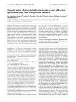
Báo cáo khoa học: Characterization of a b-N-acetylhexosaminidase and a b-N-acetylglucosaminidase/b-glucosidase from Cellulomonas fimi potx
Bạn đang xem bản rút gọn của tài liệu. Xem và tải ngay bản đầy đủ của tài liệu tại đây (1.61 MB, 13 trang )
Characterization of a b-N-acetylhexosaminidase and a
b-N-acetylglucosaminidase/b-glucosidase from
Cellulomonas fimi
Christoph Mayer
1,2,3
, David J. Vocadlo
1,
*, Melanie Mah
2
, Karen Rupitz
1
, Dominik Stoll
2
,
R. A. J. Warren
2
and Stephen G. Withers
1
1 Department of Chemistry, University of British Columbia, Vancouver, Canada
2 Department of Microbiology & Immunology, University of British Columbia, Vancouver, Canada
3 Department of Biology, University of Konstanz, Germany
Most enzymes catalyzing the hydrolysis of terminal
b-N-acetylglucosaminide linkages belong to families 3
and 20 of the glycoside hydrolases ([1,2] and the glyco-
side hydrolases database at URL s-mrs.
fr/CAZY/). Members of the two families greatly differ
in structure, enzyme mechanism, substrate specificity,
and physiologic function (for a review see [3] and
references cited therein). The enzymes in family 20 are
designated as N-acetylhexosaminidases (EC 3.2.1.52)
because they hydrolyze b-N-acetylgalactosaminides and
b-N-acetylglucosaminides, with about a four-fold greater
activity on the latter ([1] , and references cited therein).
b-N-Acetylglucosaminidases (EC 3.2.1.52) in family 3
are much more specific for the gluco-configuration,
Keywords
bifunctional glycosidase; cell wall recycling;
chitin metabolism; murein; peptidoglycan
Correspondence
C. Mayer, Department of Biology, University
of Konstanz, 78457 Konstanz, Germany
Fax: +49 7531 88 3356
Tel: +49 7531 88 4854
E-mail:
*Present address
Department of Molecular Biology and
Biochemistry, Simon Fraser University,
Burnaby, BC, Canada
Database
The nucleotide sequences listed in this
paper have been submitted to the
DDBJ ⁄ EMBL ⁄ GenBank database under the
accession numbers AF478459 and
AF478460
(Received 22 February 2006, revised 3 May
2006, accepted 4 May 2006)
doi:10.1111/j.1742-4658.2006.05308.x
The Gram-positive soil bacterium Cellulomonas fimi is shown to produce at
least two intracellular b-N-acetylglucosaminidases, a family 20 b-N-acetyl-
hexosaminidase (Hex20), and a novel family 3-b-N-acetylglucosamini-
dase ⁄ b-glucosidase (Nag3), through screening of a genomic expression
library, cloning of genes and analysis of their sequences. Nag3 exhibits
broad substrate specificity for substituents at the C2 position of the gly-
cone: k
cat
⁄ K
m
values at 25 °C were 0.066 s
)1
Æmm
)1
and 0.076 s
)1
Æmm
)1
for
4¢-nitrophenyl b-N-acetyl-d-glucosaminide and 4¢-nitrophenyl b-d-glu-
coside, respectively. The first glycosidase with this broad specificity to be
described, Nag3, suggests an interesting evolutionary link between b-N-ace-
tylglucosaminidases and b-glucosidases of family 3. Reaction by a double-
displacement mechanism was confirmed for Nag3 through the identification
of a glycosyl–enzyme species trapped with the slow substrate 2¢,4¢-dinitro-
phenyl 2-deoxy-2-fluoro-b-d-glucopyranoside. Hex20 requires the acetami-
do group at C2 of the substrate, being unable to cleave b-glucosides, since
its mechanism involves an oxazolinium ion intermediate. However, it is
broad in its specificity for the d-glucosyl ⁄ d-galactosyl configuration of the
glycone: K
m
and k
cat
values were 53 lm and 482.3 s
)1
for 4¢-nitrophenyl
b-N-acetyl-d-glucosaminide and 66 lm and 129.1 s
)1
for 4¢-nitrophenyl
b-N-acetyl-d-galactosaminide.
Abbreviations
DNP-2FGlc, 2¢,4¢-dinitrophenyl 2-deoxy-2-fluoro-b-
D-glucopyranoside; Dp, degree of polarization; IPTG, isopropyl thiogalactopyranoside;
4MU-GlcNAc, 4¢-methylumbelliferyl b-N-acetyl-
D-glucosaminide; pNP, 4-nitrophenol; pNP-Glc, 4¢-nitrophenyl b-D-glucopyranoside; pNP-GlcNAc,
4¢-nitrophenyl b -N-acetyl-
D-glucosaminide; pNP-GalNAc, 4¢-nitrophenyl b-N-acetyl-D-galactosaminide; PVDF, polyvininylidene difluoride.
FEBS Journal 273 (2006) 2929–2941 ª 2006 The Authors Journal compilation ª 2006 FEBS 2929
exhibiting little if any activity on galactosyl substrates
[1,4–6]. Family 3 primarily comprises b-glucosidases
(EC 3.2.1.21) and exo-b-glucanases (EC 3.2.1.58 and
3.2.1.74). However, b-N-acetylglucosaminidases form a
subgroup within family 3, characterized by the
sequence pattern K-H-(FI)-P-G-(HL)-G-x(4)-D-(ST)-H,
which is believed to be involved in binding of the
N-acetyl group [1,7].
The b-N-acetylglucosaminidases and hexosaminidases
in families 3 and 20 are both retaining enzymes, yet
they have different mechanisms [8]. The family 20
enzymes do not form covalent glycosyl–enzyme inter-
mediates because they lack a nucleophilic carboxylate;
hydrolysis involves the anchimeric assistance of the
acetamido group of the substrate [8–11]. By contrast,
family 3 enzymes do contain a nucleophilic carboxylate
and catalyze hydrolysis by a double-displacement
mechanism via a covalent glycosyl–enzyme intermedi-
ate [7,13–15]. This mechanism is found in most retain-
ing glycosidases, e.g. in lysozyme, an enzyme that
catalyzes an endo-type cleavage of the N-acetylglucosa-
mine-containing bacterial cell wall peptidoglycan [12].
The mechanism of family 3 exoglucanase ExoI from
Hordeum vulgare is understood in some detail [16]. The
enzyme consists of two modules, one an (a ⁄ b)
8
-barrel,
and the second a six-stranded b-sandwich [17,18]. The
substrate binds to a pocket formed between the two
modules, with Asp285 of the first domain being the
catalytic nucleophile and Glu491 of the second domain
the acid–base catalyst, which accelerates the departure
of the aglycon by protonation of the glycosidic oxygen
[19]. The catalytic nucleophile of a family 3 b-N-acetyl-
glucosaminidase (ExoII) from Vibrio furnissii was
identified using the slow substrate N-acetyl-5-fluoro-
a-l-idopyranosaminyl fluoride [7]. This residue is con-
served throughout family 3. An amino acid acting as
an acid–base catalyst in this enzyme is apparently
missing, since ExoII and other family 3 b-N-acetyl-
glucosaminidases of Gram-negative bacteria comprise
only a single (a ⁄ b)
8
-barrel module. Generally, they
have molecular masses of about 35 kDa and are pre-
dicted to be cytoplasmic: the b-N-acetylglucosamini-
dase of Escherichia coli (NagZ) is a cytoplasmic
enzyme involved in peptidoglycan recycling [20,21].
Similar enzymes in other Gram-negative bacteria may
have the same function. To date, only one family 3
b-N-acetylglucosaminidase-encoding gene (nagA) has
been cloned from a Gram-positive bacterium, namely
Streptomyces thermoviolaceus [6]. This enzyme, like
most putative family 3 b-N-acetylglucosaminidases of
Gram-positive bacteria, has a molecular mass of about
60 kDa and comprises two modules. It is extracellular
and thought to be involved in chitin degradation.
Chitin is degraded by the concerted action of chi-
tinase(s) (EC 3.2.1.14) and b-N-acetylhexosamini-
dase(s), which may involve other proteins [22–27].
As part of an analysis of the mechanisms and func-
tions of N-acetylglucosaminidases of Gram-positive
bacteria, this article reports the cloning and sequencing
of two genes from the Gram-positive soil bacterium
Cellulomonas fimi that encode enzymes acting on ter-
minal b-N-acetylglucosamine residues: a family 20
b-N-acetylhexosaminidase (Hex20) and a novel family
3 b-N-acetylglucosaminidase ⁄ b-glucosidase (Nag3).
Nag3 is the first b-glycosylase to be described that
lacks specificity for substituents at C-2.
Results
Detection of b-N-acetylglucosaminidase activity
in Cellulomonas fimi cell extracts
Cellulomonas fimi grows on minimal medium supple-
mented with 0.2% (w ⁄ v) chitin as the sole source of
carbon and it secretes a chitinase (C. Mayer, unpub-
lished results). However, b-N-acetylglucosaminidase
activity assayed with chromogenic substrates could
only be detected in the soluble cell fraction; a specific
activity of 0.20 ± 0.05 UnitsÆmg
)1
with 4¢-methylum-
belliferyl b-N-acetyl-d-glucosaminide (4MU-GlcNAc)
was determined within the soluble cell extract. The
intracellular b-N-acetylglucosaminidase(s) of Cellulo-
monas fimi could not be induced by addition of chitin
or chitosan (0.2% w ⁄ v) to the growth medium. How-
ever, significantly higher b-N-acetylglucosaminidase
activity (0.34 ± 0.05 UÆmg
)1
) was measured when
0.05% (w ⁄ v) N-acetylglucosamine was added to the
growth medium. Glucose in the culture medium had
no catabolic repression effect. To identify and clone
the gene(s) encoding for intracellular b-N-acetylglu-
cosaminidase(s), a Cellulomonas fimi genomic expres-
sion library was screened.
Screening of a Cellulomonas fimi genomic library
A Cellulomonas fimi genomic library was prepared pre-
viously by inserting genomic DNA fragments (2–
5 kbp) into the EcoRI site of the multiple cloning site
of lambda ZAPII (Stratagene [28,29]). This created
fusions of the genomic inserts with the first 36 amino
acids of the E. coli b-galactosidase coding sequence
transcribed from the lacZ promoter. E. coli XLOLR
cells transformed with the excised phagemid library
were screened for isopropyl thiogalactopyranoside
(IPTG)-inducible expression of b-N-acetylglucosamini-
dase activity using 4MU-GlcNAc. Five positive clones
Cellulomonas fimi b-N-acetylglucosaminidases C. Mayer et al.
2930 FEBS Journal 273 (2006) 2929–2941 ª 2006 The Authors Journal compilation ª 2006 FEBS
were isolated from two independent screenings. Three
clones (designated CF2, 3 and 10) produced intensely
fluorescent halos, whereas the other two colonies (CF5
and 13) produced weakly fluorescent haloes. Restric-
tion endonuclease digestion showed that the plasmids
in the clones carried inserts of the following sizes:
2.8 kb (pCF5), 2.2 kb (pCF2 and pCF3), 2.0 kb
(pCF10), and 1.7 kb (pCF13). By restriction mapping,
pCF2 and pCF3 were found to contain an identical
2.2 kb insert, which contained the 2.0 kb insert of
pCF10. DNA sequencing of the inserts revealed the
2.0 kb insert to be an incomplete ORF missing 20 bp
at the 5¢ end and a 200 bp portion at the 3¢ end. Plas-
mid pCF5 carried a 2.8 kb insert containing the com-
plete insert (1.7 kb) in pCF13.
Sequence alignment and classification to family
20 glycoside hydrolases
The 2.2 kb Cellulomonas fimi genomic DNA fragment
of pCF2 carries a 1491 kb ORF with a G ⁄ C content
of 73.3% that starts with a GTG codon and ends with
a TGA stop codon. A putative ribosome-binding site
(Shine–Dalgarno sequence) was found six bases
upstream of the start codon. The deduced amino acid
sequence of the encoded protein, designated Hex20,
had high similarity (38% overall sequence identity
according to the blast sequence alignment tool) to a
b-N-acetylhexosaminidase from Streptomyces plicatus
(UniProt database identifier O85361) as well as other
family 20 glycoside hydrolases. Recently, the crystal
structure of Streptomyces plicatus b-N-acetylhexosa-
minidase was determined ([9]; structure identifier
1HP4): the catalytic C-terminal module forms a
(b ⁄ a)
8
-barrel-type (TIM-barrel) structure, first elucida-
ted for the Serratia marcescens chitobiase [30], and the
N-terminal module forms a a + b sandwich structure.
A multiple sequence alignment of the b-N-acetylhexos-
aminidases from Cellulomonas fimi, Streptomyces plica-
tus and Streptomyces thermoviolaceus (NagB, Q9RHV6
[31]), as well as a highly similar putative enzyme from
Streptomyces coelicolor (Q9L068), along with the sec-
ondary structural elements of 1HP4, are given in
Fig. 1. Regions within Hex20 that differ strongly from
comparable regions within the Streptomyces plicatus
enzyme are found in the N-terminal module of
unknown function and within the following regions of
the catalytic module: a-helix 4 and the loops after
b-strands 4 and 6. These parts of the catalytic (ba)
8
-
barrel are believed to constitute the aglycon-binding
site of the enzymes. However, we do not know if these
differences in sequence lead to distinct aglycon specifi-
cities of the enzymes.
Sequence alignment and classification to family 3
glycoside hydrolases
The 2.8 kb Cellulomonas fimi genomic DNA fragment
of pCF4 (¼ pCF13) contained a 1695 bp open reading
frame (ORF) with a G ⁄ C content of 70.3%, starting
with an ATG codon and ending with a TGA stop
codon. A putative ribosome-binding site (Shine–Dalg-
arno sequence) was found upstream of the start codon.
The deduced amino acid sequence of the protein, enco-
ded by the 1695 bp ORF, designated Nag3, had some
25% overall sequence identity to b-N-acetylglucosa-
minidase NagA from Steptomyces thermoviolaceus
(O82840) and similarity to other members of the
b-N-acetylglucosaminidase subfamily of family 3 glyco-
side hydrolases (Figs 2,3). Nag3 may be part of an
operon; there are putative ORFs upstream and down-
stream of the 1695 bp ORF. The upstream ORF
showed similarities to ABC transport proteins and the
downstream ORF showed similarities to haloacid deh-
alogenase-like hydrolases (HAD superfamily). The stop
codon (TGA) of the putative upstream ORF overlaps
the start codon of the 1695 bp ORF.
Subcloning, overexpression and N-terminal
protein sequencing
The genes hex20 and nag3 were subcloned into the
expression vector pET29b, which allowed heterologous
overexpression of the Cellulomonas fimi enzymes in
E. coli BL21(DE3) cells. Typically, about 100 mg of
pure His6-tag fusion proteins (Hex20 and Nag3) were
obtainable from 1 L of LB culture. Overexpression of
Nag3 was enhanced by growth of E. coli cells at
reduced temperature (25 °C) after induction with IPTG.
The N-terminal amino acid sequences of the purified
proteins were identical to those deduced from the nuc-
leotide sequences (italics in Figs 1 and 2). It should be
noted that the GTG start codon obtained for hex20 was
exchanged with ATG for expression in E. coli (Fig. 1).
Characterization of the purified enzymes
Purified Hex20 and Nag3 His6-fusion proteins were
active on 4MU-GlcNAc, which is the fluorogenic sub-
strate used for the screening. In addition, they released
4-nitrophenol (pNP) from the chromogenic substrate
4¢-nitrophenyl b-N-acetyl-d-glucosaminide (pNP-Glc-
NAc). The huge differences in activity already observed
throughout the screening were confirmed with purified
protein. The kinetic parameters of Hex20 and Nag3
for pNP-glycosides are presented in Table 1. Hex20
was highly active on both b-N-acetylglucosaminide
C. Mayer et al. Cellulomonas fimi b-N-acetylglucosaminidases
FEBS Journal 273 (2006) 2929–2941 ª 2006 The Authors Journal compilation ª 2006 FEBS 2931
and b-N-acetylgalactosaminide (Fig. 4A): K
m
and k
cat
values were 53 lm and 482.3 s
)1
for pNP-GlcNAc,
and 66 lm and 129.1 s
)1
for p-nitrophenyl b-N-acetyl-
galactosaminide (pNP-GalNAc) at 25 °C. An activity
ratio (pNP-GlcNAc ⁄ pNP-GalNAc) of 3.7 was deter-
mined, a value in the range commonly observed for
hydrolysis of these substrates by family 20 b-N-acetyl-
hexosaminidases [1].
A high K
m
value and a low k
cat
value were deter-
mined for Nag3 with pNP-GlcNAc: the K
m
value was
2.7 mm and the k
cat
value at 25 °C was 0.18 s
)1
.
These values are in the range observed for other fam-
ily 3 N-acetylglucosaminidases [4,6], which generally
have very low specific activity. Nag3 was also found
to be active on 4¢-nitrophenyl b-d-glucopyranoside
(pNP-Glc) (Fig. 4B). However, there was a linear rela-
tionship of enzyme velocity with pNP-Glc concen-
tration up to 24 mm, the limit of solubility of the
substrate, so the K
m
and k
cat
values could not be deter-
mined for pNP-Glc. Interestingly, the values reflecting
Fig. 2. Multiple amino acid sequence alignment of Nag3 of Cellulomonas fimi (Q7WUL4_CELFI) and selected family 3 b-N-acetylglucosami-
nidases: NagA of Streptomyces thermoviolaceus (Q82840_STRTH) and HexA from Alteromonas sp. (P48823_ALTSO) and the sequences of
three putative b-N-acetylglucosaminidases from Bacillus subtilis (P40406_BACSU), Streptomyces colicolor (Q9RDG9_STRCO) and Clostridium
perfringens (HEXA_CLOPE). The conserved catalytic nucleophile residue (r) identified in ExoII from Vibrio furnissii (31) and the sequence
identifier (16) of the N-acetylglucosaminidase subgroup of family 3 glycoside hydrolases (*, bold letters) are indicated. For definitions see
also legend to Fig. 1.
Fig. 1. Multiple amino acid sequence align-
ment of Hex20 of Cellulomonas fimi
(Q7WUL4_CELFI) and selected family 20
b-N-acetylhexosaminidases: NagB of Strep-
tomyces thermoviolaceus (Q9RHV6_STRTL),
Hex of Streptomyces plicatus (O85361_
STRPL), and a putative b-N-acetylhexos-
aminidase of Streptomyces coelicolor
(Q9L068_STRCO). The abbreviations used
reference the accession numbers of the
UniProt database and the organism codes.
Dark shading indicates highly conserved res-
idues, and light shading indicates conserved
similar residues. Alignment was generated
using
CLUSTALW [46], and shading was per-
formed with version 3.21 of
BOXSHADE (by
K. Hofmann and M. Baron). The N-terminal
amino acid sequence of Hex20 from Cellulo-
monas fimi that was confirmed by sequen-
cing is shown in italics; the GTG start codon
obtained for the native hex20 was
exchanged with ATG for expression in
Escherichia coli. Underlined are the (puta-
tive) cleavage sites of the signal sequences.
Secondary structural elements of the Strep-
tomyces plicatus enzyme [9] are indicated:
b-sheet (¼), a-helix (//) and the structural
elements of the N-terminal catalytic (ab)
8
-
barrel. The conserved catalytic acid ⁄ base
residue (r) and the cysteine residues form-
ing an intramolecular disulfide bridge in the
b-N-acetylglucosaminidase of Streptomyces
plicatus (*) are indicated.
Cellulomonas fimi b-N-acetylglucosaminidases C. Mayer et al.
2932 FEBS Journal 273 (2006) 2929–2941 ª 2006 The Authors Journal compilation ª 2006 FEBS
C. Mayer et al. Cellulomonas fimi b-N-acetylglucosaminidases
FEBS Journal 273 (2006) 2929–2941 ª 2006 The Authors Journal compilation ª 2006 FEBS 2933
the catalytic efficiency (k
cat
⁄ K
m
) determined for pNP-
GlcNAc and pNP-Glc were about the same for Nag3
(Table 1).
Hex20 hydrolyzed N-acetylchitooligomers (degree of
polarization (Dp) 2–6) at about the same rate, as ana-
lyzed by TLC (supplementary Fig. S1). However,
Nag3 did not release GlcNAc from chitobiose ⁄ N-ace-
tylchitooligomers and Glc from cellobiose ⁄ b-glucan-ol-
igomers (data not shown).
Stability and pH effect
Hex20 was stable at pH 6.0–9.5, retaining its activity
for several months when stored in the elution buffer
used for nickel chelate chromatography (20 mm
sodium phosphate ⁄ 80 mm imidazole pH 7.5 and
300 mm NaCl) at 4 °C. However, the enzyme was
rapidly inactivated above pH 9.5. Nag3 precipitated
below pH 6.0; it was resonably stable between pH 6.8
0.1
Q9XEI3/EXOI HORVU
P33363/BGLX ECOLI
Q9P8F4/BGLA ASPNG
P96157/EXOII VIBFU
P75949/NAGZ ECOLI
P48823/HEXA ALTSO
P40406/YBBD BACSU
082840/NAGA STRTL
Q9RDG9 STRCO
Q8XP12 CLOPE
Q7WUL3/NAG3 CELFI
Q8W012/ARAI HORVU
Q8W011/XYLA HORVU
Q42835/EXOII HORVU
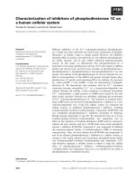
Fig. 3. Cladogram showing the evolutionary relationship of Nag3 of Cellulomonas fimi (Q7WUL3 ⁄ NAG3_CELFI) and selected members of
family 3 of glycoside hydrolases. The abbreviations used reference the accession numbers of the UniProt database and the organism codes:
NagA of Streptomyces thermoviolaceus (Q82840 ⁄ NAGA_STRTH) and HexA from Alteromonas sp. (P48823 ⁄ HEXA_ALTSO) and the
sequences of three putative b-N-acetylglucosaminidases from Bacillus subtilis (P40406 ⁄ YBBD_BACSU), Streptomyces colicolor
(Q9RDG9_STRCO) and Clostridium perfringens (HEXA_CLOPE) (see Fig. 3); the b-N-acetylglucosaminidases of two Gram-negative bacteria,
NagZ of Escherichia coli (P75949 ⁄ NAGZ_ECOLI) and ExoII of Vibrio furnisii (P96157 ⁄ EXOII_VIBFU); members of the b-glucosidase subfamily,
b-glucosidase X of Escherichia coli (P33363 ⁄ BGLX_ECOLI) and b-glucosidase A of Aspergillus niger (Q9P8F4 ⁄ BGLA_ASPNG), the two
exoglucanases ExoI and ExoII of Hordeum vulgare (Q9XEI3 ⁄ EXOI_HORVU and Q42835 ⁄ EXOII_HORVU), and a b-xylosidase and an a-
L-arabi-
ofuranosidase ⁄ b-xylosidase of Hordeum vulgare (Q8W011 ⁄ XYLA_HORVU and (QW012 ⁄ ARAI_HORVU).Nag3 and the putative family 3
N-acetylglucosaminidase of Clostridium perfringens (Q8XP12) form an intermediate branch between b-glucosidases and b-N-acetylglucosami-
nidases of family 3. The phylogenetic tree was created with the program
TREEVIEW (by R. D .M. Page).
Table 1. Kinetic parameters for the reactions of Cellulomonas fimi b-N-acetylhexosaminidase (Hex20) and b-N-acetylglucosaminidases (Nag3)
with pNP glycosides. The enzymic reaction was carried out in 50 m
M sodium phosphate buffer (pH 7.08) at 25 °C. The molar extinction coef-
ficient (
M
)1
Æcm
)1
) at 400 nm for pNP was 7280. Standard errors for the values of K
m
and k
cat
measured here were less than 5%, except
where standard error values are indicated.
a
Not determined due to a reaction being too slow to be detected.
b
Not determined due to the
linear relationship of enzyme velocity with substrate concentration. pNP-GlcNAc, 4¢-nitrophenyl b-N-acetyl-
D-glucosaminide; pNP-GalNAc,
4¢-nitrophenyl b-N-acetyl-
D-galactosaminide; pNP-Glc, 4¢-nitrophenyl b-D-glucopyranoside.
Substrate
Hex20 Nag3
K
m
(lM) k
cat
(s
)1
) k
cat
⁄ K
m
(s
)1
ÆlM
)1
) K
m
(lM) k
cat
(s
)1
) k
cat
⁄ K
m
(s
)1
ÆlM
)1
)
pNP-GlcNAc 53 482 9.09 2.7 ± 0.2 0.18 0.067
pNP-GalNAc 66 129 1.95 ND
a
ND
a
ND
a
pNP-Glc ND
a
ND
a
*ND
a
*ND
b
ND
b
0.076
Cellulomonas fimi b-N-acetylglucosaminidases C. Mayer et al.
2934 FEBS Journal 273 (2006) 2929–2941 ª 2006 The Authors Journal compilation ª 2006 FEBS
and 8.4; however, it lost its catalytic activity in diluted
buffers within a day at 4 °C. The half-life of Nag3 at
room temperature was only a few hours. Addition of
sodium chloride (0.5 m), dithiothreitol (0.1 and 1 mm),
BSA (0.5 mgÆmL
)1
), sucrose and trehalose (20%) had
no huge effect on Hex3 stability (Table 2). However,
adding glycerol and ⁄ or phosphate stabilized the
enzymes, and Nag3 retained its activity for several
0.00
0.05
0.10
A
400
/ min
1/v (A
400
/ min)
–1
0.15
0.20
0.00 0.25 0.50 0.75 1.00 1.25
[S] (m
M
)
-20 0 20 40 60 80 100 120 140 160
25
50
75
100
1 / [S] (m
M
)
-1
A
A
400
/ min
1/v (A
400
/ min)
–1
[S] (m
M
)
1 / [S] (m
M
)
-1
02468
0.00
0.05
0.10
0.15
0.20
0.25
0.30
0.35
-101234
50
100
150
B
Fig. 4. Michaelis–Menten plot of initial rates of hydrolysis of (A) 4¢-nitrophenyl b-N-acetyl- D-glucosaminide (pNP-GlcNAc) (d)and4¢-nitrophe-
nyl b-N-acetyl-
D-galactosaminide (pNP-GalNAc) (s)byCellulomonas fimi Hex20 (5.59 · 10
)5
mg ⁄ mL) at 25.2 °C and pH 7.08 and (B) pNP-
GlcNAc (n) and pNP-Glc (h)byCellulomonas fimi Nag3 (3.09 · 10
)3
mg ⁄ mL) at 25.2 °C and pH 7.08. Inset: graphical analysis of K
m
and k
cat
by Lineweaver–Burk linearization.
Table 2. Effects of various reagents on the stability of Cellulomonas fimi Nag3 dithiothreitol.
Reagents Concentration
% Remaining relative
activity
a
(18 h incubation)
b
Remaining relative
activity
a
(90 h incubation)
b
NaCl 500 mM 10 0
Dithiothreitol 0.1 m
M 35 0
Dithiothreitol and glycerol 0.1 m
M and 20% 100 90
BSA 5% 35 0
Sucrose 20% 66 25
Trehalose 20% 0 0
Tris pH 7.3 330 m
M 80
Phosphate pH 7.3 330 m
M 100 90
Glycerol 20% 80 60
Glycerol 20% 95 95
60 m
M imidazol pH 7.5
Glycerol
c
20% 100 mM phosphate pH 7.3 100 100
a
The enzymic reaction was carried out in 20 mM Tris ⁄ HCl buffer (pH 7.3) at 25 °C with 4¢-nitrophenyl b-N-acetyl-D-glucosaminide (pNP-Glc-
NAc) (6.5 m
M).
b
Before assaying, Nag3 was incubated for 18 and 90 h with the indicated supplement;
c
the activity measured after incubation
for the indicated time with the supplement shown in bold was set at 100%.
C. Mayer et al. Cellulomonas fimi b-N-acetylglucosaminidases
FEBS Journal 273 (2006) 2929–2941 ª 2006 The Authors Journal compilation ª 2006 FEBS 2935
months when stored in glycerol (20% final concentra-
tion) and phosphate buffer at pH 7.3 and ) 20 °C. Ex-
oII, a family 3 N-acetylglucosaminidase from Vibrio
furnissi, is activated by 400–700 mm sodium chloride
[4]. However, sodium chloride up to 700 mm had no
effect on Hex3; there was a 10% decrease in activity
with 1 m NaCl.
The pH dependence of Hex20 and Nag3 was investi-
gated using pNP-GlcNAc and pNP-Glc, respectively,
over a pH range of 6.2–9.2 and 6.8–8.5, respectively.
Hex20 showed a broad, bell-shaped pH optimum curve
with a maximum between pH 7.3 and 8.7 with pNP-
GlcNAc and half-maximal rate at about pH 7 and 9
(Fig. 5A). By contrast, the family 20 b-N-acetylhexosa-
minidase from Streptomyces plicatus has a pH
optimum of 5 on pNP-GlcNAc [11]. The k
cat
⁄ K
m
for
Hex20 was dependent on two ionizable groups with
pK
a
values of 6.9 and 8.8 (Fig. 6B). Nag3 gave a com-
plex pH profile on pNP-Glc, with a narrow maximal
rate at pH 7.3 and half-maximal rates at about pH 6.8
and 8.0 (Fig. 5). This is consistent with the pH opti-
mum determined for the b-N-acetylglucosaminidase
(ExoII) from Vibrio furnissii [4]. The pK
a
of the lower
ionization constant was 6.7; however, a value for an
upper ionization could not be determined from the
data (Fig. 5B).
MS and labeling
The mass of purified Hex20 was 54 186 Da, as ana-
lyzed by ESI ⁄ MS, which is in perfect agreement with
0.00
0.02
0.04
0
2500
5000
7500
A
k
ca
t
/K
m
(s
–1
m
M
–1
)
p(K
cat
/K
m
)
6.5 7.0 7.5 8.0 8.5 9.0 9.5
-1
0
1
2
3
-4
-3
-2
-1
B
pH
Fig. 5. pH dependence of k
cat
⁄ K
m
for the Nag3- and Hex20-cata-
lyzed reaction. (A) The pH profiles of Nag3 (d, left scale) and
Hex20 (s, right scale) were determined using pNP-Glc and 4¢-nitro-
phenyl b-N-acetyl-
D-glucosaminide (pNP-GlcNAc), respectively, at
25 °C. The reaction buffers were 100 m
M sodium citrate ⁄ phos-
phate (pH 6.0–7.3), 100 m
M sodium phosphate (pH 7.0–8.2) and
100 m
M glycine ⁄ HCl (pH 7.8–10). (B) Shows the same data used to
fit Eqn (1); the lines represent the best fit of the equation to the
pk
cat
⁄ K
m
data (Nag3, pK
a1
¼ 6.70 ± 0.33; pK
a2
could not be deter-
mined from the data; Hex20, pK
a1
¼ 6.91 ± 0.10; pK
a2
¼
8.79 ± 0.12).
100
A
B
C
60971.0
61126.0
61135.0
60000
61000
mass (Da)
62000
50
0
20
10
0
20
10
0
relative intensity (%)
Fig. 6. Transform of the electrospray mass spectrum of (A) Nag3,
and (B) and (C) Nag3 incubated at room temperature with 10 m
M
2¢,4¢-dinitrophenyl-2-deoxy-2-fluoro-b-glucose for 4 h and 20 h,
respectively. The mass shifts (157 and 166) of peaks shown in (B)
and (C) compared to the peak shown in (A) correspond to a 2-de-
oxy-2-fluoro-b-glucosyl residue (162 Da) covalently bound to Nag3.
Cellulomonas fimi b-N-acetylglucosaminidases C. Mayer et al.
2936 FEBS Journal 273 (2006) 2929–2941 ª 2006 The Authors Journal compilation ª 2006 FEBS
the theoretical mass of the cloned enzyme (54 186 Da).
The mass of the purified Nag3 protein was determined
by ESI ⁄ MS to be 60 971 Da, close to the theoretical
mass of the cloned enzyme (60 945 Da). After incuba-
tion with 2¢,4¢-dinitrophenyl 2-deoxy-2-fluoro-b-
d-glucopyranoside (DNP-2FGlc), two species are
observed: the native, unlabeled enzyme, and another
species with a mass of 61 126–61 135 Da (Fig. 6). The
mass difference observed between the native and inhib-
ited enzyme is 164 Da, a value that is consistent,
within error, with the addition of a single 2-deoxy-2-
fluoroglucosyl label (162 Da). The rate of the labeling
was consistent with the expectation of slow inactiva-
tion by the inhibitor when the low apparent k
cat
values
for Nag3 with chromogenic glucosides are kept in
mind. Prolonged incubation of the enzyme with the
inhibitor leads to almost complete inactivation of the
native enzyme. The observation of a covalent glycosyl
intermediate provides strong evidence for a mechanism
involving an enzymic nucleophile, as shown previously
for two family 3 glycoside hydrolases: the single
domain b-N-acetylglucosaminidase from V. furnissii [7]
and the two domain b-glucosidases from Aspergillus
niger [13]. Sequence alignment using the clustal w
algorithm revealed a conserved aspartate residue
within the sequence GLVVS
DS to be the putative cat-
alytic nucleophile. By contrast, the hydrolytic mechan-
ism of retaining family 20 b-N-acetylhexosaminidases
involves the assistance of the acetamido group of the
substrate [8,9,11].
Discussion
Cellulomonas fimi is strongly cellulolytic, producing a
complex cellulose degradative system. The system,
comprising mostly extracellular enzymes, is understood
in considerable detail (e.g. [29,32–38]). Cellulomonas
fimi also degrades chitin (C. Mayer, unpublished
observation), a homopolymer of GlcNAc similar to
cellulose, but nothing is known of its chitinolytic sys-
tem. Recently, a chitinase was isolated from culture
supernatant of Cellulomonas flavigena [39] and a chi-
tinase-encoding gene was cloned from Cellulomonas
uda [40]. Cellulomonas fimi also secretes one (or more)
chitinase(s) (C. Mayer, unpublished observation), but
N-acetylglucosaminidase activity is present only in the
soluble cell extract. Of the two enzymes described
here, only Hex20 degrades N-acetylchitooligomers and
may be involved in chitin degradation. The function
of Nag3 is unclear. It has low catalytic activity relative
to Hex20 on chromogenic substrates: the catalytic effi-
ciency (k
cat
⁄ K
m
value) for hydrolysis of pNP-GlcNAc
by the two enzymes differs by a factor of 10
5
(Table 1). In this respect, Nag3 resembles family 3
N-acetylglucosaminidases of Gram-negative bacteria,
which are involved in cell wall (peptidoglycan) recyc-
ling [1,7,20,21,41]. However, Nag3 is unusual in that it
acts on b-N-acetyl-d-glucosaminides and b-d-gluco-
sides, so it should be referred to as a b-N-acetyl-d-glu-
cosaminidase ⁄ b-d-glucosidase. The catalytic efficiencies
against pNP-Glc and pNP-GlcNAc were similar, seem-
ingly a consequence of much higher apparent values
for both k
cat
and K
m
for the b-glucoside. Unfortu-
nately, the exact kinetic parameters for hydrolysis of
pNP-Glc by Nag3 could not be determined because the
enzyme was not saturated with the substrate within the
limits of its solubility. Although a family 3 enzyme
from barley was characterized that was referred to as
a ‘bifunctional’ a-l-arabinofuranosidase ⁄ b-d-xylopyra-
nosidase [42], there is, to our knowledge, no previous
report on an enzyme with equivalent b-glucosidase and
b-N-acetylglucosaminidase activity.
Glycosyl hydrolases of family 3 form two distinct
subgroups: a b-glucosidase subfamily and a b-N-acetyl-
glucosaminidase subfamily (Fig. 3). Being a ‘bifunc-
tional’ b-N-acetyl-d-glucosaminidase ⁄ b-d-glucosidase,
Nag3 of Cellulomonas fimi represents an interesting
link between the b-N-acetylglucosaminidase and the
b-glucosidase branch of family 3 of glycoside hydrolas-
es. A conserved sequence motif in the b-N-acetylglu-
cosaminidase subgroup within family 3 may represent
the N-acetyl group-binding site [1,7]. Interest-
ingly, Nag3, as well as the uncharacterized family 3
enzyme (Q8XP12) within the genome of the recently
sequenced bacterium Clostridium perfringens [43], show
a significant alteration within this motif: the K-H-
(FI)-P-G-(
HL)-G-x(4)-D-(ST)-H motif is changed to
K-H-(FI)-P-G-
D-G-x(4)-D-Q-H (Fig. 2). It can be spe-
culated that changes within this motif (underlined) are
responsible for the broad substrate specificity of Cellu-
lomonas fimi Nag3 for substitution of the C2 position,
and further studies in order to confirm this thesis are
under way. A hint for a possible function of Nag3
comes from a gene neighbor analysis of a putative
b-N-acetylglucosaminidase of Clostridium perfringens
using the European Molecular Biology Laboratory
Search Tool for the Retrieval of Interacting Genes ⁄
Proteins (string). Analysis revealed that the encoding
gene is connected to a cluster of genes similar to
known genes involved in the uptake and metabolism
of glucuronides. We speculate that the putative N-ace-
tylglucosaminidase of Clostridium perfringens and poss-
ibly also Nag3 of Cellulomonas fimi might be involved
in the degradation of glucuronic acid-containing gly-
cosaminoglycans such as hyaluronic acid. Preliminary
experiments, however, could not confirm this hypothesis;
C. Mayer et al. Cellulomonas fimi b-N-acetylglucosaminidases
FEBS Journal 273 (2006) 2929–2941 ª 2006 The Authors Journal compilation ª 2006 FEBS 2937
hydrolysis of hyaluronic acid by Nag3 could not be
detected by TLC analysis. b-N-Acetylglucosaminidases
involved in hyaluronic acid degradation have been
placed into family 84 of glycoside hydrolases rather
than family 3 [44]. N-Acetylglucosaminidases in family
84, like family 20 enzymes, use a catalytic mechanism
involving anchimeric assistance of the 2-acetamido
group of the substrate. However, N-acetylglucosami-
nidases in family 3 are retaining enzymes that use a
double-displacement mechanism involving the partici-
pation of a catalytic nucleophilic group in the enzyme
active site [7,13]. Our data confirm that Cellulomonas
fimi Nag3 acts by participation of a catalytic nucleo-
phile: incubation of Nag3 with DNP-2FGlc permits
the observation by ESI ⁄ MS of a high steady-state
population of a 2-deoxy-2-fluoroglucosyl–enzyme inter-
mediate. The identification of a bifunctional b-N-ace-
tyl-d-glucosaminidase ⁄ b-d-glucosidase is an interesting
example of divergent evolution towards new substrate
specificity within family 3 of glycoside hydrolases. Fur-
ther structural and mutational studies are required to
elucidate the basis of substrate specificity in this family
of glycoside hydrolases.
Experimental procedures
Materials
Chemicals, reagents and materials were purchased as fol-
lows: growth media components from Difco (Sparks, ML);
DNA purification kits from Qiagen (Hilden, Germany);
restriction endonucleases, DNA ligase and DNA poly-
merase from New England Biolabs (Beverly, MA) and
Roche-Boehringer Mannheim (Germany); and His-bind
metal chelation resin from Novagen (Madison, WI). Oligo-
nucleotides were synthesized, and DNA and protein
sequences determined by the Nucleic Acids and Peptide
Service (NAPS) Unit of the Biotechnology Laboratory at
the University of British Columbia. N-Acetylchitooligosac-
charides (Dp 2–6) were from Seikagaku America (Fal-
mouth, MA, USA). Chromogenic substrates and
hyaluronic acid were from Sigma. 4MU-GlcNAc and DNP-
2FGlc were synthesized by standard procedures.
Bacterial strains, plasmids and phages
E. coli strain BL21(DE3) and pET29b were from Novagen
(Madison, WI). E. coli XLOLR and the library (2–5 kbp
fragment length) of genomic DNA from Cellulomonas fimi
in k zapii (18, 19) were from Stratagene (La Jolla, CA).
Cultures were grown in LB medium supplemented with
50 mgÆL
)1
ampicillin, or TYP medium (tryptone 16 gÆL
)1
,
yeast extract 16 gÆL
)1
, NaCl 5 gÆL
)1
,K
2
HPO
4
2.5 gÆL
)1
)
containing 50 mgÆL
)1
kanamycin.
Screening and isolation of Cellulomonas fimi
genes encoding N-acetylglucosaminidases
Plasmid isolations, restriction enzyme digests, ligations and
transformations were performed using standard techniques.
Phagemids (pBluescript SK) were excised from the k ZAPII
library using a helper phage and transferred to E. coli
XLOLR according to the supplier’s protocol. Sufficient
cells to yield about 500 colonies per plate were spread on
LB ampicillin agar. After incubation for two days at 37 °C,
colonies were replicated on LB ampicillin agar supplemen-
ted with 4MU-GlcNAc (200 mgÆL
)1
) and isopropyl thiogal-
actopyranoside (IPTG; 1 mm). It was necessary to screen
replicas because the 4-methylumbelliferone product released
by enzyme action appeared to be toxic to the cells. Colonies
were screened for fluorescence at 366 nm using a UV trans-
illuminator. Nucleotide sequences of inserts were deter-
mined by primer walking and confirmed by sequencing the
complementary strand. The nucleotide sequences of hex20A
and nag3A have been submitted to the DDBJ ⁄ EMBL ⁄
GenBank databases under the accession numbers
AF478459 and AF478460. The UniProt database accession
numbers are Q7WUL4 and Q7WUL3, repectively. The
GenBank and SWISS-PROT databases were used for nuc-
leotide and amino acid sequence searches using the basic
local alignment search tool (blast).
Construction of pETcfnag3 and pETcfhex20
The putative N-acetylglucosaminidase genes within the
inserts in pCF2 and pCF5 were amplified by PCR using
oligonucleotide primers based on the ORF sequences (under-
lined are the restriction sites NdeI, NotI and XhoI introduced
by the primer): CF2NdeI 5¢-CC
CAT ATG CCC GAC
GTC GCC GTC ATC C-3¢; CF2NotI 5¢-TT
GCG GCC
GCG CCC GGC GCG GAA CCC-3¢; CF5NdeI 5¢-AA
CAT ATG ATC GAC CTG ACC GCA GCC-3¢; CF5XhoI
5¢-AA
CTC GAG GTG GGT GTC CCA CTG GCC-3 ¢.
PCR mixtures contained 10 lm primers, 1 mm each deoxyri-
bonucleoside triphosphate, $ 50 ng of phagemid DNA, 5 U
of Pwo polymerase and 4% DMSO in 100 lL of DNA
polymerase buffer. Thirty PCR cycles (45 s at 94 °C, 45 s at
63 °C, and 120 s at 72 °C) were performed in a thermal
cycler (Perkin Elmer Applied Biosystems, Boston, MA,
USA, GeneAmp PCR System 2400). The amplified frag-
ments were cloned into pET29b according to a protocol des-
cribed previously [45].
Production, purification and N-terminal
sequencing of recombinant proteins
E. coli BL21(DE3) carrying pET29cfnag3 or pET29cfhex20
was grown at 37 °C in TYP kanamycin to a D
600 nm
value
of 0.6–0.8, IPTG was added to a concentration of 1 mm,
and incubation was continued for a further 12 h at 28 °C.
Cellulomonas fimi b-N-acetylglucosaminidases C. Mayer et al.
2938 FEBS Journal 273 (2006) 2929–2941 ª 2006 The Authors Journal compilation ª 2006 FEBS
The cells were collected by centrifugation (10 min, 3000 g,
4 °C) and resuspended in a minimal volume of buffer
(20 mm sodium phosphate, 300 mm NaCl, pH 7.5). The
resuspended cells were ruptured by passing them three times
through a French pressure cell. Debris and unbroken cells
were removed by centrifugation (15 min, 27 000 g,4°C).
The supernatant was passed through a column of His-bind
resin [7]. Adsorbed proteins were desorbed with imidazole
(linear gradient from 0 to 250 mm imidazole in 20 mm
sodium phosphate, 300 mm NaCl, pH 7.5), and fractions of
3 mL were collected. Fractions containing the highest activ-
ity were pooled and then concentrated using an Amicon
YM30 centriprep device (Amicon, Millipore, Bedford, MA).
Purified proteins were subjected to SDS ⁄ PAGE, and then
electroblotted onto polyvinylidene difluoride (PVDF) mem-
branes (Millipore). Protein bands were visualized by Coo-
massie staining (0.1% Coomassie in 40% MeOH) before
sequencing by automated Edman degradation in a Perkin
Elmer ⁄ Applied Biosystems 476 A gas-phase sequenator.
Biochemical characterization
Enzymatic reaction rates with chromogenic substrates were
determined by following changes in UV ⁄ visible absorbance
using matched quartz cells (1 cm path length) in a Phar-
macia Biotech (Freiburg, Germany) Ultrospec3000 spec-
trophotometer with temperature control unit or in a
Pye-Unicam (Angleton, TX, USA) PU-8800 spectropho-
tometer equipped with a circulating water bath to main-
tain the cells at 25 °C. The buffer employed for all kinetic
experiments (apart from those involving pH variation) was
50 mm sodium phosphate buffer, pH 7.0. The molar
extinction coefficient at 400 nm for the liberated aglycone
4¢-nitrophenol was 7280 in phosphate buffer at pH 7.0.
Kinetic measurements involving pH variation were per-
formed using 100 mm citrate ⁄ 50 mm phosphate (pH 6.0–
7.3), 100 mm sodium phosphate buffer (pH 7.0–8.2), and
100 mm glycine ⁄ NaOH buffer (pH 7.8–10). Product release
was measured at 400 nm using pNP-GlcNAc, pNP-Gal-
NAc, and pNP-Glc. pH curves for Hex20 were derived by
full measurement of k
cat
and K
m
at each pH value. The
pH dependence of k
cat
⁄ K
m
for Nag3 was determined using
pNP-Glc at a final concentration of 6.5 mm, which is well
below the apparent K
m
. The change of absorbance with
time was fitted to a first-order rate equation using the
program prism 3.0 (Graph Pad Software, San Diego, CA,
USA), which yielded values for the pseudo-first-order rate
constant at each pH value. In order to ensure that the pH
had not changed during the reaction, the pH of each reac-
tion mixture was measured after recording the rates of
hydrolysis. For pK
a
determination, the k
cat
⁄ K
m
values
determined within the pH range 6.2–9.2 for Hex20 and
within the pH range 6.8–7.2 for Nag3 were analyzed by
fitting the negative log
10
(i.e. pk
cat
⁄ K
m
) to a two-ionization
titration curve, Eqn (1).
Y ¼ A þ log
10
ð1 þð10
ÀpH
=10
ÀpKa1
Þþð10
ÀpKa2
=10
ÀpH
ÞÞ ð 1Þ
In this equation, Y represents pk
cat
⁄ K
m
at different pH
values, and A represents the maximum value of Y.
Enzymatic reaction products released from N-acetylchi-
tooligomers and b-glucan oligomers (DP 2–6: 3–10 mm,
pH 7.0 incubated for 30 min at 25 °C with Hex20 or Nag3)
were analyzed by TLC using Merck Kieselgel 60 F254
aluminum-backed sheets with butanol ⁄ methanol ⁄ water
(7:2:1v⁄ v⁄ vol) as solvent. After development, the sheets
were soaked for a short time in 5% ammonium molybdate,
0.1% cerium(IV) sulfate, and 10% H
2
SO
4
, and then heated
at 150 °C until blue spots appeared.
Trapping the glycosyl–enzyme intermediate with
DNP-2FGlc
Nag3 (20 lm) was incubated with 10 mm DNP-2FGlc for 4
and 20 h at room temperature in 20 mm sodium phos-
phate ⁄ 300 mm NaCl ⁄ 80 mm imidazole, pH 7.5. Samples
were analyzed by ESI ⁄ MS as described previously [7,14].
Acknowledgements
This work was supported by the Protein Engineering
Network of Centers of Excellence of Canada, the
Natural Sciences and Engineering Research Council of
Canada, the Swiss National Science Foundation, the
Deutsche Forschungsgemeinschaft and Neose Technol-
ogies. We thank Shouming He for technical assist-
ance.
References
1 Mayer C, Vocadlo DJ & Withers SG (2000) b-N-Acetyl-
hexosaminidases: two enzyme families, two mechanisms.
In Advances in Chitin Science (Peter MG, Domard A
& Muzzarelli RAA, eds), pp. 612–619. University of
Potsdam, Potsdam.
2 Henrissat B (1998) Glycosidase families. Biochem Soc
Trans 26, 153–156.
3 Horsch M, Mayer C, Sennhauser U & Rast DM (1997)
b-N-Acetylhexosaminidase: a target for the design of
antifungal agents. Pharmacol Ther 76 , 187–218.
4 Chitlaru E & Roseman S (1996) Molecular cloning and
characterization of a novel b-N-acetyl-d-glucosamini-
dase from Vibrio furnissii. J Biol Chem 271, 33433–
33439.
5 Tsujibo H, Fujimoto K, Tanno H, Miyamoto K, Kim-
ura Y, Imada C, Okami Y & Inamori Y (1995) Molecu-
lar cloning of the gene which encodes b-N-
acetylglucosaminidase from a marine bacterium, Altero-
monas sp. strain O-7. Appl Environ Microbiol 61, 804–
806.
C. Mayer et al. Cellulomonas fimi b-N-acetylglucosaminidases
FEBS Journal 273 (2006) 2929–2941 ª 2006 The Authors Journal compilation ª 2006 FEBS 2939
6 Tsujibo H, Hatano N, Mikami T, Hirasawa A, Miya-
moto K & Inamori Y (1998) A novel b-N-acetylglucosa-
minidase from Streptomyces thermoviolaceus OPC-520:
gene cloning, expression, and assignment to family 3 of
the glycosyl hydrolases. Appl Environ Microbiol 64,
2920–2924.
7 Vocadlo DJ, Mayer C, He S & Withers SG (2000)
Mechanism of action and identification of Asp242 as
the catalytic nucleophile of Vibrio furnisii N-acetyl-b-
d-glucosaminidase using 2-acetamido-2-deoxy-5-fluoro-
alpha-1-idopyranosyl fluoride. Biochemistry 39, 117–
126.
8 Vocadlo DJ & Withers SG (2005) Detailed comparative
analysis of the catalytic mechanisms of b-N-acetylglu-
cosaminidases from families 3 and 20 of glycoside
hydrolases. Biochemistry 44 , 12809–12818.
9 Mark BL, Vocadlo DJ, Knapp S, Triggs-Raine BL,
Withers SG & James MN (2001) Crystallographic
evidence for substrate-assisted catalysis in a bacterial
b-hexosaminidase. J Biol Chem 276, 10330–10337.
10 Mark BL, Vocadlo DJ, Zhao D, Knapp S, Withers SG
& James MN (2001) Biochemical and structural assess-
ment of the 1-N-azasugar GalNAc-isofagomine as a
potent family 20 b-N-acetylhexosaminidase inhibitor.
J Biol Chem 276, 42131–42137.
11 Williams SJ, Mark BL, Vocadlo DJ, James MN &
Withers SG (2002) Aspartate 313 in the Streptomyces
plicatus hexosaminidase plays a critical role in substrate-
assisted catalysis by orienting the 2-acetamido group
and stabilizing the transition state. J Biol Chem 277,
40055–40065.
12 Vocadlo DJ, Davies GJ, Laine R & Withers SG (2001)
Catalysis by hen egg-white lysozyme proceeds via a
covalent intermediate. Nature 412, 835–838.
13 Dan S, Marton I, Dekel M, Bravdo BA, He S, Withers
SG & Shoseyov O (2000) Cloning, expression, charac-
terization, and nucleophile identification of family 3,
Aspergillus niger b-glucosidase. J Biol Chem 275, 4973–
4980.
14 Vocadlo DJ & Withers SG (2000) Identification of
active site residues in glycosidases by use of tandem
mass spectrometry. Methods Mol Biol 146, 203–222.
15 Zechel DL & Withers SG (2000) Glycosidase mechan-
isms: anatomy of a finely tuned catalyst. Acc Chem Res
33, 11–18.
16 Hrmova M, Harvey AJ, Wang J, Shirley NJ, Jones GP,
Stone BA, Hoj PB & Fincher GB (1996) Barley b-d-glu-
can exohydrolases with b-d-glucosidase activity. Purifi-
cation, characterization, and determination of primary
structure from a cDNA clone. J Biol Chem 271, 5277–
5286.
17 Harvey AJ, Hrmova M, De Gori R, Varghese JN &
Fincher GB (2000) Comparative modeling of the three-
dimensional structures of family 3 glycoside hydrolases.
Proteins 41, 257–269.
18 Varghese JN, Hrmova M & Fincher GB (1999) Three-
dimensional structure of a barley b-d-glucan exohydro-
lase, a family 3 glycosyl hydrolase. Structure Fold Des 7,
179–190.
19 Hrmova M, De Gori R, Smith BJ, Vasella A, Varghese
JN & Fincher GB (2004) Three-dimensional structure of
the barley b-d-glucan glucohydrolase in complex with a
transition state mimic. J Biol Chem 279, 4970–4980.
20 Cheng Q, Li H, Merdek K & Park JT (2000) Molecular
characterization of the b-N-acetylglucosaminidase
of Escherichia coli and its role in cell wall recycling.
J Bacteriol 182, 4836–4840.
21 Vo
¨
tsch W & Templin MF (2000) Characterization of a
b-N-acetylglucosaminidase of Escherichia coli and eluci-
dation of its role in muropeptide recycling and b-lacta-
mase induction. J Biol Chem 275, 39032–39038.
22 Keyhani NO & Roseman S (1999) Physiological aspects
of chitin catabolism in marine bacteria. Biochim Biophys
Acta 1473, 108–122.
23 Horn SJ, Sorbotten A, Synstad B, Sikorski P, Sorlie M,
Va
˚
rum KM & Eijsink VG (2006) Endo ⁄ exo mechanism
and processivity of family 18 chitinases produced by
Serratia marcescens. FEBS J 273, 491–503.
24 Watanabe T, Kimura K, Sumiya T, Nikaidou N, Suzuki
K, Suzuki M, Taiyoji M, Ferrer S & Regue
´
M (1997)
Genetic analysis of the chitinase system of Serratia
marcescens 2170. J Bacteriol 179, 7111–7117.
25 Bassler BL, Yu C, Lee YC & Roseman S (1991) Chitin
utilization by marine bacteria. Degradation and catabo-
lism of chitin oligosaccharides by Vibrio furnissii. J Biol
Chem 266, 24276–24286.
26 de la Cruz J, Hidalgo-Gallego A, Lora JM, Benitez T,
Pintor-Toro JA & Llobell A (1992) Isolation and char-
acterization of three chitinases from Trichoderma harzia-
num. Eur J Biochem 206 , 859–867.
27 Mitsutomi M, Kidoh H, Tomita H & Watanabe T
(1995) The action of Bacillus circulans WL-12 chitinases
on partially N-acetylated chitosan. Biosci Biotechnol
Biochem 59, 529–531.
28 Meinke A, Gilkes NR, Kilburn DG, Miller RC Jr &
Warren RA (1993) Cellulose-binding polypeptides from
Cellulomonas fimi: endoglucanase D (CenD), a family A
b-1,4-glucanase. J Bacteriol 175, 1910–1918.
29 Meinke A, Gilkes NR, Kwan E, Kilburn DG, Warren
RA & Miller RC Jr (1994) Cellobiohydrolase A (CbhA)
from the cellulolytic bacterium Cellulomonas fimi is a
b-1,4-exocellobiohydrolase analogous to Trichoderma
reesei CBH II. Mol Microbiol 12, 413–422.
30 Tews I, Perrakis A, Oppenheim A, Dauter Z, Wilson
KS & Vorgias CE (1996) Bacterial chitobiase structure
provides insight into catalytic mechanism and the basis
of Tay–Sachs disease. Nat Struct Biol 3, 638–648.
31 Tsujibo H, Hatano N, Mikami T, Izumizawa Y, Miya-
moto K & Inamori Y (1998) Cloning, characterization
and expression of b-N-acetylglucosaminidase gene from
Cellulomonas fimi b-N-acetylglucosaminidases C. Mayer et al.
2940 FEBS Journal 273 (2006) 2929–2941 ª 2006 The Authors Journal compilation ª 2006 FEBS
Streptomyces thermoviolaceus OPC-520 (1). Biochim
Biophys Acta 1425, 437–440.
32 Damude HG, Ferro V, Withers SG & Warren RA
(1996) Substrate specificity of endoglucanase A from
Cellulomonas fimi: fundamental differences between
endoglucanases and exoglucanases from family 6.
Biochem J 315, 467–472.
33 Damude HG, Withers SG, Kilburn DG, Miller RC Jr
& Warren RA (1995) Site-directed mutation of the puta-
tive catalytic residues of endoglucanase CenA from
Cellulomonas fimi. Biochemistry 34, 2220–2224.
34 Gilkes NR, Claeyssens M, Aebersold R, Henrissat B,
Meinke A, Morrison HD, Kilburn DG, Warren RA &
Miller RC Jr (1991) Structural and functional relation-
ships in two families of b-1,4-glycanases. Eur J Biochem
202, 367–377.
35 Meinke A, Schmuck M, Gilkes NR, Kilburn DG, Mil-
ler RC Jr & Warren RA (1992) The tertiary structure of
endo-b-1,4-glucanase B (CenB), a multidomain cellulase
from the bacterium Cellulomonas fimi . Glycobiology 2 ,
321–326.
36 Shen H, Tomme P, Meinke A, Gilkes NR, Kilburn
DG, Warren RA & Miller RC Jr (1994) Stereochemical
course of hydrolysis catalysed by Cellulomonas fimi
CenE, a member of a new family of b-1,4-glucanases.
Biochem Biophys Res Commun 199, 1223–1228.
37 Shen H, Gilkes NR, Kilburn DG, Miller RC Jr &
Warren RA (1995) Cellobiohydrolase B, a second exo-
cellobiohydrolase from the cellulolytic bacterium
Cellulomonas fimi. Biochem J 311, 67–74.
38 Tomme P, Kwan E, Gilkes NR, Kilburn DG & Warren
RA (1996) Characterization of CenC, an enzyme from
Cellulomonas fimi with both endo- and exoglucanase
activities. J Bacteriol 178, 4216–4223.
39 Chen H-C, Hsu M-F & Jiang S-T (1997) Purification
and characterization of an exo-N,N¢ -diac-
etylchitobiohydrolase-like enzyme from Cellulomonas
flavigena NTOU1. Enzyme Microb Technol 20, 191–197.
40 Reguera G & Leschine SB (2003) Biochemical and
genetic characterization of ChiA, the major enzyme
component for the solubilization of chitin by Cellulomo-
nas uda. Arch Microbiol 180, 434–443.
41 Mayer C & Boos W (2005) Hexose ⁄ hexitol and
pentose ⁄ pentiol metabolism. In EcoSal-Escherichia coli
and Salmonella: Cellular and Molecular Biology (Curtiss
R III, ed.), . ASM Press, Wash-
ington, DC.
42 Lee RC, Hrmova M, Burton RA, Lahnstein J &
Fincher GB (2003) Bifunctional family 3 glycoside
hydrolases from barley with a-1-arabinofuranosidase
and b-d-xylosidase activity. Characterization, primary
structures, and COOH-terminal processing. J Biol Chem
278, 5377–5387.
43 Shimizu T, Ohtani K, Hirakawa H, Ohshima K,
Yamashita A, Shiba T, Ogasawara N, Hattori M,
Kuhara S & Hayashi H (2002) Complete genome
sequence of Clostridium perfringens, an anaerobic flesh-
eater. Proc Natl Acad Sci USA 99, 996–1001.
44 Macauley MS, Whitworth GE, Debowski AW, Chin D
& Vocadlo DJ (2005) O-GlcNAcase uses substrate-
assisted catalysis: kinetic analysis and development of
highly selective mechanism-inspired inhibitors. J Biol
Chem 280, 25313–25122.
45 Mayer C, Zechel DL, Reid SP, Warren RA & Withers
SG (2000) The E358S mutant of Agrobacterium sp.
b-glucosidase is a greatly improved glycosynthase. FEBS
Lett 466, 40–44.
46 Chenna R, Sugawara H, Koike T, Lopez R, Gibson TJ,
Thompson JD & Higgins DG (1994) Multiple sequence
alignment with the Clustal series of programs. Nucleic
Acids Res 22, 4673–4680.
Supplementary material
The following supplementary material is available
online:
Fig. S1. TLC analysis of the degradation of N-acetylchito-
oligomers (Dp 2–6) by Hex20. The reaction mixtures
contained 0.1 lg of enzyme and substrate: 4 mm
N-acetylchitobiose (lanes 1, 6 and 11), 6 mm N-acetyl-
chitotriose (lanes 2, 7 and 12), 4 mm N-acetyl-
chitotetraose (lanes 3, 8 and 13), 3 mm
N-acetylchitopeptaose (lanes 4, 9 and 14) and 3 mm
N-acetylchitohexaose (lane 5, 10, 15). The reactions
were analyzed at three time points: 5 min (lanes 1–5),
10 min (lanes 6–10) and 30 min (lanes 11–15).
This material is available as part of the online article
from
C. Mayer et al. Cellulomonas fimi b-N-acetylglucosaminidases
FEBS Journal 273 (2006) 2929–2941 ª 2006 The Authors Journal compilation ª 2006 FEBS 2941