Báo cáo khoa học: Mechanical regulation of the Cyr61/CCN1 and CTGF/CCN2 proteins Implications in mechanical stress-associated pathologies pptx
Bạn đang xem bản rút gọn của tài liệu. Xem và tải ngay bản đầy đủ của tài liệu tại đây (547.38 KB, 11 trang )
REVIEW ARTICLE
Mechanical regulation of the Cyr61/CCN1 and CTGF/CCN2
proteins
Implications in mechanical stress-associated pathologies
Brahim Chaqour
1
and Margarete Goppelt-Struebe
2
1 Department of Anatomy and Cell Biology, SUNY Downstate Medical Center, Brooklyn, NY, USA
2 Department of Nephrology and Hypertension, University Erlangen-Nuremberg, Germany
Introduction
Basic physiological processes ranging from blood cir-
culation and the micturition reflex to the sense of
touch and cell movement are primarily initiated by
forces rather than molecules acting on cell surface
receptors and initiating cascades of biochemical reac-
tions. There is increasing evidence that mechanical
strain plays an important role in maintaining normal
tissue architecture by influencing cell function and
behavior. Under extremely or even moderately strained
conditions (i.e., hypertension, obstruction), the cellular
Keywords
actin cytoskeleton; bladder obstruction;
fibrosis; hypertrophy; mechanical overload;
mechanotransduction; RhoA signaling; shear
stress; smooth muscle cells
Correspondence
B. Chaqour, Department of Anatomy and
Cell Biology, State University of New York
Medical Center, 450 Clarkson Avenue,
Box 5, Brooklyn, NY 11203, USA
Fax: +1 718 0270 3732
Tel: +1 718 270 8285
E-mail:
M. Goppelt-Struebe, Department of
Nephrology and Hypertension, University
Erlangen-Nuremberg, Loschgestrasse 8,
91054 Erlangen, Germany
Fax: +49 9131 8539202
Tel: +49 9131 8539201
E-mail: Goppelt-Struebe@rzmail.
uni-erlangen.de
(Received 10 April 2006, revised 1 June
2006, accepted 6 June 2006)
doi:10.1111/j.1742-4658.2006.05360.x
Cells in various anatomical locations are constantly exposed to mechanical
forces from shear, tensile and compressional forces. These forces are signifi-
cantly exaggerated in a number of pathological conditions arising from
various etiologies e.g., hypertension, obstruction and hemodynamic over-
load. Increasingly persuasive evidence suggests that altered mechanical
signals induce local production of soluble factors that interfere with the
physiologic properties of tissues and compromise normal functioning of
organ systems. Two immediate early gene-encoded members of the family
of the Cyr61/CTGF/Nov proteins referred to as cysteine-rich protein 61
(Cyr61 ⁄ CCN1) and connective tissue growth factor (CTGF ⁄ CCN2), are
highly expressed in several mechanical stress-related pathologies, which
result from either increased externally applied or internally generated forces
by the actin cytoskeleton. Both Cyr61 and CTGF are structurally related
but functionally distinct multimodular proteins that are expressed in many
organs and tissues only during specific developmental or pathological
events. In vitro assessment of their biological activities revealed that Cyr61
expression induces a genetic reprogramming of angiogenic, adhesive and
structural proteins while CTGF promotes distinctively extracellular matrix
accumulation (i.e., type I collagen) which is the principal hallmark of fibro-
tic diseases. At the molecular level, expression of the Cyr61 and CTGF
genes is regulated by alteration of cytoskeletal actin dynamics orchestrated
by various components of the signaling machinery, i.e., small Rho GTPas-
es, mitogen-activated protein kinases, and actin binding proteins. This
review discusses the mechanical regulation of the Cyr61 and CTGF in var-
ious tissues and cell culture models with a special attention to the cytoskel-
etally based mechanisms involved in such regulation.
Abbreviations
CTGF, connective tissue growth factor; Cyr61, cysteine-rich protein 61; MAP, mitogen-activated protein; SRF, serum response factor;
SSRE, shear stress-responsive elements; VEGF, vascular endothelial growth factor; CCN, Cyr61/CTGF/Nov.
FEBS Journal 273 (2006) 3639–3649 ª 2006 The Authors Journal compilation ª 2006 FEBS 3639
components of organ systems, particularly fibroblasts,
endothelial and smooth muscle cells, become subjected
to mechanical inputs beyond a normally acceptable
range. This may lead to an inappropriate response of
the cells to altered types of mechanical signals. The
transfer of such an excessive strain results in the
production of various growth factors, cytokines, and
hormones, ultimately leading to hypertrophic, hyper-
proliferative and ⁄ or fibrotic responses. For instance,
mechanical stress imposed on the vascular wall by the
intraluminal blood pressure is critical for regulating its
growth and phenotypic differentiation as shown by ex
and in vivo studies [1]. Similarly, urethral obstruction
induced experimentally results in altered pattern of
stretch within the bladder wall, which triggers hyper-
trophic and fibrotic responses [2]. Consistent with these
in vivo observations, in vitro studies have shown that
mechanical forces applied to and⁄ or generated by the
cells results in profound alterations of the histo-
morphometry, phenotype and function of the cells
[3–7]. The onset of this process is characterized by the
activation of a cascade of signaling events coupled to
progressive and perhaps, interdependent changes of
gene expression.
The cysteine rich protein 61 (Cyr61) and connective
tissue growth factor (CTGF) belong to the family of
Cyr61/CTGF/Nov (CCN) proteins, structurally related
secreted matricellular proteins with functions in adhe-
sion, migration, proliferation and extracellular matrix
synthesis [8,9]. While being minimally expressed in
normally functioning quiescent adult tissues, the Cyr61
and CTGF genes are strongly up-regulated in mechan-
ically challenged organ systems from various etiologies
including hypertension, hemodynamic overload, meta-
bolic injury and obstruction. These observations led to
the hypothesis that mechanical factors typified by
shear stress, tension, stretch and hydrostatic pressure
might be primary inducers of the Cyr61 and CTGF
genes in these pathological conditions [10].
Evidence in the literature indicates that the CTGF
and Cyr61 genes are rapidly induced in cultured cells
in response to physical and chemical stimuli, and that
the early expression of these genes is the precursor to
long-term modification in the cell’s phenotypical and
synthetic features [8,11]. In most cases, Cyr61 and
CTGF are coinduced upon exposure of the cells to
various hormones, growth factors, inflammatory mole-
cules and apoptotic agents. In particular, coinduction
of Cyr61 and CTGF occurs upon stimulation of
connective tissue type cells with transforming growth
factor-b1 (TGF-b1), vascular endothelial growth factor
(VEGF), fibroblast growth factor (FGF), angiotensin II,
prostaglandins, bioactive lipids, thrombin, factor IX,
estrogens and apoptotic agents [2,12–15]. The Cyr61
and CTGF genes are also coinduced by mechanical
stretch, but a higher strain level is required for induc-
tion of CTGF than Cyr61 suggesting that under
mechanically strained conditions their genes may not
be coordinately regulated [16,17]. This notion is sup-
ported by the observation that CTGF gene induction is
delayed compared to that of Cyr61 in the bladder wall
experiencing mechanical overload through urethral
obstruction [2]. Differential pattern of expression of
these genes underlies their distinctively nonredundant
functions despite their relatively high structural homol-
ogy (40% at the amino acid level). Correspondingly,
Cyr61- and CTGF-deficient mice show different
phenotypes: loss of Cyr61 expression leads to early
embryonic lethality due to placental insufficiency and
compromised vessel integrity, while lack of the CTGF
expression affects primarily the skeletal development
as a result of impaired chondrocyte proliferation and
extracellular matrix production and turnover [18,19].
In this review we describe in vivo and in vitro evi-
dence relating CTGF and Cyr61 to mechanical stress
and discuss the molecular mechanisms of mechano-
transduction leading to the induction of these multi-
functional proteins. The readers are referred to other
reviews describing in detail the structural and biologi-
cal activities of these proteins [8,9,11].
Mechanical modulation of CTGF and
Cyr61 gene expression in vitro and
in vivo
Mechanical regulation in bone and cartilage
Cartilage and bone provide ideal tissues for the study
of the mechanical regulation and function of Cyr61
and CTGF, because these tissues experience a wide
range of strains during normal use, due to both their
own cytoskeletally generated tension and external load-
ing. Additionally, endochondral ossification is regula-
ted by many factors, including mechanical stimuli,
which can suppress or accelerate chondrocyte matur-
ation. The role of CTGF in bone was investigated by
the group of Takigawa who provided evidence that
CTGF is a prohypertrophic chondrocyte-specific gene
product, implicated in proliferation and differentiation
of chondrocytes, and in skeletal growth and mode-
ling ⁄ remodeling [20]. Mechanical strain is also implica-
ted in cartilage biology as either cyclic tensile strains
or shear promote cartilage growth and ossification
[21]. In an in vitro study, Wong et al. compared the
effects of tensile strain and cyclic hydrostatic pressure
on CTGF expression in primary chondrocytes [22].
Mechanical regulation of Cyr61 ⁄ CCN1 and CTGF ⁄ CCN2 B. Chaqour and M. Goppelt-Struebe
3640 FEBS Journal 273 (2006) 3639–3649 ª 2006 The Authors Journal compilation ª 2006 FEBS
Their data indicated that tensile strain induced CTGF,
whereas hydrostatic pressure was without effect,
which is in contrast to the up-regulation of CTGF in
mesangial cells exposed to hydostatic pressure [23].
Meanwhile, continuous application of mechanical sti-
mulation was also performed in vivo in experimental
tooth movement, a model for mechanical-dependent
bone growth [24]. CTGF mRNA expression was
increased in osteocytes at both the compressed and the
stretched side of the teeth, indicative of complex signa-
ling pathways in both types of stress, i.e., tension and
compression.
With regard to Cyr61 expression, there is evidence
showing that Cyr61 is down-regulated during differen-
tiation of mesenchymal stem cells into chondrocytes or
osteoblasts, but it is up-regulated during fracture heal-
ing, suggesting a role for Cyr61 in chondrogenesis and
bone formation [25–27]. In this case, it has been postu-
lated that Cyr61 contributes to bone healing through
its angiogenic potential. However, in-depth analyses of
the mechanical regulation and functional significance
of Cyr61 expression in cartilage and bone remain to be
performed given the important role of Cyr61 in skel-
etal development [28].
Tensile forces in skin disorders: role of
myofibroblasts
During wound healing, skin fibroblasts at the edge of
the wound differentiate into myofibroblasts known for
their contractile capability and their capacity to prolif-
erate and migrate generating strong contractile forces
that permit wound tissue edge closing. Interestingly,
increased levels of CTGF and Cyr61 were found in
fibroblasts in closing wounds [29,30]. In addition, the
specialized cases of keloids, which apparently develop
in regions of the body that are subjected to relatively
higher mechanical strain than others, are lesions highly
enriched in CTGF [31,32]. The scar that persists is itself
a tissue under increased mechanical strain and contains
abnormally high levels of CTGF. Thus, keloids repre-
sent another example of situation in which the mechan-
ical regulation of Cyr61 and CTGF is of relevance.
TGF-b which is a major profibrotic and fibrogenic
molecule, is one of the potent inducers of CTGF gene
expression in wound healing and in various pathophys-
iological situations. In scleroderma, the initial transac-
tivation of CTGF is mediated through TGF-b specific
smad signaling pathway, whereas the maintenance of
CTGF expression is independent of TGF- b signaling
[33,34]. However, up-regulation of the CTGF gene is
neither always preceded nor systematically accompan-
ied by a concomitant increase of TGF-b expression. In
particular, CTGF expression is increased in patients
with radiation enteritis with established fibrosis with-
out a concomitant up-regulation of TGF-b [35]. These
observations indicate that even though TGF-b is the
major regulator of CTGF, additional factors must be
considered to understand the physiological and patho-
physiological relevance of this protein in the skin.
Three-dimentional collagen-1 matrices are a com-
mon model system to investigate the influence of
mechanical stress on the cell biology of fibroblasts [36].
In this model system, increased mechanical stress was
shown to up-regulate the CTGF gene while the release
of mechanical stress led to a rapid down-regulation of
CTGF expression [37,38]. Similarly, CTGF was down-
regulated when renal fibroblasts were cultured on top
of soft collagen matrices allowing a relaxed phenotype
compared to cells cultured on rigid surfaces [39]. The
flexible adaptation of CTGF synthesis to differences in
mechanical stress argues in favor of an important role
of CTGF in the cell’s response to both externally
imposed and internally generated mechanical stress.
Moreover, TGF-b-mediated fibroblast differentiation
was enhanced when mechanical tension was applied to
cells [40]. TGF-b-mediated differentiation and subse-
quent matrix contraction were dependent on CTGF
expression, but it was not promoted by CTGF alone
[41]. Fibroblast differentiation may thus be an example
of an effective cooperation between soluble mediators
and environmental cues.
Modulation of CTGF and Cyr61 expression
by hemodynamic forces
Altered hemodynamic forces are primarily responsible
for the initiation of early atherosclerotic lesions, which
are located preferentially in specific regions of the
arterial wall subjected to nonuniform blood flow
[42,43]. CTGF and Cyr61 are strongly expressed in
endothelial cells of atherosclerotic lesions although a
definite role for CTGF in the pathogenesis of athero-
sclerotic lesions has not yet been established [44–47].
In vitro studies showed that CTGF and Cyr61 belong
to the group of genes which are strongly up-regulated
in endothelial cells exposed to nonuniform shear stress
[48]. Conversely, constant shear stress reduced CTGF
and Cyr61 mRNA expression in primary human umbi-
lical vein endothelial cells (HUVEC) [49,50]. These
observations are consistent with the notion that
physiological shear stress protects the lining endothe-
lium against fibrotic and atherosclerotic diseases which
are predominantly initiated in areas of turbulent flow.
However, other studies reported conflicting data. In
particular, Eskin et al. have shown CTGF mRNA
B. Chaqour and M. Goppelt-Struebe Mechanical regulation of Cyr61 ⁄ CCN1 and CTGF ⁄ CCN2
FEBS Journal 273 (2006) 3639–3649 ª 2006 The Authors Journal compilation ª 2006 FEBS 3641
expression remained unaltered when laminar flow
was applied for 24 h in cultured HUVEC or bovine
endothelial cells, while our own data showed a down-
regulation of CTGF protein in HUVEC (Cicha and
Goppelt-Struebe, unpublished results and [51]). A
microarray analysis showed that CTGF mRNA was
up-regulated by turbulent as well as laminar flow,
which is in contrast to the in vivo situation, where
CTGF is not expressed in normal vessels exposed to
uniform laminar shear stress [52]. Utilization of differ-
ent types of cells and apparatus, and various shear
stress regimens may account for the discrepancies
among data from various laboratories.
Investigations of the biological effects of mechanical
forces have focused originally on endothelial cells, as
the layer of endothelial cells lining blood vessels pro-
tects the smooth muscle from the direct shearing effects
of the flowing blood. However, the pulsing blood
clearly stretches the entire vascular wall including the
underlying smooth muscle layers. Using an animal
model of pulmonary hypertension, Lee et al. have
shown that CTGF gene expression was up-regulated in
vascular smooth muscle cells of arteries and arterioles
[53]. Studies with cultured smooth muscle cells from
various tissue beds showed that Cyr61 is also strongly
but transiently up-regulated upon the application of up
to 7.5% cyclic biaxial strain to cultured monolayer
smooth muscle cells while the expression of CTGF was
unaffected at this strain level [16]. The minimal strain
required to trigger CTGF up-regulation was 10% [17].
Strain-mediated up-regulation of Cyr61 in bladder
smooth muscles cells regulates the expression of several
mechano-sensitive genes including VEGF, a-actin and
a
v
integrin subunit genes [54]. Cyr61 is also one of the
earliest genes whose expression is turned on in smooth
muscle-rich tissues (e.g., aorta and bladder) with the
onset of, and throughout the time period of, hyperten-
sion or bladder outlet obstruction [2,55].
Mechanical regulation of the CTGF gene in
kidney disorders
Altered hemodynamics have an impact on end organs
such as the kidney, and result in significant alterations
of its filtering units, the glomeruli. CTGF expression
has been extensively studied in renal diseases [56–58].
Hypertension which often precipitates the development
of diabetic nephropathy in hyperglycemic individuals
was associated with increased cardiac and renal levels
of CTGF [59]. High glucose and TGF-b were identi-
fied as major inducers of CTGF under these condi-
tions. However, it is noteworthy that the synthesis of
renal glomerular proteins is also modulated by mesan-
gial cell stretch. In particular, systemic arterial hyper-
tension and conditions of impaired glomerular
pressure autoregulation lead to excessive expansion
and repetitive cycles of distension-contraction of the
elastic glomeruli [60]. An enhanced glomerular capil-
lary pressure in experimental animal models was asso-
ciated with an increased synthesis of extracellular
matrix proteins and inflammatory mediators [61].
Increased capillary plasma flow rates may add to the
up-regulation of CTGF in glomeruli of diabetic rats or
patients, although the increased glucose levels are con-
sidered to be the major pathophysiological cause of
diabetic alterations of gene expression [62,63].
Increased glomerular capillary pressure and wall ten-
sion are transmitted to resident glomerular cells. This
process was investigated by exposing mesangial cells to
cyclic stress in vitro, which transiently up-regulated
CTGF [58]. In another study, sustained up-regulation
of CTGF was attributed to increased hydrostatic pres-
sure and was associated with the induction of program
cell death of mesangial cells [23]. Thus, both in vivo
and in vitro studies stress an important role of mechan-
ical strain in the kidney and associated pathologies.
However, the molecular details of mechano-transduc-
tion in glomerular cells have not been investigated yet.
Of particular interest is that the podocytes, forming an
epithelial layer enveloping the glomerular capillaries,
produce increased amounts of Cyr61 and CTGF in
animal models of glomerulonephritis and diabetic
nephropathy [64,65]. Whether mechanical factors are
the primary inducers of Cyr61 and CTGF under these
conditions is unknown.
How is mechanical stress translated
into Cyr61 and CTGF gene expression?
Mechanistically, the transfer of excessive strain results
in the activation of multiple signaling cascades, cul-
minating in the reprogramming of gene expression
and the production of growth factors such as Cyr61
and CTGF. Understanding the mechanisms whereby
mechanical forces induce Cyr61 and CTGF gene
expression is important so that mechano-transduction-
based therapies and ⁄ or pharmacological intervention
can be formulated to prevent ⁄ reverse the deleterious
effects of excessive strain and mechanical overload.
However, the notion of separate and linear pathways
linking mechanical stimuli to the expression of a mec-
hano-sensitive gene is an oversimplification. Instead,
complex and interdependent signaling networks are
probably involved.
The most important sensors of mechanical stress
are integrins linking extracellular matrix proteins to
Mechanical regulation of Cyr61 ⁄ CCN1 and CTGF ⁄ CCN2 B. Chaqour and M. Goppelt-Struebe
3642 FEBS Journal 273 (2006) 3639–3649 ª 2006 The Authors Journal compilation ª 2006 FEBS
intracellular signaling. Organization of integrins into
focal complexes is dependent on the type of matrix
molecule and it is modulated by the physical state of
the matrix [66]. Integrins are coupled via adaptor mole-
cules, such as integrin linked kinase, to the actin cyto-
skeleton and to various signaling molecules including
mitogen-activated protein (MAP) kinases and small
GTPases [67,68]. The small GTPases of the Rho family
are central in mechano-transduction, mediating the
formation of focal complexes [69], and also as trans-
ducers of signals leading to changes in gene expression
and cellular shape and morphology [70]. The impact of
altered cell morphology, rearrangement of focal adhe-
sion complexes and changes in F-actin structures on
CTGF expression was demonstrated when fibroblasts
were cultured in 3D collagen gels [38].
There is a clear evidence that the Cyr61 gene is regu-
lated through mechano-transduction pathways that
appear to converge at the level of cytoskeletal actin
dynamics [16]. Transduction mechanisms involving
protein kinase C and phosphatidyl inositol 3-kinase
activation partly blocked stretch-induced Cyr61 gene
expression in smooth muscle cells [71]. Selective inhibi-
tion of Rho ⁄ actin signaling pathways altered this
stretch effect as well, and a superinduction of the
Cyr61 gene was observed upon treatment of the cells
with actin polymerization-inducing drugs alone. The
Cyr61 gene appears to be particularly sensitive to the
physiological state of G-actin because the sole treat-
ment of the cells with swinholide, which induces actin
dimerization, was sufficient to induce up-regulation in
the expression of the Cyr61 gene [71]. In line with these
results it was shown in NIH 3T3 fibroblasts that the
Cyr61 gene belongs to a group of target genes of serum
response factor (SRF), which are dependent on RhoA-
actin signaling [72]. Additionally, the promoter region
of the Cyr61 gene contains so-called shear stress-respon-
sive elements (SSRE) representing the core sequence of
NF-jB binding sites found previously in shear stress-
responsive genes in endothelial cells [73]. A study by
Grote et al . [74] has shown that mechanical stretch of
vascular smooth muscle cells leads to enhanced expres-
sion of the Cyr61 gene via the mechano-sensitive tran-
scription factor early growth response factor-1 (Egr-1),
a transcription factor which is up-regulated independ-
ently of cytoskeletal actin remodeling [72]. Therefore,
additional studies are needed to determine the contri-
bution of both stretch-responsive and actin dynamic-
sensitive elements within the Cyr61 promoter and their
cognate transcription factors, and the relevance of these
findings in pathological conditions.
While data related to the mechanical regulation of
Cyr61 are still limited, more detailed studies focused
on the regulation of CTGF. In the network of interact-
ing signaling mediators, RhoA GTPase seems to play
a major role in maintaining the basal turnover of
CTGF mRNA and also in the stimulated expression of
CTGF. Interference with RhoA signaling by toxin B
or more specifically C3 exoenzyme prevented up-regu-
lation of CTGF by lysophosphatidic acid, a known
activator of RhoA [75]. Similarly, disruption of micro-
tubuli by colchicine, which activates RhoA in a recep-
tor-independent way, also activated CTGF in a
toxinB-sensitive manner [76]. Involvement of RhoA in
CTGF expression was confirmed by overexpression
of constitutively active RhoA or dominant negative
RhoA ([39] and S. Muehlich & M. Goppelt-Struebe,
unpublished results). RhoA signaling interacts with
other signaling pathways involved in CTGF expres-
sion. Inhibition of RhoA-associated kinase inhibited
TGF-b-mediated up-regulation of CTGF, which is
primarily mediated via the Smad 3⁄ 4 signaling path-
way [77,78]. Similarly, angiotensin-mediated induction
of CTGF requires signaling through MAP kinases
and RhoA GTPase. Angiotensin II-induced activation
of MAP kinase and adhesion-dependent activation of
RhoA signaling converged at the level of CTGF
mRNA expression renal fibroblasts [78]. Furthermore,
RhoA can be an important target for pharmacologi-
cal interference with CTGF expression. By inhibition
of the post-translational modification of RhoA,
statins (hydroxymethyl glutaryl CoA reductase inhibi-
tors) inhibit CTGF induction in vitro and in vivo [79–
82]. The Rho-kinase inhibitors, Y27632 or fasudil,
which inhibit CTGF expression in vitro, may be
another way to interfere with overexpression of
CTGF in vivo.
Activation of RhoA increases the formation of
F-actin stress fibers via downstream mediators, among
them RhoA-associated kinase (ROCK) [83]. Given
the inhibition of CTGF expression by inhibitors of
ROCK, it was obvious to investigate the direct effect
of changes in actin organization on CTGF expression
as a potential molecular mechanism of mechano-sens-
ing. Changes in the ratio of G- and F-actin were not
only observed in experimental in vitro settings, but also
detectable in vivo. In diabetic glomeruli, which are
exposed to increased mechanical strain, actin was
found to be disorganized and the structure of the
fibrillar F-actin was disrupted [84].
Recruitment of G-actin into F-actin stress fibers by
jasplakinolide increased CTGF expression, whereas
disruption of F-actin by latrunculin B reduced CTGF
expression [76]. Unexpectedly, cytochalasin D, which
also rapidly disintegrated actin stress fibers, transi-
ently increased CTGF [85]. Both cytochalasin D and
B. Chaqour and M. Goppelt-Struebe Mechanical regulation of Cyr61 ⁄ CCN1 and CTGF ⁄ CCN2
FEBS Journal 273 (2006) 3639–3649 ª 2006 The Authors Journal compilation ª 2006 FEBS 3643
latrunculin B enhance the cellular content of G-actin
[86], however, the availability of G-actin as modula-
tor of gene expression seems to be different upon
treatment with both agents: cytochalasin D was
shown to sequester and thus reduce the effective level
of G-actin [74,87]. These data indicate that rather
than being regulated by F-actin stress fibers, the
expression of CTGF seems to be sensitive to changes
in the level of G-actin. In line with this hypothesis,
overexpression of mutant G-actin that is no longer
able to polymerize into F-actin [88] significantly
reduced the expression of CTGF in endothelial cells
(unpublished result). There is increasing evidence that
G-actin plays a role as regulator of cellular traffic
and also gene transcription [89]. Coactivator proteins
such as myocardin-related transcription factors bind
to G-actin as well as the transcription factor SRF.
These coactivators thus connect proteins that until
recently were considered to belong to functionally
unrelated families such as transcription factors and
structural cytoskeletal proteins. SRF, by interacting
with a response element located about 4 kb upstream
of the transcription start site in the CTGF promoter
seems to be involved as a link between cytoskeletal
rearrangement and CTGF transcription in endothelial
cells (Goppelt-Struebe, unpublished result).
We have recently studied the molecular mechanisms
whereby externally applied mechanical strain or stretch
regulates the expression of the CTGF gene. We found
that an altered pattern of mechanical stretch in either
cultured bladder smooth muscle cells or the bladder
wall in vivo as a result of urethral obstruction induces
translocation and binding of NF-jB to a highly con-
served NF-jB binding site in the proximal promoter
region of the CTGF gene [17]. Our data also indicated
that nuclear translocation of NF-jB and transactiva-
tion of the CTGF promoter can be blocked upon
disruption of actin stress fibers by a cell-penetrating
peptide containing the N-terminal sequence Ac-EEED
of smooth muscle a-actin. The mechanical activation
of NF-jB appears as a consistent theme linking
mechanical stimuli to activation of various stretch- or
shear stress-sensitive genes and is associated with
destabilization of IjB, an NF-jB inhibitor. The stabil-
ity of IjB in resting cells depends on its anchorage to
the actin cytoskeleton, possibly via its ankyrin repeat
domain. Interestingly, stretch-dependent activation of
the CTGF promoter was also inhibited by the RhoA-
associated kinase inhibitor, Y-27632, which has been
shown both to alter the actin network and to inhibit
NF-jB binding activity by inducing cytosolic stabiliza-
tion of IjBa [90]. Therefore, stretch-mediated activa-
tion of the CTGF gene promoter is coupled to
dynamic rearrangement of the actin cytoskeleton asso-
ciated with IjB destabilization in bladder smooth mus-
cle cells.
Which biological activities do Cyr61
and CTGF manifest in mechanical
stress conditions?
Mechanical stress experiments in vitro help to under-
stand how internally generated and ⁄ or externally
imposed forces on the cells lead to changes in gene
expression. In these types of experiments, comparisons
are made between cells cultured under static conditions
and cyclically stretched or shear deformed cells. How-
ever, as reported for Cyr61 and CTGF, their expres-
sion declined rapidly and even disappeared after a
short period of mechanical deformation, when the
stretching environment became the cell’s new nor-
malcy. The rapid reestablishment of basal expression
might be indicative of an adaptive mechanism in which
compensatory signaling pathways are activated to
allow gene transcription to return to normal levels in
the stimulated cells. At these later time points, cells
may more accurately represent those in vivo, which
normally exist in a mechanically active environment.
In pathological conditions, however, such compensa-
tory mechanisms do not seem to take place because
the up-regulation of Cyr61 and CTGF appeared to be
both rapid and long lasting in the affected tissues
[2,57]. Identification of all factors that either prevent
or allow down-regulation of Cyr61 and CTGF in vivo
should provide new clues on how to interfere with
their uncontrolled overexpression in pathological con-
ditions.
However, the expression, even transient, of Cyr61 or
CTGF may have long-term implications. Previous
studies suggested that Cyr61 can regulate the expres-
sion of genes involved in angiogenesis and matrix
remodeling [18,29]. In agreement with this, interference
with Cyr61 in mechanically stimulated cells markedly
reduced mechanical strain-induced VEGF, av integrin
and smooth muscle a-actin gene expression but had no
effect on type I collagen, fibronectin and myosin heavy
chain isoform expression [54]. An intact cytoskeleton is
required for Cyr61-dependent regulation of gene
expression, indicating that cytoskeleton integrity is
required for both Cyr61 expression and activity. There-
fore, Cyr61 may well be an integral part of the
mechano-transduction process by promoting the
expression of mechano-sensors such as integrins
and ⁄ or by propagating the mechanical signal to neigh-
boring cells via the expression of autocrine and para-
crine factors such as VEGF.
Mechanical regulation of Cyr61 ⁄ CCN1 and CTGF ⁄ CCN2 B. Chaqour and M. Goppelt-Struebe
3644 FEBS Journal 273 (2006) 3639–3649 ª 2006 The Authors Journal compilation ª 2006 FEBS
CTGF is a protein which exerts its effects character-
istically by interaction with other proteins in a syner-
gistic or inhibitory manner [91,92]. Furthermore,
CTGF itself is able to modulate cytoskeletal structures
[93]. Regulation of CTGF by mechanical forces may
thus add to the complexity and variability of the regu-
lation of cellular communication. Collectively, both
the expression and function of the immediate early
genes Cyr61 and CTGF cannot be separated from the
mechanically dynamic structures inside and outside the
cells and tissues.
Conclusion
Further analysis of Cyr61 and CTGF gene activation
by mechanical forces will require obtaining more infor-
mation on the mechanical receptors involved in sensing
and converting the mechanical signals into chemical
ones (Fig. 1). In particular, it is not clear whether
there are any specific regulators at the receptor level of
signal transmission, or whether targeting of Cyr61 and
CTGF is achieved by specific cofactors at the level of
cellular signaling molecules or transcription factors.
The complex interactions among signaling molecules
and the actin cytoskeleton in mechanically challenged
cells probably implicate general as well as specific or
selective interactions among coactivators and corepres-
sors. This needs to be addressed in future studies.
Much more detailed studies are also necessary to
decipher the various levels of complexity in the regula-
tion of the Cyr61 and CTGF genes by mechanical
signals in different cell types and in various mechanical
stress conditions. In particular, it is critically important
to determine (i) whether or not the mechanisms
involved in the mechanical regulation of the Cyr61
and CTGF genes are cell type-specific and ⁄ or vary as a
function of the type of mechanical stimuli, e.g., ten-
sion, compression, shear deformation, etc.; (ii) whether
such mechanisms operate in native cells and in the
whole tissue in response to an altered pattern of
mechanical signals; (iii) the extent to which mechanical
signals override or cooperate with chemical signals ori-
ginating from growth factors and cytokines; (iiii) the
potential feed back or feed forward mechanisms, which
either perpetuate the mechanical signals in pathological
conditions or allow their quick resolution as demon-
strated in the transient expression of both the Cyr61
and CTGF genes. These types of investigations will
provide the necessary information to more adequately
reverse ⁄ prevent the deleterious effects of Cyr61 and
CTGF expression in various mechanical stress-associ-
ated pathologies.
Acknowledgements
This work was supported by grants from the National
Institutes of Health and National Institute of Diabetes,
digestive and kidney diseases R01-DK060572 and R21-
DK068483 (to B.C.) and the Deutsche Forschungsg-
emeinschaft SFB423-B3 (to M. G S.).
References
1 Hill MA, Davis MJ, Meininger GA, Potocnik SJ &
Murphy TV (2006) Arteriolar myogenic signalling
mechanisms: Implications for local vascular function.
Clin Hemorheol Microcirc 34, 67–79.
2 Chaqour B, Whitbeck C, Han JS, Macarak E, Horan P,
Chichester P & Levin R (2002) Cyr61 and CTGF are
molecular markers of bladder wall remodeling after out-
let obstruction. Am J Physiol Endocrinol Metab 283,
E765–E774.
3 Gunst SJ, Tang DD & Opazo SA (2003) Cytoskeletal
remodeling of the airway smooth muscle cell: a mechan-
ism for adaptation to mechanical forces in the lung.
Respir Physiol Neurobiol 137, 151–168.
4 Janmey PA & Weitz DA (2004) Dealing with
mechanics: mechanisms of force transduction in cells.
Trends Biochem Sci 29, 364–370.
5 Knoll R, Hoshijima M & Chien K (2003) Cardiac
mechanotransduction and implications for heart disease.
J Mol Med 81, 750–756.
6 Silver FH, DeVore D & Siperko LM (2003) Role of
mechanophysiology in aging of ECM: effects of changes
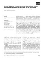
Fig. 1. Schematic model of the mechanical regulation of Cyr61 and
CTGF indicating different regulatory levels and open questions (see
Conclusions). Mediators, which have been related to mechanical
stimulation of Cyr61 or CTGF gene induction, are shown in the mid-
dle panel; details are outlined in the text.
B. Chaqour and M. Goppelt-Struebe Mechanical regulation of Cyr61 ⁄ CCN1 and CTGF ⁄ CCN2
FEBS Journal 273 (2006) 3639–3649 ª 2006 The Authors Journal compilation ª 2006 FEBS 3645
in mechanochemical transduction. J Appl Physiol 95,
2134–2141.
7 Ingber DE (2002) Mechanical signaling and the cellu-
lar response to extracellular matrix in angiogenesis
and cardiovascular physiology. Circ Res 91 , 877–
887.
8 Brigstock DR (2003) The CCN family: a new stimulus
package. J Endocrinol 178, 169–175.
9 Takigawa M (2003) CTGF ⁄ Hcs24 as a multifunctional
growth factor for fibroblasts, chondrocytes and vascular
endothelial cells. Drug News Perspect 16, 11–21.
10 Chaqour B & Goppelt-Struebe M (2005) Regulation of
CCN proteins by alterations of the cytoskeleton. In
CCN Proteins: a New Family of Cell Growth and Differ-
entiation Regulators (Perbal B & Takigawa M, eds),
pp. 177–196. Imperial College Press, London.
11 Perbal B (2004) CCN proteins: multifunctional signal-
ling regulators. Lancet 363, 62–64.
12 Brigstock DR (2002) Regulation of angiogenesis and
endothelial cell function by connective tissue growth
factor (CTGF) and cysteine-rich 61 (CYR61). Angiogen-
esis 5, 153–165.
13 Kireeva ML, Latinkic BV, Kolesnikova TV, Chen CC,
Yang GP, Abler AS & Lau LF (1997) Cyr61 and
Fisp12 are both ECM-associated signaling molecules:
activities, metabolism, and localization during develop-
ment. Exp Cell Res 233, 63–77.
14 Liang Y, Li C, Guzman VM, Evinger AJ III, Protzman
CE, Krauss AH & Woodward DF (2003) Comparison
of prostaglandin F2alpha, bimatoprost (prostamide),
and butaprost (EP2 agonist) on Cyr61 and connective
tissue growth factor gene expression. J Biol Chem 278,
27267–27277.
15 Pendurthi UR, Allen KE, Ezban M & Rao VM (2000)
Factor VIIa and thrombin induce the expression Cyr61
and connective tissue growth factor, extracellular matrix
signaling proteins that could act as possible downstream
mediators in factor VIIa tissue factor-induced signal
transduction. J Biol Chem 275, 14632–14641.
16 Tamura I, Rosenbloom J, Macarak E & Chaqour B
(2001) Regulation of Cyr61 gene expression by mechani-
cal stretch through multiple signaling pathways. Am J
Physiol Cell Physiol 281, C1524–C1532.
17 Chaqour B, Yang R & Sha Q (2006) Mechanical Stretch
Modulates the Promoter Activity of the Profibrotic Fac-
tor CCN2 Through Increased Actin Polymerization and
NF-Kappa B Activation. J Biol Chem, doi:10.1074/
jbc.M600214200.
18 Mo FE, Muntean AG, Chen CC, Stolz DB, Watkins
SC & Lau LF (2002) CYR61 (CCN1) is essential for
placental development and vascular integrity. Mol Cell
Biol 22, 8709–8720.
19 Ivkovic S, Yoon BS, Popoff SN, Safadi FF, Libuda
DE, Stephenson RC, Daluiski A & Lyons KM (2003)
Connective tissue growth factor coordinates chondro-
genesis and angiogenesis during skeletal development.
Development 130, 2779–2791.
20 Takigawa M, Nakanishi T, Kubota S & Nishida T
(2003) Role of CTGF ⁄ HCS24 ⁄ ecogenin in skeletal
growth control. J Cell Physiol 194, 256–266.
21 Wong M & Carter DR (2003) Articular cartilage func-
tional histomorphology and mechanobiology: a research
perspective. Bone 33, 1–13.
22 Wong M, Siegrist M & Goodwin K (2003) Cyclic tensile
strain and cyclic hydrostatic pressure differentially regu-
late expression of hypertrophic markers in primary
chondrocytes. Bone 33, 685–693.
23 Hishikawa K, Oemar BS & Nakaki T (2001) Static pres-
sure regulates connective tissue growth factor expression
in human mesangial cells. J Biol Chem 276, 16797–
16803.
24 Yamashiro T, Fukunaga T, Kobashi N, Kamioka H,
Nakanishi T, Takigawa M & Takano-Yamamoto T
(2001) Mechanical stimulation induces CTGF expres-
sion in rat osteocytes. J Dent Res 80, 461–465.
25 Hadjiargyrou M, Ahrens W & Rubin CT (2000) Tem-
poral expression of the chondrogenic and angiogenic
growth factor CYR61 during fracture repair. J Bone
Miner Res 15, 1014–1023.
26 Lienau J, Schell H, Epari DR, Schutze N, Jakob F,
Duda GN & Bail HJ (2005) CYR61 (CCN1) Protein
Expression during Fracture Healing in an Ovine Tibial
Model and Its Relation to the Mechanical Fixation Sta-
bility. J Orthop Res 24, 254–262.
27 Schutze N, Noth U, Schneidereit J, Hendrich C &
Jakob F (2005) Differential expression of CCN-family
members in primary human bone marrow-derived
mesenchymal stem cells during osteogenic, chondrogenic
and adipogenic differentiation. Cell Commun Signal 3,
doi:10.1186/1478-811X-3-5.
28 O’Brien TP & Lau LF (1992) Expression of the growth
factor-inducible immediate early gene cyr61 correlates
with chondrogenesis during mouse embryonic develop-
ment. Cell Growth Differ 3, 645–654.
29 Chen CC, Mo FE & Lau LF (2001) The angiogenic fac-
tor Cyr61 activates a genetic program for wound heal-
ing in human skin fibroblasts. J Biol Chem 276, 47329–
47337.
30 Leask A, Denton CP & Abraham DJ (2004) Insights
into the molecular mechanism of chronic fibrosis: the
role of connective tissue growth factor in scleroderma.
J Invest Dermatol 122, 1–6.
31 Igarashi A, Nashiro K, Kikuchi K, Sato S, Ihn H,
Fujimoto M, Grotendorst GR & Takehara K (1996)
Connective tissue growth factor gene expression in tis-
sue sections from localized scleroderma, keloid, and
other fibrotic skin disorders. J Invest Dermatol 106,
729–733.
32 Ito Y, Aten J, Bende RJ, Oemar BS, Rabelink TJ,
Weening JJ & Goldschmeding R (1998) Expression of
Mechanical regulation of Cyr61 ⁄ CCN1 and CTGF ⁄ CCN2 B. Chaqour and M. Goppelt-Struebe
3646 FEBS Journal 273 (2006) 3639–3649 ª 2006 The Authors Journal compilation ª 2006 FEBS
connective tissue growth factor in human renal fibrosis.
Kidney Int 53, 853–861.
33 Leask A, Holmes A, Black CM & Abraham DJ (2003)
Connective tissue growth factor gene regulation.
Requirements for its induction by transforming growth
factor-beta 2 in fibroblasts. J Biol Chem 278 , 13008–
13015.
34 Holmes A, Abraham DJ, Chen Y, Denton C, Shi-wen
X, Black CM & Leask A (2003) Constitutive connective
tissue growth factor expression in scleroderma fibro-
blasts is dependent on Sp1. J Biol Chem 278, 41728–
41733.
35 Vozenin-Brotons MC, Milliat F, Sabourin JC, de Gou-
ville AC, Francois A, Lasser P, Morice P, Haie-Meder
C, Lusinchi A, Antoun S, Bourhis J, Mathe D, Girinsky
T & Aigueperse J (2003) Fibrogenic signals in patients
with radiation enteritis are associated with increased
connective tissue growth factor expression. Int J Radiat
Oncol Biol Phys 56, 561–572.
36 Grinnell F (2003) Fibroblast biology in three-dimen-
sional collagen matrices. Trends Cell Biol 13, 264–269.
37 Schild C & Trueb B (2004) Three members of the con-
nective tissue growth factor family CCN are differen-
tially regulated by mechanical stress. Biochim Biophys
Acta 1691, 33–40.
38 Schild C & Trueb B (2002) Mechanical stress is required
for high-level expression of connective tissue growth
factor. Exp Cell Res 274, 83–91.
39 Graness A, Cicha I & Goppelt-Struebe M (2006) Con-
tribution of Src-FAK signaling to the induction of con-
nective tissue growth factor in renal fibroblasts. Kidney
Int 68, 1341–1349.
40 Arora PD, Narani N & McCulloch CA (1999) The
compliance of collagen gels regulates transforming
growth factor-beta induction of alpha-smooth muscle
actin in fibroblasts. Am J Pathol 154, 871–882.
41 Garrett Q, Khaw PT, Blalock TD, Schultz GS, Groten-
dorst GR & Daniels JT (2004) Involvement of CTGF in
TGF-beta1-stimulation of myofibroblast differentiation
and collagen matrix contraction in the presence of
mechanical stress. Invest Ophthalmol Vis Sci 45, 1109–
1116.
42 Nerem RM (1992) Vascular fluid mechanics, the arterial
wall, and atherosclerosis. J Biomech Eng 114, 274–282.
43 Cunningham KS & Gotlieb AI (2005) The role of shear
stress in the pathogenesis of atherosclerosis. Lab Invest
85, 9–23.
44 Oemar BS, Werner A, Garnier JM, Do DD, Godoy N,
Nauck M, Marz W, Rupp J, Pech M & Luscher TF
(1997) Human connective tissue growth factor is
expressed in advanced atherosclerotic lesions. Circula-
tion 95, 831–839.
45 Schober JM, Chen N, Grzeszkiewicz TM, Jovanovic I,
Emeson EE, Ugarova TP, Ye RD, Lau LF & Lam SC
(2002) Identification of integrin alpha(M) beta(2) as an
adhesion receptor on peripheral blood monocytes for
Cyr61 (CCN1) and connective tissue growth factor
(CCN2): immediate-early gene products expressed in
atherosclerotic lesions. J Lipid Mediat Cell Signal 99,
4457–4465.
46 Cicha I, Yilmaz A, Klein M, Raithel D, Brigstock DR,
Daniel WG, Goppelt-Struebe M & Garlichs CD (2005)
Connective tissue growth factor is overexpressed in
complicated atherosclerotic plaques and induces mono-
nuclear cell chemotaxis in vitro. Arterioscler Thromb
Vasc Biol 25, 1008–1013.
47 Panutsopulos D, Arvanitis DL, Tsatsanis C, Papalam-
bros E, Sigala F & Spandidos DA (2005) Expression
of heregulin in human coronary atherosclerotic lesions.
J Vasc Res 42, 463–474.
48 Yoshisue H, Suzuki K, Kawabata A, Ohya T, Zhao H,
Sakurada K, Taba Y, Sasaguri T, Sakai N, Yamashita
S, Matsuzawa Y & Nojima H (2002) Large scale isola-
tion of non-uniform shear stress-responsive genes from
cultured human endothelial cells through the prepara-
tion of a subtracted cDNA library. Atherosclerosis 162,
323–334.
49 McCormick SM, Eskin SG, McIntire LV, Teng CL, Lu
CM, Russell CG & Chittur KK (2001) DNA microar-
ray reveals changes in gene expression of shear stressed
human umbilical vein endothelial cells. Proc Natl Acad
Sci USA 98, 8955–8960.
50 McCormick SM, Frye SR, Eskin SG, Teng CL, Lu
CM, Russell CG, Chittur KK & McIntire LV (2003)
Microarray analysis of shear stressed endothelial cells.
Biorheology 40, 5–11.
51 Eskin SG, Turner NA & McIntire LV (2004) Endothe-
lial cell cytochrome P450 1A1 and 1B1: up-regulation
by shear stress. Endothelium 11, 1–10.
52 Garcia-Cardena G, Comander J, Anderson KR, Black-
man BR & Gimbrone MA Jr (2001) Biomechanical acti-
vation of vascular endothelium as a determinant of its
functional phenotype. Proc Natl Acad Sci USA 98,
4478–4485.
53 Lee YS, Byun J, Kim JA, Lee JS, Kim KL, Suh YL,
Kim JM, Jang HS, Lee JY, Shin IS, Suh W, Jeon ES &
Kim DK (2005) Monocrotaline-induced pulmonary
hypertension correlates with upregulation of connective
tissue growth factor expression in the lung. Exp Mol
Med 37, 27–35.
54 Zhou D, Herrick DJ, Rosenbloom J & Chaqour B
(2005) Cyr61 mediates the expression of VEGF, alphav-
integrin, and alpha-actin genes through cytoskeletally
based mechanotransduction mechanisms in bladder
smooth muscle cells. J Appl Physiol 98, 2344–2354.
55 Unoki H, Furukawa K, Yonekura H, Ueda Y, Katsuda
S, Mori M, Nakagawara K, Mabuchi H & Yamamoto
H (2003) Up-regulation of cyr61 in vascular smooth
muscle cells of spontaneously hypertensive rats. Lab
Invest 83, 973–982.
B. Chaqour and M. Goppelt-Struebe Mechanical regulation of Cyr61 ⁄ CCN1 and CTGF ⁄ CCN2
FEBS Journal 273 (2006) 3639–3649 ª 2006 The Authors Journal compilation ª 2006 FEBS 3647
56 Goldschmeding R, Aten J, Ito Y, Blom I, Rabelink T &
Weening JJ (2000) Connective tissue growth factor: just
another factor in renal fibrosis? Nephrol Dial Transplant
15, 296–299.
57 Abdel WN & Mason RM (2004) Connective tissue
growth factor and renal diseases: some answers, more
questions. Curr Opin Nephrol Hypertens 13, 53–58.
58 Riser BL & Cortes P (2001) Connective tissue growth
factor and its regulation: a new element in diabetic glo-
merulosclerosis. Ren Fail 23, 459–470.
59 Peng H, Carretero OA, Brigstock DR, Oja-Tebbe N &
Rhaleb NE (2003) Ac-SDKP reverses cardiac fibrosis in
rats with renovascular hypertension. Hypertension 42 ,
1164–1170.
60 Cortes P, Riser BL, Yee J & Narins RG (1999)
Mechanical strain of glomerular mesangial cells in the
pathogenesis of glomerulosclerosis: clinical implications.
Nephrol Dial Transplant 14, 1351–1354.
61 Ingram AJ & Scholey JW (2000) Stress-responsive signal
transduction mechanisms in glomerular cells. Curr Opin
Nephrol Hypertens 9, 49–55.
62 Wahab NA, Yevdokimova N, Weston BS, Roberts T,
Li XJ, Brinkman H & Mason RM (2001) Role of con-
nective tissue growth factor in the pathogenesis of dia-
betic nephropathy. Biochem J 359, 77–87.
63 Zatz R, Meyer TW, Rennke HG & Brenner BM (1985)
Predominance of hemodynamic rather than metabolic
factors in the pathogenesis of diabetic glomerulopathy.
Proc Natl Acad Sci USA 82, 5963–5967.
64 Sawai K, Mori K, Mukoyama M, Sugawara A, Suga-
nami T, Koshikawa M, Yahata K, Makino H, Nagae
T, Fujinaga Y, Yokoi H, Yoshioka T, Yoshimoto A,
Tanaka I & Nakao K (2003) Angiogenic protein Cyr61
is expressed by podocytes in anti-Thy-1 glomerulone-
phritis. J Am Soc Nephrol 14, 1154–1163.
65 Roestenberg P, van Nieuwenhoven FA, Joles JA, Tris-
chberger C, Martens PP, Oliver N, Aten J, Hoppener
JW & Goldschmeding R (2006) Temporal expression
profile and distribution pattern indicate a role of con-
nective tissue growth factor (CTGF ⁄ CCN-2) in diabetic
nephropathy in mice. Am J Physiol Renal Physiol 290,
F1344–F1354.
66 Katz BZ, Zamir E, Bershadsky A, Kam Z, Yamada
KM & Geiger B (2000) Physical state of the extracellu-
lar matrix regulates the structure and molecular compo-
sition of cell-matrix adhesions. Mol Biol Cell 11, 1047–
1060.
67 Li C & Xu Q (2000) Mechanical stress-initiated signal
transductions in vascular smooth muscle cells. Cell Sig-
nal 12, 435–445.
68 Attwell S, Mills J, Troussard A, Wu C & Dedhar S
(2003) Integration of cell attachment, cytoskeletal locali-
zation, and signaling by integrin-linked kinase (ILK),
CH-ILKBP, and the tumor suppressor PTEN. Mol Biol
Cell 14, 4813–4825.
69 Balaban NQ, Schwarz US, Riveline D, Goichberg P,
Tzur G, Sabanay I, Mahalu D, Safran S, Bershadsky A,
Addadi L & Geiger B (2001) Force and focal adhesion
assembly: a close relationship studied using elastic
micropatterned substrates. Nat Cell Biol 3, 466–472.
70 Ridley AJ (2001) Rho family proteins: coordinating cell
responses. Trends Cell Biol 11, 471–477.
71 Han JS, Macarak E, Rosenbloom J, Chung KC & Cha-
qour B (2003) Regulation of Cyr61 ⁄ CCN1 gene expres-
sion through RhoA GTPase and p38MAPK signaling
pathways. Eur J Biochem 270, 3408–3421.
72 Miralles F, Posern G, Zaromytidou A & Treisman R
(2003) Actin dynamics control SRF activity by regula-
tion of its coactivator MAL. Cell 113, 329–342.
73 Resnick N, Collins T, Atkinson W, Bonthron DT,
Dewey CF Jr & Gimbron MA Jr (1993) Platelet-derived
growth factor B chain promoter contains a cis-acting
fluid shear-stress-responsive element. Proc Natl Acad Sci
USA 90, 4591–4595.
74 Grote K, Bavendiek U, Grothusen C, Flach I, Hilfiker-
Kleiner D, Drexler H & Schieffer B (2004) Stretch-indu-
cible expression of the angiogenic factor CCN1 in vas-
cular smooth muscle cells is mediated by Egr-1. J Biol
Chem 279, 55675–55681.
75 Hahn A, Heusinger-Ribeiro J, Lanz T, Zenkel S &
Goppelt-Struebe M (2000) Induction of connective tis-
sue growth factor by activation of heptahelical recep-
tors. Modulation by rho proteins and the actin
cytoskeleton. J Biol Chem 275, 37429–37435.
76 Ott C, Iwanciw D, Graness A, Giehl K & Goppelt-
Struebe M (2003) Modulation of the expression of con-
nective tissue growth factor by alterations of the cytos-
keleton. J Biol Chem 278, 44305–44311.
77 Goppelt-Struebe M, Hahn A, Iwanciw D, Rehm M &
Banas B (2001) Regulation of connective tissue growth
factor (ccn2; ctgf) gene expression in human mesangial
cells: modulation by HMG CoA reductase inhibitors
(statins). Mol Pathol 54, 176–179.
78 Iwanciw D, Rehm M, Porst M & Goppelt-Struebe M
(2003) Induction of connective tissue growth factor by
angiotensin II: integration of signaling pathways. Arter-
ioscler Thromb Vasc Biol 23, 1782–1787.
79 Eberlein M, Heusinger-Ribeiro J & Goppelt-Struebe M
(2001) Rho-dependent inhibition of the induction of
connective tissue growth factor (CTGF) by HMG CoA
reductase inhibitors (statins). Br J Pharmacol 133, 1172–
1180.
80 Muehlich S, Schneider N, Hinkmann F, Garlichs CD
& Goppelt-Struebe M (2004) Induction of
connective tissue growth factor (CTGF) in human
endothelial cells by lysophosphatidic acid, sphingosine-
1-phosphate, and platelets. Atherosclerosis 175, 261–
268.
81 Watts KL & Spiteri MA (2004) Connective Tissue
Growth Factor expression and induction by Transform-
Mechanical regulation of Cyr61 ⁄ CCN1 and CTGF ⁄ CCN2 B. Chaqour and M. Goppelt-Struebe
3648 FEBS Journal 273 (2006) 3639–3649 ª 2006 The Authors Journal compilation ª 2006 FEBS
ing Growth Factor {beta}, is abrogated by Simvastatin
via a Rho signalling mechanism. Am J Physiol Lung
Cell Mol Physiol 287, L1323–L1332.
82 Song Y, Li C & Cai L (2004) Fluvastatin prevents
nephropathy likely through suppression of connective
tissue growth factor-mediated extracellular matrix accu-
mulation. Exp Mol Pathol 76, 66–75.
83 Riento K & Ridley AJ (2003) Rocks: multifunctional
kinases in cell behaviour. Nat Rev Mol Cell Biol 4, 446–
456.
84 Cortes P, Mendez M, Riser BL, Guerin CJ, Rodriguez-
Barbero A, Hassett C & Yee J (2000) F-actin fiber dis-
tribution in glomerular cells: structural and functional
implications. Kidney Int 58, 2452–2461.
85 Goppelt-Struebe M, Heusinger-Ribeiro J & Fischer B
(2002) Dual role of cytochalasin D in the regulation of
connective tissue growth factor. Ann N Y Acad Sci 973,
57–59.
86 Cipolla MJ, Gokina NI & Osol G (2002) Pressure-
induced actin polymerization in vascular smooth muscle
as a mechanism underlying myogenic behavior. FASEB
J 16, 72–76.
87 Sotiropoulos A, Gineitis D, Copeland J & Treisman R
(1999) Signal-regulated activation of serum response
factor is mediated by changes in actin dynamics. Cell
98, 159–169.
88 Posern G, Sotiropoulos A & Treisman R (2002) Mutant
actins demonstrate a role for unpolymerized actin in
control of transcription by serum response factor. Mol
Biol Cell 13, 4167–4178.
89 Gettemans J, Van Impe K, Delanote V, Hubert T, De
Vandekerckhove J & V (2005) Nuclear actin-binding
proteins as modulators of gene transcription. Traffic 6,
847–857.
90 Bourgier C, Haydont V, Milliat F, Francois A, Holler V,
Lasser P, Bourhis J, Mathe D & Vozenin-Brotons MC
(2005) Inhibition of Rho kinase modulates radiation
induced fibrogenic phenotype in intestinal smooth muscle
cells through alteration of the cytoskeleton and connec-
tive tissue growth factor expression. Gut 54, 336–343.
91 Abreu JG, Ketpura NI, Reversade B & De Robertis
EM (2002) Connective-tissue growth factor (CTGF)
modulates cell signalling by BMP and TGF-beta. Nat
Cell Biol 4, 599–604.
92 Inoki I, Shiomi T, Hashimoto G, Enomoto H,
Nakamura H, Makino K, Ikeda E, Takata S,
Kobayashi K & Okada Y (2002) Connective tissue
growth factor binds vascular endothelial growth factor
(VEGF) and inhibits VEGF-induced angiogenesis.
FASEB J 16, 219–221.
93 Crean JK, Furlong F, Finlay D, Mitchell D, Conway B,
Brady HR, Godson C & Martin F (2004) Connective
tissue growth factor [CTGF] ⁄ CCN2 stimulates mesangial
cell migration through integrated dissolution of focal
adhesion complexes and activation of cell polarization.
FASEB J 18, 1541–1543.
B. Chaqour and M. Goppelt-Struebe Mechanical regulation of Cyr61 ⁄ CCN1 and CTGF ⁄ CCN2
FEBS Journal 273 (2006) 3639–3649 ª 2006 The Authors Journal compilation ª 2006 FEBS 3649