Báo cáo khoa học: Probing protein–chromophore interactions in Cph1 phytochrome by mutagenesis potx
Bạn đang xem bản rút gọn của tài liệu. Xem và tải ngay bản đầy đủ của tài liệu tại đây (869.06 KB, 15 trang )
Probing protein–chromophore interactions in Cph1
phytochrome by mutagenesis
Janina Hahn
1
, Holger M. Strauss
1
, Frank T. Landgraf
2
, Hortensia Faus Gimene
`
z
2
,Gu
¨
nter Lochnit
3
,
Peter Schmieder
1
and Jon Hughes
2
1 Forschungsinstitut fu
¨
r Molekulare Pharmakologie, Berlin, Germany
2 Pflanzenphysiologie, Fachbereich Biologie & Chemie, Justus-Liebig-Universita
¨
t, Giessen, Germany
3 Biochemisches Institut, Fachbereich Medizin, Justus-Liebig-Universita
¨
t, Giessen, Germany
Phytochrome photoreceptors play a central role in the
regulation of plant development. Phytochromes are
red ⁄ far-red photochromic proteins with a covalently
bound linear tetrapyrrole (bilin) prosthetic group. In
the Pr ground state the chromophore preferentially
absorbs red light, this leading to a Z fi E isomeriza-
tion around the C
15
–C
16
double bond between rings C
and D. Further conformational changes culminate in
the formation of Pfr, the signalling state. This prefer-
ably absorbs far-red light which converts the pigment
back to Pr [1]. The active photoreceptor is formed by
the apoprotein taking up and covalently attaching an
Keywords
biliprotein; photoreceptor; phytochrome; site
directed mutagenesis; structure–function
studies
Correspondence
J. Hahn, Forschungsinstitut fu
¨
r Molekulare
Pharmakologie, Robert Ro
¨
ssle Str. 10,
D-13125 Berlin, Germany
Fax: +49 30 94793169
Tel: +49 30 94793316
E-mail:
(Received 28 October 2005, revised 27
January 2006, accepted 3 February 2006)
doi:10.1111/j.1742-4658.2006.05164.x
We have investigated mutants of phytochrome Cph1 from the cyanobacter-
ium Synechocystis PCC6803 in order to study chromophore–protein inter-
actions. Cph1D2, the 514-residue N-terminal sensor module produced as a
recombinant His6-tagged apoprotein in Escherichia coli, autoassembles
in vitro to form a holoprotein photochemically indistinguishable from the
full-length product. We generated 12 site-directed mutants of Cph1D2,
focusing on conserved residues which might be involved in chromophore–
protein autoassembly and photoconversion. Folding, phycocyanobilin-bind-
ing and Pr fi Pfr photoconversion were analysed using CD and UV–visible
spectroscopy. MALDI-TOF-MS confirmed C259 as the chromophore
attachment site. C259L is unable to attach the chromophore covalently but
still autoassembles to form a red-shifted photochromic holoprotein. H260Q
shows UV–visible properties similar to the wild-type at pH 7.0 but both Pr
and Pfr (reversibly) bleach at pH 9.0, indicating that the imidazole side
chain buffers chromophore protonation. Mutations at E189 disturbed fold-
ing but the residue is not essential for chromophore–protein autoassembly.
In D207A, whereas red irradiation of the ground state leads to bleaching
of the red Pr band as in the wild-type, a Pfr-like peak does not arise, impli-
cating D207 as a proton donor for a deprotonated intermediate prior to
Pfr. UV-Vis spectra of both H260Q under alkaline conditions and D207A
point to a particular significance of protonation in the Pfr state, possibly
implying proton migration (release and re-uptake) during Pr fi Pfr photo-
conversion. The findings are discussed in relation to the recently published
3D structure of a bacteriophytochrome fragment [Wagner JR, Brunzelle
JS, Forest KT & Vierstra RD (2005) Nature 438, 325–331].
Abbreviations
BV, biliverdin IXa; Cph1D2, the N-terminal 1–514 residue sensory module of Cph1 from Synechocystis PCC6803; e, extinction coefficient;
FTRR, Fourier transform resonance Raman spectroscopy; FWHM, full width half maximum; IPTG, isopropyl thio-b-
D-galactoside; LED, light-
emitting diode; MeOH, methanol; Pr ⁄ Pfr, red ⁄ far-red absorbing form of phytochrome; PCB, phycocyanobilin; PFB, phytochromobilin; SAR,
specific absorbance ratio; SEC, size-exclusion chromatography; k
max
, wavelength of the absorbance maximum.
FEBS Journal 273 (2006) 1415–1429 ª 2006 The Authors Journal compilation ª 2006 FEBS 1415
appropriate bilin from the cytoplasm: this process is
called autoassembly [2]. Phytochromes are exceedingly
effective photoreceptors on account of their high
extinction coefficients in the red⁄ far-red region, low
fluorescence losses, high resistance to photobleaching
and use of a thermodynamically stable signalling state
to activate their response pathway. The molecular pro-
cesses underlying autoassembly, hyper- and photochro-
micity and signal transduction are thus of considerable
interest.
The unexpected discovery of a prokaryotic phyto-
chrome, Cph1 [3,4], fundamentally changed our view
of evolution and function of this class of photorecep-
tors, relating them to histidine sensor kinases, a pro-
tein family involved in a wide variety of perception
systems in prokaryotes, fungi and plants [5]. Cph1 has
numerous features in common with plant phyto-
chromes. Furthermore, large amounts of pure, highly
concentrated holoCph1 can easily be produced by apo-
protein overexpression in Escherichia coli and in vitro
autoassembly with an appropriate bilin [6]. HoloCph1
can also be produced in E. coli by coexpressing haem
oxygenase and appropriate bilin reductase genes
together with Cph1 [7,8]. Cph1 is thereby well suited
to studies of autoassembly as well as of the photocon-
version mechanism. Numerous related photoreceptors
have subsequently been identified in prokaryotes, nota-
bly bacteriophytochrome from Deinococcus radiodu-
rans, DrBphP, the 3D structure of whose N-terminal
domain was recently published [9].
Phytochrome sequences show highly conserved
regions probably representing functionally essential
subdomains [6,10]. The UV-Vis absorbance and vibra-
tional spectroscopic characteristics of phytochromes
assembled with the same chromophore are remarkably
similar, whereas significant and characteristic changes
are associated with subtle changes in the bilin pros-
thetic group. It was thus expected that the pocket in
which the chromophore is held is constructed from
various functionally conserved subdomains reflected at
the sequence level in all phytochromes. The new X-ray
structure [9] indeed bears this out although it must be
born in mind that DrBphP differs functionally from
plant-like phytochromes in many respects and that the
fragment crystallized is photochemically impotent.
In oat phytochrome A the phytochromobilin (PFB)
chromophore is attached by a thioether link to C322
#380
[11–13], a residue conserved in plant-type phytochromes
including Cph1 but not in bacteriophytochromes (the
residue number is that of the named phytochrome,
#
indicating its position in the alignment at www.
uni-giessen.de/gf1251/Phytochrome/align2x.htm). Phyco-
cyanobilin (PCB) is probably the native Cph1
chromophore [14], but no direct evidence for its expec-
ted attachment at C259
#380
has been published [5]. A
substitution at this putative ligation site should abolish
covalent attachment, but not necessarily other protein–
bilin interactions, as studies with blocking reagents and
of autoassembly kinetics have implied [15–17]. In free
PCB at neutral pH 7 the two central ring nitrogens
share a single proton, but a second is added under acid
conditions. Protonation occurs during phytochrome
autoassembly too, but the donor is unknown. Con-
versely, homology studies implied that a basic residue
homologous to R254
#375
close to the presumed chromo-
phore attachment site interacts with the propionate side
chain of chromophore ring B [18,19]. Additionally, the
strength and position of the dominant red and far-red
absorbance bands of Pr and Pfr, respectively, are
pH-dependent, an H residue near the chromophore
being implicated [20]. H260
#381
adjacent to the putative
ligation site is perfectly conserved and hence a prime
candidate for this function.
Such conserved interactions probably central to
phytochrome action can be probed by modifying the
protein moiety via site-directed mutagenesis of the cog-
nate gene [21–25], with the important proviso that,
except in the case of null phenotypes, all conclusions
based purely on site-directed mutagenesis are confoun-
ded by unknown possible side-effects on folding. Ide-
ally, the mutations are guided by 3D structural data.
Such information for phytochrome [9] were not avail-
able at the time of this study.
The N-terminal 514 residue sensory module of
recombinant Cph1 ) that is, Cph1D2 ) is photochemi-
cally autonomous. We generated 12 amino acid
replacement mutants in Cph1D2 and analysed their
expression, autoassembly, UV-Vis absorbance, photo-
chromicity and thermal reversion properties. We also
used CD spectroscopy to detect gross changes in sec-
ondary structure: only correctly folded products were
considered to offer interpretable information. We pro-
vide the first direct evidence that the PCB chromo-
phore is ligated to C259
#380
, analogously to oat phyA,
and also describe the effects of mutations at this resi-
due. The H260
#381
Q mutant showed massive, reversible
pH effects on the absorbance spectra, obliterating the
characteristic Pfr peak, implying that the imidazole
side chain buffers chromophore protonation, partic-
ularly in the case of Pfr. A perhaps related effect was
seen for the conserved acidic residue D207
#328
: when
this was replaced by A, although Pr (reversibly) photo-
bleached, no Pfr-like peak was formed in its place.
Mutations of R254
#375
had similar small effects on
UV-Vis properties: that the R residue is nevertheless
perfectly conserved implies a role in signal transduc-
Protein–chromophore interactions in Cph1 J. Hahn et al.
1416 FEBS Journal 273 (2006) 1415–1429 ª 2006 The Authors Journal compilation ª 2006 FEBS
tion. We also show that E189
#130
is not required for
covalent autoassembly, as had been proposed. We dis-
cuss these findings in relation to phytochrome function
and the bacteriophytochrome X-ray structure [9] which
has subsequently become available.
Results
Characterization of Cph1 D1 and D2
At the start of this work two deletion clones with
C-terminal His6-tags were created, Cph1D1 and
Cph1D2, in which the N-terminal 492 and 514 resi-
dues, respectively, of Cph1 were overproduced as apo-
proteins in E. coli , autoassembled with and purified
by nickel affinity chromatography. Although most
Cph1D1 was expressed as insoluble inclusion bodies
so that the final yield of soluble apoprotein was only
500 lgÆL
)1
culture, addition of PCB resulted in
covalent autoassembly (as apparent from Zn
2+
-
induced bilin fluorescence in SDS ⁄ PAGE) and red ⁄
far-red photochromicity. The Pfr-like band was weak
and significantly blue-shifted, however, in comparison
to full-length Cph1 (Fig. 1); an effect was also seen
in a similar deletion mutant [26]. In contrast,
Cph1D2 yielded up to 80 mg apoproteinÆL
)1
culture
and showed a difference spectrum almost identical to
that of full-length Cph1 (Fig. 1), confirming the
results of Yeh et al. [4]. Coomassie-stained SDS ⁄
PAGE indicated a purity of 80% for Cph1D2at
this stage. Further purification via Superose 200
(Amersham Pharmacia ⁄ GE) size-exclusion chromato-
graphy (SEC) yielded essentially pure holoprotein.
Cph1D2, unlike full-length Cph1, shows no tendency
to aggregate in vitro (data not shown).
The extinction coefficient of Cph1D2 Pr was
82 mm
)1
Æcm
)1
at 654 nm (kmax), a value of
85 mm
)1
Æcm
)1
for full-length Cph1 confirming that
earlier reported [16]. Further UV-Vis data are summar-
ized in Table 1. Extinction coefficients for free PCB
were 16, 20, 29, 30 and 46 mm
)1
Æcm
)1
at each kmax in
Tris ⁄ HCl pH 7.8, MES pH 5.5, sodium acetate
pH 3.0, 0.5 m HCl pH 0.3 and CH
3
Cl ⁄ HCl (1 : 19),
respectively. The relevant UV-Vis spectra are shown in
Fig. 2. The maximal 654 nm ⁄ 280 nm specific absorb-
ance ratio (SAR) of Cph1D2 Pr obtained was 1.3, sig-
nificantly higher than that for full-length Cph1 at
equivalent purity (1.0 [16]). e
280 nm
was calculated to
be 59 and 83 mm
)1
Æcm
)1
for Cph1D2 and full-length
Cph1 apoproteins, respectively (Vector NTI, Infor-
max). The contribution of PCB attached to the holo-
protein is about 5 mm
-1
Æcm
-1
[16], yielding 64 and
88 mm
)1
Æcm
)1
at 280 nm for the holoproteins. Taking
the Pr e
kmax
in the red region from Table 1, the max-
imal SAR would be 1.28 and 0.98, respectively, in
close agreement with that of our purest samples.
Quantum efficiencies of photoconversion were not
measured directly, but kinetics under red and far-red
irradiation were similar for Cph1D2 and full-length
Cph1 holoproteins. As the e-values are similar, we thus
expect quantum efficiencies to be similar too, that is
0.16 in each direction [6,20]. A maximal 0.70 mole
fraction of Pfr at photoequilibrium in red light is seen in
Cph1D2 as in full-length Cph1: the calculated UV-Vis
spectrum for 100% Pfr derived from this is identical to
that of purified Cph1D2 Pfr (unlike full-length Cph1,
Cph1D2 as Pr is monomeric except at very high
(> 10 mm) concentrations; the Pfr form, however,
homodimerises, readily allowing it to be purified by
SEC [27]). Dark reversion is insignificant: none was
detected after 2 weeks at 20 °C (data not shown).
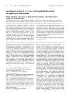
λ [nm]
∆A
500 600 700 800
-1.0
-0.5
0.0
0.5
1.0
Fig. 1. UV-Vis difference spectra of full-length Cph1 (n) and deletion
mutants Cph1D1(d) and Cph1D2(m).
Table 1. Summary of UV-Vis absorbance data for full-length Cph1
and the deletion mutant Cph1D2. ND, not determined; ibp, isosbe-
stic point.
Parameter
Cph1 (full-length) Cph1D2 (N514D)
Pr Pfr Pr Pfr
k
max, red
656 nm 704 nm 654 nm 702 nm
k
max, UV ⁄ A
359 nm ND 358 nm ND
e at k
max, red
86 mM
)1
Æcm
)1
ND 82 mM
)1
Æcm
)1
ND
k
DA,max
655 nm 707 nm 655 nm 707 nm
k
DA,0
, ibp 677 nm 677 nm
SAR (A
655
⁄ A
280
) 1.0 1.3
Molecular mass
(Apoprotein
+ His tag)
85.3 kDa 58.7 kDa
J. Hahn et al. Protein–chromophore interactions in Cph1
FEBS Journal 273 (2006) 1415–1429 ª 2006 The Authors Journal compilation ª 2006 FEBS 1417
Direct determination of the chromophore
attachment site
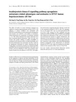
Tryptic fragments of PCB-Cph1D2 holoprotein were
separated by HPLC, the chromopeptide eluting as
a single peak with a spectrum closely fitting that expec-
ted for protonated PCB covalently linked via ring A to
the peptide (Fig. 3A). The chromopeptide showed weak
214 nm absorbance, implying a poor release efficiency.
MALDI-TOF MS of this fraction showed major peaks
([M + H]
+
) with a typical isotopic profile correspond-
ing to predicted tryptic fragments 98–112 (m ⁄ z 1753.849
and in the methionine oxidized form at m ⁄ z 1769.851),
64–80 (m ⁄ z 1951.986) and 399–420 (m ⁄ z 2534.271) of
Cph1 (Fig. 3B). The expected PCB-coupled chromopep-
tide SAYHC*HLTYLK (residues 255–279) is predicted
to have a molecular mass of 1920.923 Da. A small but
distinct double peak corresponding to the expected
[M + H]
+
at m ⁄ z 1921.9384 and to [M–H]
+
at m ⁄ z
1919.929 was seen, the latter probably presenting an
oxidized derivative (a similar effect was seen in a study
of Agp1 where the Biliverdin IXa (BV) chromopeptide
ion detected was also 2 Da lighter than expected [28]).
MS
2
analysis of the double peak showed fragment ions
at m ⁄ z 585.991 ⁄ 587.939 (reflecting the expected and
A
B
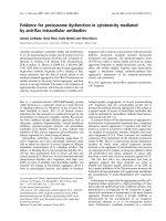
Fig. 2. pH dependence of PCB UV-Vis absorption spectra. (A)
Absorption spectra of free PCB in different buffers at different
pH-values. Spectra are plotted for PCB in 100 m
M Tris ⁄ HCl pH 7.7
(n), 10 m
M MES pH 5.5 (d), 5 mM sodium acetate pH 3.0 (m),
0.5
M HCl pH 0.3 (.), HCl ⁄ MeOH (1 : 19) (e) and CH
3
Cl ⁄ HCl
(1 : 19) (n). For comparison the absorption spectrum of Cph1D2in
the Pr state is shown (dotted line). (B) pH difference spectra for
free PCB. Absorbance changes are plotted for pH values 5.5, 3.0,
0.3 (solid lines) and HCl ⁄ MeOH (dashed line) after subtraction of
the pH 7.7 spectrum.
A
B
C
Fig. 3. Tryptic profiles and MALDI spectra of Cph1D2. (A) HPLC
elution profiles at 214 nm (peptide absorbance, upper panel) and
370 nm (bilin UV ⁄ A absorbance, lower panel); inset: UV-Vis-spec-
trum of chromopeptide peak. (B) MALDI-TOF spectrum of chromo-
peptide fraction; inset: enlarged. (C) MALDI-TOF ⁄ TOF spectrum;
inset: enlarged.
Protein–chromophore interactions in Cph1 J. Hahn et al.
1418 FEBS Journal 273 (2006) 1415–1429 ª 2006 The Authors Journal compilation ª 2006 FEBS
oxidized forms of the cleaved chromophore) and
1336.915 (reflecting the peptide backbone) (unlike full-
length Cph1, Cph1D2 as Pr is monomeric except at very
high (> 10 mm) concentrations; the Pfr form, however,
homodimerizes readily allowing it to be purified by SEC
[28]). Edman microsequence data (not shown) from the
same fraction were consistent with the sequences of
the fragments identified in MALDI: the fifth residue of
the chromopeptide ) C259 ) was absent, as would be
expected for a cysteinyl–PCB complex. Taken together
these data show that the PCB chromophore is ligated to
C259 via a thioether bond.
Characterization of Cph1 D2 site-directed mutants
To determine the role of specific conserved residues in
the Synechocystis phytochrome Cph1, 12 site-directed
mutations were introduced into the N-terminal sensory
module Cph1D2. The mutants were heterologously
expressed as C-terminally His6-tagged apoproteins in
E. coli, purified and tested for PCB-binding, apopro-
tein folding, Pr–Pfr photochromicity and thermal
reversion using SDS ⁄ PAGE ⁄ zinc fluorescence and CD
and UV-Vis spectroscopy. The appropriate data is
summarized in Table 2 and in Figs 4 and 5.
Y257
#378
, h258
#379
, l261
#382
These residues lie close to the chromophore binding
site but are not conserved in other phytochromes and
are thus, in contrast to conserved residues, probably
not functionally important. Indeed, the Y257H,
H258F and L261A holoproteins showed no significant
differences in chromophore autoassembly, UV-Vis or
CD properties relative to the wild-type (Table 2).
C259
#380
As MALDI studies showed, this is the residue in Cph1
to which PCB becomes attached via a thioether bond.
Thus mutations at C259 should abolish covalent attach-
ment and have dramatic effects on photochemistry.
Both C259M and C259L mutants autoassembled with
PCB to give red ⁄ far-red photochromic holoproteins
although, as expected, covalent attachment did not
occur (Figs 4 and 6). The autoassembly reaction was
much slower than in the wild-type especially under
nonreducing conditions, taking many hours for
chromophore binding and photochromicity to become
saturated even with a large PCB molar excess (as seen
in [29]). After brief incubation of apoprotein with a
small molar excess of PCB under nonreducing condi-
tions, holoC259L showed an almost symmetrical differ-
ence spectrum following red irradiation, with lowest
energy bands at 674 and 735 nm, representing a
25 nm bathochromic shift relative to the wild-type.
Subsequent irradiation with FR did not repopulate the
Pr-like species, however (Fig. 6A,B). HoloC259L
allowed to assemble to completion under reducing con-
ditions showed similarly shifted lowest energy bands.
Table 2. Characterization of Cph1D1, Cph1D2 and Cph1D2-mutants. ND, not determined; ibp, isosbestic point.
Mutation
Soluble expression
relative to Cph1D2
a
CD like wild-type
Cph1D2
Covalent PCB
attachment
Difference spectrum Absorption spectrum
k
max
(Pr)
[nm]
k
DA,0 ibp
[nm]
k
max
(Pfr)
[nm]
k
max
(Pr)
[nm]
k
max
(Pfr)
[nm]
Cph1D1 + ND Yes 649 676 695 ND ND
Cph1D2 + + + Yes Yes 655 677 706 654 702
E189A – – – – – – – –
E189Q + No Yes – – – 665 –
D207A + + Yes Yes 653 – – 653 –
D207N + ND Yes 653 – – ND ND
(+ E196G)
R254A + Yes Yes 645 667 702 645 705
R254K + + Yes Yes 644 668 704 647 702
Y257H + Yes Yes 654 677 707 664 702
H258F + ND yes 651 676 706 ND ND
C259L + Yes no 674 704 736 683 734
C259M + ND no 664 702 731 ND ND
H260F – – – – – – – –
H260Q + + Yes Yes 643 673 698 639 700
L261A + ND Yes 644 674 705 ND ND
a
Expression yield of Cph1D2: 80 mgÆL
)1
culture: + + +, 100 – 50%; + +, 40–10%; +, < 10%; –, insoluble expression.
J. Hahn et al. Protein–chromophore interactions in Cph1
FEBS Journal 273 (2006) 1415–1429 ª 2006 The Authors Journal compilation ª 2006 FEBS 1419
The Pr peak was much weaker than that of Pfr, but this
photochromicity was now stable (Fig. 6C,D). Attempts
to remove unbound bilins by chromatography lead to
chromophore escape as all reversibility was lost.
H260
#381
This residue is perfectly conserved in all phytochromes,
even those in which the canonical C
#380
attachment
site itself is missing. The H260Q mutant of Cph1 D2
bound PCB covalently (Fig. 4A) to give an only
slightly blue-shifted Pr absorbance maximum at
639 nm (Fig. 4B). As an H residue imidazole side
chain was expected to be involved in (de)protonation
of the chromophore [20], we measured absorption
spectra of Cph1D2 wild-type and H260Q holoproteins
at different pH values following far-red and red irradi-
ation, uncovering a remarkable phenotype (Fig. 7). At
pH 7 the spectra of the mutant and wild-type Pfr
forms were similar (kmax 700 nm and 703 nm for the
lowest energy bands, respectively) while mutant Pr was
14 nm downshifted (kmax 641 nm and 656 nm,
respectively). At pH 9, however, the mutant behaved
differently from the wild-type: Pr-typical red absorb-
ance band weakened almost 10-fold while that of Pfr
disappeared completely. Weaker bands at 549 nm and
577 nm, respectively, appeared in their place. The
effect was fully reversed by returning the pigment to
pH 7. Not surprisingly, far-red irradiation of the
bleached form at pH 9 induced no photochemistry,
whereas 550 nm irradiation of the bleached ground
state did lead to photoconversion as the product
revealed itself as Pfr once the pH 7 was restored (data
not shown).
D207
#328
This acidic amino acid is conserved in all phyto-
chromes and might be involved in chromophore proto-
nation. CD spectroscopy showed that the D207A
replacement was similarly folded to the wild-type,
binding PCB covalently to form an apparently normal
Pr state (k
max
at 653 nm, Figs 4 and 5). Upon red irra-
diation the Pr band bleached as in the wild-type, but
no Pfr-like peak appeared. The Pr-like form reap-
peared in darkness, however, reversion being complete
within an hour (Fig. 8). The extinction coefficient of
D207A was estimated to be 62.9 mm
)1
Æcm
)1
.A
D207N ⁄ E196G double mutant behaved similarly
(Table 2).
R254
#375
As this residue is perfectly conserved in plant as well
as prokaryotic phytochromes, it is likely to be func-
tionally important. Therefore R254 was mutated to K
and to A. Whereas the CD spectrum of the conserva-
tive K mutant was almost identical to that of the wild-
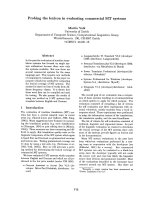
Coomassie
wt D207A R254K H260Q C259L E189Q
Zn-fluorescence
A
B
Fig. 4. Cph1D2 and site-directed mutants. (A) Coomassie stain and
Zn
2+
fluorescence of Cph1D2 and of selected Cph1D2 site-directed
mutants after SDS ⁄ PAGE. (B) UV-Vis difference spectra of Cph1D2
and of selected Cph1D2 site-directed mutants. Absorbance differ-
ence maxima and isosbestic points are given. The dotted vertical
lines are drawn through the absorption maxima of the Pr and Pfr
state of Cph1D2 to highlight shifts in the mutants.
Protein–chromophore interactions in Cph1 J. Hahn et al.
1420 FEBS Journal 273 (2006) 1415–1429 ª 2006 The Authors Journal compilation ª 2006 FEBS
type, the CD spectrum of R254A implied slight folding
differences (Fig. 5). Nevertheless, both bound PCB
covalently to form a red⁄ far-red photochromic holo-
protein with the red kmax of Pr 10 nm downshifted
but Pfr spectra indistinguishable from that of the wild-
type.
E189
#310
This acidic residue was the focus of earlier mutagenesis
studies which inferred a central role in bilin ligation
[25]. Our E189A mutant was expressed as insoluble
protein bodies, attempts at refolding by solubilization
in urea followed by slow dialysis proving unsuccessful.
The E189Q mutation was better tolerated although the
expression yield of soluble protein was much lower
than for the Cph1D2 wild-type. CD spectroscopy
implied, furthermore, that folding was significantly dif-
ferent from that of the wild-type. However, when this
mutant apoprotein was presented with PCB, a low
level of covalent attachment accompanied by a weak
Pr-like band at 665 nm was seen (Figs 4A and 9),
implying normal protonation of a thioether-linked
bilin. No photochromicity signal associated with red ⁄
far-red irradiation was measurable, however.
Discussion
In this study we focused on Cph1D2, the N-terminal
514 residue sensory module of Cph1. The smaller dele-
tion product, Cph1D1 (N1–492) was functionally
compromised (Table 1 and Fig. 1), showing very
poor solubility and a weak Pfr-like absorbance typical
of phytochromes in which the PHY subdomain (see
is incom-
plete. On the other hand the UV-Vis absorbance prop-
erties of holoCph1D2 ) whose C-terminus corresponds
exactly to that of the PHY subdomain ) closely resem-
ble those of full-length holoCph1 (see Table 1 and
Fig. 1). Thus the sensory module is photochemically
autonomous, as implied in an earlier study [25].
Cph1D2 can therefore be used as a convenient model
for investigating phytochrome functions, as ) unlike
full-length Cph1 ) it does not aggregate under normal
in vitro conditions. Indeed, as pure Pfr can be obtained
by SEC [27], it might also be possible to obtain struc-
tural data for that form too.
UV-Vis absorbance properties of bilins and other
tetrapyrroles are determined both by their protonation
state and by the extent and linearity of the conjugated
p orbital system. Coiled bilins (like free PCB) show a
Fig. 5. Circular dichroism spectra of selec-
ted mutants. For comparison Cph1D2-wt
spectra are shown (dotted lines) and molar
ellipticities were calculated.
J. Hahn et al. Protein–chromophore interactions in Cph1
FEBS Journal 273 (2006) 1415–1429 ª 2006 The Authors Journal compilation ª 2006 FEBS 1421
strong UVA band but weak absorbance at longer
wavelengths, while in linear bilins the situation is
reversed, the dipole moment perpendicular to the long
molecular axis giving strong red absorbance at the
expense of the UV ⁄ A band (the UV ⁄ A and lowest
energy red ⁄ far-red bands are sometimes called Soret
Fig. 6. UV-Vis absorbance properties of
Cph1D2-C259L in the presence of excess
PCB. Absorbance and difference spectra
under nonreducing (A,B) and reducing (C,D)
conditions. (A,C) (n) Pr[1], after autoassem-
bly with PCB in the dark; (d) Pfr (7 ⁄ 3Pfr⁄ Pr
mixture) after R irradiation; (h) Pr[2], after
FR irradiation. (B, D) (n) Pr[1]–Pfr; (d) Pfr–
Pr[2].
A
B
Fig. 7. pH-dependence of UV-Vis absorbance properties of Cph1D2
and Cph1D 2-H260Q after far-red (100% Pr) and after red (7 ⁄ 3
Pfr ⁄ Pr photoequilibrium) irradiation. (A) Cph1D2 wild-type at pH 7
(after far-red n, after red d) and pH 9 (after far-red h, after red s).
(B) Cph1D2-H260Q at pH 7-start (after far-red n, after red, m), pH 9
(after far-red h, after red n ) and at pH 7-end (after far-red r, after
red .).
A
B
Fig. 8. UV-Vis absorbance properties of Cph1D2-D207A. (A) Spectra
recorded after autoassembly with PCB in the dark (n), after 90 s
red irradiation (d) and after 90 s far red irradiation (m). (B) Thermal
reversion of Cph1D2-D207A. After PCB assembly in the dark
(n) and saturating red irradiation (d), the sample was kept in the
dark and absorption spectra were recorded after 10 (m), 20 (.), 30
(r) and 45 (b) min.
Protein–chromophore interactions in Cph1 J. Hahn et al.
1422 FEBS Journal 273 (2006) 1415–1429 ª 2006 The Authors Journal compilation ª 2006 FEBS
and Qy in analogy to closed-ring tetrapyrroles; this
possibly misleading terminology has been avoided
here) [30,31]. While the ratio of the two ‘oscillator
strengths’ changes with uncoiling, the total absorptivity
remains constant. FTRR (e.g. [32] for oat phyA) gives
more specific information about the conformation of
the chromophore, as of course can more direct meth-
ods like NMR (e.g. [33,34] and Rohmer T. and
Matysik J., University of Leiden, the Netherlands,
unpublished data). and X-ray crystallography (e.g. [9]).
On the other hand, protonation has only subtle effects
in the UV ⁄ A region, while the red peak strengthens
approximately threefold at low pH [34,35]. In fact, in
PCB a new band at 688 nm appears and strengthens
with protonation, but its k
max
does not shift, while the
broad, weaker shoulder centred at 615 nm (also seen
in phytochrome spectra) remains unchanged (Fig. 2).
Recently, Go
¨
ller et al. (2005) [36] have successfully
modelled the role of protonation in strengthening e
red
by enhancing electronic coupling between PCB rings,
while NMR studies [33] have proved that all four rings
are fully protonated in both Pr and Pfr states of Cph1.
The current model for the autoassembly reaction
[15,16,37], important to the present study, envisages
three-steps: (1) an initial chromophore recognition pro-
cess (< 1 ms) of the unprotonated, coiled bilin with
weak absorbance in the orange region; (2) entrance
into a pocket within the protein (100–200 ms) during
which uncoiling and protonation occur, leading to a
fourfold hyperchromicity of the lowest energy absor-
bance band in red ⁄ far-red and the appearance of
photochromicity in that region; (3) a final covalent
ligation (1–10 S) to a C residue through the formation
of a thioether bond. While pioneering studies showed
that PUB is attached to oat phyA at C322
#380
[13] at
least some bacteriophytochromes attach a BV chromo-
phore at a C residue close to the N-terminus [9,28,38],
contradicting earlier data [39]. A further important dif-
ference is that bacteriophytochromes covalently ligate
to the ring A vinyl side chain of BV, forming a two-
carbon linker, while in oat phyA the ring A ethylidene
side chain of PFB yields a single-carbon linker. Here
we present direct evidence that in Cph1 PCB is simi-
larly ligated to C259
#380
(Fig. 3). As shown by our
C259L mutant, if step 3 of autoassembly is prevented
by mutating this residue, many holophytochrome-like
features appear, but k
max
values are shifted 25 nm
bathochromically (Fig. 4) as would be expected if the
ethylidene group double bond was left intact to contri-
bute to the PCB delocalized p-electron system. This is
seen also in the wild-type if C residues are blocked
nonspecifically by iodacetamide [16]. No effect is seen
with blocked Agp1 and BV, however, in accordance
with the vinyl group double bond in that case not
being connected to the p-system [40]. Redox conditions
seem to be important in autoassembly steps 1 and ⁄ or
2. PCB binding was weak under nonreducing condi-
tions, requiring at least 15 lm PCB, and even then
only a single round of R ⁄ FR photoconversion was
possible – as though the chromophore was lost as a
consequence of photoconversion (Fig. 6A,B). Under
reducing conditions (Fig. 6C,D) the relative strengths
of the UV ⁄ A and red bands were approximately equal,
implying a more chiral chromophore conformation
than in the wild-type, a conclusion consistent with
experiments using methoxy-PCB [41]. Thus conforma-
tional changes leading to uncoiling are associated
with both step 2 and step 3 of autoassembly. The
long wavelength band in the Pfr-like state of the
C259L mutant was even weaker than that of the
ground state.
R
#375
near the ligation site is perfectly conserved
amongst all known phytochromes, bacteriophyto-
chromes and even several other biliproteins. It thus
might be expected to be important in chromophore
binding and conformation. Indeed, X-ray structural
data shows that it forms a salt bridge with the pro-
pionate side chain of ring B, apparently pulling on the
chromophore from deep within the protein [9,17–19].
However, R
#375
I and T mutants of pea phyA showed
only 5 nm hypsochromic shifts [22]. Here we
mutated R254
#375
to K (likewise a basic residue) and
to A (a smaller, moderately hydrophobic residue).
Both mutants fold similarly to the wild-type and bind
PCB effectively (Figs 4A and 5). Whereas their Pfr
absorbance characteristics match those of the wild-type
almost exactly, the Pr peak shows a 10-nm hypsochro-
mic shift in both cases (Table 2, Fig. 4B). This might
Fig. 9. UV-Vis absorbance properties of Cph1D2-E189Q. Spectra
recorded after autoassembly with PCB in the dark (n) and subse-
quent red irradiation (d).
J. Hahn et al. Protein–chromophore interactions in Cph1
FEBS Journal 273 (2006) 1415–1429 ª 2006 The Authors Journal compilation ª 2006 FEBS 1423
arise from a rotation around the C5–C6 bond specific
to the Pr form (H. Scheer, LMU Munich, personal
communication), although such a shift is also predicted
for (de)protonation of the propionate [36]. Either way,
the UV-Vis shift is much too subtle to explain the
degree of conservation seen, thus it is very likely that
R254
#375
instead plays a central role in signal trans-
duction: indeed, the 1ZTU structure implies that even
a slight movement of the chromophore would break
the salt bridge, fitting with the UV-Vis phenotype of
our R254K mutant. Overlooked to date, the X-ray
structure shows an intriguing 2.5-A
˚
diameter channel
leading from the salt bridge to the other side of the
protein, wide enough for water molecules or hydroxo-
nium ions. It would in any case be worthwhile investi-
gating R254
#375
mutants at the physiological level.
Go
¨
ller et al. [36] also calculated that ring B ⁄ C pro-
tonation of chiral PCB leads to dramatically increased
electron coupling associated with a bathochromic shift
of e
max
of 130 nm: we observe a shift of 160 nm on
acidification of free PCB compared with 150 nm for
holoCph1 prior to ligation (i.e. in C259L), a reason-
able fit considering likely conformation differences.
Protonation requires a donor with a pKa of < 4.6,
and is thus just possible for E or D carboxyl side
chains [35]. None of the 12 highly conserved E or D
residues are near C259
#380
in the primary sequence,
furthermore, the recent 1ZTU structure shows that
only two of these E189
#310
and D207
#328
are posi-
tioned anywhere near the bilin. Coincidentally, these
are the two residues we had focused on in the present
study. As we show, E189Q mutations are better toler-
ated than others [25,42], UV-Vis properties being con-
sistent with a ground state resembling protonated Pr
(Figs 4 and 9, Table 2). Thus the proposed central role
for E189
#310
in ligation [25] is unlikely, neither is it
likely to be the proton donor in autoassembly step 2.
Unfortunately, D207
#328
too is most unlikely to fulfil
this role. The mutant apoprotein bound PCB covalent-
ly (Fig. 4A) to yield a ground state with a similar e
max
and k
red
compared to wild-type, implying a partially
coiled, protonated chromophore. Furthermore, 1ZTU
shows that the carboxyl group of D207
#328
is directed
away from the chromophore, forming a hydrophilic
acid patch exposed to the solvent, at least in this BphP
deletion mutant. The main chain carbonyl oxygen of
D207
#328
interacts with the nitrogens of rings A, B and
C, so that they might share their protons – but this is
a proton acceptor, not a donor, and of course any
mutation at this site could fulfil this role. Our mutants
imply that D207
#328
is important in Pfr formation.
Red irradiation leads to bleaching (as in the wild-type),
but no Pfr-like band appeared, rather a broad peak
centred at 590 nm remained. Not surprisingly, FR irra-
diation had no effect, but the bleached form reverts
thermally to the Pr-like state. D207
#328
N behaved simi-
larly (Table 2). As proton exchange is probably associ-
ated with photoconversion (see below), D207
#328
might
be involved in reprotonation prior to Pfr formation.
Such a role would not be apparent from 1ZTU
because this cannot form bona fide Pfr. It seems clear,
however, that neither E189
#310
nor D207
#328
can be
the proton donor in step 2 of autoassembly. Thus the
donor for bilin protonation, even in the light of the
1ZTU structure, remains unknown.
Although it is now certain that all four chromophore
nitrogens are protonated in neutral buffers in both Pr
and Pfr states [33], transient deprotonation of the chro-
mophore seems to be a feature of Pr fi Pfr photocon-
version [20,43,44]. Significant deprotonation of both Pr
and Pfr can be induced by shifting the pH, however, one
pKa component being close to neutral, thus possibly
representing an imidazole (H side chain) and ⁄ or a direct
effect on PCB [20]. In the present study we show that
H260
#381
plays a crucial role in this process. Mutagen-
esis of H260
#381
to L, R, F, G and Q has already been
reported for recombinant phytochrome A from pea and
oat. In the first four cases covalent ligation and photo-
chromicity were obliterated probably because of
misfolding, whereas H260
#381
Q retained covalent
attachment and photochromicity [21,22,24]. While Q
resembles H regarding its steric demands and its hydro-
gen bonding ability [45], its buffering capacity is very
weak. While our H260Q mutant is wild-type like in its
folding, photochromicity and covalent autoassembly
under normal conditions (Table 2; Figs 4 and 5), it
shows dramatically increased sensitivity to buffer pH
(Fig. 7), the long wavelength absorbance peak of Pr
weakening drastically and that of Pfr disappearing com-
pletely at pH 9.0, much weaker, broader bands centred
at 549 nm and 577 nm, respectively, appearing in their
place. The UV ⁄ A bands show smaller changes for both
states. These effects are fully pH-reversible. We con-
clude that H260
#381
plays a crucial role in buffering both
Pr and Pfr protonation. As it is easy to titrate chromo-
phore protonation in the mutant, this offers a poten-
tially useful degree of freedom for more sophisticated
analytical methods.
H260
#381
is likely to be important according to the
1ZTU X-ray structure [9]. The chromophore nitrogens
of rings A, B and C are hydrogen bonded to the
D207
#328
backbone nitrogen, perhaps sharing their
protons. On the other side of the pocket, the d1 nitro-
gen of H260
#381
and chromophore ring C nitrogen are
separated by 3.3 A
˚
, just outside van der Waals’ con-
tact, but a hydrogen bonding bridge is provided by
Protein–chromophore interactions in Cph1 J. Hahn et al.
1424 FEBS Journal 273 (2006) 1415–1429 ª 2006 The Authors Journal compilation ª 2006 FEBS
Wat12. The H260
#381
e2 nitrogen hydrogen bonds to
an oxygen of the C-ring propionate, the other propio-
nate oxygen hydrogen bonding to the side chains of
S274
#395
and the conserved S272
#393
and, via Wat18,
to the H290
#411
e2 nitrogen ) which is similarly bon-
ded to the carbonyl oxygen of chromophore ring
D ) at least in the Pr form. In H260
#381
Q the side
chain carbonyl oxygen might simulate the H260
#381
d1
nitrogen functionality (at neutral pH) but not the con-
nection to the rest of the intricate hydrogen-bond net-
work. That the observed pH effect on the UV-Vis
properties of wild-type Cph1 arises from deprotonation
of the chromophore nitrogens is consistent with: (a)
the positioning of the imidazole side chain of H260
#381
adjacent and hydrogen-bonded to the ring C nitrogen
in 1ZTU; (b) the role of B ⁄ C ring nitrogen protona-
tion in electronic coupling [36]; and (c) the pKa values
for both the H side chain (imidazole) and the chromo-
phore nitrogens being between 4.5 and 7. Of course,
the buffering capacity of H260
#381
is not offered by
the Q mutant. The 12-nm hypsochromic shift in
H260
#381
Q at pH 7 relative to the wild-type is consis-
tent with decoupling of the C ring propionate [36] but
might also derive from p-electron stacking effects or
still incomplete protonation.
While it thereby seems clear that at least one func-
tion of H260
#381
is to buffer the chromophore, the imi-
dazole side chain would not under normal conditions
be acidic enough to protonate the chromophore: how-
ever, given the hydrogen bonding network in 1ZTU,
the environment of this residue is highly unusual. A
role for H260
#381
in ligation is also possible, not via
Schiff-base formation [39] because imidazole is not an
amine (nor a ketone ⁄ aldehyde), but rather by the imi-
dazole nitrogens increasing the thiol nucleophilicity of
C259
#380
by hydrogen bonding. This possibility is
offered in H260Q too: indeed, our mutant shows wild-
type autoassembly.
It is apparent from numerous studies including
several of the mutants reported here that the charac-
teristic lowest energy absorbance band of Pfr is more
labile than that of the Pr ground state. Pfr seems
to require PHY domain functions (which can, of
course, be affected by changes elsewhere): if these
are compromised red irradiation still depopulates the
Pr-like ground state but a bleached form (Pbl)
appears in place of Pfr. Pbl probably represents a
deprotonated intermediate (sometimes called Ibl or
metaRc) which has been detected in some kinetic
and freeze-trapping studies [35,43,46]. Both
H260
#381
Q at elevated pH and D207
#328
A produce a
Pbl-like form after red irradiation (Figs 7 and 8),
either because they indirectly compromise PHY
domain function or because they play a central role
in reprotonation itself.
Numerous islands of homology seen in plant-like
phytochromes [6] are conserved in BphP and, as the
ground state spectrum even of the 1ZTU crystal
itself ) at least at pH 4.9 ) is Pr-like, we expect many
features of the new structure to be typical for all
phytochromes. As studies of the phytochrome molecule
will now come to be based on the template provided
by 1ZTU, it is important to recognize that this struc-
ture describes only a portion of the BphP sensory mod-
ule (the N-terminal chromophore binding PAS ⁄ GAF
domains), the missing PHY domain precluding the for-
mation of bona fide Pfr. There may also be other prob-
lems in extrapolating from 1ZTU to the phytochrome
family. For example, the BV chromophore in 1ZTU is
modelled as ZZZssa (but with a 44° C–D ring rota-
tion), more chiral than the ZZZasa expected from
recent resonance Raman data for oat phyA [32]. Inter-
estingly: (a) RRR ring A stereochemistry of PCB or
PFB would be required for nucleophilic attack from
the sulphur of C
#380
according to the X-ray structure,
but this is provided by ZZZssa and not ZZZasa; (b)
mutants with C residues at
#
380 in bacteriophyto-
chromes show de novo ligation to PCB ([47] for Agp1
and [23] for CphB from Calothrix sp. PCC7601). Thus
either the FTRR data for plant-type phytochromes is
interpreted wrongly or a C residue at this point induces
a major change in the chromophore pocket relative to
that seen in 1ZTU. Of the five residues interacting with
the D ring in 1ZTU, two are not conserved in plant-
type phytochromes. Moreover, while mutation of one
of these (Y176
#297
H) in Cph1 gives rise to strong fluor-
escence [48], this is not seen in bacteriophytochromes
[42]. The 1ZTU structure is also notable in that, while
the A, B and C rings interact intimately with neigh-
bouring residues, ring D is left ample room for the
Z fi E isomerization associated with lumiR formation.
How then does this induce the rearrangements associ-
ated with the molecular action of Pfr? We emphasize
that despite these caveats we fully expect 1ZTU to pro-
vide new and valuable insights into phytochrome func-
tions.
Experimental procedures
Mutagenesis
Escherichia coli XL1-blue (Stratagene, La Jolla, CA, USA)
overexpression clones in pQE12 (Qiagen, Hilden, Germany)
were generated by PCR using error-checking DNA poly-
merase (TakaraEx, Otsu, Japan). pF10.His (full-length
Cph1 with a C-terminal His-tag) has been described else-
J. Hahn et al. Protein–chromophore interactions in Cph1
FEBS Journal 273 (2006) 1415–1429 ª 2006 The Authors Journal compilation ª 2006 FEBS 1425
where [6]. p920.B3 and p926.5 (Cph1D1 and Cph1D2,
respectively) were derived from genomic DNA of Synecho-
cystis PCC6083 (kindly provided by A. Wilde, Humboldt
University, Berlin, Germany) following 25 amplification
cycles using appropriate header-primers, restriction and
ligation into EcoRI and BamHI of pQE12, thereby gener-
ating clones to overexpress the N-terminal sensory module
of Cph1 with a His6-tag followed by a stop codon immedi-
ately downstream of V492 and E514, respectively.
Site-directed mutagenesis of Cph1D2 was achieved by
PCR around the 4984 bp p926.5 parent plasmid using ap-
propriate back-to-back primer pairs carrying the necessary
mismatched base(s) followed by destruction of the template
with DpnI, polynucleotide kinase treatment, blunt ligation at
low ATP and template concentrations (500 lm and
1ngÆlL
)1
, respectively) and electrotransformation into
XL1-blue. The resulting clones were screened by SDS ⁄
PAGE [49] of isopropyl thio-b-d-galactoside (IPTG)-induced
minipreps and sequencing of the entire reading frame.
Apophytochrome
Apoproteins were produced in liquid Luria–Bertani cul-
tures with ampicillin at 50 lgÆmL
)1
by addition of IPTG
to 20 lm and overnight incubation at 18 °C. The cells
were harvested and washed in cold TESb buffer (50 mm
Tris ⁄ HCl pH 7.8, 5 mm EDTA, 300 mm NaCl, 1 mm
b-mercaptoethanol), lysed in a French pressure cell at a
difference pressure of 120 MPa, and clarified at 50 000 g
for 5 min at 4 °C. The supernatant was then concentrated
by ammonium sulphate precipitation and resuspended in
TISI10 (50 mm Tris ⁄ HCl pH 7.8, 1 mm imidodiacetic
acid, 300 mm NaCl, 10 mm imidazole). Affinity purifica-
tion utilized FPLC-grade Ni–NTA Superflow (Qiagen),
washing and eluting at 20 mm and 150 mm imidazole,
respectively. Subsequently the buffer was exchanged by
overnight dialysis against 2 L of TES buffer at 4 °C and
the apophytochrome concentrated by ultrafiltration (Ultra-
free, Millipore, Billerica, MA, USA).
Phycocyanobilin
Phycocyanobilin was prepared from Synechocystis sp. PCC
6803 grown in liquid BG11 medium under constant light
at room temperature. Cells were collected by centrifuga-
tion and lysed in a French press at 120 MPa. Phycobili-
somes were purified on sucrose gradients [50] and PCB
freed by methanolysis in a Soxhlet apparatus. Further
PCB purification was carried out by first binding the
crude PCB in 50 : 50 : 1 methanol ⁄ H
2
O ⁄ acetic acid to a
C18 Sep-Pak cartridge (Waters, Mitford, MA, USA) elut-
ing with methanol (MeOH) followed by isocratic reverse-
phase HPLC over a C18 column (UltraSep ES Pharm
RP18E, 250 · 8 mm, Sepserv, Berlin, Germany) again with
50 : 50 : 1 MeOH ⁄ H
2
O ⁄ acetic acid using an A
¨
kta FPLC
system (Amersham Pharmacia ⁄ GE, Uppsala, Sweden)
equipped with a flow-cell coupled to a xenon-flash-driven
dual-channel diode array spectrometer (Ocean Optics,
Dunedin, FL, USA). The eluate was finally concentrated
using a C18 Sep-Pak cartridge and the PCB was stored at
)80 °C in darkness. PCB was quantified by UV-Vis
absorbance spectroscopy in methanol ⁄ 5%HCl, using the
extinction coefficient (e) of 37.9 mm
)1
Æcm
)1
at the 690 nm
k
max
[51].
A5-lL aliquot of HPLC-purified PCB (in Tes) was dilu-
ted to 500 lL in each of the following solutions:
HCl ⁄ CH
3
Cl (1 : 19); HCl ⁄ MeOH (1 : 19); 0.5 m aqueous
HCl, pH 0.3; 50 mm sodium acetate pH 3.0, 100 mm MES
pH 5.5 and 100 mm Tris ⁄ HCl pH 7.7. UV-Vis spectra were
then recorded and the corresponding e
kmax
determined rel-
ative to the known value in HCl ⁄ MeOH.
Holophytochrome
Holoproteins were prepared in darkness by mixing the
apoprotein at 5–100 lm with a 10-fold molar excess of
PCB in TES. Subsequent operations were carried out
with minimal exposure to 520 nm LED safelight. After
10 min incubation free PCB was removed by gel filtration
over PD-10 columns (Amersham Pharmacia ⁄ GE) and
holoprotein concentrated by ultrafiltration as necessary.
The products were examined by UV-Vis absorbance
spectroscopy with either Lambda-9 (Perkin-Elmer, Welles-
ley, MA, USA) or 8453 diode-array (Agilent, Palo Alto,
CA, USA) instruments. Actinic irradiation was from red
and far-red LED sources (B5-436-30D, kmax 664 nm and
SMC735, k
max
735 nm; both 40 nm FWHM, Roithner,
Vienna). Covalent attachment of the chromophore was
examined by SDS ⁄ PAGE followed by Zn
2+
-induced
fluorescence [6,52].
The C259L mutant was handled somewhat differently.
Incubation of the purified apoprotein with PCB for 10 min
resulted in low levels of holoprotein in which Pr fi Pfr
photoconversion was compromised. Following prolonged
incubation in the presence of b-mercaptoethanol, however,
fully reversible holoprotein was obtained. As subsequent
attempts to remove excess PCB by gel filtration led to chro-
mophore loss from the holoprotein, PCB was added only in
a small excess. Irradiation for UV-Vis analysis was per-
formed with appropriate red or far-red LED sources (ELD-
670-524, k
max
670 nm and ELD-770-324, k
max
775 nm;
both 40 nm FWHM; Roithner, Vienna).
Holoprotein extinction coefficients were measured as des-
cribed previously [16] by assembling a known concentration
of pure PCB with excess full-length Cph1 or Cph1D2 apo-
protein. The reaction was carried out in TESb. UV-Vis spec-
tra were recorded after autoassembly was complete (10 min
at 20 °C). To check that excess apoprotein had been presen-
ted, a further aliquot of PCB was added, leading to a doub-
ling of the Pr signal. To check that all PCB had been
Protein–chromophore interactions in Cph1 J. Hahn et al.
1426 FEBS Journal 273 (2006) 1415–1429 ª 2006 The Authors Journal compilation ª 2006 FEBS
consumed, the final reaction mixture was spun through
a PCB-permeable ultrafilter (10 kDa, Millipore): filtrate
absorbance in the visible region was negligible.
The pH dependence of H260Q UV-Vis properties was
investigated by re-buffering over G25 Sephadex (NAP10,
Amersham Pharmacia ⁄ GE) under safelight. The absorbance
values were corrected for dilution associated with each pas-
sage.
To check the folding of the mutated apoproteins, CD
spectra were recorded using a J-715 (Jasco, Gross-Umstadt,
Germany) instrument. Sample concentration was about
0.1 mgÆmL
)1
in 10 mm potassium phosphate buffer pH 7.8.
Spectra at 200–260 nm were measured 10-fold and averaged
by the spectrometer software.
Chromopeptide analyses
Five micrograms of PCB-HoloCph1D2 was digested over-
night under argon with 0.5 lg Trypsin (Promega, Man-
nheim, Germany). Peptide fragments were separated using
a HP1090M (Agilent) system on an Acclaim 300 column
(C18, 3 lm, 300 A
˚
, 2.1 · 150 mm; Dionex, Idstein, Ger-
many) at a flow rate of 200 lLÆmin
)1
at 40 °C. A linear
gradient from 100% solvent A (5% acetonitrile, 1% tri-
fluoroacetic acid) to 50% solvent B (80% acetonitrile, 1%
trifluoroacetic acid) in 30 min was applied. UV-Vis spectra
at 200–600 nm were recorded with a photodiode-array
detector (G1306A, Agilent). Fractions of 200 lL were col-
lected, dried in a SpeedVac (Juan, GMI, Ramsey, MN,
USA) and stored at )20 °C for further analysis. MALDI-
TOF-MS and MS
2
analyses were performed with an Ultra-
flex TOF ⁄ TOF (Bruker Daltonik, Bremen, Germany) oper-
ating under FlexControl 2.2 (Bruker) in the positive-ion
reflectron mode using a-cyano-4-cinnamic acid prespotted
targets (Bruker). Acceleration voltages of 25 kV were
applied. Ions ranging from 500 to 4000 Da were registered.
For external calibration a peptide calibration mixture (Bru-
ker) was used. Approximately 100–300 single spectra were
averaged. Data analysis was performed with FlexAnalysis
2.2 and BioTools 3.0 (Bruker). N-terminal Edman degrada-
tion was performed with a 492A Procise sequencer (Applied
Biosystems) under standard conditions.
Acknowledgements
We gratefully acknowledge the contributions of Tilman
Lamparter (FU Berlin) to the early stages of this work
and of Pill-Soon Song (Kumho Life & Environmental
Sciences, Kwangju, Korea) and Hugo Scheer (LMU
Munich) for expert advice. We thank Sabine Buchert,
Norbert Michael, Tina Lang and Sabine Kaltofen
for their assistance. We are grateful for the financial
support of the Deutsche Forschungsgemeinschaft
(Hu702 ⁄ 2 and Sfb498).
References
1 Borthwick HA, Hendricks SB, Parker MW, Toole EH
& Toole VK (1952) A reversible photoreaction control-
ling seed germination. Proc Natl Acad Sci USA 38, 662–
666.
2 Lagarias JC & Lagarias DM (1989) Self-assembly of
synthetic phytochrome holoprotein in vitro. Proc Natl
Acad Sci USA 86, 5778–5780.
3 Hughes J, Lamparter T, Mittmann F, Hartmann E,
Ga
¨
rtner W, Wilde A & Bo
¨
rner T (1997) A prokaryotic
phytochrome. Nature 386, 663.
4 Yeh KC, Wu SH, Murphy JT & Lagarias JC (1997) A
cyanobacterial phytochrome two-component light sen-
sory system. Science 277, 1505–1508.
5 Hughes J & Lamparter T (1999) Prokaryotes and phy-
tochrome: the connection to chromophores and signal-
ing. Plant Physiol 121, 1059–1068.
6 Lamparter T, Mittmann F, Ga
¨
rtner W, Bo
¨
rner T,
Hartmann E & Hughes J (1997) Characterization of
recombinant phytochrome from the cyanobacterium
Synechocystis. Proc Natl Acad Sci USA 94, 11792–11797.
7 Landgraf FT, Forreiter C, Hurtado Pico
´
A, Lamparter
T & Hughes J (2001) Recombinant holophytochrome in
Escherichia coli. FEBS Lett 508, 459–462.
8 Wahleithner JA, Li LM & Lagarias JC (1991) Expression
and assembly of spectrally active recombinant holophyto-
chrome. Proc Natl Acad Sci USA 88, 10387–10391.
9 Wagner JR, Brunzelle JS, Forest KT & Vierstra RD
(2005) A light-sensing knot revealed by the structure of
the chromophore-binding domain of phytochrome.
Nature 438, 325–331.
10 Hughes J, Lamparter T & Mittmann F (1996) Cerpu;
PHY0; 2, a ‘normal’ phytochrome in Ceratodon. Plant
Physiol 112, 446.
11 Hershey HP, Barker RF, Idler KB, Lissemore JL &
Quail PH (1985) Analysis of cloned cDNA and genomic
sequences for phytochrome: complete amino acid
sequences for two gene products expressed in etiolated
Avena. Nucl Acids Res 13, 8543–8559.
12 Lagarias DM, Crepeau MW, Maines MD & Lagarias
JC (1997) Regulation of photomorphogenesis by expres-
sion of mammalian biliverdin reductase in transgenic
Arabidopsis plants. Plant Cell 9, 675–688.
13 Lagarias JC & Rapoport H (1980) Chromopeptides
from phytochrome. The structure and linkage of the Pr
form of the phytochrome chromophore. J Am Chem
Soc 102, 4821–4828.
14 Hu
¨
bschmann T, Bo
¨
rner T, Hartmann E & Lamparter T
(2001) Characterization of the Cph1 holo-phytochrome
from Synechocystis sp. PCC6803. Eur J Biochem 268,
2055–2063.
15 Borucki B, Otto H, Rottwinkel G, Hughes J, Heyn MP
& Lamparter T (2003) Mechanism of Cph1 phyto-
J. Hahn et al. Protein–chromophore interactions in Cph1
FEBS Journal 273 (2006) 1415–1429 ª 2006 The Authors Journal compilation ª 2006 FEBS 1427
chrome assembly from stopped-flow kinetics and circu-
lar dichroism. Biochemistry 42, 13684–13697.
16 Lamparter T, Esteban B & Hughes J (2001) Phyto-
chrome Cph1 from the cyanobacterium Synechocystis
PCC6803. Purification, assembly, and quaternary struc-
ture. Eur J Biochem 268, 4720–4730.
17 Ru
¨
diger W & Thu
¨
mmler F (1994) The phytochrome
chromophore. In Photomorphogenesis in Plants (Ken-
drick, RE & Kronenberg, GHM, eds), pp. 51–69.
Kluwer, Dortrecht.
18 Parker MW, Goebel P, Ross CR, Song PS & Stezowski
JJ (1993) Molecular modeling of phytochrome using con-
stitutive C- phycocyanin from Fremyella diplosiphon as a
putative structural template. Bioconjug Chem 5, 21–30.
19 Partis MD & Grimm R (1990) Computer analysis of
phytochrome sequences from five species: implications
for the mechanism of action. Z Naturforsch C 45 , 987–
998.
20 van Thor JJ, Borucki B, Crielaard W, Otto H, Lam-
parter T, Hughes J, Hellingwerf KJ & Heyn MP (2001)
Light-induced proton release and proton uptake reac-
tions in the cyanobacterial phytochrome Cph1. Bio-
chemistry 40, 11460–11471.
21 Bhoo SH, Hirano T, Jeong H-Y, Lee J-G, Furuya M &
Song PS (1997) Phytochrome photochromism probed
by site-directed mutations and chromophore esterifica-
tion. J Am Chem Soc 119, 11717–11718.
22 Deforce L, Furuya M & Song PS (1993) Mutational
analysis of the pea phytochrome A chromophore
pocket: chromophore assembly with apophytochrome A
and photoreversibility. Biochemistry 32, 14165–14172.
23 Jorissen HJ, Quest B, Remberg A, Coursin T, Braslav-
sky S, Schaffner K, Tandeau de Marsac N & Ga
¨
rtner
W (2002) Two independent, light-sensing two-compo-
nent systems in a filamentous cyanobacterium. Eur J
Biochem 269, 2662–2671.
24 Remberg A, Schmidt P, Braslavsky SE, Gartner W &
Schaffner K (1999) Differential effects of mutations in
the chromophore pocket of recombinant phytochrome
on chromoprotein assembly and Pr-to-Pfr photoconver-
sion. Eur J Biochem 266, 201–208.
25 Wu S-H & Lagarias JC (2000) Defining the bilin lyase
domain: lessons from the extended phytochrome super-
family. Biochemistry 39, 13487–13495.
26 Park CM, Shim JY, Yang SS, Kang JG, Kim JI, Luka
Z & Song PS (2000) Chromophore–apoprotein interac-
tions in Synechocystis sp. PCC6803 phytochrome cph1.
Biochemistry 39, 6349–6356.
27 Strauss HM, Schmieder P & Hughes J (2005) Light-
dependent dimerisation in the N-terminal sensory mod-
ule of cyanobacterial phytochrome 1. FEBS Lett 579,
3970–3974
28 Lamparter T, Carrascal M, Michael N, Martinez E,
Rottwinkel G & Abian J (2004) The biliverdin chromo-
phore binds covalently to a conserved cysteine residue
in the N-terminus of Agrobacterium phytochrome Agp1.
Biochemistry 43, 3659–3669.
29 Quest B & Ga
¨
rtner W (2004) Chromophore selectivity
in bacterial phytochromes. Eur J Biochem 271, 1117–
1126.
30 Falk H (1989) The Chemistry of Linear Oligopyrroles
and Bile Pigments. Springer-Verlag, Wien.
31 Scheer H (1981) Biliproteine. Angew Chem 93, 230–250.
32 Mroginski MA, Murgida DH, von Stetten D, Kneip C,
Mark F & Hildebrandt P (2004) Determination of the
chromophore structures in the photoinduced reaction
cycle of phytochrome. J Am Chem Soc 126, 16734–
16735.
33 Strauss HM, Hughes J & Schmieder P (2005) Hetero-
nuclear solution-state NMR studies of the chromophore
in cyanobacterial phytochrome Cph1. Biochemistry 44,
8244–8250.
34 Mizutani Y, Tokutomi S, Aoyagi K, Horitsu K & Kita-
gawa T (1991) Resonance Raman study on intact pea
phytochrome and its model compounds: evidence for
proton migration during the phototransformation. Bio-
chemistry 30, 10693–10700.
35 Stanek M & Grubmayr K (1998) Protonated 2,3-dihydro-
bilindiones – models for the chromophores of phycocya-
nin and the red-absorbing form of phytochrome. Chem
Eur J 4, 1653–1659.
36 Go
¨
ller AH, Strehlow D & Hermann G (2005) The
excited-state chemistry of phycocyanobilin: a semiempi-
rical study. Chem Phys Chem 6, 1259–1268.
37 Sineshchekov VA, Koppel L, Esteban B, Hughes J &
Lamparter T (2002) Fluorescence investigations of the
recombinant cyanobacterial phytochrome (Cph1) and its
C-terminally truncated monomeric species (Cph1D2):
implication for holoprotein assembly, chromophore–
apoprotein interaction and photochemistry. J Photo-
chem Photobiol B 67, 39–50.
38 Tasler R, Moises T & Frankenberg-Dinkel N (2005)
Biochemical and spectroscopic characterization of the
bacterial phytochrome of Pseudomonas aeruginosa.
FEBS J 272, 1927–1936.
39 Davis SJ, Vener AV & Vierstra RD (1999) Bacteriophy-
tochromes: phytochrome-like photoreceptors from non-
photosynthetic eubacteria. Science 286, 2517–2520.
40 Lamparter T, Michael N, Mittmann F & Esteban B
(2002) Phytochrome from Agrobacterium tumefaciens
has unusual spectral properties and reveals an N-term-
inal chromophore attachment site. Proc Nat Acad Sci
USA 99, 11628–11633.
41 Lindner I, Braslavsky SE, Schaffner K & Ga
¨
rtner W
(2000) Model studies of phytochrome photochromism:
protein-mediated photoisomerization of a linear tetra-
pyrrole in the absence of covalent binding. Angew Chem
Int Ed 39, 3269–3271.
42 Fischer AJ, Rockwell NC, Jang AY, Ernst LA,
Waggoner AS, Duan Y, Lei H & Lagarias JC (2005)
Protein–chromophore interactions in Cph1 J. Hahn et al.
1428 FEBS Journal 273 (2006) 1415–1429 ª 2006 The Authors Journal compilation ª 2006 FEBS
Multiple roles of a conserved GAF domain tyrosine
residue in cyanobacterial and plant phytochromes. Bio-
chemistry 44, 15203–15215.
43 Borucki B, von Stetten D, Seibeck S, Lamparter T,
Michael N, Mroginski MA, Otto H, Murgida DH,
Heyn MP & Hildebrandt P (2005) Light-induced proton
release of phytochrome is coupled to the transient
deprotonation of the tetrapyrrole chromophore. J Biol
Chem 280, 34358–34364.
44 Remberg A, Lindner I, Lamparter T, Hughes J, Kneip
K, Hildebrandt P, Braslavsky SE, Ga
¨
rtner W & Schaff-
ner K (1997) Raman spectroscopic and light-induced-
kinetic characterization of a recombinant phytochrome
of the cyanobacterium Synechocystis. Biochemistry 36,
13389–13395.
45 Bordo D & Argos P (1991) Suggestions for ‘safe’ resi-
due substitutions in site-directed mutagenesis. J Mol
Biol 217, 721–729.
46 Mizutani Y, Tokutomi S & Kitagawa T (1994) Reso-
nance Raman spectra of the intermediates in photo-
transformation of large phytochrome: deprotonation of
the chromophore in the bleached intermediate. Biochem-
istry 33, 153–158.
47 Lamparter T, Michael N, Caspani O, Miyata T, Shirai
K & Inomata K (2003) Biliverdin binds covalently to
Agrobacterium phytochrome AgP1 via its ring A vinyl
side-chain. J Biol Chem 278, 33786–33792.
48 Fischer AJ & Lagarias JC (2004) Harnessing phyto-
chrome’s glowing potential. Proc Natl Acad Sci USA
101, 17334–17339.
49 Laemmli UK (1970) Cleavage of structural proteins dur-
ing the assembly of the head of bacteriophage T4. Nat-
ure 227, 680–685.
50 Glazer AN (1988) Phycobiliproteins. Methods Enzymol
167, 291–303.
51 Cole WJ, Chapman DJ & Siegelman HW (1967) The
structure of phycocyanobilin. J Am Chem Soc 89, 3642–
3645.
52 Berkelman TR & Lagarias JC (1986) Visualization of
bilin-linked peptides and proteins in polyacrylamide
gels. Anal Biochem 156, 194–201.
J. Hahn et al. Protein–chromophore interactions in Cph1
FEBS Journal 273 (2006) 1415–1429 ª 2006 The Authors Journal compilation ª 2006 FEBS 1429