Báo cáo khoa học: Neuropeptide S as a novel arousal promoting peptide transmitter pdf
Bạn đang xem bản rút gọn của tài liệu. Xem và tải ngay bản đầy đủ của tài liệu tại đây (146.49 KB, 5 trang )
MINIREVIEW
Neuropeptide S as a novel arousal promoting peptide
transmitter
Rainer K. Reinscheid and Yan-Ling Xu
Department of Pharmacology, University of California, Irvine, CA, USA
Introduction
The importance of neuropeptides for the regulation of
sleep-wake cycles has only become visible in the recent
past. For many years, sleep neurobiology focused on
the major small molecule transmitters in the brain,
however, this work has produced a complex picture of
how sleep and wakefulness might be modulated at the
neurochemical level. Basically, aminergic transmitters
such as noradrenaline, histamine, acetylcholine, dop-
amine and serotonin are responsible for particular sta-
ges of wakefulness or its maintenance [1,2]. Also, the
excitatory transmitter, glutamate, is involved in arousal
and therefore stabilizes an awake state. On the other
hand, the major inhibitory transmitter in the brain,
GABA, is necessary to reduce cortical activity and
plays an important role in sleep onset and mainten-
ance. Acetylcholine (ACh) appears to serve a dual role:
ACh release coincides with elevated arousal as well as
the onset of paradoxical sleep, also known as rapid eye
Keywords
anxiety; brainstem; locus coeruleus;
neuropeptide; sleep ⁄ wakefulness
Correspondence
R. K. Reinscheid, Department of
Pharmacology, University of California Irvine,
360 Med Surge II, Irvine, CA 92697-4625,
USA
Fax: +1 949 824 4855
Tel: +1 949 824 9228
E-mail:
(Received 21 June 2005, accepted 18
August 2005)
doi:10.1111/j.1742-4658.2005.04982.x
Behavioral arousal requires integration of multiple neurotransmitter and
neuromodulatory systems. Identifying these systems is the key to not only
a better understanding of the neurobiology of sleep ⁄ wakefulness but may
also lead to the discovery of potential therapeutic targets for various sleep
disorders. We review here a novel arousal promoting neuropeptide system,
neuropeptide S (NPS) and its receptor. Pharmacologically, NPS activates
NPS receptors at low nanomolar concentration to increase concentrations
of intracellular Ca
2+
. Anatomically, both NPS precursor and receptor
mRNAs are found predominately in the central nervous system. NPS pre-
cursor mRNA is expressed only in several discrete regions located mainly
in the brainstem. In particular, it is highly expressed in a previously undes-
cribed group of neurons localized between locus coeruleus and Barring-
ton’s nucleus. NPS receptor mRNA is widely distributed in many brain
areas with high expression levels in cortex, hypothalamus, amygdala and
multiple midline thalamic nuclei. Functionally, central administration of
NPS increases locomotor activity in both naı
¨
ve and habituated mice. It
also significantly increases wakefulness and decreases paradoxical (rapid
eye movement) sleep and slow wave sleep in rats. In addition, NPS sup-
presses anxiety-like behaviors in mice exposed to different behavioral para-
digms measuring responses to novelty or stress. These studies indicate that
the NPS system is a newly discovered transmitter system that regulates
vigilance and emotional states. NPS appears to possess a unique pharmaco-
logical profile in producing both anxiolytic-like and hypervigilant effects.
Abbreviations
ACh, acetylcholine; CRF, corticotropin-releasing factor; GPCR, G-protein coupled receptor; NPS, neuropeptide S; NPSR, neuropeptide S
receptor; TH, tyrosine hydroxylase.
FEBS Journal 272 (2005) 5689–5693 ª 2005 FEBS 5689
movement sleep [3]. In addition to these major neuro-
chemical systems, subtle roles for prostaglandins and
adenosine have been described in the modulation of
sleep and wakefulness [4]. Despite this detailed descrip-
tion of the neurobiological basis of sleep-wakefulness
regulation, many aspects are still incompletely under-
stood. For example, the neuronal mechanisms orches-
trating the transition between sleep and wakefulness,
and vice versa, or disorders such as narcolepsy were not
explained by these neurotransmitter systems. Also, the
function of sleep for metabolic homeostasis, immune
function or complex brain processes such as learning
and memory are under intense investigation [5].
The work on orphan G-protein coupled receptors
(GPCRs) during the last decade has greatly, and unex-
pectedly, advanced our knowledge about neurobio-
logical mechanisms underlying sleep-wakefulness
modulation. The first step was marked by the discov-
ery that the neuropeptide hypocretin ⁄ orexin could
potently induce wakefulness, and its absence or a null-
mutation in one of its receptors was associated with
narcolepsy [6–8]. Another important, and even less
expected, finding was the discovery that another pep-
tide, termed prokineticin 2 was signaling the circadian
clock rhythm from the suprachiasmatic nucleus in
order to control circadian behavior [9]. Both of these
peptides were initially discovered as ligands of orphan
GPCRs. The neuropeptide cortistatin, which activates
somatostatin receptors, was found to suppress cortical
activity and antagonize ACh-induced cortical excita-
tion, indicating that it might be involved in cortical
synchronization [10].
The newest example of yet another novel ligand of
an orphan GPCR involved in sleep-wakefulness regula-
tion is Neuropeptide S (NPS) [11]. This paper will
summarize our current knowledge about the pharma-
cology, distribution and behavioral effects of NPS and
will outline some strategies for future research.
Structure, biosynthesis, distribution
and pharmacology of NPS
Bioinformatic analysis showed that the primary struc-
ture of NPS is highly conserved among vertebrates. At
the time of writing this review, genomic DNA
sequences corresponding to parts of the NPS precursor
were available from the following species: human,
chimpanzee, macaque, bovine, dog, elephant, mouse,
rat, rabbit, guinea pig, chicken, frog (Xenopus tropicalis)
and opossum. However, the gene appears to be absent
from the currently available fish genomes (zebrafish
and fugu), indicating that the NPS precursor gene
occurred late during vertebrate evolution. The amino-
terminal residue of NPS in all species is always serine
(single amino acid code ‘S’) and therefore we termed
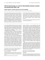
this molecule neuropeptide S, or NPS. The NPS pre-
cursor protein contains the typical structural features
of a neuropeptide precursor. A hydrophobic signal
peptide immediately follows the initiator methionine.
The immature peptide is preceded by a pair of basic
amino acids (Lys, Arg) that might serve as processing
sites for proteolytic cleavage (Fig. 1). The NPS recep-
tor is a typical GPCR containing seven membrane-
spanning domains. It shares moderate homology
with other members of the GPCR supergene family,
especially neuropeptide receptors. The highest degree
of similarity is found with vasopressin or oxytocin
receptors.
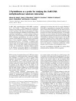
Using in situ hybridization we studied the distribu-
tion of NPS precursor and receptor mRNA in rat
brain (Fig. 2). These experiments showed that the
NPS receptor (NPSR) mRNA is widely expressed
throughout the nervous system, with highest levels
found in cortex, thalamus, hypothalamus, and amy-
gdala. Low levels of NPSR mRNA were detected in
brainstem. In contrast, the NPS precursor mRNA
was mainly expressed in brainstem nuclei such as the
locus coeruleus area, the principle 5 sensory nucleus
and the lateral parabrachial nucleus of the brain-
stem. A small number of scattered NPS-positive cells
were found in other brain areas, such as amygdala
and hypothalamus.
The NPS-producing neurons in the locus coeruleus
area were found to define a novel nucleus that lies
between the noradrenergic locus coeruleus proper and
Barrington’s nucleus. Double in situ hybridization
revealed that NPS precursor mRNA is neither colocal-
ized with tyrosine hydroxylase (TH; a marker of
noradrenergic neurons) nor with corticotropin-releas-
ing factor (CRF; a marker for neurons of Barrington’s
nucleus). This unique anatomical pattern of NPS
expressing neurons defines a previously unrecognized
population of cells in the brainstem. It is also evident
from our in situ hybridization data that there are still
other cells in this area that express none of these neuro-
chemical markers (TH, CRF or NPS) and thus might
contain other known or novel transmitters.
Fig. 1. Primary structure of the human NPS precursor. The hydro-
phobic signal peptide is shown by broken underlining. Endopro-
tease cleavage at a pair of basic amino acids (KR; double
underlined) is presumed to release the mature NPS peptide (single
underlined).
NPS produces arousal and anxiolysis R. K. Reinscheid and Y L. Xu
5690 FEBS Journal 272 (2005) 5689–5693 ª 2005 FEBS
Cells stably expressing NPSR were used to charac-
terize the in vitro pharmacology of NPS. Nanomolar
concentrations of NPS produce a transient increase in
intracellular free Ca
2+
, indicating that NPS might be
an excitatory transmitter in vivo by elevating intracellu-
lar Ca
2+
. A radiolabled analog of NPS (
125
I-labeled
Tyr
10
-NPS) shows displaceable binding with high affin-
ity (K
d
¼ 0.3 nm) [11]. High affinity receptor binding
and high potency to evoke intracellular second messen-
ger responses are important pharmacological para-
meters to classify NPS as a typical neuropeptide
transmitter which is active at low concentrations.
NPS promotes arousal and reduces
anxiety-like behavior in rats and mice
The first step in studying the physiological functions of
NPS in the nervous system was a detailed analysis of
locomotor behavior produced by central administra-
tion of NPS in mice. Mice that were naı
¨
ve to the test
chamber showed a profound increase in locomotion,
measured as the total distance traveled over one hour.
It is well known that animals naturally show increased
exploratory activity when they are exposed to a novel
environment and therefore the NPS-induced locomo-
tion seen in these naı
¨
ve mice could have two possible
reasons: (a) NPS might enhance the exploratory com-
ponent by increasing the response to novelty, or (b)
the stimulatory effect might be independent of novelty
and thus genuine arousal. To distinguish between these
two possibilities we injected mice that had been habitu-
ated to the test chamber for one hour before adminis-
tration of the drug. In habituated mice, injection of
saline (control) did not produce any increase in loco-
motion because they had already explored the test
chamber extensively before. However, low concentra-
tions (0.1 or 1 nmole) of NPS were able to reinstate
exploration in habituated mice that lasted for almost
one hour. In both naı
¨
ve and habituated mice NPS sig-
nificantly reduced inactivity, i.e., time the animals rest.
These experiments show that NPS produces profound
arousal that is independent of novelty [11].
Because arousal is an important component of
wakefulness, we also analyzed the effect of NPS on
sleep patterns in rats during their normal period of
inactivity, i.e., during the light phase. Low doses of
NPS significantly increased wakefulness and conversely
suppressed all stages of sleep during the first hour post
administration. These studies indicate that NPS might
be involved in the induction or maintenance of wake-
fulness. The arousal-promoting effect of NPS might be
partially mediated by NPSRs expressed in thalamic
Fig. 2. Schematic drawings of NPS receptor mRNA expression in the rat brain. Representative regions with high levels of NPS receptor
mRNA signals (small circles) are depicted in the drawings. Numbers at the bottom left of each drawing correspond to the anteroposterior
distance of the plate relative to bregma according to the rat brain atlas of Paxinos & Watson [20]. Strong NPS receptor mRNA expression is
found in the anterior olfactory nucleus, endopiriform nucleus, piriform cortex, motor cortex, retrosplenial cortex and subiculum. Multiple tha-
lamic nuclei including the midline nuclei of the thalamus (indicated by an arrow) show significant levels of NPSR expression. Substantial
expression of NPSR mRNA is also observed in the hypothalamus and the amygdala complex. DEn, dorsal endopiriform nucleus; AON, anter-
ior olfactory nucleus; En, endopiriform nucleus (dorsal and ventral); M2, motor cortex 2; Hyp, hypothalamus; Amg, amygdala; RSA, retrosple-
nial agranular cortex; S, subiculum; Prc, precommissural nucleus, Pvp, paraventricular thalamic nucleus; PH, posterior hypothalamus.
R. K. Reinscheid and Y L. Xu NPS produces arousal and anxiolysis
FEBS Journal 272 (2005) 5689–5693 ª 2005 FEBS 5691
midline nuclei, as this brain structure is known to act
as a relay between arousal centers of the brainstem
and the cortex [12].
High levels of NPS receptor expression were also
found in the amygdala. The amygdala is a brain struc-
ture that is closely involved in the processing of emo-
tional behavior and memories [13]. The well-established
role of the amygdala in modulation of fear and anxiety
led us to hypothesize that the NPS system might also
be involved in emotional behavior. Therefore, the effect
of NPS administration was tested in mice using four
different paradigms which are able to measure fearful
responses and have been validated using anxiolytic
drugs such as diazepam. We found that centrally
administered NPS could produce an anxiolytic-like pro-
file that was independent of the motor-activating effects
of the peptide [11]. NPS increased the time the animals
spent exploring the less protected or brighter areas of
the different test chambers (open field, light-dark box,
elevated plus maze) similar to classical anxiolytic drugs.
In order to control for possible confounding effects
of the NPS-induced hyperlocomotion, we used the
marble burying test. This is a behavioral paradigm in
which anxiolytic drugs have been shown to selectively
reduce a natural defensive behavior [14]. NPS adminis-
tration reduced the time mice engaged in burying the
unfamiliar objects placed in their cages [11]. In
summary, the behavioral studies showed that NPS can
produce arousal independent of novelty while also
alleviating anxiety responses triggered by stressful or
unfamiliar environments.
Comparison with other neuro-
transmitters involved in arousal and
anxiety
The present examples demonstrate that NPS can
potently modulate arousal and stress. This pharmaco-
logical spectrum of NPS is quite unique as compared
to other transmitters or drugs that influence sleep
and ⁄ or emotional behavior. For example, stimulants
such as amphetamine or cocaine promote arousal and
suppress sleep but appear to have anxiogenic-like
effects in tests of emotional behavior [15,16]. Hypocre-
tin ⁄ orexin is able to suppress sleep and induce pro-
found wakefulness, but the peptide shows no effects
on anxiety-like behavior [17]. The antinarcolepsy drug
modafinil (ProvigilÒ), whose mechanism of action is
still unknown, induces long-lasting wakefulness but
does not modulate anxiety [18]. Typical anxiolytic
drugs such as benzodiazepines (diazepam, ValiumÒ)
do not affect locomotion at anxiolytic doses but tend
to inhibit motor activity at higher doses [19]. These
examples show that NPS produces a unique spectrum
of behavioral effects. Future research will have to dem-
onstrate how release of endogenous NPS is involved in
modulating sleep-wake states and emotional behavior.
NPS agonists could have unique applications in the
treatment of hypersomnia and anxiety disorders while
NPS antagonists might be novel therapeutic tools to
treat insomnia. Synthetic NPS agonists and antago-
nists will also be crucial to discover and study further
physiological functions of NPS and validate its poten-
tial as a drug target.
NPS and its receptor are a very recent example for
the impact of orphan receptor research on neuro-
science and our understanding of brain functions. The
identification of NPS as a modulator of arousal and
anxiety represents a first step to elucidate its complete
spectrum of physiological functions and sheds new
light on the neurochemistry and biological basis of
sleep-wakefulness regulation and fear.
Acknowledgements
R.K.R. and Y.L.X. were supported in part by grants
from the National Institutes of Mental Health (NIMH).
R.K.R. was also supported by a Young Investigator
Award from the National Alliance for Research on
Schizophrenia and Depression (NARSAD).
References
1 Siegel JM (2004) The neurotransmitters of sleep. J Clin
Psychiatry 65 (Suppl. 16), 4–7.
2 Jones BE (2003) Arousal systems. Front Biosci 8, 438–
451.
3 Jones BE (2004) Activity, modulation and role of basal
forebrain cholinergic neurons innervating the cerebral
cortex. Prog Brain Res 145, 157–169.
4 Gerashchenko D, Okano Y, Urade Y, Inoue S &
Hayaishi O (2000) Strong rebound of wakefulness fol-
lows prostaglandin D2- or adenosine A2a receptor
agonist-induced sleep. J Sleep Res 9, 81–87.
5 Greene R & Siegel J (2004) Sleep: a functional enigma.
Neuromolecular Med 5, 59–68.
6 Chemelli RM, Willie JT, Sinton CM, Elmquist JK,
Scammell T, Lee C, Richardson JA, Williams SC,
Xiong Y, Kisanuki Y et al. (1999) Narcolepsy in orexin
knockout mice: molecular genetics of sleep regulation.
Cell 98, 437–451.
7 Lin L, Faraco J, Li R, Kadotani H, Rogers W, Lin X,
Qiu X, de Jong PJ, Nishino S & Mignot E (1999) The
sleep disorder canine narcolepsy is caused by a mutation
in the hypocretin (orexin) receptor 2 gene. Cell 98, 365–
376.
NPS produces arousal and anxiolysis R. K. Reinscheid and Y L. Xu
5692 FEBS Journal 272 (2005) 5689–5693 ª 2005 FEBS
8 de Lecea L & Sutcliffe JG (2005) The hypocretins and
sleep. FEBS J 272, 5675–5688.
9 Cheng MY, Bullock CM, Li C, Lee AG, Bermak JC,
Belluzzi J, Weaver DR, Leslie FM & Zhou QY (2002)
Prokineticin 2 transmits the behavioural circadian rhythm
of the suprachiasmatic nucleus. Nature 417, 405–410.
10 de Lecea L, Criado JR, Prospero-Garcia O, Gautvik
KM, Schweitzer P, Danielson PE, Dunlop CL, Siggins
GR, Henriksen SJ & Sutcliffe JG (1996) A cortical neuro-
peptide with neuronal depressant and sleep-modulating
properties. Nature 381, 242–245.
11 Xu YL, Reinscheid RK, Huitron-Resendiz S, Clark SD,
Wang Z, Lin SH, Brucher FA, Zeng J, Ly NK, Henrik-
sen SJ, de Lecea L & Civelli O (2004) Neuropeptide S:
a neuropeptide promoting arousal and anxiolytic-like
effects. Neuron 43, 487–497.
12 van der Werf YD, Witter MP & Groenewegen HJ
(2002) The intralaminar and midline nuclei of the thala-
mus. Anatomical and functional evidence for participa-
tion in processes of arousal and awareness. Brain Res
Rev 39, 107–140.
13 Pare D, Quirk GJ & Ledoux JE (2004) New vistas on
amygdala networks in conditioned fear. J Neurophysiol
92, 1–9.
14 N’junge K & Handley SL (1991) Evaluation of marble-
burying behavior as a model of anxiety. Pharmacol
Biochem Behav 38, 63–67.
15 Hascoet M & Bourin M (1998) A new approach to the
light ⁄ dark test procedure in mice. Pharmacol Biochem
Behav 60, 645–653.
16 Paine TA, Jackman SL & Olmstead MC (2002)
Cocaine-induced anxiety: alleviation by diazepam, but
not buspirone, dimenhydrinate or diphenhydramine.
Behav Pharmacol 13, 511–523.
17 Hagan JJ, Leslie RA, Patel S, Evans ML, Wattam TA,
Holmes S, Benham CD, Taylor SG, Routledge C, Hem-
mati P et al. (1999) Orexin A activates locus coeruleus
cell firing and increases arousal in the rat. Proc Natl
Acad Sci USA 96, 10911–10916.
18 Simon P, Panissaud C & Costentin J (1994) The stimu-
lant effect of modafinil on wakefulness is not associated
with an increase in anxiety in mice. A comparison with
dexamphetamine. Psychopharmacology 114, 597–600.
19 Chaouloff F, Durand M & Mormede P (1997) Anxiety-
and activity-related effects of diazepam and chlordiazep-
oxide in the rat light ⁄ dark and dark ⁄ light tests. Behav
Brain Res 85, 27–35.
20 Paxinos G & Watson C (1997) The Rat Brain in Stereo-
taxic Coordinates . Compact 3rd edn. Academic Press,
San Diego, CA.
R. K. Reinscheid and Y L. Xu NPS produces arousal and anxiolysis
FEBS Journal 272 (2005) 5689–5693 ª 2005 FEBS 5693