Báo cáo khoa học: Human airway trypsin-like protease induces amphiregulin release through a mechanism involving protease-activated receptor-2-mediated ERK activation and TNF a-converting enzyme activity in airway epithelial cells doc
Bạn đang xem bản rút gọn của tài liệu. Xem và tải ngay bản đầy đủ của tài liệu tại đây (439.43 KB, 13 trang )
Human airway trypsin-like protease induces amphiregulin
release through a mechanism involving protease-activated
receptor-2-mediated ERK activation and TNF a-converting
enzyme activity in airway epithelial cells
Manabu Chokki, Hiroshi Eguchi, Ichiro Hamamura, Hiroaki Mitsuhashi and Takashi Kamimura
Pharmaceutical Discovery Research Laboratories, Institute for Bio-Medical Research, Teijin Pharma Limited, Tokyo, Japan
Human airway trypsin-like protease (HAT) is a novel
serine protease that can be purified from the sputum
of patients with chronic airway diseases, such as chro-
nic bronchitis and bronchial asthma, based on its pro-
tease activity [1]. It exists in the sputum as a monomer
with a molecular size of 27 kDa as estimated by gel fil-
tration chromatography [1]. HAT cDNA has been iso-
lated from a tracheal tissue cDNA library; analysis of
this cDNA suggests that HAT is originally translated
as a precursor with a molecular size of 48 kDa and
Keywords
amphiregulin; extracellular signal-regulated
kinase; human airway trypsin-like protease;
protease-activated receptor-2; tumour
necrosis factor a-converting enzyme
Correspondence
M. Chokki, Pharmaceutical Discovery
Research Laboratories, Institute for Bio-
Medical Research, Teijin Pharma Limited,
Tokyo, Japan
Tel: +81 42 586 8134
Fax: +81 42 587 5515
E-mail:
(Received 15 September 2005, revised 20
October 2005, accepted 26 October 2005)
doi:10.1111/j.1742-4658.2005.05035.x
Human airway trypsin-like protease (HAT), a serine protease found in the
sputum of patients with chronic airway diseases, is an agonist of protease-
activated receptor-2 (PAR-2). Previous results have shown that HAT
enhances the release of amphiregulin (AR); further, it causes MUC5AC
gene expression through the AR-epidermal growth factor receptor pathway
in the airway epithelial cell line NCI-H292. In this study, the mechanisms
by which HAT-induced AR release can occur were investigated. HAT-
induced AR gene expression was mediated by extracellular signal-regulated
kinase (ERK) pathway, as pretreatment of cells with ERK pathway inhib-
itor eliminated the effect of HAT on AR mRNA. Both HAT and PAR-2
agonist peptide (PAR-2 AP) induced ERK phosphorylation; further, desen-
sitization of PAR-2 with a brief exposure of cells to PAR-2 AP resulted in
inhibition of HAT-induced ERK phosphorylation, suggesting that HAT
activates ERK through PAR-2. Moreover, PAR-2 AP induced AR gene
expression subsequent to protein production in the cellular fraction
through the ERK pathway indicating that PAR-2-mediated activation of
ERK is essential for HAT-induced AR production. However, in contrast
to HAT, PAR-2 AP could not cause AR release into extracellular space; it
appears that activation of PAR-2 is not sufficient for HAT-induced AR
release. Finally, HAT-induced AR release was eliminated by blockade of
tumour necrosis factor a-converting enzyme (TACE) by the TAPI-1 and
RNA interference, suggesting that TACE activity is necessary for HAT-
induced AR release. These observations show that HAT induces AR pro-
duction through the PAR-2 mediated ERK pathway, and then causes AR
release by a TACE-dependent mechanism.
Abbreviations
AR, amphiregulin; EGFR, epidermal growth factor receptor; ERK, extracellular signal-regulated kinase; HAT, human airway trypsin-like
protease; HGF, hepatocyte growth factor; MEK, ERK kinase; PAR, protease-activated receptor; MMP, matrix metalloprotease; PAR-2 AP,
PAR-2 agonist peptide; siRNA, small interfering RNA; TACE, tumour necrosis factor a-converting enzyme.
FEBS Journal 272 (2005) 6387–6399 ª 2005 The Authors Journal compilation ª 2005 FEBS 6387
possesses a hydrophobic transmembrane domain near
the N terminus [2]. Based on this derived structure,
HAT is thought to be a member of the type-II trans-
membrane serine protease family, which includes corin,
enteropeptidase, MT-SP1 (also known as matriptase)
and hepsin [3]. Northern blotting results using RNA
that was collected from 17 human tissues showed that
HAT mRNA is most prominently expressed in tra-
cheal tissue, suggesting that HAT is localized in the
airway [2]. Additional evidence supporting this was
obtained from the results of using a HAT-specific
mAb to conduct an immunohistochemical analysis of
the airway tissue isolated from healthy subjects. These
results show that HAT is specifically found in ciliated
epithelial cells; however, it is not found in the basal
and goblet cells in the epithelium or in the submucosal
gland cells [4]. Therefore, it is thought that HAT might
be responsible for regulating certain biological proces-
ses in airway cells.
It has been shown that protease-activated receptor
(PAR)-2 functions as a target protein for HAT in
bronchial epithelial cells [5]. PAR-2 is a member of
the PAR protein family; this family includes PAR-1,
PAR-3 and PAR-4 [6]. PARs are heterotrimeric guan-
ine nucleotide-binding protein-coupled receptors that
are activated by the cleavage of their N-terminal
domain. The proteolytic cleavage of the N-terminal
region of each PAR reveals a new N terminus. This
newly revealed terminus acts as a tethered ligand that
binds to the receptor and autoactivates it. PAR-2 is
activated by trypsin and mast cell tryptase and is
known to mediate airway inflammation both in vitro
[7,8] and in vivo [9]. Furthermore, it has been reported
that the PAR-2 mRNA expression in airway epithe-
lium increases in bronchial asthmatic patients [10].
These observations suggest that HAT might mediate
airway inflammation by PAR-2 activation.
In addition to airway inflammation, hypersecretion
of airway mucus is a characteristic sign of chronic
obstructive airway diseases, which include bronchitis,
bronchial asthma and cystic fibrosis [11,12]. Such
excessive mucus secretion causes airway obstruction,
which contributes to the morbidity and mortality due
to these diseases [11,12]. MUC5AC, a prominent pro-
tein in the airway, is known to participate in the path-
ogenesis of mucus hypersecretion in patients with
chronic airway diseases [13–15]. In a previous study
[16], HAT was shown to increase MUC5AC gene
expression, leading to mucus production by the airway
epithelial cell line (NCI-H292) in a range of samples
obtained from the sputum of patients with chronic air-
way disease such as bronchial asthma or bronchitis. In
addition, the effect of HAT was completely negated by
treating these cells with a neutralizing antibody of
either amphiregulin (AR) or its receptor, i.e. epidermal
growth factor receptor (EGFR). Further, the treatment
of cells with HAT induced AR gene expression, and
subsequently, AR protein release. Although the PAR-2
activation in NCI-H292 cells, inferred by intracellular
calcium mobilization, is thought to occur after NCI-
H292 stimulation by either HAT or the PAR-2 agonist
peptide (PAR-2 AP), neither MUC5AC gene expres-
sion nor AR protein release was affected by treatment
with PAR-2 AP. From these observations, HAT-
induced MUC5AC gene expression appears to be
mediated by the AR-EGFR pathway, and PAR-2 acti-
vation alone cannot account for the effect of HAT
[16]. It has been shown that EGFR plays an important
role in the induction of mucin gene expression and
mucin production [17–19]. Further, the activation of
the AR-EGFR pathway has been observed in airway
epithelial cells exposed to cigarette smoke extract
[20,21] and fine particulate matter [22], which are well
known as agents causing airway diseases. These obser-
vations suggest that the AR-EGFR pathway plays an
important role in the induction of mucus overproduc-
tion; however, the mechanism by which HAT induces
AR release remains unknown. In the present study,
HAT-induced AR release and the target molecule of
HAT were investigated.
Results
HAT regulates AR release at the transcriptional
level
Results of a previous study, which focused on the
effect of HAT on AR mRNA level 2–24 h after the
treatment, indicated that a statistically significant
increase in the AR mRNA level began 2 h after the
start of HAT treatment (P<0.01), and that these
levels returned to the basal level after 24 h [16]. To
evaluate the kinetics of HAT-induced AR release, the
time course of this release was investigated. As shown
in Fig. 1A, HAT stimulated a time-dependent AR
release. A statistically significant effect of HAT was
observed as early as 2 h after treatment (Fig. 1A;
P<0.05), suggesting that the effect of HAT on the
AR mRNA level occurred earlier than 2 h. To define
the onset time of HAT-induced increase in AR mRNA
level more accurately, changes in the AR mRNA level
of HAT-treated cells were evaluated at 0.5–2 h after
stimulation. As shown in Fig. 1B, the AR mRNA level
appeared to increase almost immediately, i.e. 0.5 h
after the HAT treatment, with a statistically significant
difference (P<0.01) from the control. To evaluate
HAT-induced AR release by PAR-2 and TACE M. Chokki et al.
6388 FEBS Journal 272 (2005) 6387–6399 ª 2005 The Authors Journal compilation ª 2005 FEBS
the requirement of transcriptional regulation of the AR
gene on HAT-induced AR release, the effects of
actinomycin D and cycloheximide, which are transcrip-
tion and protein synthesis inhibitors, respectively, on
HAT-induced AR release were determined. As shown
in Fig. 1C, HAT-induced AR release was significantly
and almost completely inhibited by both actino-
mycin D and cycloheximide treatments. These obser-
vations indicate the HAT-induced AR release is
regulated at the transcriptional level.
HAT induces tyrosine phosphorylation of EGFR
mediated by AR
To examine whether HAT-induced autocrine AR
release activates EGFR, HAT-induced tyrosine phos-
phorylation of EGFR and the involvement of AR in
this signalling cascade were investigated. Western blots
probed with antiphospho-EGFR antibodies were used
to determine HAT-induced tyrosine phosphorylation of
EGFR. Since EGFR is known to be phosphorylated by
its intrinsic receptor kinase through homo- and hetero-
dimerization following ligand binding (autophosphory-
lation) and by nonreceptor tyrosine kinases such as Src
family kinases [23,24], phosphorylation of EGFR at
Tyr845 (known to be phosphorylated by Src [24]) and
Tyr1068 (known as an autophosphorylation site [23])
were investigated. In HAT-treated cells, phosphoryla-
tion of EGFR at Tyr845 and Tyr1068 was not observed
until 30 min after treatment (Fig. 2A), whereas imme-
diate phosphorylation (i.e. 3 min after the stimulation)
of these tyrosine residues occurred during treatment
with AR. However, 120 min after HAT treatment,
EGFR phosphorylation at these tyrosine residues had
increased and the extent of phosphorylation continued
to increase until 480 min after the treatment and
decreased to the basal level by the last time point meas-
ured, 24 h after treatment (Fig. 2B). Next, the effect of
anti-AR neutralizing antibody on HAT-induced phos-
phorylation of EGFR was assessed. The extent of
EGFR phosphorylation at the Tyr1068 residue was
used to evaluate the EGFR activation because phos-
phorylation on this tyrosine residue functions as the
direct binding site for the signal-transducing adapter
molecule Grb2 [25], leading to ERK activation [26,27]
following MUC5AC gene expression in NCI-H292 cells
[19]. As shown in the time course analysis in Fig. 2C,
HAT-induced EGFR phosphorylation was almost
completely inhibited in the presence of anti-AR neutral-
izing antibody, from the onset time of HAT-induced
EGFR phosphorylation (120 min after treatment) to
the peak of phosphorylation (480 min after treatment).
Results of a previous study suggest that AR is involved
in HAT-induced MUC5AC gene expression [16]. In this
study, the effect of AR on HAT-induced MUC5AC
production was also investigated at the protein level.
As shown in Fig. 2D, the MUC5AC protein content of
NCI-H292 cells increased twofold 24 h after the HAT
treatment; however, this effect was almost completely
negated in the presence of anti-AR neutralizing anti-
body. These results indicate that the HAT-induced
EGFR phosphorylation almost completely depends on
AR, and HAT-induced MUC5AC production is medi-
ated through the AR-EGFR pathway.
Fig. 1. HAT regulates AR production at the transcriptional level. (A,
B) NCI-H292 cells were stimulated with HAT (200 n
M) for indicated
durations. (C) NCI-H292 cells were pretreated with the vehicle alone
(Veh), cycloheximide (CHX; 5 lgÆmL
)1
) or actinomycin D (ActD;
10 lgÆmL
)1
) for 20 min and then stimulated with HAT (200 nM) for
2 h in the presence of these inhibitors. (A, C) ELISA was used to
determine the AR concentrations in the culture supernatant. (B)
Total RNA was extracted, and quantitative real-time RT ⁄ PCR (Taq-
Man
TM
) was used to determine the AR and b-actin mRNA amounts.
The results are expressed as the mean ± SD (n ¼ 3). *P < 0.05,
**P < 0.01 when compared with vehicle-treated cells at the same
time point and
#
P < 0.05,
##
P < 0.01 when compared with HAT-
treated cells in the absence of the inhibitors, Dunnett’s test.
M. Chokki et al. HAT-induced AR release by PAR-2 and TACE
FEBS Journal 272 (2005) 6387–6399 ª 2005 The Authors Journal compilation ª 2005 FEBS 6389
Involvement of extracellular-signal regulated kinase
pathway in HAT-induced AR gene expression
The cellular mechanism responsible for HAT-induced
AR gene expression was examined. As it has been repor-
ted that the extracellular-signal regulated kinase (ERK)
pathway involves AR release from airway epithelial cells
exposed to fine particulate matter [22], the role of the
ERK signal transduction pathway in HAT-induced AR
gene expression was investigated using PD98059 and
U0126, which are potent and selective chemical inhibi-
tors of ERK kinase (MEK). As shown in Fig. 3A, pre-
treatment with PD98059 completely eliminated the
stimulatory effect of HAT on AR mRNA level. Simi-
larly, and consistent with findings showing that HAT-
induced AR release is regulated at the transcriptional
level, the HAT-induced AR release was also completely
inhibited by treatment with either PD98059 or U0126
(Fig. 3B). These results suggest that HAT induces AR
gene expression through the MEK-ERK pathway.
HAT induces biphasic ERK activation through
AR-dependent and -independent pathways
To determine whether HAT activates the MEK-ERK
pathway, western blotting using antiphospho-MEK and
A
B
C
D
Fig. 2. HAT induces phosphorylation of EGFR mediated by AR in NCI-
H292 cells. NCI-H292 cells were stimulated with HAT (200 n
M)orAR
(3 ngÆmL
)1
) for the indicated durations (A, B). NCI-H292 cells were
pretreated with the vehicle alone or anti-AR neutralizing antibody
(aAR; 10 lgÆmL
)1
) for 20 min and then either (C) stimulated with
HAT (200 n
M) for the indicated durations or (D) stimulated with HAT
(300 n
M) for 24 h in the presence of the antibodies. (A, B, C) Immuno-
blotting, with repeated probing using the antibodies indicated on the
left side of the figure, was used to analyse the cell lysates. (D)
MUC5AC protein level in cell lysates was determined by ELISA. The
results are presented as mean ± SD (n ¼ 3). **P < 0.01 when com-
pared with vehicle-treated cells and
##
P < 0.01 when compared with
HAT-treated cells in the absence of the antibodies, Dunnett’s test.
Fig. 3. Involvement of ERK in HAT-induced AR gene expression.
(A, B) NCI-H292 cells were pretreated with the vehicle alone (Veh),
PD98059 (PD; 10 l
M) or U0126 (U; 5 lM) for 20 min. (A) Cells were
then stimulated with HAT (200 n
M) for 1 h in the presence of the
inhibitor or vehicle, total RNA was extracted and quantitative real-
time RT ⁄ PCR (TaqMan
TM
) analysis was used to determine the
amounts of AR and b-actin mRNA. (B) Cells were stimulated with
HAT (200 n
M) for 2 h in the presence of the inhibitor or vehicle and
ELISA was used to determine the AR concentrations in the culture
supernatant. The results are presented as mean ± SD (n ¼ 3).
*P < 0.05, **P < 0.01 when compared with vehicle-treated cells
and
#
P < 0.05,
##
P < 0.01 when compared with HAT-treated cells
in the absence of inhibitors, Dunnett’s test.
HAT-induced AR release by PAR-2 and TACE M. Chokki et al.
6390 FEBS Journal 272 (2005) 6387–6399 ª 2005 The Authors Journal compilation ª 2005 FEBS
antiphospho-ERK antibodies was performed. Time-
dependent effects of HAT on phosphorylation of MEK
and ERK were examined until 24 h after the treatment.
Phosphorylation of MEK and ERK was observed
within 5 min of the HAT treatment; however, de-
phosphorylation occurred 30 min after treatment. After
completion of the rapid transient phosphorylation of
MEK and ERK, a second, less extensive round of
MEK and ERK phosphorylation was observed, which
began 120 min after the HAT stimulation and lasted
until 480 min after treatment (Fig. 4A). In order to
assess the effect of HAT on the kinase activity of ERK,
phosphorylation of the downstream kinase p90RSK at
Ser359 and Thr363 (residues known to be directly
phosphorylated by ERK [28]) was examined. Similar
to HAT-induced MEK-ERK phosphorylation, biphasic
phosphorylation of p90RSK was induced by HAT
treatment (Fig. 4B). To confirm this result, the effect of
PD98059 on HAT-induced p90RSK phosphorylation at
5 min and 480 min was examined independently by the
following steps. For the 5-min time point, NCI-H292
cells were pretreated with PD98050 20 min before the
treatment, while for the 480 min time point, NCI-H292
cells were treated with PD98059 30 min after HAT sti-
mulation; at this time, the first round of phosphoryla-
tion is completed (Fig. 4A). When assessed in this
manner, HAT-induced p90RSK phosphorylation at
5 min and 480 min was inhibited in the presence
of PD98059 (Fig. 4C), suggesting that p90RSK was
directly phosphorylated by activated ERK. Thus, the
HAT-induced biphasic ERK phosphorylation is accom-
panied by the enhancement of its kinase activity.
Next, the involvement of AR in the HAT-induced
biphasic ERK activation was investigated using an anti-
AR neutralizing antibody. The HAT-induced initial
ERK activation (5 min after stimulation with HAT)
was inhibited by a PD98059 treatment but not by the
anti-AR neutralizing antibody treatment (Fig. 5A),
while the HAT-induced second round of ERK activa-
tion (480 min after stimulation with HAT) was inhib-
ited by the PD98059 treatment or the anti-AR
neutralizing antibody treatment (Fig. 5B). A time-
course study of HAT-induced ERK activation in the
presence or absence of anti-AR neutralizing antibody
was also conducted in order to confirm the involvement
of AR. ERK activation at 5 min after HAT treatment
was not affected by the anti-AR neutralizing antibody;
however, the activation of ERK was completely inhib-
ited by the anti-AR neutralizing antibody at 120 min
and 240 min after HAT treatment (Fig. 5C). In addi-
tion, AR-induced ERK phosphorylation occurred
within 5 min of the stimulation and sustained up to
480 min (Fig. 5D); these kinetics were similar to those
of the HAT-induced second round of ERK activation
(Fig. 4A). Considered together, these observations sug-
gest that the HAT increases AR gene expression
through initial ERK activation and that a second round
of ERK activation is induced through EGFR that is
activated by autocrine AR stimulation. In addition,
these events appear to occur in the airway of patients
with chronic airway diseases since the HAT-induced
initial ERK activation was observed at a low HAT
concentration of 6.6 nm (equivalent to 10.8 mUÆmL
)1
,
Fig. 5E), which is similar to the concentration observed
in mucoid sputum from patients with either chronic
bronchitis (23.46 ± 18.03 mUÆmL
)1
) or bronchial
asthma (46.96 ± 43.96 mUÆmL
)1
[29]).
Desensitization of PAR-2 blocks HAT-induced
ERK phosphorylation
HAT and PAR-2 AP-induced activation of PAR-2 has
been observed in NCI-H292 cells [16]. In addition, it
has been reported that the activation of PAR-2 causes
ERK phosphorylation [8,30–34]. To clarify whether
the HAT-induced initial ERK activation is mediated
A
B
C
Fig. 4. HAT induces biphasic activation of ERK. (A, B) NCI-H292
cells were stimulated with HAT (200 n
M) for the indicated dura-
tions. (C) NCI-H292 cells were pretreated with the vehicle alone or
with PD98059 (PD; 10 l
M) for 20 min and then stimulated with
HAT (200 n
M) for 5 min. During evaluation at a culture period of
480 min, NCI-H292 cells were stimulated with HAT for 30 min and
then treated with the vehicle alone or with PD98059 (PD; 10 l
M).
Further, they were cultured up to 480 min. Immunoblotting, with
repeated probing using the antibodies indicated on the left side of
the figure, was used to analyse the cell lysates.
M. Chokki et al. HAT-induced AR release by PAR-2 and TACE
FEBS Journal 272 (2005) 6387–6399 ª 2005 The Authors Journal compilation ª 2005 FEBS 6391
by PAR-2, experiments using PAR-2 AP, which specif-
ically activates PAR-2 [6], were conducted. First, the
effect of PAR-2 AP on the extent and pattern of ERK
activation was examined. In PAR-2 AP-treated cells,
the ERK activation was observed within 5 min of
treatment (Fig. 6A); however, unlike the HAT-induced
biphasic ERK activation (Fig. 4A), PAR-2 AP-induced
ERK activation was transient, and a second round of
ERK activation was not observed within 480 min of
treatment (Fig. 6A). Consistent with findings that
show that a second round of HAT-induced ERK acti-
vation is mediated by the AR-EGFR pathway (Fig. 5B
and C), the activation of EGFR was not observed
within 480 min of PAR-2 AP treatment (Fig. 6B).
Next, the effect of a brief exposure to PAR-2 AP (to
desensitize PAR-2 [30,31]); prior to the HAT stimula-
tion, on the HAT-induced initial ERK activation was
examined. As shown in Fig. 6C, brief exposure of
NCI-H292 cells to PAR-2 AP resulted in specific inhi-
bition of PAR-2 AP-induced ERK activation whereas
hepatocyte growth factor (HGF)-induced ERK activa-
tion was unaffected. Further, HAT-induced initial
ERK activation was completely inhibited by pretreat-
ment with PAR-2 AP (Fig. 6C). These observations
suggest that at least a part of the HAT-induced initial
ERK activation was mediated by PAR-2.
ERK activation through PAR-2 induces AR protein
production but not protein release into the
culture supernatant
Since HAT-induced initial ERK activation results in
induction of AR gene expression, the effect of PAR-2
A
B
C
D
E
Fig. 5. HAT induces biphasic activation of ERK through AR-depend-
ent and -independent pathways. (A, C) NCI-H292 cells were pre-
treated with the vehicle alone (Veh), PD98059 (PD; 10 l
M), anti-AR
neutralizing antibody (aAR; 10 lgÆmL
)1
) or normal mouse IgG1
(IgG; 10 lgÆmL
)1
) for 20 min. The cells were then stimulated with
HAT (200 n
M) for 5 min (A) or the indicated durations (C) in the
presence of the indicated inhibitors or antibody. (B) For the
480 min culture period, NCI-H292 cells were stimulated with HAT
for 30 min and then treated with the vehicle alone or with
PD98059 (PD; 10 l
M), anti-AR neutralizing antibody (aAR;
10 lgÆmL
)1
) or normal mouse IgG1 (IgG; 10 lgÆmL
)1
) and further
cultured for 480 min. (D) Cells were stimulated with AR (3 lgÆmL
)1
)
for the indicated durations. (E) Cells were stimulated with increas-
ing concentrations of HAT for 5 min. Immunoblotting, with repea-
ted probing using the antibodies indicated on the left side of the
figure, was used to analyse the cell lysates.
A
B
C
Fig. 6. Desensitization of PAR-2 blocks HAT-induced initial ERK
activation. (A, B) NCI-H292 cells were stimulated with PAR-2 AP
(300 l
M) or HAT (200 nM) for the indicated durations. (C) NCI-H292
cells were pretreated with the vehicle alone or with PAR-2 AP
(300 l
M) for 20 min. (C) Cells were then stimulated with HAT
(200 n
M), PAR-2 AP (300 lM) or HGF (20 ngÆmL
)1
) for 5 min in the
presence of inhibitors or the vehicle. Immunoblotting, with repea-
ted probing using the antibodies indicated on the left side of the
figure, was used to analyse the cell lysates.
HAT-induced AR release by PAR-2 and TACE M. Chokki et al.
6392 FEBS Journal 272 (2005) 6387–6399 ª 2005 The Authors Journal compilation ª 2005 FEBS
AP on the AR gene expression during the 0.5–2-h per-
iod after the treatment was examined. In PAR-2
AP-treated NCI-H292 cells, AR mRNA level signifi-
cantly increased at 0.5 h after the treatment (Fig. 7A).
In contrast to the continuous increase in AR mRNA
level observed in HAT-treated cells until 4 h after the
treatment (Fig. 1B), the AR mRNA level returned to
the basal level 1 h after treatment in PAR-2 AP-trea-
ted cells and did not significantly increase until 2 h
after the treatment (Fig. 7A). These results demon-
strate that the activation of PAR-2 causes a rapid
transient increase in AR mRNA level, although no sta-
tistically significant change in AR protein concentra-
tion has been observed in the culture supernatant of
PAR-2 AP-treated NCI-H292 cells in a previous study
[16]. To investigate whether PAR-2 AP has any effect
on AR protein production, the AR protein concentra-
tion in the culture supernatant and cellular lysates was
evaluated in cell cultures treated with PAR-2 AP. Sim-
ilar to the results of a previous study [16], PAR-2 AP
treatment had no effect on the AR protein concen-
tration in the culture supernatant 2 and 4 h after
treatment (Fig. 7B). However, the AR protein con-
centration in cell lysates prepared from PAR-2
AP-treated cells showed a statistically significant
increase (P<0.01) between 2 and 4 h after the treat-
ment. The effect of PAR-2 AP on AR protein produc-
tion was mediated by the ERK pathway since the
PAR-2 AP-induced increase in AR protein concentra-
tion in cellular lysate was completely eliminated by
pretreatment with PD98059 (Fig. 7C). These observa-
tions suggest that activation of PAR-2 mediated ERK
pathway results in the induction of AR gene expression
subsequent to the production of AR protein that is
bound to, or otherwise associated with, the cells, for
example, bound to the cell surface.
Tumour necrosis factor a-converting enzyme
activity is required for HAT-induced AR release
that prolongs HAT-induced AR gene expression
by a positive feedback loop
Results of the present study suggest that the activation
of PAR-2 is sufficient to induce AR protein produc-
tion, but cannot account for AR release. Thus, the
mechanism of HAT-induced AR release from a cell
was also investigated. The effect of GM6001, a broad-
spectrum metalloprotease inhibitor, and TAPI-1, a rel-
atively selective metalloprotease inhibitor for tumour
necrosis factor a-converting enzyme (TACE), on
HAT-induced AR release was determined as it is well
known that metalloproteases, such as matrix metallo-
protease (MMP) and TACE, cause AR release by
proteolytic cleavage of the transmembrane precursor
[20,35–37]. As shown in Fig. 8A, HAT-induced AR
protein release was significantly inhibited (P<0.01)
by pretreatment of cells with GM6001 and TAPI-1
(Fig. 8A). In addition, these inhibitors did not affect
the protease activity of HAT at the concentration used
in this study (data not shown). Next, to evaluate whe-
ther TACE is required for HAT-induced AR release,
endogenous TACE expression was blocked using a
small interfering RNA (siRNA). Silencing of TACE
Fig. 7. Activation of PAR-2 causes AR gene expression and AR pro-
tein production but does not evoke AR release. (A, B) NCI-H292
cells were stimulated with PAR-2 AP (300 l
M) for the indicated
durations. (C) NCI-H292 cells were pretreated with the vehicle
alone (Veh) or with PD98059 (PD; 10 l
M) for 20 min and then sti-
mulated with PAR-2 AP (300 l
M) for 2 h. (A) Total RNA was extrac-
ted, and quantitative real-time RT ⁄ PCR (TaqMan
TM
) analysis was
used to determine the amounts of AR and b-actin mRNA. (B, C)
ELISA was used to determine the AR concentrations in the culture
supernatants and cellular lysates. The results are presented as
mean ± SD (n ¼ 3). **P < 0.01 when compared with vehicle-trea-
ted cells at the same time point,
##
P < 0.01 when compared with
PAR-2 AP-treated cells in the absence of inhibitors, Dunnett’s test.
M. Chokki et al. HAT-induced AR release by PAR-2 and TACE
FEBS Journal 272 (2005) 6387–6399 ª 2005 The Authors Journal compilation ª 2005 FEBS 6393
was confirmed by flow cytometry using a mouse mAb
that recognizes an ectodomain of TACE. TACE was
detected on the surface of NCI-H292 cells but was
almost reduced to the background level by transfecting
with TACE siRNA (Fig. 8B). Inhibition of TACE
expression significantly suppressed HAT-induced AR
release (Fig. 8C). These data suggest that HAT causes
AR protein release mediated by TACE activity. Fur-
ther, it has been reported that the activation of EGFR
by EGF induces an autocrine EGFR ligand expression
in bronchial epithelial cells [21]; therefore, it is possible
that the prolonged effect of HAT on AR mRNA
expression is mediated by autocrine stimulation of AR.
To test this hypothesis, the involvement of AR with
HAT-induced AR mRNA expression was assessed
using the anti-AR neutralizing antibody. As shown in
Fig. 8D, treatment of exogenous AR causes AR
mRNA expression after 1 h stimulation and this effect
was inhibited by pretreatment with PD98059, suggest-
ing that AR induces AR gene expression through the
ERK pathway. Further, the HAT-induced increase in
AR mRNA level occurring 2 h after the HAT treat-
ment was significantly and almost completely negated
when NCI-H292 cells were treated with the anti-AR
neutralizing antibody (Fig. 8E). Considered together,
these results suggest that HAT induces AR release
through TACE activity and the released AR prolongs
HAT-induced AR gene expression by a positive feed-
back loop.
Discussion
Recently, the EGFR signalling pathway has been
shown to function as a common pathway through
which many stimuli induce MUC5AC production
in vitro [17–19]. Further, in the airway epithelium of
asthmatic patients, the activation of EGFR was sug-
gested to be involved in mucus hypersecretion [38],
which can have profound effects on health [11,12]. In
another study, HAT was originally found in the spu-
tum of patients with diseases causing airway mucus
hypersecretion [1]. Subsequent investigations revealed
that EGFR and its ligand AR are involved in the
HAT-induced MUC5AC gene expression. As a result,
HAT appeared to prefer EGFR as an activator. There-
fore, finding a mechanism by which HAT regulates
AR-EGFR activation might elucidate the basic mecha-
nisms of airway disease pathogenesis. Further, this
finding may also provide an additional benefit in terms
of leading to the development of new therapeutic strat-
egies to treat diseases marked by airway mucus hyper-
secretion. The results of the present study showed that
HAT activates EGFR through a pathway that includes
A
B
C
D
E
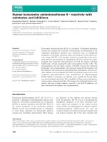
Fig. 8. TACE is involved in HAT-induced AR release, which pro-
longs HAT-induced AR gene expression by positive feedback loop.
(A, D, E) NCI-H292 cells were pretreated with the vehicle alone
(Veh), GM6001 (GM; 3 l
M), TAPI-1 (TAPI; 3 lM), PD98059 (PD;
10 l
M) or anti-AR neutralizing antibody (aAR; 10 lgÆmL
)1
) for
20 min. (B, C) NCI-H292 cells were transfected with siRNA for
TACE or control siRNA (cont) and cultured for 72 h. (A, C) Cells
were then stimulated with HAT (200 n
M) for 2 h and ELISA was
used to determine the AR concentration in culture supernatant.
(B) Cells were then collected and stained with anti-TACE antibody
or normal mouse IgG1 (background) and analysed for cell surface
TACE density by flow cytometry. (D, E) Cells were then stimulated
with AR (3 ngÆmL) for 1 h (D) or HAT (200 n
M) for 2 h (E) and
the total RNA was extracted, and quantitative real-time RT ⁄ PCR
(TaqMan
TM
) analysis was used to determine the amounts of AR
and b-actin mRNA. The results are presented as the mean ± SD
(n ¼ 3). *P < 0.05, **P < 0.01 when compared with vehicle-treated
cells and
##
P < 0.01 when compared with HAT (A, C) or AR
(B)-treated cells in the absence of inhibitors, Dunnett’s test.
HAT-induced AR release by PAR-2 and TACE M. Chokki et al.
6394 FEBS Journal 272 (2005) 6387–6399 ª 2005 The Authors Journal compilation ª 2005 FEBS
PAR-2 mediating ERK-dependent AR gene expression
and TACE-dependent AR protein release.
In the time course analysis of tyrosine phosphoryla-
tion of EGFR, AR-induced EGFR activation could be
detected within 3 min of stimulation; however, HAT-
induced EGFR activation was observed 2 h after sti-
mulation. This observation was in good agreement
with the results of our previous study showing that
HAT-mediated increase in MUC5AC mRNA level
occurs after the EGF-mediated increase in MUC5AC
mRNA level, although both cause MUC5AC gene
expression through EGFR activation. In the present
study, the time course of HAT-induced EGFR activa-
tion corresponded to that of HAT-induced AR release;
further, the activation of EGFR and the induction of
MUC5AC protein production were completely inhib-
ited in the presence of anti-AR neutralizing antibody.
Considered along with our previous observation that
among several EGFR ligands (EGF, HB-EGF, trans-
forming growth factor-a, AR), only anti-AR neutral-
izing antibody completely inhibited HAT-induced
MUC5AC gene expression, the results of the present
study strongly suggest that AR may be the initial
EGFR ligand responsible for HAT-induced EGFR
activation in NCI-H292 cells.
Different steps of the pathway leading to HAT-
induced AR production have been analysed using
pharmacological enzyme inhibitors. The following
results suggest the involvement of PAR-2-mediated
activation of ERK: (a) the MEK inhibitor PD98059
and U0126 inhibited HAT-induced AR release; (b)
activation of PAR-2 by PAR-2 AP induced ERK
phosphorylation and desensitization of PAR-2 resulted
in inhibition of HAT-induced initial ERK activation;
and (c) PD98059 inhibited PAR-2 AP-induced AR
protein production. Although the mechanisms by
which PAR-2 activates ERK were unknown, our study
aimed to show that PAR-2 mediated ERK activation
is an essential step in HAT-induced EGFR activation
since the induction of gene expression and subsequent
protein production of EGFR ligands by PAR-2 agon-
ists have not yet been reported in any cells.
It has been reported that the EGFR ligand family,
which includes AR, is originally translated as precur-
sors with transmembrane domains, and these proteins
are located on the exterior surface of the cytoplasmic
membrane. In response to an appropriate stimulation,
these precursors are proteolytically cleaved to obtain
their mature forms by metalloproteases such as MMP
and TACE and are released into the extracellular space
[20,35–37]. Although membrane-bound EGFR ligands
can engage in juxtacrine signalling [39,40], the TACE-
dependent release of AR has been shown to function
as a key step in transactivating EGFR in tobacco
smoke-stimulated bronchial epithelial cells [20]. In the
present study, HAT and PAR-2 AP stimulate AR gene
expression and subsequent AR protein production;
however, an increase in AR protein release in NCI-
H292 cells was only observed by HAT stimulation.
Further, the increase in the AR content in PAR-2
AP-treated cells did not result in the induction of
EGFR activation (Fig. 6B). Moreover, HAT-induced
AR protein release, which could induce EGFR activa-
tion, was eliminated by blocking TACE using siRNA
(Fig. 8C). These observations indicate that the activa-
tion of ERK through PAR-2 results in the production
of the AR precursor; however, this is not responsible
for the release of active AR. HAT stimulates AR
release by TACE activity mediated by a PAR-2-inde-
pendent mechanism. Although the cellular process of
TACE activation has not been defined, the mecha-
nisms that cause immediate activation of TACE are
probably not responsible for HAT-induced activation
of TACE, because time-course analysis results of this
study show that the HAT-induced AR-dependent acti-
vation of EGFR occurred 2 h after treating cells with
HAT (Fig. 2B). One possible mechanism is that HAT
may increase TACE expression. Recently, it has been
reported that in alveolar macrophage, lipopolysaccha-
ride increases TACE expression which correlates with
the catalytic activity of this enzyme [41]. In contrast to
the results from our study, it is reported that in colon
cancer cells, PAR-2 activation induces an MMP-
dependent release of transforming growth factor-a,
thus suggesting that PAR-2 activation caused the
MMP activation [34]. These observations suggest that
mechanisms that provoke the release of EGFR ligands
appear to be heterogeneous and depend upon specific
components of signalling molecules expressed within a
cell type. Thus, further investigation is needed to clar-
ify the mechanisms that lead to HAT-induced AR
release, including TACE activity regulation. In conclu-
sion, Fig. 9 depicts the mechanism of HAT-induced
AR in NCI-H292 cells. It schematically reflects the
major findings of the present study, which are as fol-
lows: (a) HAT induces AR gene expression subsequent
to AR protein release through ERK activation; (b) at
least a part of HAT-induced initial ERK activation is
mediated through PAR-2; (c) only HAT, and not
PAR-2 AP, causes AR protein release through TACE
activity; and (d) prolonged effect of HAT on AR
mRNA is mediated through a positive feedback loop
stimulated by autocrine AR. The results of the present
study shed light on a complex mechanisms of AR
release, further suggest that excess HAT activity
directly leads to the pathogenesis of chronic airway
M. Chokki et al. HAT-induced AR release by PAR-2 and TACE
FEBS Journal 272 (2005) 6387–6399 ª 2005 The Authors Journal compilation ª 2005 FEBS 6395
diseases through its effect on the PAR-2 and EGFR
signalling pathways.
Experimental procedures
Reagents and antibodies
Recombinant HAT (60 UÆmg
)1
protein) was prepared as
previously described [1,2,5]. In brief, HAT was expressed in
insect cells infected with a recombinant baculovirus carry-
ing the HAT cDNA [2]. Benzamidine affinity chromatogra-
phy was used to purify recombinant HAT from the cell
lysate [5], and the specific activity of the purified protein
was measured with Boc-Phe-Ser-Arg-MCA as a substrate,
as previously described [1]. PAR-2 AP consisting of Ser-
Leu-Ile-Gly-Lys-Val-NH
2
[6] was from Bachem AG (Bub-
endorf, Switzerland). Cycloheximide was from Wako Pure
Chemicals (Osaka, Japan); PD98059 and actinomycin D,
Biomol (Plymouth Meeting, PA, USA); U0126, Promega
(Madison, WI, USA); and GM6001 and TAPI-1, Calbio-
chem (San Diego, CA, USA). Human recombinant AR,
anti-TACE mAb (clone 111633) and nonimmune mouse
IgG
1
, used as negative controls, were from R&D Systems
Inc. (Minneapolis, MN, USA). Anti-p90RSK antibody was
from Upstate Biotechnologies (Lake Placid, NY, USA) and
neutralizing mouse mAb against AR (clone 31221.111) and
goat polyclonal antibody against AR were from Genzyme
(Minneapolis, MN, USA). The antiphospho-MEK (Ser217 ⁄
221), antiphospho-ERK (Thr202 ⁄ Tyr204), anti-ERK, anti-
phospho-p90RSK (Thr359 ⁄ Ser363), antiphospho-EGFR
(Tyr845) and antiphospho-EGFR (Tyr1068) antibodies
were from Cell Signaling (Beverly, MA, USA). The mouse
anti-EGFR mAb was from Transduction Laboratories
(Lexington, KY, USA) and the mouse anti-MUC5AC mAb
(Clone 45M1) were from LAB VISION (Fremont, CA,
USA).
Cell culture
NCI-H292 cells were from the American Type Culture Col-
lection (Rockville, MD, USA) and were cultured in RPMI
1640 medium supplemented with 10% (v ⁄ v) fetal bovide
serum, 50 UÆmL
)1
penicillin and 50 lgÆmL
)1
streptomycin
(Gibco BRL, Grand Island, NY, USA) in a humidified
incubator at 37 °C in an atmosphere of 5% CO
2
. Prior to
the experiments, confluent NCI-H292 cells were cultured in
a serum-free medium composed of RPMI 1640 medium
containing only 0.1% (w ⁄ v) BSA (Sigma, St. Louis, MO,
USA) for 24 h, unless otherwise indicated.
Immunoblotting
NCI-H292 cells were incubated with the appropriate condi-
tions, quickly placed on ice, and washed twice in ice-cold
NaCl ⁄ P
i
. The cells were then lysed in M-PER
Ò
Mammalian
Protein Extraction Reagent (Pierce, Rockford, IL, USA)
containing 1% (v ⁄ v) each of protease inhibitor cocktail and
phosphatase inhibitor cocktail (Sigma), while gently stirring
the cells for 5 min at room temperature. Each lysate was
transferred to a separate centrifuge tube, and the lysates
were centrifuged at 4 °C for 10 min at 15 000 g. The
cleared supernatants were collected separately, and a Bio-
Rad protein assay system (Bio-Rad, Hercules, CA, USA)
was used to determine the protein content in each superna-
tant by using the Bradford technique. Separate samples
containing approximately equal amounts of cellular protein
were mixed with SDS ⁄ PAGE sample buffer containing
dithiothreitol, heated for 5 min at 99 °C, and loaded on
10–20% or 3–10% gradient SDS ⁄ polyacrylamide gels.
Electrophoresis was performed at a constant current
(25 mA ⁄ 0.75-mm thick gel). After electrophoresis, the pro-
teins were electroblotted (100 mA constant current per
100 cm
2
gel) onto a polyvinylidene difluoride membrane
(Hybond-P; Amersham Biosciences, Piscataway, NJ, USA).
The membrane was blocked with 5% (v ⁄ v) nonfat milk
TBST [10 mm Tris ⁄ HCl pH 7.4, 150 mm NaCl and 0.1%
(v ⁄ v) Tween 20] solution for 1 h, washed three times for
5 min with TBST and treated with one of the following
antibody preparations at 4 °C overnight: antiphospho ERK
antibody (diluted to 1 : 2000 in 5% nonfat milk TBST),
mouse anti-EGFR mAb (diluted to 1 : 2500 in 5% nonfat
milk TBST), antiphospho-EGFR (Tyr845) antibody, anti-
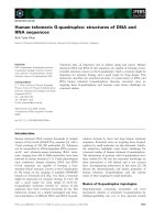
Fig. 9. Proposed mechanisms of HAT-induced activation of
AR-EGFR pathway in airway epithelial cell lines. According to this
model, HAT-induced activation of AR production is mediated by
PAR-2 dependent or PAR-2 independent mechanisms. A PAR-2-
mediated initial ERK activation causes the production of AR
precursor, and then, biologically active AR is released by PAR-2-
independent mechanism involving TACE.
HAT-induced AR release by PAR-2 and TACE M. Chokki et al.
6396 FEBS Journal 272 (2005) 6387–6399 ª 2005 The Authors Journal compilation ª 2005 FEBS
phospho-EGFR (Tyr1068) antibody, anti-ERK antibody,
antiphospho-MEK antibody, or antiphospho-p90RSK anti-
body (diluted to 1 : 1000 in 5% BSA TBST). Following
treatment with the antibody preparations, the blots were
washed three times for 5 min in TBST, and incubated for
1 h with peroxidase-conjugated secondary IgG (Amersham
Biosciences; diluted to 1 : 10000 in 5% nonfat milk TBST).
The bands were detected using a chemiluminescence detec-
tion kit (ECL-plus; Amersham Biosciences). In some cases,
blots were separated from the bound antibody using the
Restore
TM
Western Blot Stripping Buffer (Pierce) in
accordance to the manufacturer’s instructions.
Real-time RT/PCR to measure AR mRNA
A GeneAmp 5700 Sequence Detection System (Applied
Biosystems, Foster City, CA, USA) was used to conduct
real-time PCR to measure the expression of AR mRNA by
a previously described method [16]. In brief, an RNeasy
Mini Kit (Qiagen, Hilden, Germany), which included a
DNase I digestion step (RNase-free DNase set; Qiagen),
was used to extract total cellular RNA. Approximately
0.5 lg of each total RNA preparation was reverse tran-
scribed with Omniscript RT Kit (Qiagen); random hexa-
mers were used to prime the reactions. PCR was
performed using a TaqMan
TM
Universal PCR Master Mix
Kit (Applied Biosystems) with specific primers and probes
for AR and b-actin.
Determination of the amount of AR protein
The amount of AR in the culture supernatant of NCI-
H292 cells and in the cellular lysates of NCI-H292 cells
treated with the vehicle HAT or PAR-2 AP was measured
as follows. Anti-AR mAb (clone 12111.333, Genzyme) was
used as a capture antibody, and a biotinylated antihuman
AR polyclonal antibody (Genzyme) was used as a detection
antibody for ELISA. Sulpho-NHS-LC-Biotin (Pierce) was
used to biotinylate the antibody. Cellular lysates of treated
NCI-H292 cells were prepared as described previously. The
capture antibody (2 lgÆmL
)1
in NaCl ⁄ P
i
) was used to coat
the bottom of the wells of 96-well plates (NUNC Maxisorp;
Fisher Scientific, Santa Clara, CA, USA) by incubating the
plates containing the capture antibody overnight at 4 °C.
The wells were washed with PBST [NaCl ⁄ P
i
, 0.05% (v ⁄ v)
Tween 20], blocked by the addition of NaCl ⁄ P
i
containing
1% (w ⁄ v) BSA for 1 h at 37 °C and washed again. The cul-
ture medium, cellular lysates or standards (human recom-
binant AR in NaCl ⁄ P
i
containing 1% BSA) were then
added to the wells. The plates were incubated for 1 h at
room temperature, washed and incubated with biotinylated
detection antibody (150 ngÆmL
)1
in NaCl ⁄ P
i
containing 1%
BSA and 0.05% Tween 20) at room temperature for 1 h.
After washing, peroxidase-labelled streptavidin (diluted
1 : 6000 in NaCl ⁄ P
i
containing 1% BSA and 0.05% Tween
20) was added to each well, incubated for 15 min at room
temperature, and after washing, incubated with the per-
oxidase substrate (TMB substrate; Kirkegaard & Perry
Laboratories, Gaithersburg, MD, USA) for 30 min at room
temperature. The reaction was terminated by the addition
of 1 molÆL
)1
sulphuric acid, and a microplate reader (Ther-
momax; Molecular Devices, Sunnyvale, CA, USA) was
used to determine the optical density at 450 nm. The con-
centration of AR in each sample was determined by inter-
polation from the standard curve using softmax pro
software (Molecular Devices). The limit of assay sensitivity
is 4.096 pg Æ mL
)1
.
Determination of the amount of MUC5AC protein
The method reported by Takeyama et al. [17], with some
modifications, was used to determine the amount of
MUC5AC protein in the cellular lysates of NCI-H292 cells.
In brief, the cell lysates were prepared as described previ-
ously. Each lysate was diluted with bicarbonate-carbonate
buffer, applied to a separate well of a 96-well plate (NUNC
Maxisorp) and incubated at 40 °C until it was dry. The
plates were washed three times with NaCl ⁄ P
i
and blocked
with NaCl ⁄ P
i
containing 1% (w ⁄ v) BSA for 1 h at room
temperature. The plates were again washed for five times
with PBST and mouse anti-MUC5AC mAb [0.5 lgÆmL
)1
in
NaCl ⁄ P
i
containing 1% BSA and 0.05% (v ⁄ v) Tween 20]
was added to the wells. The plates were incubated for 2 h
at room temperature. The plates were then washed again,
and biotinylated antimouse antibody (Dako, Glostrup,
Denmark; diluted to 1 : 10000 in NaCl ⁄ P
i
containing 1%
BSA and 0.05% Tween 20) was added to each well. The
plates were then incubated for 1 h at room temperature.
After washing, streptavidin-conjugated HRP was added to
each well and the plates were incubated for 15 min at room
temperature. After washing, addition of peroxidase sub-
strate, termination of reaction and the measuring the opti-
cal density were done as described previously. Data are
expressed as ratio of the observed value to the mean value
of the control group.
Transfection of siRNA
Predesigned human TACE siRNA (siRNA ID no. 113003)
was purchased from Ambion (Austin, TX, USA). The
sequences of the 21-nucleotide siRNAs were sense: GCAG
CAUUCGGUAAGAAAAtt and antisense: UUUUCU
UACCGAAUGCUGCtg. Silencer Negative Control no. 1
siRNA (Ambion) was used as a control siRNA. NCI-H292
cells in 6-well plates were transfected for 72 h prior to the
HAT treatment with 100 pmol TACE or control siRNA by
using the Lipofectamine 2000 Reagent (Invitrogen) accord-
ing to the manufacturer’s instructions. Specific silencing of
TACE was confirmed by flow cytometry as described
below.
M. Chokki et al. HAT-induced AR release by PAR-2 and TACE
FEBS Journal 272 (2005) 6387–6399 ª 2005 The Authors Journal compilation ª 2005 FEBS 6397
Flow cytometry
To detect cell-surface TACE, flow cytometry was per-
formed. NCI-H292 cells were nonenzymatically collected by
incubation in NaCl ⁄ P
i
containing 5 mm EDTA for 5 min
at 37 °C. After washing in NaCl ⁄ P
i
containing 1% FBS,
the cells were stained with anti-TACE mAb or control IgG
at 4 °C for 30 min. After washing with NaCl ⁄ P
i
containing
1% FBS, cells were incubated with fluorescein isothiocya-
nate-conjugated goat antimouse IgG (DAKO) for 30 min
at 4 °C and washed again with NaCl ⁄ P
i
containing 1%
FBS. The cells were analysed on a FACS Calibur cytometer
(BD Bioscience, Mountain View, CA, USA).
Statistical analysis
Data are presented as the mean ± SD of at least three
separate experiments. For statistical analysis, Dunnett’s
two-tailed test was used to test the statistically significant
differences. The commercial statistical software Super
anova ver. 1.1 (Abacus Concepts Inc., Berkeley, CA, USA)
was used to perform the tests. Tests that returned P -values
< 0.05 were considered to represent significant differences.
Acknowledgements
The authors would like to thank Dr S. Yasuoka
(Department of Nutrition, University of Tokushima
School of Medicine) for his valuable advice and
support.
References
1 Yasuoka S, Onishi T, Kawano S, Tsuchihashi S, Ogaw-
ara M, Masuda K, Yamaoka K, Takahashi M & Sano
T (1997) Purification, characterization, and localization
of a novel trypsin-like protease found in the human air-
way. Am J Respir Cell Mol Biol 16, 300–308.
2 Yamaoka K, Masuda K, Ogawa H, Takagi K, Umemo-
to N & Yasuoka S (1998) Cloning and characterization
of the cDNA for human airway trypsin-like protease.
J Biol Chem 273, 11895–11901.
3 Hooper JD, Clements JA, Quigley JP & Antalis TM
(2001) Type II transmembrane serine proteases-insights
into an emerging class of cell surface proteolytic
enzymes. J Biol Chem 276 , 857–860.
4 Takahashi M, Sano T, Yamaoka K, Kamimura T,
Umemoto N, Nishitani H & Yasuoka S (2001) Locali-
zation of human airway trypsin-like protease in the air-
way: an immunohistochemical study. Histochem Cell
Biol 115, 181–187.
5 Miki M, Nakamura Y, Takahashi A, Nakaya Y, Eguchi
H, Masegi T, Yoneda K, Yasuoka S & Sone S (2003)
Effect of human airway trypsin-like protease on
intracellular free Ca
2+
concentration in human bron-
chial epithelial cells. J Med Invest 50, 95–107.
6 Schmidlin F & Bunnett NW (2001) Protease-activated
receptors: how proteases signal to cells. Curr Opin
Pharmacol 1, 575–582.
7 Asokananthan N, Graham PT, Fink J, Knight DA,
Bakker AJ, McWilliam AS, Thompson PJ & Stewart
GA (2002) Activation of protease-activated receptor
(PAR) -1, PAR-2, and PAR-4 stimulates IL-6, IL-8,
and prostaglandin E
2
release from human respiratory
epithelial cells. J Immunol 168, 3577–3585.
8 Temkin V, Kantor B, Weg V, Hartman M & Levi-
Schaffer F (2002) Tryptase activates the mitogen-acti-
vated protein kinase ⁄ activator protein-1 pathway in
human peripheral blood eosinophils, causing cytokine
production and release. J Immunol 169, 2662–2669.
9 Scmidlin F, Amadesi S, Dabbagh K, Lewis DE, Knott P,
Bunnett NW, Gater PR, Geppetti P, Bertrand C &
Stevens ME (2002) Protease-activated receptor 2 mediates
eosinophil infiltration and hyperreactivity in allergic
inflammation of the airway. J Immunol 169, 5315–5321.
10 Knight DA, Lim S, Scaffidi AK, Roche N, Chung KF,
Stewart GA & Thompson PJ (2001) Protease-activated
receptors in human airways: upregulation of PAR-2 in
respiratory epithelium from patients with asthma.
J Allergy Clin Immunol 108, 797–803.
11 Lundgren JD & Shelhamer JH (1990) Pathogenesis of
airway mucus hypersecretion. J Allergy Clin Immunol
85, 399–417.
12 Aikawa T, Shimura S, Sasaki H, Ebina M & Takishima
T (1992) Marked goblet cell hyperplasia with mucus
accumulation in the airways of patients who died of
severe acute asthma attack. Chest 101, 916–921.
13 Reid CJ, Gould S & Harris A (1997) Developmental
expression of mucin genes in the human respiratory
tract. Am J Respir Cell Mol Biol 17, 592–598.
14 Alimam MZ, Piazza FM, Selby DM, Letwin N, Huang
L & Rose MC (2000) Muc-5 ⁄ 5ac mucin messenger
RNA and protein expression is a marker of goblet cell
metaplasia in murine airways. Am J Respir Cell Mol
Biol 22, 253–260.
15 Ordonez CL, Khashayar R, Wong HH, Ferrando R,
Wu R, Hyde DM, Hotchkiss JA, Zhang Y, Novikov A,
Dolganov G & Fahy JV (2001) Mild and moderate
asthma is associated with airway goblet cell hyperplasia
and abnormalities in mucin gene expression. Am J
Respir Crit Care Med 163, 517–523.
16 Chokki M, Yamamura S, Eguchi H, Masegi T, Horiu-
chi H, Tanabe H, Kamimura T & Yasuoka S (2004)
Human airway trypsin-like protease increases mucin
gene expression in airway epithelial cells. Am J Respir
Cell Mol Biol 30, 470–478.
17 Takeyama K, Dabbagh K, Lee HM, Agusti C, Lausier
JA, Ueki IF, Grattan KM & Nadel JA (1999) Epidermal
HAT-induced AR release by PAR-2 and TACE M. Chokki et al.
6398 FEBS Journal 272 (2005) 6387–6399 ª 2005 The Authors Journal compilation ª 2005 FEBS
growth factor system regulates mucin production in
airways. Proc Natl Acad Sci USA 96, 3081–3086.
18 Burgel PR & Nadel JA (2004) Roles of epidermal
growth factor receptor activation in epithelial cell repair
and mucin production in airway epithelium. Thorax 59,
992–996.
19 Perrais M, Pigny P, Copin MC, Aubert JP & Van Seu-
ningen I (2002) Induction of MUC2 and MUC5AC
mucins by factors of the epidermal growth factor (EGF)
family is mediated by EGF receptor ⁄ Ras ⁄ Raf ⁄ extracel-
lular signal-regulated kinase cascade and Sp1. J Biol
Chem 277, 32258–32267.
20 Lemjabbar H, Li D, Gallup M, Sidhu S, Drori E &
Basbaum C (2003) Tobacco smoke-induced lung cell
proliferation mediated by tumor necrosis factor a-con-
verting enzyme and amphiregulin. J Biol Chem 278,
26202–26207.
21 Richter A, O’Donnell RA, Powell RM, Sanders MW,
Holgate ST, Djukanovic R & Davies DE (2002) Auto-
crine ligands for the epidermal growth factor receptor
mediate interleukin-8 release from bronchial epithelial
cells in response to cigarette smoke. Am J Respir Cell
Mol Biol 27, 85–90.
22 Blanchet S, Ramgolam K, Baulig A, Marano F &
Baeza-Squiban A (2004) Fine particulate matter induces
amphiregulin secretion by bronchial epithelial cells. Am
J Respir Cell Mol Biol 30, 421–427.
23 Downward J, Parker P & Waterfield MD (1984) Auto-
phosphorylation sites on the epidermal growth factor
receptor. Nature 311, 483–485.
24 Tice DA, Biscardi JS, Nickles AL & Parsons SJ (1999)
Mechanism of biological synergy between cellular Src
and epidermal growth factor receptor. Proc Natl Acad
Sci USA 96, 1415–1420.
25 Okutani T, Okabayashi Y, Kido Y, Sugimoto Y, Saka-
guchi K, Matuoka K, Takenawa T & Kasuga M (1994)
Grb2 ⁄ Ash binds directly to tyrosines 1068 and 1086 and
indirectly to tyrosine 1148 of activated human epidermal
growth factor receptors in intact cells. J Biol Chem 269,
31310–31314.
26 Schlessinger J (2000) Signaling by receptor tyrosine kin-
ases. Cell 103, 211–225.
27 Rojas M, Yao S & Lin YZ (1996) Controlling epider-
mal growth factor (EGF) -stimulated Ras activation in
intact cells by a cell-permeable peptide mimicking phos-
phorylated EGF receptor. J Biol Chem 271, 27456–
27461.
28 Frodin M & Gammeltoft S (1999) Role and regulation
of 90 kDa ribosomal S6 kinase (RSK) in signal trans-
duction. Mol Cell Endocrinol 151, 65–77.
29 Yoshinaga S, Nakahori Y & Yasuoka S (1998) Fibrino-
genolytic activity of a novel trypsin-like enzyme found
in human airway. J Med Invest 1998 (45), 77–86.
30 Belham CM, Tate RJ, Scott PH, Pemberton AD, Miller
HR, Wadsworth RM, Gould GW & Plevin R (1996)
Trypsin stimulates proteinase-activated receptor-
2-dependent and – independent activation of mitogen-
activated protein kinases. Biochem J 320, 939–946.
31 Koo BH, Chung KH, Hwang KC & Kim DS (2002)
Factor Xa induces mitogenesis of coronary artery
smooth muscle cell via activation of PAR-2. FEBS Lett
523, 85–89.
32 DeFea KA, Zalevsky J, Thoma MS, Dery O, Mullins
RD & Bunnett NW (2000) b-arrestin-dependent endo-
cytosis of proteinase-activated receptor 2 is required for
intracellular targeting of activated ERK1 ⁄ 2. J Cell Biol
148, 1267–1281.
33 Ge L, Ly Y, Hollenberg M & DeFea K (2003) A b-arr-
estin-dependent scaffold is associated with prolonged
MAPK activation in pseudopodia during protease-acti-
vated receptor-2-induced chemotaxis. J Biol Chem 278,
34418–34426.
34 Darmoul D, Gratio V, Devaud H & Laburthe M (2004)
Protease-activated receptor 2 in colon cancer. Trypsin-
induced MAPK phosphorylation and cell proliferation
are mediated by epidermal growth factor receptor trans-
activation. J Biol Chem 279, 20927–20934.
35 Gschwind A, Hart S, Fischer OM & Ullrich A (2003)
TACE cleavage of proamphiregulin regulates GPCR-
induced proliferation and motility of cancer cells.
EMBO J 22, 2411–2421.
36 Brown CL, Meise KS, Plowman GD, Coffey RJ &
Dempsey PJ (1998) Cell surface ectodomain cleavage
of human amphiregulin precursor is sensitive to a
metalloprotease inhibitor. Release of a predominant
N-glycosylated 43-kDa soluble form. J Biol Chem 273,
17258–17268.
37 Zhang Q, Thomas SM, Xi S, Smithgall TE, Siegfried
JM, Kamens J, Gooding WE & Grandis JR (2004) SRC
family kinases mediate epidermal growth factor receptor
ligand cleavage, proliferation and invasion of head and
neck cancer cells. Cancer Res 64, 6166–6173.
38 Takeyama K, Fahy JV & Nadel JA (2001) Relationship
of epidermal growth factor receptors to goblet cell pro-
duction in human bronchi. Am J Respir Crit Care Med
163, 511–516.
39 Brachmann R, Lindquist PB, Nagashima M, Kohr W,
Lipari T, Napier M & Derynck R (1989) Transmem-
brane TGF-alpha precursors activate EGF ⁄ TGF-alpha
receptors. Cell 56, 691–700.
40 Higashiyama S, Abraham JA, Miller J, Fiddes JC &
Klagsbrun M (1991) A heparin-binding growth factor
secreted by macrophage-like cells that is related to
EGF. Science 251, 936–939.
41 Armstrong L, Godinho SI, Uppington KM, Whitting-
ton HA & Millar AB (2005) Contribution of TACE
and Proteinase-3 to TNF-a processing in human
alveolar macrophages. Am J Respir Cell Mol Biol,
doi:10.1165 ⁄ rcmb.2005-0087oc.
M. Chokki et al. HAT-induced AR release by PAR-2 and TACE
FEBS Journal 272 (2005) 6387–6399 ª 2005 The Authors Journal compilation ª 2005 FEBS 6399