Báo cáo khoa học: Secretion of proteases in serglycin transfected Madin–Darby canine kidney cells ppt
Bạn đang xem bản rút gọn của tài liệu. Xem và tải ngay bản đầy đủ của tài liệu tại đây (318.42 KB, 12 trang )
Secretion of proteases in serglycin transfected
Madin–Darby canine kidney cells
Lillian Zernichow
1
, Knut T. Dalen
1
, Kristian Prydz
2
, Jan-Olof Winberg
3
and Svein O. Kolset
1
1 Department of Nutrition, Institute of Basic Medical Sciences, University of Oslo, Norway
2 Department of Molecular Biosciences, University of Oslo, Norway
3 Department of Biochemistry, Institute of Medical Biology, University of Tromsø, Norway
Studies on proteoglycans (PGs) have, to a large
extent, focused on molecules located in the extracellu-
lar matrix and on cell surfaces [1–3], and their roles
in, for example, the regulation of cell adhesion, cell
migration, proliferation and wound healing. However,
PGs located in different intracellular locations are
receiving increasing attention [4,5]. In particular, PGs
in storage and secretory granules in cells of the hae-
matopoietic lineage have been the subject of several
recent studies, as, for instance, in the mast cell, where
heparin PG is stored in secretory granules together
with histamine and proteases. Generation of mice
with a deleted version of the gene for the heparin-
synthesizing enzyme N-deacetylase ⁄ N-sulfotransferase-
2 (NDST-2) resulted in the appearance of mast cells
with large changes in secretory granule morphology
and in greatly reduced levels of the proteases nor-
mally confined to these granules [6,7]. Heparin PG in
mast cells, accordingly, seems to be of fundamental
importance for the generation of storage granules.
Recently, serglycin knockout mice were generated [8].
They developed normally and were fertile, but their
mast cells were affected in a manner similar to that
of the NDST-2 knockout mice.
Keywords
matrix metalloproteinase; MDCK;
plasminogen activator; proteoglycan;
serglycin
Correspondence
S. O. Kolset, Department of Nutrition,
Institute of Basic Medical Sciences,
University of Oslo, Box 1046 Blindern,
0316 Oslo, Norway
Fax: +47 22 851 398
Tel: +47 22 851 383
E-mail:
(Received 2 November 2005, accepted
2 December 2005)
doi:10.1111/j.1742-4658.2005.05085.x
Madin–Darby canine kidney (MDCK) cells, which do not normally express
the proteoglycan (PG) serglycin, were stably transfected with cDNA for
human serglycin fused to a polyhistidine tag (His-tag). Clones with differ-
ent levels of serglycin mRNA expression were generated. One clone with
lower and one with higher serglycin mRNA expression were selected for
this study.
35
S-labelled serglycin in cell fractions and conditioned media
was isolated using HisTrap affinity chromatography. Serglycin could also
be detected in conditioned media using western blotting. To investigate the
possible importance of serglycin linked to protease secretion, enzyme activ-
ities using chromogenic substrates and zymography were measured in cell
fractions and serum-free conditioned media of the different clones. Cells
were cultured in both the absence and presence of phorbol 12-myristate
13-acetate (PMA). In general, enzyme secretion was strongly enhanced by
treatment with PMA. Our analyses revealed that the clone with the highest
serglycin mRNA expression, level of HisTrap isolated
35
S-labelled sergly-
cin, and amount of serglycin core protein as detected by western blotting,
also showed the highest secretion of proteases. Transfection of serglycin
into MDCK cells clearly leads to changes in secretion levels of secreted
endogenous proteases, and could provide further insight into the biosynthe-
sis and secretion of serglycin and potential partner molecules.
Abbreviations
cABC, chondroitinase ABC; CS, chondroitin sulfate; DMEM, Dulbecco’s modified Eagle’s medium; FBS, fetal bovine serum; GAG,
glycosaminoglycan; HS, heparan sulfate; His-tag, polyhistidine tag; MDCK, Madin–Darby canine kidney; MMP, matrix metalloproteinase;
NDST-2, N-deacetylase ⁄ N-sulfotransferase-2; NGAL, neutrophil gelatinase-associated lipocalin; PA, plasminogen activator; PG, proteoglycan;
PMA, phorbol 12-myristate 13-acetate; PVDF, poly(vinylidene difluoride); uPA, urokinase-type plasminogen activator.
536 FEBS Journal 273 (2006) 536–547 ª 2006 The Authors Journal compilation ª 2006 FEBS
Haematopoietic cells either harbour storage granules
or granules destined for constitutive secretion. In
human monocytes and macrophages the main PG is
serglycin [9]. In these cells, the PG is secreted and acti-
vation results in increased synthesis and secretion of
PGs [9,10], suggesting that secretion of PGs is linked
to inflammatory response. Secreted serglycin may
potentially associate with other secreted products from
macrophages in the extracellular environment [11,12],
or team up intracellularly with such molecules during
constitutive secretion. In a recent study in murine
macrophages, abrogation of PG biosynthesis with b-d-
xylosides resulted in decreased enzyme secretion [13].
In particular, the secretion of urokinase-type plasmino-
gen activator (uPA) and matrix metalloproteinase-9
(MMP-9, also referred to as 92 kDa gelatinase B and
type IV collagenase) was lowered after xyloside treat-
ment.
To further study the interrelationship between ser-
glycin and proteases, both with regard to biosynthesis
and secretion, serglycin was stably transfected into
Madin–Darby canine kidney (MDCK) epithelial cells.
A polyhistidine tag (His-tag) was introduced at the
C-terminus to facilitate the isolation of serglycin.
Transfectants with vector without serglycin insert were
generated as negative controls. It has previously been
shown that MDCK cells secrete MMP-9 [14] and uPA
[15]. Secretion of these endogenous proteases was stud-
ied in the transfected cells, in both the absence and
presence of the phorbol ester phorbol 12-myristate
13-acetate (PMA), previously reported to enhance the
secretion of MMP-9 in MDCK cells [14]. Results pre-
sented show that the secretion levels of proteases cor-
relate with the levels of serglycin mRNA, HisTrap
isolated
35
S-labelled serglycin and serglycin core pro-
tein detected by western blotting. The MDCK system
with transfected serglycin is potentially a useful model
to study PGs in relation to secretion of enzymes
important in physiological and pathological condi-
tions.
Results
Transfection of serglycin
Clones of MDCK cells with serglycin–His-tag were
obtained, and the levels of serglycin mRNA deter-
mined using northern blotting. Cell clones transfected
with the vector without the serglycin insert were
used as negative controls (mock transfectants). No
serglycin mRNA was detected in these clones. The
serglycin mRNA expression level in the different
clones was related to the housekeeping gene 36B4
mRNA expression level. One clone with a lower ser-
glycin mRNA expression level (1–7), one with a
higher level (1–10) and one of the mock transfectants
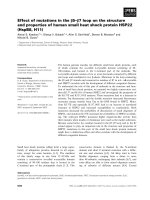
were selected for further studies. Figure 1 shows nor-
thern blots of the selected clones, using
32
P-labelled
cDNA probes of serglycin, MMP-9, uPA and 36B4,
cultured in both absence and presence of PMA. The
northern blot of the unstimulated cells in Fig. 1A
shows that the clone with the highest serglycin
mRNA level also has the highest mRNA levels of
uPA and MMP-9, although the levels are very low
for MMP-9. PMA stimulation (Fig. 1B) was shown
to lead to an upregulation of the mRNA levels of
serglycin, MMP-9 and uPA.
Proteoglycan analyses
35
S-labelled macromolecules
According to Svennevig et al. [16] and Erickson &
Couchman [17], PGs synthesized by MDCK cells are
perlecan, agrin, collagen XVIII, biglycan, bamacan
AB
Fig. 1. mRNA expression levels of serglycin, MMP-9, uPA and
36B4 in serglycin-transfected MDCK clones. Total RNA was isola-
ted from MDCK clones and subjected to northern blotting onto the
same membrane and hybridized using
32
P-labelled human serglycin,
canine MMP-9 and uPA and murine 36B4 cDNA probes. (A) Un-
stimulated clones. (B) PMA-stimulated clones. The experiment was
repeated independently three times, and the result shown is typical
of the three experiments. The northern blots in (A) and (B) for
MMP-9 and uPA have been exposed for different periods, due to
large differences in expression levels.
L. Zernichow et al. Serglycin and proteases
FEBS Journal 273 (2006) 536–547 ª 2006 The Authors Journal compilation ª 2006 FEBS 537
and versican. To investigate the extent to which
expression of serglycin influenced total PG biosynthe-
sis, cells were labelled with
35
S sulfate for 24 h. The
labelled macromolecules were separated from unin-
corporated
35
S-labelled sulfate by Sephadex G-50
Fine gel chromatography. It has previously been
shown that the major fraction of
35
S-labelled macro-
molecules in MDCK cells are of PG nature [16].
The level of
35
S-labelled macromolecules therefore
indicates the level of PG synthesis. As can be seen
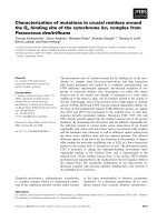
in Fig. 2A, the introduction of serglycin into MDCK
cells increased the amount of
35
S-labelled macromole-
cules in the medium, but did not significantly
increase the total amount of
35
S-labelled macromole-
cules. Radiolabelling was also performed in the
presence of PMA. Figure 2B shows that in PMA-
stimulated cells the distribution of PGs changed, so
the majority (78–90%) was secreted into the medium,
whereas the corresponding value for unstimulated
cells was 30–45%. The total increase of
35
S-labelled
macromolecules in clones 1–7 and 1–10, relative to
the mock, was approximately the same in unstimu-
lated and PMA-stimulated cells.
HisTrap isolation of His-tagged serglycin
His-tagged serglycin in cell fractions and condi-
tioned media from unstimulated clones was isolated by
HisTrap affinity chromatography using
35
S-labelled
macromolecules obtained by Sephadex G-50 Fine gel
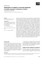
chromatography. Figure 3A shows the total amount
of
35
S-labelled macromolecules loaded onto the
HisTrap column, whereas Fig. 3B shows the total
amount of
35
S-labelled macromolecules with affinity
for the column. The highest level of
35
S-labelled
macromolecules with affinity for the HisTrap column,
in both cell fractions and conditioned media, was
measured in clone 1–10. This level was 2–3 times
higher than for clone 1–7, whereas the level for the
mock transfected clone was found to be negligible.
In both clone 1–10 and 1–7 the highest level of
incorporated
35
S-labelled sulfate was measured in the
conditioned media. As can be seen in Fig. 3, serglycin
contributes very little to the total incorporation of
35
S-labelled sulfate. When comparing the amount of
35
S-labelled macromolecules before and after HisTrap
isolation, it was found that only 1 ⁄ 60 of the radio-
activity was associated to serglycin in clone 1–7,
whereas the corresponding value for clone 1–10 was
1 ⁄ 20. Superose 6 gel chromatography of HisTrap
isolated
35
S-labelled serglycin from conditioned media
showed that serglycin from clone 1–10 eluted at a
slightly more retarded position compared with clone
1–7 (not shown). Furthermore, analyses of
35
S-labelled
glycosaminoglycan (GAG) chains obtained from the
same material showed a similar trend, with K
av
values
of 0.53 and 0.57 for clone 1–7 and 1–10, respectively.
These findings indicate that the GAG chains of
0
1
2
3
4
5
6
7
8
9
0
1
2
3
4
5
6
7
8
9
1-7
Cell
Medium
Total
35
S sulfate (cpmx10
6
)
35
S sulfate (cpmx10
6
)
A
B
Cell
Medium
Total
1,42
1,35
1,0
1,38
1,32
1,0
1-10 Mock
1-7 1-10 Mock
Fig. 2.
35
S-labelled macromolecules in serglycin-transfected MDCK
clones. Confluent MDCK clones were labelled with
35
S sulfate for
24 h in both absence and presence of PMA, whereupon the cells
fractions and conditioned media were harvested and subjected to
Sephadex G-50 Fine gel chromatography to remove unincorporated
35
S sulfate. Each point represents the mean ± SD of measurement
on material from three separate wells. The number on top of the
black columns, representing the total
35
S sulfate incorporation, is
relative to the mock transfectant. (A) Unstimulated clones. (B)
PMA-stimulated clones. The experiment was repeated independ-
ently three times, and the result shown is typical of the replicate
experiments.
Serglycin and proteases L. Zernichow et al.
538 FEBS Journal 273 (2006) 536–547 ª 2006 The Authors Journal compilation ª 2006 FEBS
serglycin from clone 1–7 are slightly longer than those
of clone 1–10. Analysis of the composition of the
35
S-labelled GAGs showed that both clone 1–7 and
1–10 contained 70% chondroitin sulfate (CS) and
30% heparan sulfate (HS) (not shown), indicating that
CS is the dominating GAG, in agreement with previ-
ous findings [18].
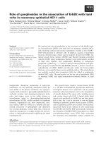
Western blotting of serglycin
To further analyse for the presence of serglycin, condi-
tioned media of the different clones were subjected
to western blotting after chondroitinase ABC (cABC)
treatment, using a rabbit polyclonal antibody to
human serglycin. As evident in Fig. 4, clones 1–7 and
1–10 contained the serglycin core protein, with the
highest amount in the latter. The molecular mass of
the serglycin core protein was 35 kDa, in accordance
with another study [5]. The same results were observed
with a mouse monoclonal antibody to the His-tag (not
shown).
Enzyme analyses
The possible relationship between serglycin and prote-
ase secretion was analysed in the different clones using
serum-free conditioned media, the chromogenic sub-
strates S-2288 and S-2444, and gelatin and plasmino-
gen–gelatin zymography. Cell fractions were also
analysed. All cell culture experiments were carried out
in both the absence and presence of PMA, to ensure
0
100
200
300
400
500
600
700
Cell
Medium
Total
0
5
10
15
20
25
30
35
Cell
Medium
Total
A
B
35
S sulfate (cpm x 10
4
)
35
S sulfate (cpm x 10
4
)
1-7 1-10 Mock
1-7
1-10
Mock
Fig. 3. HisTrap isolation of
35
S sulfate-labelled His-tagged sergly-
cin from serglycin-transfected MDCK clones. Cell fractions and
conditioned media from cells exposed to
35
S sulfate for 24 h
were buffer exchanged to binding buffer by Sephadex G-50
Fine gel chromatography. The samples were further applied to a
1 mL HisTrap column, pre-equilibrated with binding buffer (20 m
M
phosphate, 1 M NaCl, 8 M urea and 20 mM imidazole pH 8.0).
After a washing step, the samples were eluted with a solution
containing 20 m
M phosphate, 1 M NaCl, 8 M urea and 500 mM
imidazole (pH 8.0). (A) Before HisTrap isolaton. (B) After HisTrap
isolation. The experiment was repeated independently more than
three times, and the result shown is typical of all the replicate
experiments.
Fig. 4. Western blot of serglycin core protein in serglycin-trans-
fected MDCK clones. Conditioned media from unstimulated MDCK
clones were desalted against Milli-Q water by Sephadex G-50 Fine
gel chromatography. After freeze-drying, the samples were dis-
solved in a small volume of Milli-Q water and treated with cABC as
described in Experimental Procedures. Furthermore, the samples
were subjected to SDS ⁄ PAGE followed by western blotting using a
rabbit polyclonal antibody to human serglycin. The data shown are
from a single experiment that was repeated three times with the
same results.
L. Zernichow et al. Serglycin and proteases
FEBS Journal 273 (2006) 536–547 ª 2006 The Authors Journal compilation ª 2006 FEBS 539
comparison of basal and stimulated secretion of pro-
teases.
Chromogenic substrates
Cell fractions and serum-free conditioned media were
analysed with respect to a broad spectrum of serine
proteases using the chromogenic substrate S-2288
(Fig. 5). Media from clone 1–10 showed the highest
enzyme activity in unstimulated cells. When cells
were treated with PMA, the enzyme activities in the
media were strongly enhanced. Compared with the
media, enzyme activities in cell fractions were found
to be low. Media from PMA-treated clones con-
tained 100-fold more protease activity than
medium from unstimulated clones. Also after PMA
treatment, medium from clone 1–10 showed the
highest activity, but the difference between the clones
was much less distinct than observed for the unstim-
ulated clones.
Further experiments were performed using the uro-
kinase substrate S-2444. Cell fractions and serum-free
conditioned media from the various clones all con-
tained activity towards this substrate, as can be seen in
Fig. 6. Again, media from the clone 1–10 had the high-
est enzyme activity. Here also PMA treatment resulted
in an 100-fold increase in enzyme activity in the
clones tested.
Clearly, the enzyme activities in serum-free condi-
tioned media towards the chromogenic substrates
S-2288 and S-2444 were related to the levels of sergly-
cin, MMP-9 and uPA mRNA expression in the
MDCK-transfected cells. The clone with the highest
levels of serglycin, MMP-9 and uPA mRNA and level
of HisTrap isolated
35
S sulfate-labelled serglycin, and
amount of serglycin core protein detected by western
blotting, in conditioned media, i.e. clone 1–10, also
had the highest secretion of the enzymes analysed with
chromomogenic substrates, both basal and after PMA
treatment.
To investigate the nature of the enzyme activities
measured, serum-free conditioned media were incuba-
ted in both the absence and presence of the enzyme
inhibitors amiloride and Pefabloc. The enzyme activity
recognizing the substrate S-2288 was inhibited 90%
in the presence of 2 mm Pefabloc, demonstrating that
this activity is of a serine protease nature (Table 1).
Furthermore, an inhibition of 70% of the enzyme
activity was observed in the presence of 2 mm amilo-
ride, indicating that a major part of the serine protease
activity measured with S-2288 is due to plasminogen
activators (PAs). To investigate whether the activity
recognizing the substrate S-2444 is indeed uPA, we
made use of the inhibitor amiloride, considered to be a
specific inhibitor of uPA [19]. When serum-free condi-
tioned media from the different clones were incubated
in the presence of 2 mm amiloride, the activities were
inhibited 95%, demonstrating that this activity is of
uPA nature (Table 2).
A
B
Fig. 5. Analysis of enzyme activities in serglycin-transfected MDCK
clones using the chromogenic substrate S-2288. Confluent MDCK
clones were cultured for 24 h under serum-free conditions in both
absence and presence of PMA. Cell fractions and conditioned
media were harvested and analysed for enzyme activities by using
the chromogenic substrate S-2288. Each point represents the mean
and standard deviation of measurement on material from three sep-
arate wells. (A) Unstimulated clones. (B) PMA-stimulated clones.
The S-2288 activity assay was repeated independently more than
three times, and the result shown is typical of all the replicate
experiments. Note the difference in the scales of the vertical axis
for the unstimulated and PMA-stimulated clones.
Serglycin and proteases L. Zernichow et al.
540 FEBS Journal 273 (2006) 536–547 ª 2006 The Authors Journal compilation ª 2006 FEBS
Gelatin and plasminogen–gelatin zymography
To further investigate the relationship between sergly-
cin and protease secretion in MDCK clones, the
possible presence of gelatinases in the serum-free con-
ditioned media of the different clones was investigated
by gelatin zymography. The rationale for measuring
gelatinase activities is that these enzymes are known to
interact with PGs [20,21].
No gelatinolytic bands could be detected in the
serum-free conditioned media of any of the clones tes-
ted (Fig. 7A). In contrast, when the cells were treated
with PMA, several gelatinolytic bands were detected
(Fig. 7B). By comparing the gelatinolytic bands with
those of MMP-9 and MMP-2 standards, it is likely
that the 225 and 92 kDa gelatinolytic bands are
dimeric and monomeric MMP-9, respectively [22]. The
highest degree of gelatinolytic activity was evident in
clone 1–10, although no particular difference in inten-
sity of the 92 kDa (proform) band could be demon-
strated between the different clones. However, the
78 kDa band, probably an active form, had higher
intensity in media from clone 1–10 than from 1–7 and
the mock transfected clone. The 127 kDa band
observed for clone 1–10 may be a complex of mono-
meric MMP-9 with neutrophil gelatinase-associated
lipocalin (NGAL) [23].
A
B
Fig. 6. Analysis of enzyme activities in serglycin-transfected MDCK
clones using the chromogenic substrate S-2444. Confluent MDCK
clones were cultured for 24 h under serum-free conditions in both
absence and presence of PMA. Cell fractions and conditioned
media were harvested and analysed for enzyme activities by using
the chromogenic substrate S-2444. Each point represents the mean
and standard deviation of measurement on material from three sep-
arate wells. (A) Unstimulated clones. (B) PMA-stimulated clones.
The S-2444 activity assay was repeated independently more than
three times, and the result shown is typical of all the replicate
experiments. Note the difference in the scales of the vertical axis
for the unstimulated and PMA-stimulated clones.
Table 1. Effect of amiloride and Pefabloc on serine protease activit-
ies in transfected MDCK clones. Serum-free conditioned media
from unstimulated cells were harvested and analysed for enzyme
activities using the chromogenic substrate S-2288 in both the
absence and presence of amiloride and Pefabloc, both at a final
concentration of 2 m
M. The results are calculated as percentages
of controls. Each value represents the mean ± SD of measurement
made on three independent wells. The assay was repeated inde-
pendently three times, and the result shown is typical of the repli-
cate experiments.
Inhibitor
1–7
% remaining
activity
1–10
% remaining
activity
Mock
% remaining
activity
Control 100 100 100
Pefabloc 14 ± 5 10 ± 3 12 ± 4
Amiloride 27 ± 6 35 ± 2 29 ± 2
Table 2. Effect of amiloride on PA activities in transfected MDCK
clones. Serum-free conditioned media from unstimulated cells were
harvested and analysed for enzyme activities using the chromo-
genic substrate S-2444 in both the absence and presence of amilo-
ride (2 m
M final concentration). The results are calculated as
percentages of controls. Each value represents the mean ± SD of
measurement made on three independent wells. The assay was
repeated independently three times, and the result shown is typical
of the replicate experiments.
Inhibitor
Clone 1–7
% remaining
activity
Clone 1–10
% remaining
activity
Mock
% remaining
activity
Control 100 ± 6 100 ± 6 100 ± 3
Amiloride 0 ± 3 9 ± 4 7 ± 2
L. Zernichow et al. Serglycin and proteases
FEBS Journal 273 (2006) 536–547 ª 2006 The Authors Journal compilation ª 2006 FEBS 541
The ability to degrade gelatin is not a unique prop-
erty of MMPs. The presence of MMP activity in gela-
tin zymography of serum-free conditioned media from
the PMA-treated clones was verified by using 100 nm
galardin, an inhibitor of MMPs [24]. After incubation
with galardin, the gelatinolytic bands shown in Fig. 7B
were abolished (not shown), indicating that these gela-
tinolytic bands were MMPs.
The possible presence of PA activity was also investi-
gated using plasminogen–gelatin zymography. Gelatin
gels run without plasminogen were used as controls
against gels containing plasminogen and gelatin. Indeed,
when plasminogen was incorporated into the gels, all
the clones displayed PA activity in the 55 kDa region,
the highest activity again being evident in the clone with
the highest mRNA levels for serglycin, MMP-9 and
uPA, i.e. clone 1–10 (Fig. 7C). Here also PMA treat-
ment resulted in elevated enzyme activities, and two
additional bands with molecular masses of 67 and
35 kDa appeared in the zymogram (Fig. 7D). Upon
dilution of media from PMA-stimulated clones, 1–10
showed highest activity (not shown). The PA activity
in serum-free conditioned media from unstimulated
clones was shown to be uPA, because the 55 kDa
band was abolished when including 2 mm amiloride
in all incubation steps after gel electrophoresis. In
zymograms of serum-free conditioned media from
PMA-treated cells, both the 55 and 35 kDa bands
were abolished in the presence of amiloride, whereas
the intensity of the 67 kDa band was unaltered. The
identity of the 67 kDa band is unknown.
Western blotting of MMP-9
In an attempt to identify the nature of the gelatinolytic
bands, we performed western blotting using a MMP-9
antibody (Fig. 8). Owing to a lack of canine MMP-9
antibody we used a rabbit polyclonal antibody to
human MMP-9. As in gelatin zymography, no bands
were visualized by western blotting of serum-free con-
ditioned media from unstimulated clones (not shown).
From the western blot of media from PMA-treated
clones in Fig. 8, the presence of MMP-9 could be dem-
onstrated. As for the zymography, there was no partic-
ular difference in intensity of the MMP-9 monomer
(92 kDa) band between the different clones. The rela-
tive differences in intensity of the 67 kDa band in
the different lanes in Fig. 8 are similar to those of the
78 kDa band in Fig. 7B. These bands may or may
not represent the same protein, as the western blotting
was performed under reducing conditions, whereas
zymography was not.
Discussion
Human serglycin has been stably transfected into
MDCK cells. The results presented show related levels
of serglycin and MMP-9 and uPA, both at mRNA
and protein levels. In Figs 1, 3 and 8 it can be seen
that clone 1–10 has the highest level of serglycin,
whereas clone 1–7 has the lowest level. The levels of
35
S sulfate incorporation were not particularly high in
the clone with the highest serglycin mRNA expression,
1-7
1-10
Mock
1-7
1-10
Mock
1-7
1-10
Mock
1-7
1-10
Mock
C
D
A
B
M
r
(kDa)
~ 200
~ 127
~ 92
~ 78
~ 67
~ 55
~ 37
Fig. 7. Zymograms of serum-free condi-
tioned media from serglycin-transfected
MDCK clones. Confluent MDCK clones
were cultured for 24 h under serum-free
conditions in both absence and presence of
PMA. Conditioned media were harvested
and analysed by zymography. (A) Gelatin
zymography. Media from unstimulated
clones. (B) Gelatin zymography. Media from
PMA-stimulated clones. (C) Plasminogen–
gelatin zymography. Media from unstimu-
lated clones. (D) Plasminogen–gelatin
zymography. Media from PMA-stimulated
clones. The data shown are from single
experiments that were repeated more than
three times with the same results.
Serglycin and proteases L. Zernichow et al.
542 FEBS Journal 273 (2006) 536–547 ª 2006 The Authors Journal compilation ª 2006 FEBS
level of HisTrap isolated
35
S sulfate-labelled serglycin,
and the amount of serglycin core protein detected by
western blotting. Figure 3 clearly illustrate that high
serglycin levels do not necessarily translate into high
levels of
35
S-labelled macromolecules expressed. This
indicates that the higher release of proteases in clone
1–10 is related to the biosynthesis and release of ser-
glycin, and not the endogenous PGs. This raises inter-
esting questions concerning the functions of serglycin
in intracellular compartments. Our results suggest that
the presence and level of serglycin could be important
for the secretion of different types of proteases. Fur-
thermore, the introduction of serglycin into a cell type
not normally expressing this PG, changes the levels of
endogenous protease secretion.
We have previously shown increased secretion of
PGs, which is mainly due to increased secretion of
serglycin, in monocytes and macrophages after PMA
stimulation [9,10]. An increase in PG secretion in
monocytes and macrophages has also been observed
after stimulation with lipopolysaccharide and gamma
interferon, suggesting that increased serglycin secretion
is linked to inflammatory reactions [10]. The biological
functions linked to the release of serglycin from mono-
cytes and macrophages have not been outlined in any
great detail, but it has been suggested that serglycin
might be important for the binding and release of
important inflammatory molecules, such as chemokines
[11]. It has also recently been shown that abrogation
of PG biosynthesis in the murine macrophage cell line
J774 resulted in a decrease in MMP-9 and uPA secre-
tion [13]. It therefore seems as though further progress
in studies on the biological functions of serglycin will
depend, to a certain extent, on a more thorough
understanding of interactions with partner molecules.
It will furthermore be important to study serglycin
secretion in different cell types. The processes and
regulation leading to the formation of serglycin-
containing granules, secretory or storage type, will
probably differ to a large extent between different ser-
glycin-expressing cells.
The transfected MDCK cells generated here can be
used as a model system to study possible relations
between serglycin and different partner molecules. The
coordinated levels of serglycin and proteases are
important in relation to those cells already known to
express serglycin. These include haematopoietic cells,
such as mast cells, monocytes and macrophages, plate-
lets and also endothelial cells and pancreatic acinar
cells [4]. All these cells have granules which contain a
large variety of serglycin-binding molecules, including
histamine, chymases, gelatinase, granzymes, platelet
factor 4, lactoferrin and procarboxypeptidase. With
the established MDCK clones we are now able to
address questions concerning regulation of serglycin
release and interactions with different partner mole-
cules.
It is of interest to note that histamine, which is an
important partner molecule for heparin PG in the mast
cell granules, is also important for the genesis of gran-
ules. Inactivation of the gene encoding histidine
decarboxylase, the enzyme converting histidine to
histamine, resulted in reduced storage of PG and pro-
teases in the granules [25]. It therefore seems as though
there is cross-talk between the different granule com-
ponents during granule formation, and that lack of
one important component has serious consequences
for this process, and will also affect the amount of
partner molecules sorted to such granules. Our study
shows that introduction of serglycin to MDCK cells
leads to changes in secretion levels of proteases, via as
yet undefined mechanisms, regulated in relation to the
serglycin level. This relationship between the levels of
serglycin and protease levels could, accordingly, be in
support of cross-talk regulatory mechanisms.
The fate of serglycin released from different types of
immune cells has not been studied to any great extent.
Fig. 8. Western blot of MMP-9 in serglycin-transfected MDCK
clones. Serum-free conditioned media from PMA-stimulated MDCK
clones were subjected to SDS ⁄ PAGE followed by western blotting
using a rabbit polyclonal antibody to human MMP-9. The data
shown is from a single experiment that was repeated three times
with the same results.
L. Zernichow et al. Serglycin and proteases
FEBS Journal 273 (2006) 536–547 ª 2006 The Authors Journal compilation ª 2006 FEBS 543
It has been shown that serglycin may bind to CD44,
and thereby participate in cell–cell interactions [26],
and that it can participate in the delivery of perforin
to target cells [27]. Furthermore, it has been shown
that serglycin isolated from macrophages is not degra-
ded when it is added back to fresh cultures of macro-
phages, suggesting extracellular functions after release
[28]. It has also been shown that serglycin may bind
covalently to a fraction of the MMP-9 secreted from
the monocyte cell line THP-1 [29]. This association
was shown to alter the biochemical properties of the
enzyme.
There are several possibilities for serglycin to inter-
act with other secreted components, such as enzymes,
growth factors or cytokines, and modulate their activ-
ities [4]. Hence, both the generation of secretory com-
plexes during biosynthesis and granule formation and
interactions between secreted components, are proces-
ses in which serglycin is most probably an important
component, worthy of more detailed study.
Experimental procedures
Cell culture and transfection
Cell culture reagents were purchased from Sigma (St. Louis,
MO), unless otherwise stated. MDCK epithelial cells
(ATCC, Manassas, VA) were cultured at 37 °C, in 5%
CO
2
, in Dulbecco’s modified Eagle’s medium (DMEM)
containing 2 mml-glutamine, 50 unitsÆmL
)1
penicillin,
50 lgÆmL
)1
streptomycin and 5% (v ⁄ v) heat-inactivated
(56 °C for 30 min) fetal bovine serum (FBS). The cell cul-
tures were checked for Mycoplasma infection with Myco-
Alert mycoplasma detection assay (Cambrex, Rockland,
ME) in routine. Before each experiment the cells were
grown to confluency (4 days). Triplicate cultures were used
for all experiments.
The pcDNA3.1(–) ⁄ Myc-His A vector (Invitrogen Life
Technologies, Carlsbad, CA) was used to generate the ser-
glycin–His-tag expression vector. To obtain inframe transla-
tion into the His sequence, serglycin cDNA was amplified
from human serglycin cDNA [9] by PCR with the following
primers: upper primer (XbaI): 5¢-CTCTAGAGTCATG
ATGCAGAAGCTACTCA-3¢ and lower primer (EcoRI):
5¢-CGAATTCCTTCTAATCCATGTTGACCCAA-3¢. The
obtained PCR product was cloned into a pCRII vector
with the use of TA Cloning Kit (Invitrogen Life Technol-
ogies), cut out with XbaI and EcoRI, both purchased from
Promega (Madison, WI), and ligated into XbaI ⁄ EcoRI
restricted pcDNA3.1(–) ⁄ Myc-His vector. Correct inframe
cloning of the insert was verified by sequencing. Vectors
encoding the serglycin–His-tag [pcDNA3.1(serglycin) ⁄ Myc-
His] were stably transfected into MDCK cells with the
DNA-calcium phosphate procedure as described previously
[30,31]. Two days after transfection, cells were given select-
ive medium [geneticin (G-418), 500 lgÆmL
)1
]. After two
weeks with selective medium, stably transfected single col-
onies were picked with cloning rings to obtain homogenous
subcell lines stably expressing the serglycin construct.
Transfectants with vector without serglycin insert
(pcDNA3.1(–) ⁄ Myc-His) were generated as negative con-
trols.
Preparation and analysis of RNA
For northern blot analyses, total RNA was extracted from
confluent cells using Trizol Reagent (Invitrogen Life Tech-
nologies). Parallel cell cultures were treated with
50 ngÆmL
)1
PMA for 24 h prior to RNA extraction. DNA
fragments used for generation of serglycin and 36B4 probes
were digested and purified from vectors containing human
serglycin [pcDNA3.1(serglycin) ⁄ Myc-His] and murine acidic
ribosomal phosphoprotein PO (36B4). Partial cDNAs for
canine MMP-9 and uPA were amplified by RT-PCR using
total RNA from PMA-stimulated MDCK cells, followed
by PCR using PfuUltra (Stratagene, La Jolla, CA) and
cloned into a pPCR-Sript Amp SK(+) vector (Stratagene),
as described previously [32]. The following primers were
used: For the 5¢-human serglycin: (5¢-CTCTAGAGTCAT
GATGCAGAAGCTACTCA-3¢) and 3 ¢-human serglycin:
(5¢-CGAATTCCTTCTAATCCATGTTGACCCAA-3¢). For
the 5¢-canine MMP-9: (5¢-TTAGGGAGCACGGAGATG
GGTAT-3¢) and 3¢-canine MMP-9: (5¢-GTTGGGCAGA
AGCCGTAGAGTTT-3¢), and for the 5¢-canine uPA:
(5¢-GTCAGCGCCACACACTGCTT-3¢) and 3¢-canine uPA:
(5¢-GCCTTGGGTAGAGCAGACCA-3¢). Correct amplifi-
cation was verified by sequencing of the inserts. Fragments
containing the partial MMP-9 and uPA cDNAs were ampli-
fied with PCR using the vectors as template to generate
DNA used for labelling. Total RNA samples (20 lgÆwell
)1
)
were separated by electrophoresis in 1% agarose gels and
transferred to nylon membranes. Probes were generated by
radiolabelling of cDNAs with
32
P-labelled dCTP[aP] (Perkin
Elmer Life and Analytical Sciences, Boston, MA, USA)
using Megaprime DNA labelling systems (Amersham Bio-
sciences, Little Chalfont, Bucks, UK). After hybridization,
the nylon membranes were washed and further exposed to
autoradiography films for detection.
Proteoglycan analyses
Radiolabelling of macromolecules
For radiolabelling of macromolecules, confluent cells were
changed to sulfate-free medium (RPMI 1640) (GibcoBRL
Life Technologies, Paisley, UK), containing 2 mml-gluta-
mine, 50 unitsÆmL
)1
penicillin, 50 lgÆmL
)1
streptomycin
and 2% (v ⁄ v) FBS, and exposed to
35
S sulfate
(100 lCiÆmL
)1
) (Perkin Elmer Life and Analytical Sciences).
Serglycin and proteases L. Zernichow et al.
544 FEBS Journal 273 (2006) 536–547 ª 2006 The Authors Journal compilation ª 2006 FEBS
Parallel cell cultures were treated with 50 ngÆmL
)1
PMA
during the radiolabelling. After 24 h incubation, cell frac-
tions and conditioned media were harvested. Cell layers
were recovered by solubilization in a solution containing
4 m guanidine hydrochloride and 50 mm sodium acetate
(pH 6.0). Loose cells were separated from the conditioned
media by centrifugation at 400 g for 3 min. Unincorporated
35
S sulfate was removed from cell fractions and media by
Sephadex G-50 Fine gel chromatography. Radioactivity was
measured using a liquid scintillation counter.
Isolation of His-tagged serglycin
Cell fractions and conditioned media from cells exposed to
35
S sulfate for 24 h were buffer exchanged to binding buffer
containing 20 mm phosphate, 1 m NaCl, 8 m urea and
20 mm imidazole (pH 8.0) by Sephadex G-50 Fine gel chro-
matography. In addition, unincorporated
35
S sulfate was
removed during this step. The samples were further applied
to a 1 mL HisTrap HP column (Amersham Biosciences),
pre-equilibrated with binding buffer. After a washing step,
samples were eluted with a solution containing 20 mm
phosphate, 1 m NaCl, 8 m urea and 500 mm imidazole
(pH 8.0). Fractions containing radioactivity were pooled,
and desalted on PD-10 desalting columns (Amersham Bio-
sciences). The samples were further treated with NaOH to
release GAGs from the serglycin core protein, as described
elsewhere [33]. After desalting, the samples were subjected
to HNO
2
and cABC treatment, as described previously
[16,32]. cABC (from Proteus vulgaris) was purchased from
Seikagaku Corporation (Tokyo, Japan). The amounts of
HS and CS, degraded by HNO
2
and cABC treatment,
respectively, were calculated from the proportions of degra-
dation products by gel chromatography on Superose 6
(Amersham Biosciences), using 1 m NaCl as the mobile
phase. The elution profiles were monitored by liquid scintil-
lation counting.
Western blotting of serglycin
Conditioned media from the clones were desalted against
Milli-Q water by Sephadex G-50 Fine gel chromatogra-
phy. After freeze-drying, the samples were dissolved in a
small volume of Milli-Q water and treated with cABC at
37 °C for 2 h to release the serglycin core protein. The
cABC treatment was performed in the presence of the
serine protease inhibitor Pefabloc SC (Fluka, Buchs,
Switzerland) and the cysteine protease inhibitor N-ethyl-
maleimide (Sigma), both at a 2 mm final concentration.
SDS ⁄ PAGE and western blotting were performed accord-
ing to standard procedures. Briefly, samples were treated
with 2-mercaptoethanol and separated on 15% polyacryl-
amide gels. The proteins in the gels were transferred to
nitrocellulose membranes. After blocking with 5% skim-
med milk, the membranes were incubated with an
affinity-purified rabbit polyclonal antibody to human
serglycin, (kindly provided by C. U. Niemann and
N. Borregaard, Rigshospitalet, Department of Haemato-
logy, Copenhagen, Denmark). A mouse monoclonal anti-
body to the His-tag (Roche Diagnostics, Mannheim,
Germany) was also used to detect the serglycin core pro-
tein. Bound antibodies were detected using peroxidase-
linked secondary antibodies, followed by chemilumines-
cence detection Molecular masses were estimated using
prestained SDS⁄ PAGE standards (Amersham Biosciences).
Enzyme analyses
For enzyme analyses, confluent cells were changed to
serum-free medium (DMEM), containing 2 mml-gluta-
mine, 50 unitsÆmL
)1
penicillin and 50 lgÆmL
)1
streptomy-
cin. Parallel cell cultures were treated with 50 ngÆmL
)1
PMA. After 24 h incubation, cell fractions and serum-free
conditioned media were harvested. Cell layers were recov-
ered by solubilization in a solution containing 0.25% (v ⁄ v)
Triton X-100 and 10 mm CaCl
2
. Loose cells were separated
from the serum-free conditioned media by centrifugation at
400 g for 3 min.
Chromogenic substrates
Aliquots (100 lL) of the cell fractions and serum-free con-
ditioned media were analysed for enzyme activity using the
chromogenic substrates H-d-Ile-Pro-Arg-pNA (S-2288) and
pyro-Glu-Gly-Arg-pNA (S-2444), essentially as suggested
by the manufacturer (Chromogenix, Milan, Italy). Experi-
ments were performed at room temperature in both the
absence and presence of the enzyme inhibitors amiloride
(Sigma), a selective inhibitor of uPA, and Pefabloc SC, an
inhibitor of serine proteases, both used at 2 mm final con-
centration in analyses of serum-free conditioned media
from unstimulated cells, as previously described [13].
Absorbance was read at 405 nm. In the analyses performed,
we observed linearity up to absorbance values of 1.5. To
assure that the detected activities were within the linearity
of the assay, absorption measurements were performed
regularly up to 1 h for material from PMA-stimulated cells
and up to 24 h for material from unstimulated cells. The
activities, measured as changes in absorption with time,
were calculated from the initial linear parts of the different
curves. As the same amount of either cell fraction or condi-
tioned medium was used for the different clones, the result
of the activity measurements is presented as the change in
absorption at 405 nm h
-1
(DA
405nm ⁄ h
).
Gelatin and plasminogen–gelatin zymography
MMP and PA activities were determined by gelatin and
plasminogen–gelatin zymography, respectively. Briefly,
L. Zernichow et al. Serglycin and proteases
FEBS Journal 273 (2006) 536–547 ª 2006 The Authors Journal compilation ª 2006 FEBS 545
serum-free conditioned media were separated on 7.5%
polyacrylamide gels containing 0.1% (w ⁄ v) gelatin (type A,
from porcine skin) (Sigma) or 10 lgÆmL
)1
plasminogen
(Glu-type, from human plasma) (Calbiochem, La Jolla,
CA) and 0.1% (w ⁄ v) gelatin. After electrophoresis, gels
were washed 2 · 20 min with 2.5% (v ⁄ v) Triton X-100 and
further incubated overnight at 37 °Cin50mm Tris
(pH 7.5), 200 mm NaCl, 5 mm CaCl
2
and 0.02% (w ⁄ v) Brij
35 (Sigma). Serum-free media from a monocyte cell line
(THP-1) and osteosarcoma cells (II-11b) were used as posit-
ive controls for MMP-9 [29,34] and MMP-2 [35,36],
respectively. Because uPA is produced in the kidney, and
known to be excreted in the urine [37], urine from a healthy
dog collected at the Norwegian School of Veterinary
Science was used as a positive control for uPA. Mole-
cular masses were estimated using the positive MMP
controls and prestained SDS ⁄ PAGE standards. Experi-
ments were performed in both absence and presence of the
enzyme inhibitors galardin (3-(N-hydroxycarbamoyl)-2(R)-
isobutpropionyl-l-tryptophan methylamide) (Calbiochem),
100 nm final concentration, and amiloride, 2 m m final
concentration. The inhibitors were added in all incubation
steps after gel electrophoresis. The gels were stained
with 0.1% (w ⁄ v) Coomassie Brilliant Blue R-250. Clear
zones in the background demonstrate the presence of
protease activity.
Western blotting of MMP-9
SDS ⁄ PAGE and western blotting were performed accord-
ing to standard procedures. Briefly, serum-free conditioned
media from PMA-stimulated clones were treated with
2-mercaptoethanol and separated on 12.5% polyacrylamide
gels. Serum-free medium from a monocyte cell line (THP-1)
was used as positive control for MMP-9. The MMP-9
standard was not treated with 2-mercaptoethanol, as this
strongly reduced the band intensity. The separated proteins
in the gels were transferred to poly(vinylidene fluoride)
(PVDF) membranes. After blocking with 5% skim milk,
the membranes were incubated with a rabbit polyclonal
antibody to human MMP-9 (Ab-10, NeoMarkers, Fremont,
CA). Bound antibody was detected using a peroxidase-
linked secondary antibody (rabbit IgG) (Amersham Bio-
sciences), followed by chemiluminescence detection.
Molecular masses were estimated using prestained
SDS ⁄ PAGE standards.
Acknowledgements
We are grateful to Tommy W. Nordeng for help with
transfection of the MDCK cells, and Tuva B. Dahl for
valuable technical assistance. This study was supported
by Norwegian Cancer Society grant A88367. Lillian
Zernichow is a fellow of Norwegian Cancer Society.
References
1 Kjellen L & Lindahl U (1991) Proteoglycans: structures
and interactions. Annu Rev Biochem 60, 443–475.
2 Esko JD & Selleck SB (2002) Order out of chaos:
assembly of ligand binding sites in heparan sulfate.
Annu Rev Biochem 71, 435–471.
3 Bernfield M, Gotte M, Park PW, Reizes O, Fitzgerald
ML, Lincecum J & Zako M (1999) Functions of cell
surface heparan sulfate proteoglycans. Annu Rev Bio-
chem 68, 729–777.
4 Kolset SO, Prydz K & Pejler G (2004) Intracellular pro-
teoglycans. Biochem J 379, 217–227.
5 Schick BP, Gradowski JF & San Antonio JD (2001)
Synthesis, secretion, and subcellular localization of ser-
glycin proteoglycan in human endothelial cells. Blood
97, 449–458.
6 Forsberg E, Pejler G, Ringvall M, Lunderius C, Toma-
sini-Johansson B, Kusche-Gullberg M, Eriksson I,
Ledin J, Hellman L & Kjellen L (1999) Abnormal mast
cells in mice deficient in a heparin-synthesizing enzyme.
Nature 400, 773–776.
7 Humphries DE, Wong GW, Friend DS, Gurish MF,
Qiu WT, Huang C, Sharpe AH & Stevens RL (1999)
Heparin is essential for the storage of specific granule
proteases in mast cells. Nature 400, 769–772.
8 Abrink M, Grujic M & Pejler G (2004) Serglycin is
essential for maturation of mast cell secretory granules.
J Biol Chem 279, 40897–40905.
9 Uhlin-Hansen L, Wik T, Kjellen L, Berg E, Forsdahl F
& Kolset SO (1993) Proteoglycan metabolism in normal
and inflammatory human macrophages. Blood 82, 2880–
2889.
10 Uhlin-Hansen L, Eskeland T & Kolset SO (1989) Mod-
ulation of the expression of chondroitin sulfate proteo-
glycan in stimulated human monocytes. J Biol Chem
264, 14916–14922.
11 Kolset SO, Mann DM, Uhlin-Hansen L, Winberg JO &
Ruoslahti E (1996) Serglycin-binding proteins in acti-
vated macrophages and platelets. J Leukoc Biol 59,
545–554.
12 Graham GJ, Wilkinson PC, Nibbs RJ, Lowe S, Kolset
SO, Parker A, Freshney MG, Tsang ML & Pragnell IB
(1996) Uncoupling of stem cell inhibition from mono-
cyte chemoattraction in MIP-1alpha by mutagenesis of
the proteoglycan binding site. EMBO J 15, 6506–6515.
13 Pejler G, Winberg JO, Vuong TT, Henningsson F, Uhlin-
Hansen L, Kimata K & Kolset SO (2003) Secretion of
macrophage urokinase plasminogen activator is depen-
dent on proteoglycans. Eur J Biochem 270, 3971–3980.
14 Fiorino AS & Zvibel I (1996) Disruption of cell–cell
adhesion in the presence of sodium butyrate activates
expression of the 92 kDa type IV collagenase in MDCK
cells. Cell Biol Int 20, 489–499.
Serglycin and proteases L. Zernichow et al.
546 FEBS Journal 273 (2006) 536–547 ª 2006 The Authors Journal compilation ª 2006 FEBS
15 Canipari R, Zurzolo C, Polistina C, Garbi C, Aloj L,
Cali G, Gentile R & Nitsch L (1992) Polarized secretion
of plasminogen activators by epithelial cell monolayers.
Biochim Biophys Acta 1175, 1–6.
16 Svennevig K, Prydz K & Kolset SO (1995) Proteogly-
cans in polarized epithelial Madin–Darby canine kidney
cells. Biochem J 311, 881–888.
17 Erickson AC & Couchman JR (2001) Basement mem-
brane and interstitial proteoglycans produced by
MDCK cells correspond to those expressed in the kid-
ney cortex. Matrix Biol 19, 769–778.
18 Tveit H, Dick G, Skibeli V & Prydz K (2005) A proteo-
glycan undergoes different modifications en route to the
apical and basolateral surfaces of Madin–Darby canine
kidney cells. J Biol Chem 280, 29596–29603.
19 Vassalli JD & Belin D (1987) Amiloride selectively inhi-
bits the urokinase-type plasminogen activator. FEBS
Lett 214, 187–191.
20 Collier IE, Wilhelm SM, Eisen AZ, Marmer BL,
Grant GA, Seltzer JL, Kronberger A, He CS, Bauer
EA & Goldberg GI (1988) H-ras oncogene-trans-
formed human bronchial epithelial cells (TBE-1)
secrete a single metalloprotease capable of degrading
basement membrane collagen. J Biol Chem 263, 6579–
6587.
21 George HJ, Marchand P, Murphy K, Wiswall BH,
Dowling R, Giannaras J, Hollis GF, Trzaskos JM &
Copeland RA (1997) Recombinant human 92-kDa type
IV collagenase ⁄ gelatinase from baculovirus-infected
insect cells: expression, purification, and characteriza-
tion. Protein Expr Purif 10, 154–161.
22 Olson MW, Bernardo MM, Pietila M, Gervasi DC,
Toth M, Kotra LP, Massova I, Mobashery S & Frid-
man R (2000) Characterization of the monomeric and
dimeric forms of latent and active matrix metalloprotei-
nase-9. Differential rates for activation by stromelysin 1.
J Biol Chem 275, 2661–2668.
23 Kjeldsen L, Johnsen AH, Sengelov H & Borregaard N
(1993) Isolation and primary structure of NGAL, a
novel protein associated with human neutrophil gelati-
nase. J Biol Chem 268, 10425–10432.
24 Galardy RE (1993) Galardin. Drugs Future 18, 1109–
1111.
25 Ohtsu H, Tanaka S, Terui T, Hori Y, Makabe-Kobaya-
shi Y, Pejler G, Tchougounova E, Hellman L, Gertsen-
stein M, Hirasawa N et al. (2001) Mice lacking histidine
decarboxylase exhibit abnormal mast cells. FEBS Lett
502, 53–56.
26 Toyama-Sorimachi N, Kitamura F, Habuchi H, Tobita
Y, Kimata K & Miyasaka M (1997) Widespread expres-
sion of chondroitin sulfate-type serglycins with CD44
binding ability in hematopoietic cells. J Biol Chem 272,
26714–26719.
27 Raja SM, Wang B, Dantuluri M, Desai UR, Demeler
B, Spiegel K, Metkar SS & Froelich CJ (2002) Cyto-
toxic cell granule-mediated apoptosis. Characterization
of the macromolecular complex of granzyme B with
serglycin. J Biol Chem 277, 49523–49530.
28 Uhlin-Hansen L & Kolset SO (1988) Cell density-depen-
dent expression of chondroitin sulfate proteoglycan in
cultured human monocytes. J Biol Chem 263, 2526–
2531.
29 Winberg JO, Berg E, Kolset SO & Uhlin-Hansen L
(2003) Calcium-induced activation and truncation of
promatrix metalloproteinase-9 linked to the core protein
of chondroitin sulfate proteoglycans. Eur J Biochem
270, 3996–4007.
30 Wigler M, Pellicer A, Silverstein S, Axel R, Urlaub G &
Chasin L (1979) DNA-mediated transfer of the adenine
phosphoribosyltransferase locus into mammalian cells.
Proc Natl Acad Sci USA 76, 1373–1376.
31 Simonsen A, Stang E, Bremnes B, Roe M, Prydz K &
Bakke O (1997) Sorting of MHC class II molecules and
the associated invariant chain (Ii) in polarized MDCK
cells. J Cell Sci 110, 597–609.
32 Dalen KT, Ulven SM, Bamberg K, Gustafsson JA &
Nebb HI (2003) Expression of the insulin-responsive
glucose transporter GLUT4 in adipocytes is dependent
on liver X receptor alpha. J Biol Chem 278, 48283–
48291.
33 Cheung WF, Eriksson I, Kusche-Gullberg M, Lindhal
U & Kjellen L (1996) Expression of the mouse mastocy-
toma glucosaminyl N-deacetylase ⁄ N-sulfotransferase in
human kidney 293 cells results in increased N-sulfation
of heparan sulfate. Biochemistry (Mosc) 35, 5250–5256.
34 Winberg JO, Kolset SO, Berg E & Uhlin-Hansen L
(2000) Macrophages secrete matrix metalloproteinase 9
covalently linked to the core protein of chondroitin sul-
phate proteoglycans. J Mol Biol 304, 669–680.
35 Loennechen T, Mathisen B, Hansen J, Lindstad RI, El
Gewely SA, Andersen K, Maelandsmo GM & Winberg
JO (2003) Colchicine induces membrane-associated acti-
vation of matrix metalloproteinase-2 in osteosarcoma
cells in an S100A4-independent manner. Biochem Phar-
macol 66, 2341–2353.
36 Mathisen B, Lindstad RI, Hansen J, El Gewely SA,
Maelandsmo GM, Hovig E, Fodstad O, Loennechen T
& Winberg JO (2003) S100A4 regulates membrane
induced activation of matrix metalloproteinase-2 in
osteosarcoma cells. Clin Exp Metastasis 20, 701–711.
37 Williams JR (1951) The fibrinolytic activity of urine.
Br J Exp Pathol 32, 530–537.
L. Zernichow et al. Serglycin and proteases
FEBS Journal 273 (2006) 536–547 ª 2006 The Authors Journal compilation ª 2006 FEBS 547