Báo cáo khóa học: The C-terminal t peptide of acetylcholinesterase forms an a helix that supports homomeric and heteromeric interactions potx
Bạn đang xem bản rút gọn của tài liệu. Xem và tải ngay bản đầy đủ của tài liệu tại đây (489.26 KB, 15 trang )
The C-terminal t peptide of acetylcholinesterase forms an a helix
that supports homomeric and heteromeric interactions
Suzanne Bon
1
, Jean Dufourcq
2
, Jacqueline Leroy
1
, Isabelle Cornut
2
and Jean Massoulie
´
1
1
Laboratoire de Neurobiologie Cellulaire et Mole
´
culaire, Ecole Normale Supe
´
rieure, Paris, France;
2
Centre de Recherche Paul Pascal,
Pessac, France
Acetylcholinesterase subunits of type T (AChE
T
) possess an
alternatively spliced C-terminal peptide (t peptide) which
endows them with amphiphilic properties, the capacity to
form various homo-oligomers and to associate, as a tetra-
mer, with anchoring proteins containing a proline rich
attachment domain (PRAD). The t peptide contains seven
conserved aromatic residues. By spectroscopic analyses of
the synthetic peptides covering part or all of the t peptide of
Torpedo AChE
T
, we show that the region containing the
aromatic residues adopts an a helical structure, which is
favored in the presence of lipids and detergent micelles: these
residues therefore form a hydrophobic cluster in a sector of
the helix. We also analyzed the formation of disulfide bonds
between two different AChE
T
subunits, and between
AChE
T
subunits and a PRAD-containing protein [the
N-terminal fragment of the ColQ protein (Q
N
)] possessing
two cysteines upstream or downstream of the PRAD. This
shows that, in the complex formed by four T subunits with
Q
N
(T
4
–Q
N
)
4
, the t peptides are not folded on themselves as
hairpins but instead are all oriented in the same direction,
antiparallel to that of the PRAD
5
. The formation of disulfide
bonds between various pairs of cysteines, introduced by
mutagenesis at various positions in the t peptides, indicates
that this complex possesses a surprising flexibility.
Keywords: acetylcholinesterase; amphiphilic alpha helix;
disulfide bonds; proline rich domain.
The quaternary associations of acetylcholinesterase (AChE)
and butyrylcholinesterase (BChE) are determined by small
C-terminal domains that are distinct from the catalytic
domain [1,2]. In vertebrates, alternatively spliced exons of
the AChE gene
6
encode several C-terminal domains which
distinguish different types of subunits. However, only
subunits of type T (ÔtailedÕ) exist in the BChE and AChEs
of all vertebrates; in mammals they represent the only
AChE variant expressed in the adult nervous system and
muscles. These subunits possess specific association pro-
perties, which depend on their C-terminal t peptide. This
peptide is strongly conserved in vertebrates, with 75%
identity between cartilagenous fishes (Torpedo) and mam-
mals; it contains 40 or 41 residues, with a cysteine at )4from
the C-terminus and a series of seven conserved aromatic
residues including three tryptophans [3].
Transfected COS cells expressing subunits of type T
produce a wide array of catalytically active AChE forms,
including monomers, dimers and tetramers [4]. The mono-
mers, dimers and some tetramers are amphiphilic, as defined
by their interaction with detergent micelles, which modify
their sedimentation and their electrophoretic migration in
nondenaturing conditions [5]. These amphiphilic molecular
forms require detergents to be totally solubilized but are also
secreted when expressed in transfected COS cells [4]. The
t peptide is necessary for the amphiphilic character of
AChE and for the formation of tetramers, as deleted
subunits that lack this peptide generate only nonamphiphilic
monomers [6].
AChE subunits of type T (AChE
T
) can assemble into
tetramers with their anchoring proteins ColQ and PRiMA,
and these heteromeric associations represent the physio-
logically functional species in muscles and brain [7,8]. At the
neuromuscular junction, collagen-tailed asymmetric forms
are inserted in the basal lamina; in these molecules, one
AChE
T
tetramer (T
4
) is attached to the N-terminal region of
each of the three strands of the triple helical ColQ collagen.
In the mammalian brain, the predominant AChE species
is a tetramer, anchored at the cell surface through the
Correspondence to S. Bon, Laboratoire de Neurobiologie Cellulaire
et Mole
´
culaire, CNRS UMR 8544, Ecole Normale Supe
´
rieure,
46 rue d’Ulm, 75005 Paris, France.
Fax: + 33 1 44 32 38 87, Tel.: + 33 1 44 32 38 91,
E-mail:
Abbreviations: AChE, acetylcholinesterase; AChE
H
, AChE subunit of
type H; AChE
T
, AChE subunit of type T (ÔtailedÕ); BChE, butyryl-
cholinesterase; BChE
T
, BChE subunit of type T (ÔtailedÕ); cmc, critical
micellar concentration; CTAB, cetyltrimethylammonium bromide;
C37, C-terminal cysteine residue at position 37; GPI, glycophospha-
tidylinositol; PI-PLC, phosphatidylinositol-specific phospholipase C;
PRAD, proline rich attachment domain; Q
N
, N-terminal fragment of
the ColQ protein; SMCC, N-succinimidyl-4-(N-maleimidomethyl)
cyclohexane-1 carboxylate; t peptide, the C-terminal peptide of
AChE
T
subunits; T, AChE
T
subunits; WAT, tryptophan amphiphilic
tetramerization domain.
Note: In this paper the residues of the t peptides of AChE
T
from
different species are numbered from 1 to 40 in order to facilitate
comparisons.
(Received 31 July 2003, revised 10 October 2003,
accepted 23 October 2003)
Eur. J. Biochem. 271, 33–47 (2004) Ó FEBS 2003 doi:10.1046/j.1432-1033.2003.03892.x
transmembrane protein PRiMA (T
4
–PRiMA). The
N-terminal regions of both ColQ and PRiMA contain a
proline-rich attachment domain (PRAD) [9], which is
responsible for their interaction with AChE
T
or BChE
T
subunits; in addition, they contain cysteines that form
disulfide bonds with two cholinesterase T subunits in each
tetramer, by means of the cysteines located near their
C-terminus [10–12].
The t peptide is in fact sufficient for association with a
PRAD, as shown by the fact that it can replace a complete
AChE
T
or BChE
T
subunit in PRAD-associated tetramers,
and can induce the formation of PRAD-linked tetramers
when added at the C-terminus of foreign proteins such as
green fluorescent protein or alkaline phosphatase: it there-
fore constitutes an autonomous interaction domain,
referred to as the WAT [tryptophan amphiphilic tetra-
merization] domain [13]. The t peptide also acts as an
enhancer of degradation through the ER-associated degra-
dation pathway [14].
In the present study, we analyse the structural basis for
the hydrophobic and quaternary interactions of the t pep-
tide. In particular, we ask whether hydrophobic interactions
result from the structure of the peptide itself or require post-
translational modifications, e.g. the addition of lipidic
residues. It has been reported that membrane-bound mouse
AChE produced in transfected human embryo kidney 293
cells incorporates palmitic acid, but not mevalonate, in spite
of the resemblance of its C-terminus with an isoprenylaytion
signal [15].
The amphiphilic properties of AChE
T
subunits suggest
that the t peptide constitutes an amphiphilic ahelix, with its
seven aromatic residues located in the same sector, forming
a hydrophobic cluster [1]. Here, we present evidence that the
t peptide actually forms an amphiphilic helix and that it is
elongated, rather than folded upon itself in a hairpin as
proposed by Giles [16], in AChE
T
monomers and dimers as
well as in tetramers associated with an N-terminal fragment
of ColQ (Q
N
). We also show that the four t peptides are
parallel to each other and antiparallel
7
to the PRAD in the
T
4
–Q
N
heteromeric complex.
Materials and methods
Materials
Egg phosphatidylcholine and its lyso derivative were
prepared as described previously [17]. Phosphatidylserine
was obtained from Lipid Products (Nutfield, Surrey, UK).
The detergents used for the spectroscopic studies were from
VWR (Strasbourg, France) and Sigma
8
and were recrystal-
lized before use. A lytic tetrameric form (G
4
) derived from
collagen-tailed Electrophorus AChE was purified by affinity
chromatography on Sepharose derivatized with hexylamido-
carboxyphenyl-dimethylethylammonium, as described pre-
viously [18].
Peptide synthesis
The t
1)32
peptide was synthesized in the laboratory of
J. Vandekerckhove (Laboratorium Genetika, Gent, Bel-
gium). It was purified by preparative HPLC and analyzed in
a C-18 Vydac column (The Nest Group, Southborough,
MA, USA): the preparation contained essentially only the
monomeric peptide, with less than 10% dimers, spontane-
ously formed upon air oxidation and that could be reduced
by dithiothreitol. The t
1)40
peptide, at 85% purity, was
synthesized by Neosystem Laboratoires (Strasbourg,
France). The t
25)40
peptide was synthesized in the laboratory
of J. Igolen (Institut Pasteur, Paris, France) and was puri-
fied by preparative HPLC. Whereas the C-terminal cysteine
residue at position 37 of t
1)40
(C37) was blocked by an
acetamidomethyl group, cysteines were added at the N-ter-
minus of t
1)32
and t
1)40
, to allow their linkage to non-
amphiphilic AChE tetramers from Electrophorus electric
organs, via their N-terminal extremity, as with AChE
T
subunits.
Chemical coupling of peptides with
Electrophorus
G
4
AChE
Each of the t
1)32
,t
1)40
and t
25)40
peptides were covalently
coupled to the G
4
form of Electrophorus AChE by the
heterobifunctional reagent N-succinimidyl-4-(N-maleimido-
methyl)cyclohexane-1 carboxylate (SMCC). This method
involves the reaction of thiol groups from cysteine residues
of the peptides with a maleimido group incorporated into
AChE after reaction with SMCC. The preparation of
AChE–SMCC has been described elsewhere [19].
Subsequently to being dissolved in 0.1
M
phosphate
buffer, pH 6, the thiol content of the peptides was measured
by reaction with 5,5¢-dithiobis(2-nitrobenzoic acid) [20].
Coupling between the peptide and the enzyme was obtained
by mixing AChE–SMCC with an excess of thiol groups (the
concentration of thiol was 100-fold that of G
4
). Peptides
t
1)32
and t
1)40
were coupled using the added N-terminal
cysteine and t
25)40
was coupled through C37. After 3 h at
30 °C, the conjugate was purified by molecular sieve
chromatography in a Biogel A0.5 column (Bio-Rad
Laboratories), as described previously [21]. We observed
no significant loss in enzyme activity during the coupling
procedure.
Production of antibodies against t
25)40
peptide
Anti-(t
25)40
) polyclonal Ig was raised in rabbit against the
t
25)40
peptide covalently coupled to BSA. The t
25)40
–BSA
conjugate was obtained by reaction with glutaraldehyde, as
described previously [22]. Immunization followed the pro-
cedure described by Vaitukatis [23].
Spectroscopic analyses
Circular dichroism spectra were obtained in an AVIV 62DS
(AVIV, Zu
¨
rich, Switzerland) spectrometer at 25 °C, using
cuvettes of 0.1–1 cm path-length according to the concen-
tration of peptide. The blank was subtracted in all cases. For
evaluation of the molar ellipticity per residue (h) expressed
in degÆdmol
)1
Æcm
2
, the peptide concentration was calculated
by using an absorbance e
280
¼ 20 000
M
–l
Æcm
–l
.
Fluorescence spectra were obtained with a Fluoromax
SPEX spectrophotometer (Jobin et Yvon, Longjumeau,
France)at25°C, with an excitation wavelength of 280 nm
and a slit width of 1.7 nm. The spectra corresponding to an
average of at least two or three scans were corrected in
34 S. Bon et al. (Eur. J. Biochem. 271) Ó FEBS 2003
emission, and the background fluorescence from buffer and
detergent were subtracted.
Mutagenesis and transfections
cDNA encoding rat AChE subunits was inserted in the
pEF-BOS vector, which is under the control of the human
EF-10c promotor; this vector was used for mutagenesis and
expression in COS cells [4]. All constructs were identical,
except for the 3¢ sequence encoding the C-terminal peptides.
AChE
T
subunits were coexpressed with proteins derived
from Q
N
, containing either the natural PRAD motif with
its two adjacent cysteines upstream of the proline-rich
segment (CC-Q
N
), or a modified PRAD, in which these
cysteines were replaced by serines, and two cysteines were
introduced downstream of the prolines (Q
N
-CC). A Q
N
construct from which the PRAD was deleted (residues 70–
86) was used in control cultures, to ensure an identical level
of AChE
T
expression. In a number of experiments we used
a construct that contained a C-terminal GPI addition signal
derived from Torpedo type H AChE (AChE
H
) subunits, so
that the resulting complex, (AChE
T
)
4
–Q
N
–GPI, could be
recovered from the cell surface by treatment with phos-
phatidylinositol-specific phospholipase C (PI-PLC). For
transfections, DNA was purified on Nucleobond AX
columns (Macherey–Nagel, Hoerdt, France). COS-7 cells
were transfected by the diethylaminoethyl-dextran method,
as described previously [9]. The cells were maintained at
37 °C and were collected after three days.
Preparation of extracts and AChE assay
The cells were extracted with TMg buffer [1% (v/v) Triton
X-100; 20 m
M
Tris/HCl pH 7.5; 10 m
M
MgCl
2
]at4°C
when the AChE
T
subunits were expressed alone or with Q
N
,
and at 20 °C when they were expressed with a Q
N
–GPI
construct, because the GPI-anchored complex is associated
with sphingolipid/cholesterol microdomains which remain
partially insoluble in Triton X-100 in the cold.
The AChE activity was assayed by the colorimetric
method of Ellman [20]. Enzyme samples (10 lL) were
added to 0.2 mL of Ellman assay medium and the reaction
kinetics were monitored at 414 nm, at 15 s intervals over a
3 min period, using a Multiskan RC microplate reader
(Labsystems, Helsinki, Finland).
Sucrose gradients and nondenaturing electrophoresis
Aliquots of extracts (typically 200 lL) containing 1% (v/v)
Brij-96 buffer (10 m
M
MgCl
2
,25m
M
Tris/HCl pH 7) were
loaded on 5–20% (w/v) sucrose gradients in 1% (v/v) Brij-
96 buffer. Escherichia coli b-galactosidase (16 S) and
alkaline phosphatase (6.1 S) were included as internal
sedimentation standards. The gradients were centrifuged
for 18 h at 36 000 r.p.m. at 5 °C, in a LE80K centrifuge
using an SW-41 rotor (Beckman–Coulter, Villepinte,
France). Fractions of 300 lL were collected and assayed
for AChE, b-galactosidase and alkaline phosphatase
activities. Electrophoresis in nondenaturating polyacryl-
amide gels was performed as described previously [24] and
AChE activity was shown by the histochemical method of
Karnovsky and Roots [25].
Metabolic labeling
Two days after cotransfection of AChE
T
subunits with the
Torpedo AChE
H
C-terminal addition signal, the transfected
COS cells were preincubated for 45 min in Dulbecco’s
modified Eagle’s medium lacking cysteine and methionine,
and then labeled with [
35
S]methionine–cysteine (Amersham
Biosciences) for 3 h. The cells were then rinsed with NaCl/
P
i
, and chased overnight in a medium containing Nu-serum
(BD Biosciences, Bedford, MA, USA). The cell surface
GPI-anchored AChE was solubilized by treating intact cells
for 2 h at 37 °C with PI-PLC (1 : 600) from Bacillus
thuringiensis, kindly provided by I. Silman (Weizmann
Institute, Rehovot, Israel). Following centrifugation at
10 000 g for 15 min to remove cell debris, the soluble
enzyme (secreted and PI-PLC released) was collected for
immunoprecipitation.
Immunoprecipitation and SDS/PAGE
AChE from cell extracts or medium were immunoadsorbed
on protein G immobilized on Sepharose 4B Fast Flow
beads (Sigma). The beads were first washed and saturated
with 5% (v/v) BSA in a buffer containing 150 m
M
NaCl,
5m
M
EDTA, 50 m
M
Tris/HCl pH 7.4, 0.05% (v/v) NP40.
Samples of 1.5 mL of cell extracts or media were incubated
with 40 lL of a 10% suspension of beads for 3 h to
eliminate nonspecific adsorption and the beads were
discarded. The samples were incubated with 1 : 500 anti-
(rat AChE) serum A63 [26] overnight at 8 °C, with gentle
agitation on a rotating wheel, followed by addition of 80 lL
of a 10% suspension of BSA-saturated washed beads and
incubation for 1 h. After immunoadsorbtion, the beads
were washed and centrifuged three times with 1 mL of
buffer containing 1% Triton X-100 and centrifugations at
10 000 g for 5 min. All incubations were performed at 8 °C
under mild rotational agitation.
For polyacrylamide electrophoresis under denaturing
conditions, samples of the washed beads were resuspended
in 30 lLof0.125
M
Tris/HCl buffer pH 6.8 containing 1%
SDS, 0.002% bromophenol blue, 5% 2-mercaptoethanol
(v/v/v), heated at 98 °C for 5 min, and centrifuged at
10 000 g for 5 min at room temperature. Aliquots of 10 lL
of the supernatant were submitted to electrophoresis in
SDS/polyacrylamide gels, and the resulting bands were
revealed with the BAS 1000 Fuji Image analyzer (Fujifilm,
St Quentin-en-Yvelines, France) or by autoradiography,
and analyzed with the Fuji Image
GAUGE
software.
Prediction of secondary structure elements
The secondary structure of the C-terminal region of the
catalytic domain and of the t peptide was predicted
according to Rost [27] using
PREDICTPROTEIN
at http://
maple.bioc.columbia.edu/predictprotein.
Results
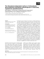
Modeling of the t peptide as an amphiphilic a helix
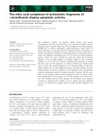
The primary sequence of the C-terminal region of Torpedo
AChE
T
is shown in Fig. 1A, including the last 12 residues of
Ó FEBS 2003 Amphiphilic a helical domain of the AChE T subunit (Eur. J. Biochem. 271)35
the catalytic domain and the t peptide. Secondary structure
prediction algorithms show that a large part of this peptide
is expected to assume an a helical structure, extending from
residue five to residue 26 or 28, with a possible interruption
at residues 14–16 that might allow a bend between two
helical segments. Giles proposed a similar arrangement, in
which a bend at residues 21–22 would bring together the
aromatic sectors of the two helices [16]; according to this
model, residues located in the N-terminal region of the
t peptide would be in close contact with the C-terminal
cysteine, C37.
If we assume an ahelical structure for the t peptide, a
lateral view shows that all the aromatic residues are oriented
on the same side (Fig. 1B), and a wheel projection [28] shows
that a sector of 100° is totally apolar (Fig. 1B). The polar
sector contains five acidic residues (one aspartic and four
glutamic acids) and four basic residues (one lysine, two
arginines and one histidine), which might form internal salt
bridges between residues D4 or E5 and R8, between E7 and
K11, and between E13 and R16, as analyzed in a further
study (S. Belbeoc’h, J. Leroy, A. Ayon, J. Massoulie
´
&
S. Bon, unpublished results). The cluster of hydrophobic side
chains in the apolar sector includes the seven aromatic
residues that are conserved in all known vertebrate AChEs
and BChEs, ranging from cartilagenous fishes (Torpedo)to
mammals. In particular, three tryptophans are evenly spaced
by seven residues and very close to each other in the wheel
diagram (Fig. 1B). This aromatic cluster could be respon-
sible for the hydrophobic interactions of AChE
T
subunits.
Chemical grafting of synthetic peptides confers
hydrophobic properties on water-soluble AChE
To characterize the interactions of the t region while
excluding possible effects of putative post-translational
modifications, we used chemically synthesized peptides, as
shown in Fig. 1C. Peptide t
1)40
corresponds to the whole
Torpedo t peptide; peptide t
1)32
corresponds to its first 32
aminoacids and contains all seven conserved aromatic
residues.
The peptides were grafted onto a water-soluble tetrameric
form (G
4
)ofElectrophorus electricus AChE, obtained by
tryptic digestion of collagen-tailed forms from the electric
organ [29,30]. We used this enzyme preparation because we
could obtain it in a highly purified form [18] and because
it was very stable, totally nonamphiphilic and could be
Fig. 1. Sequence and putative organization of
the C-terminal t peptide from AChE
T
.
(A) Primary structure of the last 12 residues of
the catalytic domain and of the t peptide.
A comparison of the Torpedo and rat
sequences shows the high degree of conserva-
tion, particularly of the seven aromatic resi-
dues, throughout vertebrates. The N-terminal
region of the human amyloid Ab peptide is
shown to indicate a 12 residue segment which
presents some homology with the t peptide
(underlined) (B) Proposed helical structure of
the N-terminal region of the t peptide: in the
side view, the distance of each residue from the
helix axis corresponds to the vertical dimen-
sion, with the central residue of the aromatic
cluster (W17) at the top. The position along
the axis corresponds to the horizontal dimen-
sion (arbitrary scales). The wheel representa-
tion corresponds to a faceview along the helix
axis of the segment of the t peptide containing
the aromatic residues. (C) Synthetic peptides
corresponding to different parts of the
t peptide. The underlined residues have been
substituted from the wildtype sequence of the
Torpedo marmorata tpeptide.
36 S. Bon et al. (Eur. J. Biochem. 271) Ó FEBS 2003
analyzed by the same methods used for the amphiphilic
AChE species. Chemical coupling of the synthetic peptides
to exposed lysine residues occurred randomly and did not
affect enzymic activity.
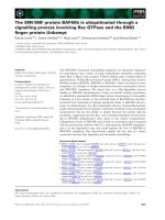
We deduced the mean number of peptides added per
tetramer from the apparent increase in molecular mass: the
modified Electrophorus G
4
AChE molecules obtained after
coupling of the peptides sedimented as fairly homogenous
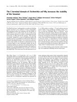
peaks, as illustrated in Fig. 2A. The sedimentation coeffi-
cient of G
4
-t
1)32
and of G
4
-t
1)40
was about 12.8 S, as
compared to 11.8 S for the original G
4
form (Fig. 2B).
Assuming that the mass of this globular protein is propor-
tional to S
3/2
, we estimate that the mass of the tetramer
increased from 320 kDa to 360 kDa, i.e. 10 kDa per
subunit, which corresponds to an average of three grafted
peptides per AChE subunit. In the case of G
4
-t
1)40
and
G
4
-t
25)40
, the formation of complexes with antibodies raised
against t
25)40
confirmed that essentially all the Electrophorus
G
4
AChE molecules had been modified (not shown). The
G
4
-t
1)32
derivative did not bind the antibodies, indicating
that the t
1)32
peptide did not contain the necessary epitopes.
The G
4
-t
25)40
derivative, like the original Electrophorus
G
4
enzyme, was not amphiphilic: its sedimentation coeffi-
cient (12.9 S) was not influenced by the presence of
detergent in the gradients. By contrast, the G
4
-t
1)32
and
G
4
-t
1)40
derivatives were clearly amphiphilic, as they
sedimented more slowly in the presence of Triton X-100
and even more slowly in the presence of Brij-96 (Fig. 2A,B).
This amphiphilic character was confirmed by charge-shift
electrophoresis under nondenaturing conditions. The
t-peptide–AChE conjugates migrated in opposite directions
in the presence of the negatively and positively charged
detergents, cetyltrimethylammonium bromide (CTAB) and
Na
+
deoxycholate (not shown).
The fact that the short t
1)32
peptide and the long t
1)40
peptide confer amphiphilic properties to Electrophorus
AChE tetramers, whereas the t
25)40
peptide does not
suggests that the 1–32 region, containing an a helix with
seven aromatic residues, is sufficient to support hydropho-
bic interactions.
Characterization of t peptide–lipid interactions
by use of circular dichroism
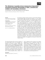
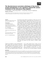
Figure 3 shows the CD spectrum in the far UV of the t
1)32
peptide under various conditions. In organic solvents, such
as methanol, the spectrum presents the characteristic
features of an a helical structure, with double minima at
210 nm and 222 nm. The h
222
value of )31 600 degÆdmol
)1
Æ
cm
2
indicates that about 85% of the polypeptide is a helical.
We obtained a similar proportion of a helical structure by
reconstituting the whole spectrum as a sum of the contri-
butions of different secondary structures, derived from a
set of known proteins [31]. This high a helical content
is comparable to that of amphiphilic peptides of similar
length, which have been characterized by various methods
as monomeric 20-residue a helical rods [32,33]. When the
peptide was dissolved in an aqueous buffer, the minima at
210 nm and 222 nm displayed ellipticities of only
h ¼ )12 210 degÆdmol
)1
Æcm
2
and h ¼ )9770 degÆdmol
–l
Æcm
2
respectively, indicating a much lower a helical content of
35%.
Fig. 2. Effect of detergents on the sedimentation of Electrophorus AChE
tetramers, chemically coupled with the t
1)40
peptide. (A) Sedimentation
patterns of a conjugate of Electrophorus AChE G
4
species with the
t
1)40
peptide, obtained in sucrose gradients containing no detergent;
0.1% Triton X-100 or 0.1% Brij-96. (B) Sedimentation coefficients
obtained in these different conditions for G
4
AChE and its conjugates.
The conjugated enzymes containing peptides t
1)32
and t
1)40
sedi-
mented faster without detergent than in the presence of Triton X-100
or Brij-96, indicating that they bind detergent micelles, in contrast with
conjugated enzyme containing peptide t
25)40
and the nonconjugated
enzyme, which sedimented in the same way under all three conditions.
Fig. 3. Far UV dichroic spectrum of peptide t
1-32
. Peptide (5 l
M
)in
1m
M
Tris/HCl buffer, pH 7.5, using a 1 cm path-length cuvette
(dotted line); the same solution after addition of lysolecithin micelles,
with a lipid/peptide molar ratio of 20 (thin line); 50 l
M
peptide in
methanol, using a 0.1 path-length cuvette (bold line).
Ó FEBS 2003 Amphiphilic a helical domain of the AChE T subunit (Eur. J. Biochem. 271)37
The CD spectrum was markedly modified by addition of
lysolecithin micelles. It approached that observed in meth-
anol when the lysolipid/peptide molar ratio was about 10,
and was not modified further at higher micelle concentra-
tions (Fig. 3). Under these conditions, the helical content
was about 68%, corresponding to 18–22 residues per
peptide organized into an a helix. Thus, lipid micelles can
induce an a helical conformation in the t peptide.
Intrinsic fluorescence of the t peptide
The t
1)32
peptide displays intrinsic fluorescence due to the
fact that it contains three tryptophans; W10, W17 and W24,
and two tyrosines, Y20 and Y31. When dissolved in aqueous
buffer and excited at 280 nm, its emission spectrum was
centered at 345 nm. The shape of the emission spectrum was
identical when excitation was at 295 nm, a wavelength at
which tyrosine residues do not absorb. Thus the fluorescence
of the peptide is totally due to tryptophan residues: Y20 and
Y31 are either totally quenched or very efficiently transfer
their energy to tryptophan residues in their neighbourhood.
In aqueous solution, the fluorescence of the tryptophan
residues showed a blue shift of 6 nm relative to
N-acetyltryptophanylamide, indicating that they are only
slightly buried. The blue shift was increased by about 2 nm
when dithiothreitol was omitted. Addition of methanol,
which decreased the polarity of the medium, prevented
aggregation and increased the a helical content; this pro-
duced an increase in quantum yield and a slight shift of the
maximum emission wavelength, indicating that the trypto-
phan residues were more exposed to the solvent.
We obtained similar results with the t
1)40
peptide, except
that it was more aggregated in aqueous solution; the t
1)32
peptide also aggregated above 1 l
M
, as indicated by an
increase in the light scattering. On the contrary, reducing the
concentration below 0.2 l
M
induced a progressive red shift
of the emission k
max
for both peptides; however, this never
reached 350 nm, which would correspond to total exposure
of tryptophan residues.
Interaction of peptides with detergents and
phospholipids as followed by fluorescence
We followed changes of the intrinsic tryptophan fluores-
cence by addition of phospholipids (Fig. 4) and detergents
(Fig. 5). The induced blue shifts in the k
max
of emission and
intensity changes were similar for t
1)32
and t
1)40
.
Figure 4 shows that addition of lipid vesicles to an
aqueous solution of the t
1)32
peptide at pH 7.5 (5 l
M
)
produced changes both in intensity and wavelength of
fluorescence. For the zwitterionic egg lecithin vesicles, the
changes did not reach a plateau even at Ri values greater
than 150, indicating a low affinity of the peptides for the
lecithin–water interface. In contrast, we observed a stronger
blue shift and more pronounced quenching upon addition
of negatively charged phosphatidylserine vesicles, and both
effects reached a plateau below an Ri value of 100. The
emission maximum at the plateau, 327 nm, indicates that
the tryptophan residues were in a very hydrophobic
environment.
Figure 5 shows that addition of 32 l
M
lysolecithin to
peptide t
1)32
(3.2 l
M
) shifted the emission maximum close
to 330 nm, and increased the intensity twofold. The affinity
of the peptide was much higher for lysolecithin micelles than
for lecithin vesicles, indicating that insertion of the peptide is
easier in the more fluid and dynamic lysolecithin micelles
than in the bilayer of lecithin vesicles, as observed for other
amphiphilic peptides [34].
At lower concentrations of the peptide (0.5 l
M
),
lysolecithin induced a similar shift in k
max
but a more
complex variation of the intensity, which first decreased,
reaching a minimum at a lipid : peptide molar ratio (Ri)
of 30–40 and then increased again (not shown). Such
biphasic curves were previously observed for lipid–peptide
interactions occurring in the concentration range of the
critical micellar concentration (cmc) [17]. For lysolecithin,
the cmc is 20 l
M
, corresponding to Ri values of 5–6 and
30–40, for peptide concentrations of 3.2 and 0.5 l
M
respectively. These observations show that the t
1)32
peptide interacts with lysolecithin both below and above
the cmc.
CTAB is a positively charged detergent with a cmc of
0.2–0.3 m
M
, and SDS is negatively charged and has a cmc
of 1–2 m
M
[35]. At neutral pH, addition of CTAB to
3.2 l
M
peptide shifted k
max
down to 334 nm, reaching a
plateau for Ri ¼ 20, and induced a large increase in the
intensity at 334 nm, attaining 260% for Ri values above
100, i.e. above the cmc that corresponds to Ri values of
60–90 (Fig. 5). In contrast, SDS did not induce any
significant change in fluorescence up to Ri ¼ 150; above
this value, we noted a gradual shift of k
max
downto330nm
for Ri ¼ 300–400, i.e. above the cmc of the detergent. We
obtained similar results at pH 5.7, in spite of a reduction in
Fig. 4. Effects of phospholipid vesicles on the intrinsic fluorescence of
peptide t
1)32
. The peptide concentration was 5 l
M
,in20m
M
Tris/
acetate buffer pH 7.5 containing 5 m
M
dithiothreitol to avoid the
formation of disulfide bonds, under a nitrogen stream, at 25 °C. (A)
Variation of the wavelength of maximum emission (k
max
) as a function
of the molar ratio of lipids to peptide (Ri). (B) Relative variation of
emission intensity at 333 nm (DI/I
0
) as a function of Ri. (m)egg
lecithin vesicles; (s) phosphatidylserine vesicles.
38 S. Bon et al. (Eur. J. Biochem. 271) Ó FEBS 2003
the negative charge of the peptide. Thus, the zwitterionic
and positively charged detergents readily interact with the
peptides even below the cmc, while the negatively charged
detergent interacts only when approaching the cmc.
Formation of disulfide bonds in homomeric oligomers
with cysteines at various positions in the C-terminal
region of rat AChE
T
subunits
The preceding studies were performed on isolated peptides
or on conjugates in which peptides were chemically coupled
at the surface of a protein. However, the t peptide is
normally linked to the C-terminus of the catalytic domain of
AChE
T
subunits and it contains a cysteine (C37) which
allows their dimerization through an intersubunit disulfide
bond. The crystallographic structure of AChE dimers
[36,37] or monomers [38] shows that the catalytic domain
terminates with an a helix (helix a
10
) constituted by residues
)18 to )1. Secondary structure predictions suggest that this
helix is separated from the a helical portion of the t peptide
by a short loop (around residues )1to2),andmaypresenta
break around residues 15–16 (Fig. 1A). An interrupted helix
could form a hairpin, as proposed by Giles [16], who
suggested that the aromatic-rich sectors of two a helical
segments would constitute a compact aromatic cluster.
According to this model, a bend at residues 21 and 22 would
bring the N-terminal and C-terminal ends into close
proximity.
To obtain information on the articulation between the
catalytic domain and the t peptide, we analyzed the
formation of intercatenary disulfide bonds by cysteine
residues located at the end of the catalytic domain of rat
AChE
T
or at the beginning of its t peptide, in the )5to6
interval; in these mutants, the original cysteine was either
retained or replaced by a serine (C37S). We also introduced
cysteine residues near the middle of the t peptide, in the
predicted a helical region containing aromatic residues (at
positions 19 and 21), and in its C-terminal region, which is
not predicted to be a helical, at positions 34 to 36.
The AChE
T
cysteine mutants were expressed in transi-
ently transfected COS cells. In the absence of any cysteine in
the C-terminal region of rat AChE, we obtained mainly
monomers, with a small proportion of tetramers, as
reported previously in the case of human AChE [39] and
rat AChE [40]. Therefore, the presence of dimers, as
observed in the case of the other mutants, indicates the
formation of an intercatenary disulfide bond.
In the hypothesis of a hairpin structure, an intracatenary
disulfide bond might be formed in mutants containing the
original cysteine or another C-terminal cysteine, together
with a cysteine in the N-terminal region of the t peptide; this
would preclude the formation of dimers, which requires an
intercatenary disulfide bond. However, we did not observe
this in any combination of N-terminal and C-terminal
cysteines (not shown). Therefore, the t peptide almost
certainly adopts an elongated conformation in AChE
T
monomers and dimers.
When the original cysteine was mutated to serine (C37S),
all mutants containing a single cysteine at positions )5to6,
19, 21, or 34 to 36, produced active AChE which was
secreted at variable levels (Fig. 6A). The cellular and
secreted enzymes contained different proportions of dimers,
sometimes with a small amount of tetramers, as indicated by
nondenaturing electrophoresis (Fig. 6B).
Sedimentation patterns illustrating the amounts of mono-
mers, dimers and tetramers are shown in Fig. 7 for cysteines
in the )5 to 6 interval. The proportion of dimers produced
was very low with cysteines in the )5to)3 interval, a region
which is predicted to be a helical. The distances between
pairs of alpha carbons corresponding to residues )5to)2
can be determined from the crystallographic structure of a
catalytic dimer [36]: they are 8.6, 13, 15 and 8.9 A
˚
respectively. A small proportion of AChE
T
subunits were
dimerized with cysteines at position )5and)2, for which
the distance is smallest but still appears too high for
establishment of a disulfide bond, which is normally < 6 A
˚
.
This indicates that, in AChE
T
subunits, the distal part of the
catalytic domain is sufficiently flexible to allow the forma-
tion of a disulfide bond in this segment, between the two
subunits in a dimer. The production of dimers was higher
than for the wildtype with cysteines at positions )2to3,
suggesting that this region, which is predicted to form a coil,
constitutes a flexible hinge between the catalytic domain and
the amphiphilic helix of the t peptide; it was lower at
positions 4 and 5 and increased again at position 6. As these
three residues are probably included in the N-terminal
region of the helix, the observed variations in the efficiency of
dimerization may be due to their orientation relative to the
Fig. 5. Effects of zwitterionic and charged detergents on the intrinsic
fluorescence of peptide t
1-32
. The peptide concentration was 3.2 l
M
,in
20 m
M
Tris/HCl buffer containing 5 m
M
dithiothreitol to avoid the
formation of disulfide bonds. (A) Variation of the wavelength of
maximum emission (k
max
) as a function of the molar ratio of detergent
to peptide (Ri). (B) Relative variation of emission intensity at 333 nm
(DI/I
0
) as a function of Ri. (h, j)SDS;(s, d) cetyl-trimethyl-
ammonium bromide (CTAB); (n, m) lysolecithin. Filled symbols
(j, d, m), pH 7.5; open symbols (h, s, n), pH 5.7.
Ó FEBS 2003 Amphiphilic a helical domain of the AChE T subunit (Eur. J. Biochem. 271)39
aromatic sector: residue 6 is in the aromatic sector, while
residues 4 and 5 are on the opposite side.
We also studied the production of dimers with cysteine
residues located at positions 19 and 21, in the center of the
predicted amphiphilic a helical region but in opposite
sectors. With a cysteine at 19, the cellular enzyme contained
dimers but their secretion was very low (Fig. 6B), suggesting
that the presence of a disulfide bond at this position induced
their degradation. In contrast, a cysteine at position 21,
within the sector containing aromatic residues, appeared
much more favorable for dimerization and secretion. In
contrast with dimers containing disulfide bonds in the
N-terminal or C-terminal regions of the t peptide, the
M21C/C37S dimers did not interact with detergent micelles
(not shown), indicating that the two aromatic clusters
occluded each other.
Dimers were as efficiently produced and secreted with
cysteines located at positions 34, 35 or 36 as with the original
cysteine (at position 37) suggesting that this C-terminal
region of the t peptide is flexible. It is noteworthy that the
level of cellular activity was markedly higher with a cysteine
at 35, corresponding to an increased amount of monomers;
the presence of a cysteine instead of an aspartic acid at this
position seems to increase the retention or decrease the
degradation of monomers.
Figures 6B and 7 show that the production of tetramers
varied with the position of the cysteine and was not
proportional to that of dimers. Tetramer production was
systematically higher with C-terminal cysteines (34–37) than
with cysteines in the N-terminal region of the t peptide ()2
to 3). This suggests that the relative organization of the
t peptides and of the catalytic domains is more favorable
for tetramerization when dimers are joined through a
C-terminal disulfide bond.
Hetero-oligomerization: orientation of the PRAD
and t peptides in the T
4
–Q
N
complex
The Q
N
protein possesses two adjacent cysteine residues
(C70 and C71) located immediately upstream of the proline-
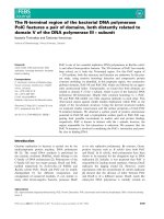
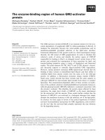
Fig. 6. Effect of cysteines at various positions
in the C-terminal region of rat AChE
T
subunits.
Cysteines were introduced into rat AChE
T
subunits at various positions at the junction of
the catalytic domain and the t peptide ()5to
6), in the middle of the t peptide (19 or 21),
and in the C-terminal part of the t peptide (34
to 36); in these mutants, the original cysteine
(C37) was replaced by a serine, so that all
mutants possessed a single cysteine. (A) Cel-
lular and secreted AChE activities: all mutants
produced and secreted active AChE when
expressed with or without Q
N
.Whencysteines
were present in the )5 to 6 interval, we used a
modified Q
N
(Q
N
-CC) with cysteines down-
stream of the proline-rich region; with the
other mutants, we used the Q
N
protein con-
taining cysteines upstream of the proline-rich
region. Activities are expressed as percentage
of the wildtype; the bars indicate the standard
errors of two to three independent experi-
ments. The shaded and hatched rectangles
correspond to mutants expressed without and
with Q
N
, respectively. (B) Nondenaturing
electrophoresis of AChE oligomers produced
by rat AChE
T
subunits containing a single
cysteine at different positions, expressed
without Q
N
.
40 S. Bon et al. (Eur. J. Biochem. 271) Ó FEBS 2003
rich motif (Fig. 8A,B) such that the disulfide linkage of two
AChE
T
subunits with one Q
N
protein produces a Ôheavy
dimerÕ that can be distinguished by SDS/PAGE under
nonreducing conditions from Ôlight dimersÕ consisting of
only two disulfide linked AChE
T
subunits [12] (Fig. 8C). In
order to study the formation of these disulfide bonds,
AChE
T
mutants were coexpressed with the natural Q
N
protein possessing two adjacent cysteines C70 and C71
upstream of the PRAD (CC-Q
N
), and with a Q
N
mutant in
which the original cysteines were mutated to serines and two
cysteines were introduced downstream of the PRAD, at
positions 87 and 88 (Q
N
-CC), as shown in Fig. 8A. The
C37S mutant that formed no intercatenary disulfide bonds
but was recruited into T
4
–Q
N
complexes, served as a
control.
Figure 8Ca illustrates the fact that CC-Q
N
formed
disulfide bonds with AChE
T
mutants that contained a
cysteine in the C-terminal region of the t peptide (as
expected, given that this corresponds to the wildtype
situation), but not with AChE
T
mutants containing an
upstream cysteine (Fig. 8Cb); in the latter case, all AChE
T
subunits were disulfide-linked in homodimers. Recipro-
cally, disulfide bonds could be formed, although less
efficiently, between Q
N
-CC and some of the AChE
T
mutants that contained a cysteine in the N-terminal region
of the t peptide (Fig. 8Cd) but not in the C-terminal
region (Fig. 8Cc). This indicates that the N- and
C-terminal extremities of the t peptides are distant in the
complex, eliminating the possibility that the peptides
would be folded in hairpins as suggested above for the
free t peptides; the same reasoning shows that the PRAD
is also elongated.
Taking into consideration that both the t peptides and
the PRAD are elongated in the heteromeric complexes, we
Fig. 7. Formation of homo-oligomers of rat
AChE
T
subunits with cysteines at various posi-
tions in their C-terminal region. Sedimentation
patterns in sucrose gradients, for mutants of
the )5 to 6 interval. The patterns obtained for
the wildtype (solid line) and for the C37S
mutant with no C-terminal cysteine (C37S)
(dotted line) are shown for comparison. The
monomers, dimers and tetramers are indicated
(T
1
,T
2
,T
4
respectively). The areas under the
sedimentation profiles are proportional to the
cellular and secreted activities, so that the
areas of the peaks represent the relative
amounts of the corresponding molecular
forms.
Ó FEBS 2003 Amphiphilic a helical domain of the AChE T subunit (Eur. J. Biochem. 271)41
can then question their respective orientations: the forma-
tion of intercatenary disulfide bonds shows that the four
t peptides are all parallel, and are oriented in the opposite
direction to the PRAD, as the N-terminal extremity of the
PRAD can only be disulfide linked to the C-terminal region
of two t peptides, and vice versa.
Exploring the association of t peptides and PRAD
in the T
4
–Q
N
complex by the formation of heterophilic
intercatenary disulfide bonds
Figure 9A shows an analysis of the complexes formed
between the various AChE
T
cysteine mutants and Q
N
,in
Fig. 8. Disulfide bonds between Q
N
and two AChE
T
subunits, in the T
4
–Q
N
complex. (A) Schematic representation of the constructs used. The T
4
–Q
N
complex was formed when AChE
T
subunits possessing a cysteine near the N- or C-terminus of the t peptide were expressed with Q
N
constructs
containing pairs of cysteines located either upstream or downstream of the PRAD. The arrows indicate the N-terminal to C-terminal orientation.
(B) Schematic representation of the different combinations of cysteine mutants; the PRAD is shown as a thick central line and the t peptides as
zigzags; the cysteines are indicated by circles and the disulfide bonds by thick lines. Scheme a corresponds to the wildtype
44
situation; b corresponds to
an association of wildtype t peptides with Q
N
-CC; c and d correspond to associations of t peptides containing an upstream cysteine (L3C/C375)
with CC-Q
N
and Q
N
-CC, respectively. (C) Analysis of disulfide-linked species by SDS/PAGE after metabolic labeling. Lanes a, b, c, d correspond
to the four diagrams in panel
45
(B). ÔHeavy dimersÕ (composed of one Q
N
protein linked to two AChE
T
subunits) were produced only when cysteines
were at opposite ends of the t peptide and PRAD.
Fig. 9. Effect of the position of cysteines in the C-terminal region of AChE
T
on the formation of hetero-oligomers (T
4
-Q
N
). (A) Nondenaturing
electrophoresis of AChE oligomers secreted by cells expressing AChE
T
subunits with the appropriate Q
N
construct (Q
N
-CC for AChE
T
subunits
containing a cysteine in the )5 to 6 interval, CC-Q
N
for AChE
T
subunits containing a cysteine at positions 19, 21 and in the 34 to 37 interval).
(B) Analysis of disulfide bonds between AChE
T
subunits and Q
N
(Ôheavy dimersÕ), by nonreducing denaturing electrophoresis after metabolic
labeling. There were no heavy dimers with cysteines at 19 or 21, with either the CC-Q
N
or Q
N
-CC construct (not shown).
42 S. Bon et al. (Eur. J. Biochem. 271) Ó FEBS 2003
nondenaturing electrophoresis. The mutant AChE
T
sub-
units were coexpressed with a Q
N
construct carrying a
C-terminal flag epitope, so that the T
4
–Q
N
-flag complexes
were characterized by their reaction with the M2 anti-flag Ig
(not shown). Every cysteine mutant described above, with
the exception of M21C/C37S, formed heteromeric non-
amphiphilic T
4
–Q
N
complexes, including the C37S mutant
which cannot form intercatenary disulfide bonds [9]. The
fact that the M21C/C37S mutant did not associate with Q
N
probably results from the formation of compact dimers in
which the aromatic sectors were mutually masked, as
discussed above. In the case of the S19C/C37S mutant, the
level of secretion was increased by coexpression with Q
N
,
indicating that a fraction of AChE
T
subunits were rescued
from degradation by the formation of T
4
–Q
N
complexes.
The formation of disulfide bonds was studied by electro-
phoresis of metabolically labelled proteins under denaturing
conditions and without reduction (Fig. 9B). The left panel
illustrates complexes formed between AChE
T
subunits
possessing an upstream cysteine ()5to6)andtheÔinvertedÕ
Q
N
-CC construct, the central and right panels illustrate
complexes formed between AChE
T
subunits possessing
either a central (19 or 21) or downstream cysteine (34 to 37,
including the wildtype), and the CC-Q
N
construct.
The distribution of unlinked subunits and disulfide-linked
dimers is consistent with the distribution of active mono-
mers and dimers, as indicated by nondenaturing electro-
phoresis and sedimentation of the enzyme produced in the
absence of Q
N
(Figs 6B and 7) or in its presence (Fig. 9A).
In particular, a large number of AChE
T
subunits were not
disulfide-linked in the case of cysteines at )5, )4and)3,
while homodimers were efficiently formed with cysteines in
the )2 to 6 and 34 to 37 regions. In the case of mutant S19C/
C37S, the level of disulfide-linked ÔlightÕ dimers was very low
without Q
N
(not shown) but increased in its presence
(Fig. 9), indicating that disulfide bonds between cysteines at
position 19 were present in the complex. Mutant M21C/
C37S produced homomeric, disulfide-linked dimers with or
without Q
N
, in agreement with the fact that it did not form
T
4
–Q
N
complexes.
With cysteines in the N-terminal region of the t peptide,
the production of ÔheavyÕ dimers with the Q
N
-CC construct
was maximal for cysteines at positions 3 and 4, bracketed by
adecreaseat2and5andasmallerincreaseat1and6.
AChE
T
subunits with cysteines at positions 19 or 21 did not
form intercatenary disulfide bonds with either CC-Q
N
(Fig. 9B central panel) or Q
N
-CC (not shown). The
formation of ÔheavyÕ dimers with CC-Q
N
was similar with
cysteines at positions 34, 35, 36 and with the natural cysteine
(C37), again suggesting that this C-terminal region of the
t peptide is flexible.
Discussion
The alternatively spliced C-terminal peptides of vertebrate
AChE largely condition the function of the enzyme [2]. They
determine both the oligomeric associations and the anchor-
ing: the ÔreadthroughÕ (R) variant produces soluble mono-
mers, the ÔhydrophobicÕ (H) variant produces GPI-anchored
dimers, and the ÔtailedÕ (T) variant produces a wide array of
homo-oligomers and hetero-oligomers. In some cases, the
C-terminal peptides have been shown to possess an intrinsic
bioactivity; for example, the C-terminal peptide of the
ÔreadthroughÕ variantofAChE,whichwasshowntobe
induced by stress in the central nervous system [41], was
called ARP: it appears to be released and to control
hematopoietic differentiation [42].
The t peptide of AChE
T
subunits is necessary for
association with the collagen protein ColQ to produce
collagen-tailed forms, and with the transmembrane protein
PRiMA to produce hydrophobic-tailed tetramers [2]. The
physiological importance of these associations is illustrated
by the fact that mutations in the human COLQ gene which
prevent this association or modify the structure of the
collagen tail result in AChE-deficient neuromuscular end-
plates [43–45].
In addition to the hydrolysis of acetylcholine, AChE may
exert nonclassical functions which also depend on its
variable C-terminal domains; for example, AChE
T
subunits
have been shown to induce neurite extension in neural cells
[46–48] and it is not certain whether other AChE variants
possess the same capacity [49]. In addition to functions
related to cell–cell interactions, AChE has been shown to be
expressed in brain tumors [50]. It is also expressed in
apoptotic cells and, quite surprisingly, is transferred to the
nucleus [51,52]; this localization probably depends on the
C-terminal t peptide, as shown by the fact that green
fluorescent protein presents a nuclear localization when
fused to a 67-residue C-terminal fragment of the AChE
T
subunit containing the t peptide, but not to a fragment
containing the ÔreadthroughÕ r peptide [50]. AChE may also
participate in the pathogenesis of Alzheimer’s disease as it
induces the aggregation of Ab amyloidogenic peptide and
increases its neurotoxicity [53,54]. It is possible that the
t peptide itself, or a fragment of the t peptide without the
catalytic domain, may be involved in this pathology. As
illustrated in Fig. 1A, fragment 12–25 of the AChE
t peptide, containing the central aromatic residues, presents
some homology with part of the Ab amyloid peptide [55]
and was shown to aggregate into amyloid fibrils, whereas
the complete t peptide or the homologous fragment of the
human BChE t peptide do not possess this property [56,57].
In addition, this peptide fragment was found to modulate
the response of N-methyl-
D
-aspartate receptors in the
guinea-pig hippocampus [58]. This indicates that fragments
derived from the t peptide may possess distinct conforma-
tions and biological activities.
The C-terminal t peptide of AChE forms an amphiphilic
a helix
In this study, we present spectroscopic evidence that at least
part of the C-terminal t peptide of acetylcholinesterase
AChE
T
subunits may adopt an a helical structure, especially
in the presence of detergents or lipids but also in heteromeric
complexes with a PRAD, in agreement with our previous
proposal that it could form an amphiphilic ahelix in which
conserved aromatic residues are grouped in a hydrophobic
cluster [1]. Secondary prediction programs suggest that the
peptidic chain that corresponds to residues 4–29 can adopt
an a helical conformation; crystallography of a complex
formed by four synthetic t peptides and a synthetic PRAD
peptide showed that residues 1–30 and/or 1–36 are definitely
a helical (M. Harel, H. Dvir, S. Bon, W.Q. Liu, C. Garbay,
Ó FEBS 2003 Amphiphilic a helical domain of the AChE T subunit (Eur. J. Biochem. 271)43
J.L. Sussman, J. Massoulie
´
& I. Silman, unpublished
results). In this structure the tyrosine residues are close to
tryptophans, which explains why we do not observe any
contribution from tyrosines in the fluorescence emission; the
tryptophans never appear to be totally exposed to the
solvent, even in the absence of lipids or detergents, but
detergent molecules could bind to the peptides below the
cmc, in agreement with the existence of an aromatic stack in
the ahelical structure when in an aqueous solution. These
results are consistent with a recent study showing that
synthetic t peptides from human AChE and BChE are
predominantly a helical according to their UV absorption
spectra [57]. Moreover, these authors present evidence,
based on recognition by conformation-sensitive antibodies,
that the t peptide is also a helical in the AChE
T
subunits.
Amphiphilic helices represent a key structural element in
many protein interactions [59,60]. We now suggest that an
a helical organization of part of the t peptide provides the
basis for the hydrophobic binding properties of AChE
T
subunits. The t peptide has a low hydrophobicity according
to Eisenberg’s scale [61,62], Htot ¼ )10.3, or <H> ¼
)0.3. However, in an a helical conformation, the hydro-
phobic moment of the helix is rather high, lH ¼ 8.55 or
<lH> ¼ 0.26. The characteristics of this helix may be
compared to those of cytotoxic peptides, such as melittin,
magainins and mastoparans [63,64] and polypeptide hor-
mones; their apolar angle (100°) is similar and the helical
structure is induced by lipids in both cases, even though the
mean hydrophobic moment of the latter peptides is
somewhat higher (0.37 and 0.54, respectively).
The fact that chemically coupled synthetic t peptides
containing the aromatic-rich segment conferred an amphi-
philic character to soluble tetramers of Electrophorus (G
4
)
AChE, shows that the amphiphilic properties of AChE
T
subunits do not require any modification of the t peptide.
However, this does not exclude the possibility that post-
translational modifications, such as acylation by fatty acids
or addition of other lipidic moieties may occur during
normal biosynthesis of AChE
T
subunits and contribute to
their hydrophobic properties. In fact, it has been reported
that palmitate was incorporated into a membrane-bound
enzyme that was produced from murine AChE
T
subunits in
transfected human embro kidney 293 cells [15]; much of this
enzyme appeared to remain monomeric, possibly because
the formation of a thioester bond with cysteine C37
prevented dimerization.
Conjugates containing the t
1)32
peptide that lack the final
eight residues, presented the same amphiphilic properties as
those containing a full length t
1)40
peptide. In contrast,
G
4
–T
25-40
, which only contains the last two aromatic
residues, does not interact with detergents. This indicates
that at least part of the first 32 residues of the t peptide
(T
1-32
) may form an amphiphilic helix that is sufficient to
explain the hydrophobic interactions of AChE
T
subunits,
and that the distal eight residues are not necessary in this
respect.
Self assembly of t peptides and intercatenary disulfide
bonds
In the second part of our study, we analyzed mutant rat
AChE
T
subunits in which we introduced cysteines at
various positions at the end of the catalytic domain ()5to
)1) and in the t peptide, at the beginning (1 to 6), in the
middle (19 or 21), or near the end (34, 35 or 36), in the
presence or in the absence of the original cysteine (C37). All
these mutants produced and secreted active AChE,
although at variable levels, indicating that they were
partially degraded [14]. The results presented here have
been confirmed for some of these positions, in the case
of homologous mutants of Torpedo AChE
T
subunits
(S. Belbeoc’h, J. Leroy, A. Ayon, J. Massoulie
´
& S. Bon,
unpublished results).
In the absence of any cysteine, AChE
T
subunits do not
form dimers, but can still form homotetramers, although at
a lower level [39,40,65]. The fact that dimers were efficiently
produced when the original cysteine was present together
with an additional cysteine in the )5 to 6 interval suggests
that the t peptide does not fold as a hairpin, as proposed by
Giles [16], because dimerization would be prevented by the
formation of an intracatenary disulfide bond.
When the original cysteine was removed, mutants
possessing a cysteine between )5 and 6 presented several
increases and decreases in the level of secreted activity and in
the proportion of dimers. This can be explained by
distinguishing three segments, in agreement with secondary
structure predictions: (a) residues )5to)3 are probably
constrained by the C-terminal a helix (a
10
)ofthecatalytic
domain, so that dimers are formed inefficiently or not at all
(at position )4), and the resulting proteins are largely
degraded. (b) Residues )2 to 3 may form a flexible hinge
between the catalytic domain and the helical part of the
t peptide, resulting in a high proportion of dimers; this
flexibility may be induced by the presence of the t peptide,
as the a
10
helix was seen to extend to the end of the common
catalytic domain (residue )1), in dimers of the AChE
H
subunits [37] and in monomers of the truncated subunits,
reduced to the catalytic domain [38]. (c) Residues 4 to 6 are
probably included in the a helical region of the t peptide; the
fact that the level of secreted dimers was higher
36
with a
cysteine at position 6 than at positions 4 and 5 may result
from a more appropriate orientation, relative to the
aromatic sector of the helix.
The effect of a cysteine in the central region of the
t peptide depended on its orientation relative to the
aromatic sector. Mutant S19C/C37S formed dimers which
were degraded rather than secreted. Mutant M21C/C37S,
with a cysteine near the middle of the aromatic sector, was
the only mutant that produced nonamphiphilic dimers,
suggesting that the two aromatic clusters occlude each other
in the dimer when linked
37
by an intracluster disulfide bond.
All subunits that possessed a cysteine in the 34 to 37
interval efficiently formed dimers and tetramers, indicating
that this C-terminal region is flexible and that the geometry
of these dimers is favorable for their assembly into
tetramers.
Association of four t peptides with a PRAD
in the T
4
–Q
N
complex
The most interesting and physiologically important prop-
erty of the t peptide is its capacity to form quaternary
complexes with a proline-rich motif or PRAD, which is
present in both anchoring proteins, ColQ and PRiMA [8,9].
44 S. Bon et al. (Eur. J. Biochem. 271) Ó FEBS 2003
The wildtype complex contains intercatenary disulfide
bonds, based on the C-terminal cysteines of the t peptides:
two AChE
T
subunits are linked together, forming a ÔlightÕ
dimer, while the other two are linked to two adjacent
cysteines located upstream of the PRAD in Q
N
, forming a
ÔheavyÕ dimer [12]. However, the complex can be assembled
without intercatenary disulfide bonds [9].
T
4
–Q
N
complexes in which four AChE
T
subunits are
associated with Q
N
, a PRAD-containing fragment of ColQ,
could be formed at variable levels with all cysteine mutants
(except M21C/C37S). The formation of heteromeric disul-
fide bonds between the PRAD and the t peptide was
revealed by the presence of ÔheavyÕ dimers in nonreducing,
denaturing electrophoresis. We found that such disulfide
bonds could only form between C-terminal cysteines in the
t peptide and N-terminal cysteines in the PRAD (CC-Q
N
),
or vice versa between N-terminal cysteines of the t peptide
and C-terminal cysteines in the PRAD (Q
N
-CC). This
excludes the hypothesis that the two ends of the t peptide
would be in close contact, so that the t peptide appears to
adopt an elongated structure in the complex, as also shown
without a PRAD. In addition, the organization of intercat-
enary disulfide bonds in the T
4
–Q
N
complex implies that
the four t peptides are parallel, and that the PRAD runs in
the opposite direction.
This organization is in perfect agreement with the
structure of a PRAD–WAT complex formed by the
spontaneous assembly of synthetic PRAD and WAT pep-
tides, which has recently been solved by X-ray crystallo-
graphy (M. Harel, H. Dvir, S. Bon, W.Q. Liu, C. Garbay,
J.L. Sussman, J. Massoulie
´
& I. Silman,
38
unpublished
results). In this very compact structure, four t peptides
(WAT) organized as elongated a helices form a cylinder
around the PRAD, which is organized as a polyproline II
helix. The seven aromatic residues of the t peptides are
oriented towards the center of the cylinder with the
tryptophan rings apposed to prolines of the PRAD, while
the charged residues are exposed at the external surface.
This confirms that residues 1 to 34 of the t peptide can
adopt an a helical structure in which all seven aromatic
residues are grouped in a hydrophobic sector, and explains
thefactthattheT
4
–Q
N
complex is nonamphiphilic.
The presence of a flexible hinge between helix a
10
of the
catalytic domain and the helical part of the t peptide, as
predicted by secondary structure algorithms, may therefore
be crucial for the assembly of AChE tetramers associated
with a PRAD-containing anchoring subunit such as ColQ
or PRiMA. This flexibility explains that cysteines intro-
duced in the )2 to 3 region can efficiently form homomeric
disulfide bonds in dimers; however, the same cysteines did
not form heteromeric disulfide bonds with the Q
N
-CC
construct in the secreted T
4
–Q
N
complexes, probably
because the distance along the axis of the cylinder of
t peptides was too large.
In the helical part of the t peptide, the orientation of
cysteines around the axis seems to determine the possibility
of intercatenary disulfide bonding: positions 3 and 6 are
more favorable than position 5 for the formation of
homomeric dimers and also for the formation of hetero-
meric disulfide bonds with the PRAD; position 4 is not
particularly favorable for dimerization, but allows disulfide
bonding with Q
N
-CC, although less than half of the AChE
T
subunits were included in the ÔheavyÕ dimers. The formation
of disulfide bonds between cysteines at positions 3 and 6
of the t peptide and cysteines introduced at positions 87 and
88 of Q
N
suggests that the PRAD can slide over the
corresponding distance, in the cylinder of t peptides.
Cysteines at positions 19 and 21, located in opposite
sectors of the a helix, had very different effects. Mutant
S19C/C37S formed dimers which were degraded rather than
secreted, and was able to produce heteromeric T
4
–Q
N
complexes containing disulfide bonds between t peptides.
Mutant M21C/C37S AChE
T
subunits were secreted as
nonamphiphilic dimers and did not associate with Q
N
,
indicating that the aromatic clusters masked each other
when a disulfide bond was present in the aromatic sector.
The formation of both ÔlightÕ and ÔheavyÕ dimers with
cysteines at positions 34, 35 or 36 appeared similar to the
wildtype (with cysteine C37), indicating that the 34 to 37
segment of the t peptide is flexible, allowing an efficient
formation of T
4
–Q
N
complexes, stabilized by heteromeric
disulfide bonds with cysteines 70 and 71 of CC-Q
N
for all
these positions. In addition, it is possible that the PRAD can
slide longitudinally in the complex, as suggested for
N-terminal cysteines. AChE
T
subunits possessing cysteines
in the 34 to 37 segment produced the highest level of
homomeric tetramers, and T
4
–Q
N
complexes were com-
posed of appproximately equal amounts of ÔlightÕ and
ÔheavyÕ dimers. Thus, the geometry of dimers possessing a
C-terminal disulfide bond is probably the most favorable
one for the assembly of both homotetramers and PRAD-
linked tetramers. The lack of amphiphilic properties in
nonamphiphilic T
4
tetramers and T
4
–Q
N
complexes sug-
gests that the t peptides are engaged in similar quaternary
interactions in both cases; in the absence of a PRAD, small
hydrophobic molecules may occupy the central channel
formed by the cylinder of four t peptides.
The present results show that the organization of the
t peptides and the PRAD domain is similar in the T
4
–Q
N
complex and in a complex of isolated peptides; however,
T
4
–Q
N
complexes were found to accommodate disulfide
bonds between residues which appear too far apart in the
very compact structure observed for the peptides, which
indicates a remarkable flexibility.
Acknowledgements
We thank Drs Jean-Luc Popot and Fadel Samatey for helpful
discussions, Professor J. Vandekerckhove for the t
1)40
peptide, Drs J.
Igolen and O. Siffert for the t
25)40
peptide, Dr Jacques Grassi for
coupling the peptides to Electrophorus G
4
AChE, Dr Jean-Louis
Dasseux and Dr Eric Thiaudie
`
re for making available their CD facilities,
Professor Israel Silman for the gift of PI-PLC, Ms Annick Ayon and
M. Jean-Pierre Desmaze
`
s for their technical help. This work was
supported by grants from the Centre National de la Recherche
Scientifique, the Association Franc¸ aise contre les Myopathies, the
Direction des Forces et de la Prospective and the European Community.
References
1. Massoulie
´
, J., Pezzementi, L., Bon, S., Krejci, E. & Vallette, F.M.
(1993) Molecular and cellular biology of cholinesterases. Prog.
Neurobiol. 41, 31–91.
2. Massoulie
´
, J. (2002) The origin of the molecular diversity and
functional anchoring of cholinesterases. Neurosignals 11, 130–143.
Ó FEBS 2003 Amphiphilic a helical domain of the AChE T subunit (Eur. J. Biochem. 271)45
3. Massoulie
´
,J.,Anselmet,A.,Bon,S.,Krejci,E.,Legay,C.,Morel,
N. & Simon, S. (1998) Acetylcholinesterase: C-terminal domains,
molecular forms and functional localization. J. Physiol. (Paris)
92, 183–190.
4. Bon, S. & Massoulie
´
, J. (1997) Quaternary associations of acetyl-
cholinesterase. I. Oligomeric associations of T subunits with and
without the amino-terminal domain of the collagen tail. J. Biol.
Chem. 272, 3007–3015.
5. Bon, S., Rosenberry, T.L. & Massoulie
´
, J. (1991) Amphiphilic,
glycophosphatidylinositol-specific phospholipase C (PI-PLC)-
insensitive monomers and dimers of acetylcholinesterase. Cell.
Mol. Neurobiol. 11, 157–172.
6. Duval, N., Massoulie
´
, J. & Bon, S. (1992) H and T subunits of
acetylcholinesterase from Torpedo, expressed in COS cells, gen-
erate all types of globular forms. J. Cell Biol. 118, 641–653.
7. Feng, G., Krejci, E., Molgo, J., Cunningham, J.M., Massoulie
´
,J.
& Sanes, J.R. (1999) Genetic analysis of collagen Q: Roles in
acetylcholinesterase and butyrylcholinesterase assembly and in
synaptic structure and function. J. Cell Biol. 144, 1349–1360.
8. Perrier, A.L., Massoulie
´
,J.&Krejci,E.(2002)PRiMA,the
membrane anchor of acetylcholinesterase in brain. Neuron. 33,
275–285.
9. Bon,S.,Coussen,F.&Massoulie
´
, J. (1997) Quaternary associa-
tions of acetylcholinesterase. II. The polyproline attachment
domain of the collagen tail. J. Biol. Chem. 272, 3016–3021.
10. Rosenberry, T.L. & Richardson, J.M. (1977) Structure of 18S and
14S acetylcholinesterase. Identification of collagen-like subunits
that are linked by disulfide bonds to catalytic subunits. Biochem-
istry 16, 3550–3558.
11. Lee, S.L., Heinemann, S. & Taylor, P. (1982) Structural char-
acterization of the asymmetric (17 + 13) S forms of acetylcholi-
nesterase from Torpedo. I. Analysis of subunit composition.
J. Biol. Chem. 257, 12282–12291.
12. Roberts, W.L., Doctor, B.P., Foster, J.D. & Rosenberry, T.L.
(1991) Bovine brain acetylcholinesterase primary sequence
involved in intersubunit disulfide linkages. J. Biol. Chem. 266,
7481–7487.
13. Simon, S., Krejci, E. & Massoulie
´
, J. (1998) A four–to–one
association between peptide motifs: four C-terminal domains from
cholinesterase assemble with one proline-rich attachment domain
(PRAD) in the secretory pathway. EMBO J. 17, 6178–6187.
14. Belbeoc’h, S., Massoulie
´
, J. & Bon, S. (2003) The C-terminal T
peptide of acetylcholinesterase enhances degradation of
unassembled active subunits through the ERAD pathway. EMBO
J. 22, 3536–3545.
15. Randall, W.R. (1994) Cellular expression of a cloned, hydrophilic,
murine acetylcholinesterase. Evidence of palmitoylated mem-
brane-bound forms. J. Biol. Chem. 269, 12367–12374.
16. Giles, K. (1997) Interactions underlying subunit association in
cholinesterases. Protein Eng. 10, 677–685.
17. Talbot, J.C., Lalanne, J., Faucon, J.F. & Dufourcq, J. (1982)
Effectofthestateofassociationofmelittinandphospholipidson
their reciprocal binding. Biochim. Biophys. Acta. 698, 106–112.
18. Massoulie
´
, J. & Bon, S. (1976) Affinity chromatography of acetyl-
cholinesterase. The importance of hydrophobic interactions. Eur.
J. Biochem. 68, 531–539.
19. McLaughlin, L.L., Wei, Y., Stockman, P.T., Leahy, K.M., Nee-
dleman, P., Grassi, J. & Pradelles, P. (1987) Development, vali-
dation and application of an enzyme immunoassay of atriopeptin.
Biochem. Biophys. Res. Comm. 144, 469–476.
20. Ellman, G.L., Courtney, K.D., Andres, V. & Featherstone, R.M.
(1961) A new and rapid colorimetric determination of acetyl-
cholinesterase activity. Biochem. Pharmacol. 7, 88–95.
21. Pradelles, P., Grassi, J. & Maclouf, J. (1985) Enzyme immuno-
assays of eicosanoids using acetylcholinesterase as label. Anal.
Chem. 57, 1170–1173.
22. Habib, A., Cre
´
minon, C., Frobert, Y., Grassi, J., Pradelles, P. &
Maclouf, J. (1993) Demonstration of an inducible cyclooxygenase
in human endothelial cells using antibodies raised against the
carboxy-terminal region of cyclooxygenase-2. J. Biol. Chem. 31,
23448–23454.
23. Vaitukaitis, J.L. (1981) Production of antisera with small doses of
immunogen: multiple intradermal injections. In Methods in Enz-
ymology. (Langone, J., & Van Vunakis, H., eds), pp. 46–52.
Academic Press, New York.
24. Bon, S., Toutant, J.P., Me
´
flah, K. & Massoulie
´
, J. (1988)
Amphiphilic and nonamphiphilic forms of Torpedo cholines-
terases. II. Electrophoretic variants and phosphatidylinositol
phospholipase C -sensitive and -insensitive forms. J. Neurochem.
51, 786–794.
25. Karnovsky, M.J. & Roots, L. (1964) A direct-coloring thiocholine
method for cholinesterase. J. Histochem. Cytochem. 12, 219–222.
26. Marsh, D., Grassi, J., Vigny, M. & Massoulie
´
, J. (1984) An
immunological study of rat acetylcholinesterase: comparison with
acetylcholinesterases from other vertebrates. J. Neurochem. 43,
204–213.
27. Rost, B. (1996) PHD: predicting one-dimensional protein structure
by profile-based neural networks. Meth. Enzymol. 266, 525–539.
28. Schiffer, M. & Edmundson, A.B. (1967) Use of helical wheels to
represent the structures of proteins and to identify segments with
helical potential. Biophys. J. 7, 121–135.
29. Bon, S., Rieger, F. & Massoulie
´
, J. (1973) Properties of elongated
acetylcholinesterase molecules in solution. Stokes radius, density,
and mass. Eur. J. Biochem. 35, 372–379.
30. Dudai, Y. & Silman, I. (1974) The molecular weight and subunit
structure of acetylcholinesterase preparations from the electric
organ of the electric eel. Biochem. Biophys. Res. Comm. 59,
117–124.
31. Provencher, S.W. & Glockner, J. (1981) Estimation of globular
protein secondary structure from circular dichroism. Biochemistry
20, 33–37.
32. Vogel, H. (1987) Comparison of the conformation and orientation
of melittin and alamethicin in lipid membranes. Biochemistry 26,
4562–4572.
33. Tappin, M.J., Pastore, A., Norton, R.S., Freer, J.H. & Campbell,
I.D. (1988) High-resolution
1
H NMR study of the solution
structure of d-hemolysin. Biochemistry 27, 1643–1647.
34. Dempsey, C.E. (1990) The actions of melittin on membranes.
Biochim. Biophys. Acta. 1031, 143–161.
35. Davies, S.S., Bruce, P., Daniels, P. & Feely, L. (1984) Study of
cmcs of alkylammonium bromides. In Surfactants in Solutions
(Mittal, K.L. & Lindman, B., eds) Plenum Press, pp. 1391–1401.
36. Sussman, J.L., Harel, M., Frolow, F., Oefner, C., Goldman, A.,
Toker, L. & Silman, I. (1991) Atomic structure of acetyl-
cholinesterase from Torpedo californica: a prototypic acetyl-
choline-binding protein. Science 253, 872–879.
37. Bar-On,P.,Millard,C.B.,Harel,M.,Dvir,H.,Enz,A.,Sussman,
J.L. & Silman, I. (2002) Kinetic and structural studies on the
interaction of cholinesterases with the anti-Alzheimer drug rivas-
tigmine. Biochemistry 41, 3555–3564.
38. Bourne, Y., Taylor, P. & Marchot, P. (1995) Acetylcholinesterase
inhibition by fasciculin: crystal structure of the complex. Cell 83,
503–512.
39. Velan, B., Grosfeld, H., Kronman, C., Leitner, M., Gozes, Y.,
Lazar, A., Flashner, Y., Marcus, D., Cohen, S. & Shafferman, A.
(1991) The effect of elimination of intersubunit disulfide bonds on
the activity, assembly, and secretion of recombinant human
acetylcholinesterase. Expression of acetylcholinesterase Cys-
580 fi Ala mutant. J. Biol. Chem. 266, 23977–23984.
40. Morel, N., Leroy, J., Ayon, A., Massoulie
´
, J. & Bon, S. (2001)
Acetylcholinesterase H and T dimers are associated through the
same contact; mutations at this interface interfere with the
46 S. Bon et al. (Eur. J. Biochem. 271) Ó FEBS 2003
C-terminal T peptide, inducing degradation rather than secretion.
J. Biol. Chem. 276, 37379–37389.
41. Kaufer, D., Friedman, A., Seidman, S. & Soreq, H. (1998) Acute
stress facilitates long-lasting changes in cholinergic gene expres-
sion. Nature 393, 373–377.
42. Grisaru, D., Deutsch, V., Shapira, M., Pick, M., Sternfeld, M.,
Melamed-Book, N., Kaufer, D., Galyam, N., Gait, M.J., Owen,
D.,Lessing,J.B.,Eldor,A.&Soreq,H.(2001)ARP,apeptide
derived from the stress-associated acetylcholinesterase variant, has
hematopoietic growth promoting activities. Mol. Med. 7, 93–105.
43. Donger,C.,Krejci,E.,PouSerradell,A.,Eymard,B.,Bon,S.,
Nicole, S., Chateau, D., Gary, F., Fardeau, M., Massoulie
´
,J.&
Guicheney, P. (1998) Mutation in the human acetylcholinesterase-
associated collagen gene, COLQ, is responsible for congenital
myasthenic syndrome with end-plate acetylcholinesterase defi-
ciency (Type Ic). Am.J.Hum.Genet.63, 967–975.
44. Ohno, K. & Engel, A.G. (2002) Congenital myasthenic syn-
dromes: genetic defects of the neuromuscular junction. Curr.
Neurol. Neurosci. Report 2, 78–88.
45. Bon,S.,Ayon,A.,Leroy,J.&Massoulie
´
, J. (2003) Trimerization
domain of the collagen tail of acetylcholinesterase. Neurochem.
Res. 28, 523–535.
46. Koenigsberger, C., Chiappa, S. & Brimijoin, S. (1997) Neurite
differentiation is modulated in neuroblastoma cells engineered for
altered acetylcholinesterase expression. J. Neurochem. 69, 1389–
1397.
47. Bigbee, J.W., Sharma, K.V., Chan, E.L. & Bogler, O. (2000)
Evidence for the direct role of acetylcholinesterase in neurite
outgrowth in primary dorsal root ganglion neurons. Brain Res.
861, 354–362.
48. Sharma, K.V., Koenigsberger, C., Brimijoin, S. & Bigbee, J.W.
(2001) Direct evidence for an adhesive function in the non-
cholinergic role of acetylcholinesterase in neurite outgrowth.
J. Neurosci. Res. 63, 165–175.
49. De Jaco, A., Augusti-Tocco, G. & Biagioni, S. (2002) Alternative
acetylcholinesterase molecular forms exhibit similar ability to
induce neurite outgrowth. J. Neurosci. Res. 70, 756–765.
50. Perry, C., Sklan, E.H., Birikh, K., Shapira, M., Trejo, L., Eldor,
A. & Soreq, H. (2002) Complex regulation of acetylcholinesterase
gene expression in human brain tumors. Oncogene 21, 8428–8441.
51. Zhang,X.J.,Yang,L.,Zhao,Q.,Caen,J.P.,He,H.Y.,Jin,Q.H.,
Guo, L.H., Alemany, M., Zhang, L.Y. & Shi, Y.F. (2002)
Induction of acetylcholinesterase expression during apoptosis in
various cell types. Cell Death Differ. 9, 790–800.
52.Zhang,H.Y.,Brimijoin,S.&Tang,X.C.(2003)Apoptosis
induced by,-amyloid
25)35
in acetylcholinesterase-overexpressing
neuroblastoma cells. Acta Pharmacol. Sin. 24, 853–838.
53. Campos, E.O., Alvarez, A. & Inestrosa, N.C. (1998) Brain acetyl-
cholinesterase promotes amyloid-beta-peptide aggregation but
does not hydrolyze amyloid precursor protein peptides. Neuro-
chem. Res. 23, 135–140.
54. Alvarez, A., Alarcon, R., Opazo, C., Campos, E.O., Munoz, F.J.,
Calderon, F.H., Dajas, F., Gentry, M.K., Doctor, B.P., De Mello,
F.G. & Inestrosa, N.C. (1998) Stable complexes involving acet-
ylcholinesterase and amyloid-b peptide change the biochemical
properties of the enzyme and increase the neurotoxicity of Alz-
heimer’s fibrils. J. Neurosci. 18, 3213–3223.
55. Greenfield, S.A. & Vaux, D.J. (2002) Parkinson’s disease, Alz-
heimer’s disease and motor neurone disease: identifying a common
mechanism. Neuroscience 113, 485–492.
56. Cottingham, M.G., Hollinshead, M.S. & Vaux, D.J. (2002)
Amyloid fibril formation by a synthetic peptide from a region of
human acetylcholinesterase that is homologous to the Alzheimer’s
amyloid-beta peptide. Biochemistry 41, 13539–13547.
57. Cottingham, M.G., Voskuil, J.L.A. & Vaux, D.J.T. (2003) The
intact human acetylcholinesterase C-terminal oligomerization
domain is a-helical in situ andinisolation,butashorterfragment
forms b-sheet-rich amyloid fibrils and protofibrillar oligomers.
42
Biochemistry 42, 10863–10873.
58. Bon, C.L.M. & Greenfield, S.A. (2003) Bioactivity of a peptide
derived from acetylcholinesterase: electrophysiological character-
ization in guinea-pig hippocampus. Eur. J. Neurosci. 17, 1991–
1995.
59. Segrest, J.P., De Loof, H., Dohlman, J.G., Brouillette, C.G. &
Anantharamaiah, G.M. (1990) Amphipathic helix motif: classes
and properties. Proteins 8, 103–117.
60. White, F.H., Wimley, W.C., Ladokhin, A.S. & Hirstova, K.
(1998) Protein folding in membranes. Methods in Enzymol. 295B,
62–87.
61. Eisenberg, D., Weiss, R.M. & Terwilliger, T.C. (1982) The helical
hydrophobic moment: a measure of the amphiphilicity of a helix.
Nature. 299, 371–374.
62. Eisenberg, D. (1984) Three-dimensional structure of membrane
and surface proteins. Annu. Rev. Biochem. 53, 595–623.
63. Cornut, I., Thiaudie
`
re, E. & Dufourcq, J. (1993) The Amphi-
pathic Helix in Cytotoxic Peptides. In The amphipathic helix.
(Epand, R.M., eds), pp. 173–219. CRC Press, Boca Raton, FL,
USA.
64. Matsuzaki, K. (1998) Magainins as paradigm for the mode of
action of pore forming polypeptides. Biochim. Biophys. Acta. 1376,
391–400.
65. Gough, N.R. & Randall, W.R. (1995) Oligomerization of chicken
acetylcholinesterase does not require intersubunit disulfide bonds.
J. Neurochem. 65, 2734–2741.
Ó FEBS 2003 Amphiphilic a helical domain of the AChE T subunit (Eur. J. Biochem. 271)47