Báo cáo khóa học: Two distinct heterodisulfide reductase-like enzymes in the sulfate-reducing archaeon Archaeoglobus profundus pptx
Bạn đang xem bản rút gọn của tài liệu. Xem và tải ngay bản đầy đủ của tài liệu tại đây (285.68 KB, 11 trang )
Two distinct heterodisulfide reductase-like enzymes
in the sulfate-reducing archaeon
Archaeoglobus profundus
Gerd J. Mander
1
, Antonio J. Pierik
2
, Harald Huber
3
and Reiner Hedderich
1
1
Max-Planck-Institut for Terrestrial Microbiology, Marburg, Germany;
2
Laboratory for Microbiology, Department of Biology,
Philipps University Marburg, Germany;
3
Department of Microbiology and Archaeenzentrum, University of Regensburg, Germany
Heterodisulfide reductase (Hdr) is a unique disulfide reduc-
tase that plays a key role in the energy metabolism of
methanogenic archaea. Two types of Hdr have been identi-
fied and characterized from distantly related methanogens.
Here we show that the sulfate-reducing archaeon Archaeo-
globus profundus cultivated on H
2
/sulfate forms enzymes
related to both types of Hdr. From the membrane fraction of
A. profundus, a two-subunit enzyme (HmeCD) composed of
a b-type cytochrome and a hydrophilic iron–sulfur protein
was isolated. The amino-terminal sequences of these sub-
units revealed high sequence identities to subunits HmeC
and HmeD of the Hme complex from A. fulgidus. HmeC
and HmeD in turn are closely related to subunits HdrE and
HdrD of Hdr from Methanosarcina spp. From the soluble
fraction of A. profundus a six-subunit enzyme complex
(Mvh:Hdl) containing Ni, iron–sulfur clusters and FAD was
isolated. Via amino-terminal sequencing, the encoding genes
were identified in the genome of the closely related species
A. fulgidus in which these genes are clustered. They encode a
three-subunit [NiFe] hydrogenase with high sequence iden-
tity to the F
420
-nonreducing hydrogenase from Methano-
thermobacter spp. while the remaining three polypeptides are
related to the three-subunit heterodisulfide reductase from
Methanothermobacter spp. The oxidized enzyme exhibited
an unusual EPR spectrum with g
xyz
¼ 2.014, 1.939 and
1.895 similar to that observed for oxidized Hme and Hdr.
Upon reduction with H
2
this signal was no longer detectable.
Keywords: Archaeoglobus; heterodisulfide reductase; Hmc
complex; iron-sulfur proteins; sulfate-reducing bacteria.
Heterodisulfide reductase (Hdr) is a unique disulfide reduc-
tase, which has a key function in the energy metabolism of
methanogenic archaea. The enzyme catalyses the reversible
reduction of the mixed disulfide (CoM–S–S–CoB) of the
two methanogenic thiol-coenzymes, called coenzyme M
(CoM-SH) and coenzyme B (CoB-SH). This disulfide is
generated in the final step of methanogenesis [1]. Two types
of Hdr have been identified and characterized from distantly
related methanogens [2–6].
One type of Hdr, which was purified and characterized
from Methanothermobacter marburgensis, is a soluble iron–
sulfur flavoprotein composed of the three subunits HdrA,
HdrB and HdrC [2,3]. For clarity this enzyme will be called
HdrABC throughout this paper. From sequence data it has
been deduced that HdrA contains an FAD-binding motif
and four binding motifs for [4Fe)4S] clusters. HdrC was
shown to contain two binding motifs for [4Fe)4S] clusters
while in subunit HdrB no characteristic binding motif of
any known cofactor could be identified. However, this
subunit contains 10 highly conserved cysteine residues
present in two Cx
31)38
CCx
33)34
Cx
2
Cmotifs.
The second type of Hdr, designated as HdrDE, is found
in Methanosarcina species [4,6]. This enzyme is tightly
membrane bound. It is composed of two subunits, a mem-
brane anchoring b-type cytochrome (HdrE) and a hydro-
philic iron–sulfur protein (HdrD). The amino-terminal
part of HdrD contains two characteristic binding motifs
for [4Fe)4S] clusters also conserved in subunit HdrC of
the Mt. marburgensis enzyme. The carboxy-terminal part
of HdrD harbours the two Cx
31)38
CCx
33)34
Cx
2
Cmotifs
also present in HdrB. Subunit HdrD of the Methanosarcina
enzyme can be regarded as a hypothetical fusion protein of
subunits HdrC and HdrB of Mt. marburgensis Hdr [5].
ThecatalyticcentremustbelocatedonMs. barkeri
HdrD and Mt. marburgensis HdrCB, which are conserved
in both enzymes [3,5]. A detailed spectroscopic character-
ization showed that the active site harbours a [4Fe)4S]
cluster [7,8], which is most probably coordinated by some of
the cysteine residues present in the Cx
31)38
CCx
33)34
Cx
2
C
motifs. With both HdrABC and HdrDE a reaction
intermediate is trapped when only coenzyme M is added
to the oxidized enzyme (in the absence of coenzyme B). It
is characterized by a unique S ¼ 1/2 EPR spectrum with
Correspondence to R. Hedderich, Max-Planck-Institute for Terrestrial
Microbiology, Karl-von-Frisch Str., D-35043 Marburg, Germany.
Fax: + 49 6421 178299, Tel.: + 49 6421 178230,
E-mail: hedderic@staff.uni-marburg.de
Abbreviations: HdrABC, soluble flavin–iron–sulfur heterodisulfide
reductase from Methanothermobacter spp.; HdrDE, heme-containing
membrane-bound heterodisulfide reductase from Methanosarcina
spp.; Hdl, HdrABC-like enzyme from Archaeoglobus spp.; Hme,
HdrDE-like menaquinol-oxidizing enzyme from Archaeoglobus spp.;
Mvh, F
420
-nonreducing hydrogenase (methylviologen-reducing
hydrogenase) from Methanothermobacter spp.; Mvh:Hdl, F
420
-
nonreducing hydrogenase:heterodisulfide reductase-like enzyme
complex; APS, adenosine 5¢-phosphosulfate; DMN,
2,3-dimethyl-1,4-naphtoquinone.
Enzyme: heterodisulfide reductase (EC 1.99.4 ).
(Received 24 September 2003, revised 10 December 2003,
accepted 26 January 2004)
Eur. J. Biochem. 271, 1106–1116 (2004) Ó FEBS 2004 doi:10.1111/j.1432-1033.2004.04013.x
principal g values ¼ 2.013, 1.991 and 1.938 for HdrDE
and g
xyz
¼ 2.011, 1.993, 1.944 for HdrABC observable at
temperatures below 50 K [7]. In this paramagnetic species,
which was designated as CoM–Hdr, coenzyme M was
shown to be directly bound to the cluster via its thiol group
[9]. Hence, the active site iron–sulfur cluster is directly
involved in the disulfide cleavage reaction.
The two types of Hdr differ with respect to their
physiological electron donor. HdrDE receives reducing
equivalents from the reduced methanophenazine pool via
its b-type cytochrome subunit. The enzyme is part of an
energy-conserving membrane-bound electron transport
chainwithH
2
or reduced coenzyme F
420
as electron donor
and the heterodisulfide as terminal electron acceptor [10,11].
HdrABC forms a tight complex with the F
420
-nonreducing
hydrogenase (Mvh). This six-subunit complex catalyses the
reduction of the heterodisulfide by H
2
. After cell lysis, this
complex is almost completely localized in the soluble
fraction. It is yet unknown how the exergonic reduction of
the heterodisulfide is coupled to energy conservation in
Mt. marburgensis [12].
An interesting result obtained from the analysis of the
genome sequence of the sulfate-reducing archaeon Archaeo-
globus fulgidus was the presence of several genes encoding
enzymes closely related to heterodisulfide reductase from
methanogens [13]. The isolation of one of these enzymes,
called Hme, has recently been reported [14]. Hme, when
purified, is composed of four subunits HmeACDE. The
encoding gene cluster predicts the presence of a fifth subunit
(HmeB). One of the Hme subunits (HmeC) is a b-type
cytochrome, a second subunit (HmeD) is closely related to
subunit HdrD of Ms. barkeri Hdr. HmeD also contains one
copy of the Cx
31)38
CCx
33)34
Cx
2
C motif, which was found
to be characteristic for Hdr. However, in HmeD this
cysteine-rich motif is composed of only four cysteine
residues, an aspartate residue replaces the last cysteine
residue.
Oxidized Hme exhibited an unusual EPR spectrum with
g-values at 2.031, 1.995, and 1.951. The paramagnetic
species could be reduced in a one-electron transfer reaction,
but could not be oxidized further. It thus shows EPR and
redox properties similar to a paramagnetic species formed
when the active-site iron–sulfur cluster of Hdr from
Ms. barkeri or Mt. marburgensis binds one of its thiol
substrates as an extra ligand during the catalytic cycle [9].
Based on the spectroscopic properties of Hme and based
on the presence of the Cx
31)38
CCx
33)34
Cx
2
C/D motif in
A. fulgidus HmeD, this subunit was proposed to have a
catalytic site similar to that of Hdr [14].
Enzymes related to Hme have also been identified in
the sulfate-reducing bacterium Desulfovibrio vulgaris,the
green sulfur bacterium Chlorobium tepidum and the purple
sulfur bacterium Allochromatium vinosum [15–18]. One of
the major open questions in understanding the energy
metabolic pathways of sulfate reducing bacteria and
archaea concerns the path of reducing equivalents gener-
ated in the oxidative branch of the metabolic pathway to
the enzymes of sulfate reduction. This electron transfer
process is thought to be coupled with energy conservation.
The A. fulgidus Hme protein has been proposed to
participate in this electron transfer reaction. Evidence
has been provided that this enzyme functions as a
menaquinol-acceptor oxidoreductase mediating the elec-
tron transfer from the quinone pool to a yet unidentified
electron carrier in the cytoplasm which in turn could
function as an electron donor of the enzymes of sulfate
reduction, adenosine 5¢-phosphosulfate (APS) reductase
and sulfite reductase [14].
In this communication we address the question whether
Hme or one of its homologues is also involved in sulfate
reduction when H
2
is the electron donor. Although
A. fulgidus hasbeenreportedtogrowwithH
2
as sole
electron donor, growth under these conditions is very poor.
Lactate-grown A. fulgidus cells do not exhibit hydrogenase
activity [19]. Therefore, the hydrogenotrophic Archaeo-
globus species, A. profundus, was used in this study.
Materials and methods
Materials
Unless otherwise stated, chemicals were from Merck
(Darmstadt, Germany) and chromatographic materials
and columns were from Amersham Biosciences.
Organism growth
A. profundus (DSMZ 5631) was grown in a 300-L fermenter
at 85 °C as described previously [20]. Cells were harvested
after shock cooling to 4 °C in a continuous flow centrifuge
(Z61, Padberg Lahr, Germany) at 17 000 g; the pellet was
frozen in liquid nitrogen and stored at )80 °C prior to use.
Purification of HmeCD
All purification steps were carried out under strictly
anaerobic conditions under an atmosphere of N
2
/H
2
(95 : 5; v/v) at 18 °C. Cells were lysed by sonication and
then centrifuged at 6400 g for 1 h. The supernatant was
ultracentrifuged at 150 000 g for 2 h. The pellet was
resuspended in 50 m
M
Mops/KOH, pH 7.0 (buffer A)
using a Teflon homogenizer. Protein was solubilized from
themembranewith15m
M
dodecyl-b-
D
-maltoside (2 mg
dodecyl-b-
D
-maltosideÆmg
)1
protein) at 4 °C for 12 h.
Proteins not solubilized after 12 h were removed by
ultracentrifugation as described above. Solubilized protein
was loaded to a Q-Sepharose column (2.6 · 10 cm) equil-
ibrated with buffer A containing 2 m
M
dodecyl-b-
D
-malto-
side (buffer A1). Protein was eluted in a stepwise NaCl
gradient (80 mL each in buffer A1): 0 m
M
,300m
M
,
400 m
M
, 500 m
M
,600m
M
and 1
M
. The fractions were
checked for their heme-content by UV/visible spectroscopy.
The majority of the heme-containing proteins eluted at
600 m
M
NaCl. These fractions were applied to a Superdex
200 gel-filtration column (2.6 · 60 cm) equilibrated in
buffer A1 with 100 m
M
NaCl. Protein was eluted with the
same buffer. The only heme-containing fraction eluted after
180 mL (peak maximum). These fractions were loaded on a
MonoQ column (1.0 · 10 cm) equilibrated with buffer A1.
Protein was eluted using a linear NaCl gradient (0–1
M
,
100 mL). Heme-containing protein(s) eluted at 600 m
M
NaCl. These fractions were pooled and concentrated by
ultrafiltration (100-kDa cut off, Molecular/Por ultrafiltra-
tion membranes, Houston) and stored in buffer A1 at 4 °C
Ó FEBS 2004 Heterodisulfide reductase-like enzymes in A. profundus (Eur. J. Biochem. 271) 1107
under N
2
. Protein was judged to be > 95% pure by SDS/
PAGE.
Purification of the Mvh:Hdl enzyme complex
from
A. profundus
All purification steps were performed as described above for
the purification of HmeCD. The 150 000 g supernatant
of cell-free extracts was applied to a Q-Sepharose
(2.6 · 10 cm) anion exchange column equilibrated with
buffer A. Protein was eluted in a stepwise NaCl gradient in
buffer A (see above). The majority of the hydrogenase
activity eluted at 400 m
M
NaCl (Table 2). These fractions
were pooled, the buffer was changed to 10 m
M
Na-phos-
phate buffer pH 7.0 by ultrafiltration (50-kDa cut-off)
and protein was loaded on a hydroxyapatite column
(1.6 · 10 cm) equilibrated with 10 m
M
Na-phosphate buf-
fer pH 7.0. Protein was eluted in a linear Na-phosphate
gradient (10 m
M
to 1
M
, 350 mL). The majority of the
hydrogenase activity eluted at 150–180 m
M
Na-phosphate.
These fractions were pooled and the buffer was changed to
buffer A by ultrafiltration. The resulting fraction was loaded
on a MonoQ column (1.0 · 10 cm). Protein was eluted in a
linear NaCl gradient in buffer A (0–700 m
M
, 150 mL). The
majority of the hydrogenase activity eluted at 400 m
M
NaCl. These fractions were concentrated by ultrafiltration
andstoredinbufferAat4°C under N
2
.
Determination of enzyme activities
Enzyme assays were routinely carried out under anoxic
conditions in 1.5-mL quartz cuvettes at 65 °C. One unit of
enzyme activity corresponds to 1 lmol H
2
consumedÆmin
)1
.
Hydrogen uptake activity with benzylviologen as electron
acceptor was determined by following the reduction of
benzylviologen at 578 nm (e ¼ 8.6 m
M
)1
Æcm
)1
). The 0.8-
mL assays contained 2 m
M
benzylviologen and 0.1 m
M
sodium dithionite in 50 m
M
Mops/KOH pH 7.0). One unit
of H
2
-oxidation activity is defined as the reduction of
2 lmol benzylviologenÆmin
)1
.
UV/visible spectroscopy
Spectra of samples in 1.5-mL Quartz cuvettes in an
anaerobic chamber under N
2
/H
2
(95/5, v/v) were recorded
using a Zeiss Specord S10 diode array spectrophotometer
connected to a quartz photoconductor (Hellma Mu
¨
hlheim,
Germany). Sodium dithionite was added to an enzyme
solution (0.7 mg proteinÆmL
)1
in buffer A) to obtain
the spectrum of the fully reduced enzyme. The spectrum
of the oxidized enzyme was obtained after oxidation by air.
The oxidation of the heme groups by DMN (2,3-dimethyl-
1,4-naphtoquinone) was followed spectrophotometri-
cally. DMN was added to the enzyme solution (1 mg
proteinÆmL
)1
in 50 m
M
Mops/KOH pH 7.0), 2 m
M
dode-
cyl-b-
D
-maltoside) to a final concentration of 150 l
M
and
spectra were recorded every 5 s.
EPR spectroscopy measurements
EPR spectra at X-band (9.45 GHz) were obtained with a
Bruker EMX spectrometer. All spectra were recorded with
a field modulation frequency of 100 kHz and a modulation
amplitude of 0.6 mT. The sample was cooled by an Oxford
Instrument ESR 900 flow cryostat with an ITC4 tempera-
ture controller. Spin quantifications were carried out under
nonsaturating conditions using copper perchlorate as
standard (10 m
M
CuSO
4
,2
M
NaClO
4
,10m
M
HCl). When
EPR signals overlapped with other signals, e.g. radical
signals from flavins, the signals were simulated, and the
simulations were double integrated to obtain the spin
intensity. Temperature dependence studies were carried
out under nonsaturating conditions where possible. For all
signals, the peak amplitude was measured at different
temperatures. These values were used to obtain Curie plots
describing the temperature behaviour of the respective
signal. EPR signals were simulated using noncommercial
programs supplied by S.P.J. Albracht based on formulas
described previously [21].
Determination of amino-acid sequences
For determination of amino-terminal amino acid
sequences, polypeptides were separated by SDS/PAGE
and blotted on to poly(vinylidene difluoride) membranes
(Applied Biosystems) as described previously [5]. Sequences
were determined using an Applied Biosystems 4774
protein/peptide sequencer and the protocol given by the
manufacturer.
Analytical methods
Iron was quantified colorimetrically with neocuproin
(2,9-dimethyl-1,10-phenanthroline) and ferrozine[3-(2-pyr-
idyl)-5,6-bis-(4-phenylsulfonate)-1,2,4-triazine] as described
previously [22]. Acid labile sulfur was analysed with the
methylene blue method [23].
Protein concentration was routinely measured by the
method of Bradford (Rotinanoquant; Roth, Karlsruhe,
Germany) using BSA as standard [24].
Nickel was determined by atomic absorption spectro-
scopy on a 3030 Perkin Elmer atomic absorption spectro-
meter fitted with a HGA-600 graphite furnace assembly and
an AS-60 autosampler.
For identification of the flavin and determination of the
flavin content of the Mvh:Hdl complex, protein (200 lL,
8.9 mgÆmL
)1
) was denatured by exposure to 10% (m/v)
trichloroacetic acid. Denatured protein was removed by
centrifugation, the resulting supernatant was adjusted to
pH 7 with K
2
HPO
4
and analysed by chromatography using
a reverse-phase HPLC column (LiChrospher 60 RP 18,
5 lm, 125 · 4 mm, Merck, Germany) equilibrated with
50 m
M
ammonium formate containing 25% methanol.
Flavins were eluted isocratically with the equilibration
buffer. FAD and FMN standards were used to identify and
quantify the flavin.
Hemes were characterized by their pyridine hemochrome
spectra [25]. Protein (500 lL, 2 mgÆmL
)1
) was mixed with
500 lL of a stock solution of 200 m
M
NaOH in 40% (v/v)
pyridine/H
2
Oand3lLof0.1
M
K
3
Fe(CN)
6
in a 1.5-mL
cuvette to record the oxidized spectrum. Solid sodium
dithionite was then added (2–5 mg) and several successive
spectra of the reduced pyridine hemochromes were
recorded.
1108 G. J. Mander et al. (Eur. J. Biochem. 271) Ó FEBS 2004
Results
Purification of a heme-containing protein
from the membrane fraction of
A. profundus
To purify heme-containing enzymes possibly related to the
Hme complex from A. fulgidus, the characteristic UV/
visible spectrum of heme proteins was followed throughout
the purification. This resulted in the isolation of the major
heme-containing protein from the membrane fraction of
A. profundus cells cultivated on H
2
/sulfate. For analysis of
the protein by SDS/PAGE samples were either boiled for
5 min in SDS sample buffer or incubated in SDS sample
buffer for 30 min at room temperature (Fig. 1). The sample
incubated at room temperature yielded two major polypep-
tide bands with apparent molecular masses of 53 kDa and
32 kDa (Fig. 1; lane 1). In the boiled sample, the 32-kDa
polypeptide was undetectable in Coomassie Brilliant
blue-stained gels (Fig. 1, lane 2). This could be due to
aggregation of the protein upon boiling, which is frequently
observed for integral membrane proteins. From 5 g of wet
cell mass ( 500 mg protein) 5 mg of purified enzyme
were obtained.
Determination of the amino-terminal sequences and
identification of homologous genes in
A. fulgidus
The amino-terminal sequences of the two polypeptides were
determined by Edman degradation (Table 1). Using these
sequences, the genome of A. fulgidus was searched for
corresponding genes [13]. The amino-terminal sequence of
the 53-kDa polypeptide shows 45% sequence identity to the
gene product of AF502, the amino-terminal sequence of
the 32-kDa polypeptide shows 50% sequence identity to the
polypeptide encoded by AF501 (Table 1). In A. fulgidus
both gene products are part of the Hme complex, which has
recently been described [14]. AF501 (HmeC) and AF502
(HmeD) were shown to share sequence identity with the two
subunits HdrE and HdrD of Hdr from Methanosarcina
species. Based on its similarity to subunits of the A. fulgidus
Hme complex the A. profundus enzyme was designated as
HmeCD.
Characterization of the heme groups by UV/visible
and EPR spectroscopy
The enzyme purified under anaerobic conditions generally
contained the heme groups in the reduced state. Fig. 2
shows the dithionite-reduced minus air-oxidized absorbance
difference spectrum. The absorbance maxima at 426 nm
(c band), 530 nm (b band) and a split a band at 557 nm and
562 nm are characteristic for hemes of the b-type [26]. A
similar splitting of the a band has been observed for other
heme proteins, for example the cytochrome b
L
of the
cytochrome bc
1
complex form Rhodopseudomonas sphaero-
ides GA [27]. Pyridine hemochrome reduced–oxidized
difference spectra show maxima for the a and b band at
553 and 521 nm. These values are blue-shifted by 4 nm
relative to the published values for protoheme IX [28]. The
same result was obtained for the heme present in Hme from
A. fulgidus [14]. This suggests that Hme in both organisms
contains a modified protoheme IX as prosthetic group.
Addition of DMN to the reduced enzyme resulted in a rapid
oxidation of the heme groups present in the enzyme. The
rates were too rapid to be resolved.
In oxidized HmeCD and at temperatures below 10 K
a sharp absorption-shaped signal with g-values at 6.06
and 5.83 characteristic for ferric high-spin (S ¼ 5/2,
Fig. 1. SDS/PAGE of purified HmeCD. Proteins were separated in a
14% slab gel (8 · 7 cm), which was subsequently stained with Coo-
massie Brilliant Blue R250. The molecular masses of marker proteins
are given on the right side, the apparent molecular masses of the
polypeptides in lanes 1 and 2 are given on the left side. M, Low-
molecular-mass marker (Amersham Biosciences). Lane 1, 10 lg
A. profundus HmeCD denatured for 30 min at room temperature in
SDS sample buffer (Laemmli buffer containing 5 m
M
dithiothreitol
and 2% SDS); lane 2, 10 lg Hme complex denatured for 5 min at
100 °C in SDS sample buffer. The polypeptide with an apparent
molecular mass of 32 kDa, identified as a b-type cytochrome by
N-terminal sequencing, is nondetectable in the boiled sample; it
probably forms aggregates that do not run into the gel (lane 2). This
behaviour is typical for integral membrane proteins.
Table 1. Amino-terminal sequences of the membrane-bound heme containing enzyme of A. profundus. Amino-terminal sequences of the A. profundus
enzyme were derived by Edman degradation, amino-terminal sequences of A. fulgidus were derived from the genome sequence [13]. In both
sequences, identical amino acids are underlined. X, No clear assignment to an amino acid could be made; –, gaps inserted to allow an alignment.
Amino-terminal
sequences
Sequence
identity (%)
Corresponding gene
A. fulgidus
A. profundus
LEALYIFY-ALPYIXFAIFVI 45 AF501 (HmeC)
A. fulgidus
MIGV-IFGVIVPYIAVAIFVI
A. profundus EVPEELXIKQKFPNWXYXL 55 AF502 (HmeD)
A. fulgidus
MEEMPERIEIKQKFPSWREML
Ó FEBS 2004 Heterodisulfide reductase-like enzymes in A. profundus (Eur. J. Biochem. 271) 1109
E/D < 0.01) heme was observed as described previously
for Hdr from M. thermophila [6]. The third g-value (g 2
region) could not observed due to the presence of other
signals (see below). The oxidized enzyme at pH 7 showed a
additional signal with g-values at 2.87 and 2.28 which is
characteristic for low-spin (S ¼ 1/2) ferric heme [29] Oxi-
dized HmeCD also showed an intense signal at a g-value of
4.3 characteristic for adventitiously bound high spin ferric
iron.
Characterization of the iron-sulfur clusters of HmeCD
by EPR spectroscopy
HmeCD was shown to contain 107 ± 3 nmol nonheme
iron and 117 ± 3 nmol acid-labile sulfurÆmg
)1
protein.
From the SDS/PAGE an apparent molecular mass of the
enzyme of 85 kDa was calculated, this corresponds to
9.2 ± 0.2 mol nonheme iron per mol enzyme and
10 ± 0.3 mol acid-labile sulfur per mol enzyme indicating
the presence of two to three [4Fe)4S] clusters. These clusters
were further characterized by EPR spectroscopy. The
sodium dithionite reduced enzyme showed at temperatures
below 15 K a broad featured spectrum around g ¼ 1.93
indicative of spin–spin coupling between different
[4Fe)4S]
1+
clusters.
In ferricyanide or duroquinone oxidized samples a
paramagnetic species was detected with g
xyz
values at
2.031, 1.995 and 1.948 (Fig. 3). The total spin concentration
of this species was 2.8 l
M
, corresponding to 0.15 spinÆmol
)1
enzyme. This signal was detectable without significant
broadening from 15 to 35 K. At temperatures below 15 K
the signal was readily power saturated and at temperatures
higher than 35 K the signal started to broaden and was
broadened beyond detection at 60 K. The EPR properties
of this paramagnetic species are very similar to that of the
paramagnetic species recently described for the oxidized
A. fulgidus Hme complex [14].
Purification of a six-subunit [NiFe] hydrogenase
from the soluble fraction of
A. profundus
Starting from cell-free extracts of A. profundus,hydro-
genase was purified by following the hydrogen uptake
activity using benzylviologen as artificial electron acceptor.
The majority (97%) of the hydrogenase activity was found
in the soluble fraction (Table 2). Further purification
resulted in an enzyme preparation consisting of six major
polypeptides with apparent molecular masses of 72, 50, 35,
31, 22 and 15 kDa (Fig. 4). It exhibited a specific
hydrogen uptake activity of 420 UÆmg
)1
protein at
65 °C. From 5 g wet cells ( 500mgprotein)12mgof
the purified enzyme were obtained. The amino-terminal
sequences of the six polypeptides showed highest sequence
identity to proteins encoded by the A. fulgidus genome
(Table 3) [13]. These genes are organized in a putative
transcriptional unit (AF1377 to AF1372) (Fig. 5). Only
the amino-terminal sequence of the AF1376 gene product
Fig. 2. Room temperature reduced/oxidized difference spectrum of the
purified HmeCD from A. profundus. The spectrum of the reduced
enzyme was recorded after reduction of Hme (0.7 mg protein per mL
in 50 m
M
Mops/KOH, pH 7.0) with sodium dithionite. The oxidized
spectrum was obtained after oxidation by air. The arrows indicate the
maxima of the split a-band at 557 and 562 nm.
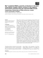
Fig. 3. EPR spectrum of A. profundus HmeCD. EPR spectrum ob-
tained after oxidation of HmeCD (2 mgÆmL
)1
with 3 m
M
K
3
Fe(CN)
6
(thin black line). EPR conditions: temperature, 20 K; microwave
power, 2 mW; microwave frequency, 9.458 GHz; modulation ampli-
tude, 0.6 mT; modulation frequency, 100 kHz. The spin concentration
was 0.15 spinÆmol
)1
enzyme as determined by double integration of the
simulated EPR signal (thick grey line). Simulation parameters:
g
1,2,3
¼ 1.948, 1.995 and 2.031; W
1,2,3
¼ 1.25, 1.15 and 1.325 mT.
Table 2. Purification of Mvh:Hdl enzyme complex from A. profundus.
Hydrogenase-uptake activity was measured after each chromato-
graphic step as described in Materials and methods. One unit of
enzyme activity corresponds to the reduction of two lmol of benzyl-
viologen per minute.
Purification step Fraction Total activity [U]
150 000 g supernatant – 30 000
Q-Sepharose 400 m
M
NaCl 21 000
Hydroxyapatite 150–180 m
M
PO
4
3–
4200
MonoQ 400 m
M
NaCl 4000
1110 G. J. Mander et al. (Eur. J. Biochem. 271) Ó FEBS 2004
did not correspond to one of the amino-terminal
sequences determined for the subunits of the purified
enzyme, however, its molecular mass corresponds to
the apparent molecular mass of the 22 kDa subunit of
the purified enzyme. The amino-terminal sequence of the
22-kDa polypeptide did not show any significant sequence
similarity to proteins in the databases. The AF1374 to
AF1372 gene products revealed high sequence identity to
the three subunits of F
420
-nonreducing hydrogenase (Mvh)
from Methanothermobacter spp. and related methanogens
[12,30], the AF1377 to AF1375 proteins showed high
sequence identity to subunits HdrA, HdrB and HdrC of
Hdr from Methanothermobacter species and related meth-
anogens [3]. Due to these sequence identities the proteins
of the A. profundus enzyme complex were designated as
MvhA (AF1372) MvhG (AF1373) and MvhD (AF1374),
HdlA (AF1377), HdlC (AF1376) and HdlB (AF1375).
Hdl stands for HdrABC-like.
A detailed sequence analysis revealed the following data
(Table 4). HdlA shows sequence similarity to subunit HdrA
of heterodisulfide reductase and shares four binding motifs
for [4Fe)4S] clusters (Cx
2
Cx
2
Cx
3
C) and one binding motif
for FAD [GxGx
2
Gx
16)19
(D/E)] with HdrA. HdlC corres-
ponds to subunit HdrC of Hdr and shares two binding
motifs for [4Fe)4S] clusters with HdrC. A multiple
sequence alignment of various members of the HdrC
protein family showed that the amino terminus of these
proteins is poorly conserved. This may explain why the
determined amino terminus of the 22-kDa polypeptide
could not be assigned to the AF1376 gene product. HdlB
shows sequence similarity to subunit HdrB of Hdr. The two
CX
31)39
CCX
35)36
CX
2
C sequence motifs present in HdrB
are also conserved in HdlB. For Hdr it has been proposed
that some of these cysteine residues ligate the active-site
iron–sulfur cluster [8,9]. MvhD from Mt. marburgensis
binds a [2Fe)2S] cluster [31]. It contains five cysteine
residues also conserved in the AF1374 gene product. MvhG
(AF1373) was identified as hydrogenase small subunit with
highest sequence identity to MvhG of Mt. thermoautotro-
phicus. This protein contains 14 cysteine residues, 12 of these
are highly conserved among the hydrogenase small subunits
of several [NiFe] hydrogenases and are predicted to ligate
three [4Fe)4S] clusters. MvhA (AF1372) was identified as
Fig. 4. SDS/PAGE of the Mvh:Hdl enzyme complex from A. profun-
dus. Proteins were separated in a 14% slab gel (8 · 7cm),whichwas
subsequently stained with Coomassie Brilliant Blue R250. Lane 1,
25 lg of purified Mvh:Hdl complex; M, low-molecular-mass marker
(Amersham Pharmacia Biotech). The molecular masses of the marker
peptides are given on the right side. The apparent molecular masses of
the polypeptides of lane 1 are given on the left side.
Table 3. Amino-terminal sequences of the soluble hydrogenase of A. profundus. Amino-terminal sequences of the A. profundus enzyme were derived
by Edman-degradation, amino-terminal sequences of A. fulgidus were derived from the genome sequence [13]. In both sequences, identical amino
acids are underlined. Annotations made are based on sequence identities of the respective polypeptides (see text). X, No clear assignment to an
amino acid could be made; –, gaps inserted to allow an alignment.
Amino-terminal
sequences
Sequence identity Gene A. fulgidus Annotation
A. profundus
GKYGLFLGCNISFNRPDVEV 55% AF1375 HdlB
A. fulgidus
MFMKYALFPGCKIAFERPDLEL
A. profundus SEEWEPNII-VAANWXTYQ 50% AF1374 MvhD
A. fulgidus
MKIIGFACQWCAYQ
A. profundus MKKIEIEPMTRLEGHXKIAI 63% AF1372 MvhA
A. fulgidus
M-KIEINPVSRIEGHAKVTI
A. profundus GEEEPKIGVYIXH 67% AF1377 HdlA
A. fulgidus
MKIGVYVCH
A. profundus LKLA-YLLVXGCGGCDM 44% AF1373 MvhG
A. fulgidus
IDVAFYIA-HGCSGCTM
A. profundus MEMHEEGVPDVINLSYLAER – – HdlC
A. fulgidus
-
Ó FEBS 2004 Heterodisulfide reductase-like enzymes in A. profundus (Eur. J. Biochem. 271) 1111
hydrogenase large subunit carrying the four cysteine ligands
of the binuclear [Ni–Fe] centre.
None of the polypeptides reported above has extended
hydrophobic regions, which could form membrane-span-
ning helices. This agrees well with the finding that the
enzyme was purified from the soluble fraction.
Cofactor analysis and characterization of the iron–sulfur
clusters by EPR spectroscopy
The enzyme preparation contained 3.7 ± 0.5 nmol NiÆ
mg
)1
protein, 214 ± 9 nmol acid-labile sulfurÆmg
)1
protein
and 207 ± 11 nmol ironÆmg
)1
protein. As predicted from
the primary structure it contains a flavin identified as
FAD ) 3.0±0.2nmolFADÆmg
)1
protein were found. A
densitometric analysis of Coomassie Brilliant blue-stained
SDS gels indicated the presence of all subunits in stoi-
chiometric amounts in the complex. From the genome
sequence of the close relative A. fulgidus the molecular mass
of the complex was calculated to be 220 kDa. Per mol
the enzyme complex thus contains 0.83 ± 0.12 mol Ni,
0.73 ± 0.05 mol FAD, 47 ± 2 mol acid-labile sulfur and
45 ± 2 mol nonheme iron. From the primary sequence the
enzyme is predicted to harbour one FAD, one [Ni–Fe]
centre, one [2Fe)2S] cluster and nine [4Fe)4S] clusters and
one active-site [Fe–S] cluster in HdlB.
A characterization of the iron–sulfur clusters by EPR
spectroscopy revealed the following results: the H
2
reduced
enzyme exhibited a spectrum dominated by a signal with
g-values at 2.036, 1.933 and 1.912 (Fig. 6A). This signal was
detectable without significant broadening at temperatures
up to 80 K. The g-values, temperature behaviour and redox
properties are reminiscent of a signal detected in purified
Mvh from Mt. marburgensis where this signal was attrib-
uted to a [2Fe)2S]
1+
cluster [31]. In the spectrum of the
H
2
-reduced enzyme a radical signal around g ¼ 2wasalso
visible. The intensity of this signal increased upon further
reduction of the enzyme by sodium dithionite (not shown).
The line width of the radical signal is 1.5 mT as determined
from a spectrum recorded at 120 K (data not shown). In the
absorption spectrum there is no maximum around 600 nm,
which would be indicative for a neutral (blue) semiquinone.
This all points to an anionic (red) flavinsemiquinone radical
(line width 1.5 mT) [32].
At temperatures below 20 K additional signals in the
reduced enzyme were detectable as a shoulder of the much
more intensive [2Fe)2S]
1+
signal at g ¼ 1.890 (Fig. 6A).
These signals are indicative of spin–spin coupling between
the different [4Fe)4S]
1+
clusters in the enzyme [33,34].
The duroquinone (2,3,5,6-tetramethyl-p-benzoquinone)-
oxidized enzyme exhibited a rhombic EPR signal with g
xyz
values at 2.014, 1.939 and 1.895. The line shape of this
spectrum was similar to the spectrum observed for oxidized
HmeCD (Fig. 3), however, the g-values are shifted to
lower values (Fig. 6B). This paramagnetic species could be
measured under nonsaturating conditions at 20 K and was
detectable without significant broadening up to 60 K. The
signal broadened beyond detection at 110 K. The total spin
concentration was 13 l
M
corresponding to 0.35 spinÆmol
)1
enzyme. When the oxidized sample was incubated under
100% H
2
the signal was no longer detectable and again the
[2Fe)2S]
+
signal described above was observed. Experi-
ments with heterodisulfide (CoM-S-S-CoB) as electron
acceptor and hydrogen as electron donor showed that the
complex has no detectable activity with these substrates
(data not shown). When the enzyme was oxidized with
K
3
Fe(CN)
6
a Ôg ¼ 2.02Õ-EPR signal indicative of a
[3Fe)4S]
+
cluster was observed. This cluster was most
probably formed by the oxidative degradation of a [Fe)4S]
cluster at high redox potentials.
Table 4. Features of the subunits of the Mvh:Hdl complex from A. profundus.
TIGR
annotation
Calculated
molecular mass
Cofactor binding sites
Mt. thermoautotrophicus
Sequence
identity to Annotation
AF1377 72.1 kDa 4 [4Fe)4S], FAD HdrA (46%) HdlA
AF1376 18 kDa 2 [4Fe)4S] HdrC (32%) HdlC
AF1375 33.9 kDa 2 · (Cx
31)39
CCx
35)36
Cx
2
C) HdrB (35%) HdlB
AF1374 15 kDa [2Fe)2S] MvhD (30%) MvhD
AF1373 32.1 kDa 3[4Fe)4S] MvhG (31%) MvhG
AF1372 50.9 kDa [Ni–Fe] MvhA (36%) MvhA
Fig. 5. Genomic organization of the genes encoding the subunits of the Mvh:Hdl enzyme complex and a putative membrane-bound hydrogenase in
A. fulgidus. The ORFs annotated by TIGR are given above the arrow representing the genes and their direction of transcription. The gene names of
genes encoding the Mvh:Hdl complex are given below the gene symbols. The genes have almost no intergenic regions or they overlap. The AF1371
gene product was not found in the enzyme preparation. It is predicted to encode a hydrogenase maturation protease. The AF1381–AF1379 genes
encode a putative membrane-bound hydrogenase closely related to the F
420
-nonreducing hydrogenases (Vho and Vht) from Mt. mazei [37].
AF1378 encodes a putative hydrogenase maturation protease.
1112 G. J. Mander et al. (Eur. J. Biochem. 271) Ó FEBS 2004
In addition, signals derived from the [NiFe] centre were
observed with g
xyz
¼ 2.338, 2.174 and 2.007 in the duro-
quinone oxidized enzyme (data not shown). This signal
most probably corresponds to the Ni(III) ready form of the
enzyme [35]. The identification of this Ni(III) derived signal
clearly shows that the enzyme is an oxidized state.
Discussion
In the present study two Hdr-like enzymes, HmeCD and the
Mvh:Hdl-complex, were isolated from H
2
/sulfate-grown
cells of A. profundus. Each enzyme contains a subunit
(HmeD or HdlB) with sequence similarity to the proposed
catalytic subunit of Hdr (HdrD from Ms. barkeri or HdrB
from Mt. marburgensis). The EPR signals observed for
both, HmeCD and the Mvh:Hdl complex from A. profun-
dus are reminiscent to the CoM–Hdr signal. However, the
two enzymes from A. profundus form this paramagnetic
species already when oxidized with either ferricyanide
or duroquinone in the absence of any added thiol. The
same result was recently obtained with the A. fulgidus
HmeACDE complex [14]. One possible explanation could
be that the enzymes contain substoichiometric amounts of a
tightly bound thiol, which becomes ligated to the active-site
[Fe–S] cluster upon oxidation. This could also explain why
the spin concentration obtained is much lower than 1 spin
per molecule. With the Mt. marburgensis enzyme the CoM–
Hdr EPR signal could also be obtained when oxidized in the
presence of nonsubstrate thiols such as b-mercaptoethanol
or cysteine. With these thiols the midpoint potential of the
signal was, however, shifted to rather nonphysiological,
high values [7]. Also in the A. profundus enzymes a
nonsubstrate thiol might induce the paramagnetic species
observed by EPR spectroscopy.
The architecture of A. profundus HmeCD resembles that
of HdrDE from Ms. barkeri (Fig. 7) [4,5]. It contains a
b-type cytochrome as membrane anchor and a hydrophilic
iron–sulfur protein, presumably carrying the active-site for
the reduction of a yet unidentified substrate. In Ms. barkeri
Hdr together with a membrane-bound [NiFe] hydrogenase
and the membrane-bound electron carrier methanophena-
zine forms an electron transport chain catalysing the
reduction of CoM-S-S-CoB by H
2
. This reaction is coupled
to the formation of a proton motive force [10,36]. Two
isoenzymes of the membrane-bound hydrogenase, called
Vho and Vht, are present in Ms. barkeri.Bothenzymes
contain a membrane anchoring b-type cytochrome in
addition to the hydrogenase large and small subunit. The
two latter subunits are predicted to extrude into the
extracytoplasmic side of the membrane [37]. Interestingly,
a closely related hydrogenase is encoded by the genome of
A. fulgidus (AF1381–1379) [13] (Fig. 5). The genes are
directly upstream of the genes encoding the Mvh:Hdl
complex. However, only 3% of the hydrogenase activity
present in cell extracts of A. profundus were localized in the
membrane fraction. This could be due to the lability of the
enzyme. Also the Vho/Vht hydrogenases from Ms. barkeri
rapidly dissociate from their membrane anchor [37,38]. It is
thus reasonable to assume that the proposed membrane-
bound hydrogenase of A. profundus became detached from
its membrane anchoring b-type cytochrome subunit upon
cell lysis and thus was released into the soluble fraction.
Fig. 6. EPR spectra of the H
2
-reduced and duroquinone-oxidized
A. profundus Mvh:Hdl enzyme complex. (A) EPR spectra obtained
after reduction of the Mvh:Hdl complex (8.9 mg proteinÆmL
)1
at
pH 7.0) with hydrogen (1.2 · 10
5
Pa)at10K(power¼ 0.2 mW)
and 25 K (power ¼ 2 mW). The upper spectrum shows a simula-
tion of the [2Fe)2S]
1+
signal at 25 K. Simulation parameters:
g
1,2,3
¼ 1.912, 1.933 and 2.036; W
1,2,3
¼ 2.0, 3.6 and 4.6 mT. The
flavin radical signal is saturated under these conditions. (B) EPR
spectrum obtained after oxidation of the Mvh:Hdl enzyme complex
(8.9 mg proteinÆmL
)1
)with3m
M
duroquinone (thin black line).
The total spin concentration was 0.35 spinÆmol
)1
enzyme as
determined by double integration of the simulated EPR signal
(thick grey line). Simulation parameters: g
1,2,3
¼ 1.895, 1.939 and
2.014; W
1,2,3
¼ 2.54, 1.62 and 1.00 mT. EPR conditions: tempera-
ture,20K;microwavepower,2mW;microwavefrequency,
9.458 GHz; modulation amplitude, 0.6 mT; modulation frequency,
100 kHz.
Ó FEBS 2004 Heterodisulfide reductase-like enzymes in A. profundus (Eur. J. Biochem. 271) 1113
During the purification of the Mvh:Hdl complex no other
major fraction with hydrogenase activity was detected.
However, chromatography on hydroxyapatite resulted in a
significant loss of hydrogenase activity. This could be due
to the inactivation or irreversible binding of this second
hydrogenase during chromatography on hydroxyapatite.
During chromatography of solubilized membrane proteins
on Q-Sepharose, in addition to the HmeCD-containing
fraction further heme-containing fractions were observed
which contained 20% of the total heme present in the
membrane fraction. These fractions, which might contain
the b-type cytochrome of the membrane-bound hydro-
genase, have not yet been analysed further. This proposed
membrane-bound hydrogenase and HmeCD could be part
of an electron transport chain very similar to that present in
Ms. barkeri with the exception that methanophenazine is
replaced by the modified menaquinone described for
A. fulgidus [39]. Unlike HmeCD from A. profundus the
A. fulgidus HmeACDE complex contains the two addi-
tional subunits HmeA and HmeC. Both subunits are
predicted to extrude into the extracytoplasmic side of the
membrane. These subunits have recently been proposed to
form a distinct module, which may mediate the electron
transfer from the menaquinone pool to alternative electron
acceptors or oxidoreductases [14]. We can currently not
exclude that these subunits are also formed in A. profundus
and in vivo form a complex with HmeCD but are lost during
the purification.
The Mvh:Hdl complex from A. profundus is closely
related to the Mvh:Hdr complex from Methanothermo-
bacter species (Fig. 7). The sequences deduced from the
AF1377–1372 (hdlACB and mvhAGD)genesofA. fulgidus
not only show high sequence identities to the corresponding
subunits of the Mvh:Hdr complex from Methanothermo-
bacter spp. but also contain all cofactor binding sites
present in the Mt. marburgensis enzyme complex. This was
also confirmed by the biochemical characterization of the
A. profundus enzyme complex. A putative transcription unit
encoding all six subunits was identified in the genome of
A. fulgidus. In the genome of Mt. thermoautotrophicus the
Fig. 7. Schematic presentation of (A) HmeCD from A. profundus in comparison to HdrDE from Ms. barkeri and the HmeABCDE complex from
A. fulgidus and (B) the Mvh:Hdl complex from A. profundus in comparison to the Mvh:Hdr complex from Methanothermobacter spp. The scheme is
based on the sequence analysis of the encoding genes from A. fulgidus. The physiological substrate of the A. profundus enzyme is yet unknown, but
based on the similarity to Hdr a disulfide is proposed. MP, methanophenazine; MQ, menaquinone.
1114 G. J. Mander et al. (Eur. J. Biochem. 271) Ó FEBS 2004
genes encoding the six subunits of the Mvh:Hdr complex are
located at three different loci [40].
Also the sulfate-reducing bacterium D. vulgaris contains a
putative six-gene operon (ORF2976–2967) encoding an
enzyme complex, designated as H
2
-heterodisulfide oxido-
reductase complex, closely related to the Mvh:Hdl complex
described here. These genes are expressed in D. vulgaris as
was shown by macroarray RNA hybridizations [41]. Expres-
sion of these genes was found to be downregulated in a
Fe-only hydrogenase mutant strain (Dhyd). Downregulation
was also observed in a strain carrying a deletion of the adh
gene, encoding an alcohol dehydrogenase. One possible
function discussed for the H
2
-heterodisulfide oxidoreductase
complex in D. vulgaris is the formation of H
2
with reducing
equivalents generated by the oxidation of ethanol to acetate,
as part of a H
2
-cycling system [41]. Thus far, the Mvh:Hdl
genetic organization is unique to sulfate reducers.
The complete understanding of the process of dissimila-
tory sulfate reduction is hampered by the high complexity of
the systems studied thus far. Many of the organisms studied
are able to utilize several electron donors for growth and
contain many different electron transfer components in
parallel. In contrast, A. profundus is obligatory hydrogeno-
trophic [20]. For this organism H
2
is the ultimate electron
donor and hence reducing equivalents generated upon H
2
oxidation have to be channelled to the enzymes of sulfate
reduction. Our finding that a major hydrogenase present in
cell extracts of A. profundus forms a tight complex with an
Hdr-like enzyme strongly supports previous assumptions
that Hdr-like enzymes play an essential role in the electron
transport chain(s) of sulfate reducing archaea and bacteria.
The Mvh:Hdl enzyme complex accounts for at least 2.5% of
the total cell protein of A. profundus indicating an important
catabolic function. In analogy to the Mvh:Hdr complex
from Methanothermobacter species, the A. profundus
enzyme complex is proposed to reduce an electron acceptor
which in turn could function as electron donor of the
enzymes of sulfate reduction.
Several questions remain to be answered. What is the
nature of the physiological electron acceptor of the two Hdr-
like enzymes present in A. profundis? The similarity to Hdr
suggests a disulfide. Do both enzymes reduce the same
electron acceptor or are different substrates used? If both
systems reduce the same substrate, why are two different
enzyme systems operating? Is the Mvh:Hdl enzyme complex
in vitro bound to the cytoplasmic membrane via additional
membrane subunits and is the reaction catalysed by this
complex coupled to energy conservation, as has been
proposed for the Mvh:Hdr complex from Methanothermo-
bacter marburgensis [12]. In future studies the identification of
the substrate(s) ofboth enzymes will beattempted. A possible
way this could be done is the addition of low molecular mass
fractions of cell extracts to purified HmeCD or Mvh:Hdl in
the presence of an oxidant. This could result in higher spin
concentrations of the paramagnetic species, assuming that
the substrate binds to an iron–sulfur cluster in the active site.
Acknowledgements
This work was supported by the Max-Planck-Gesellschaft, by the
Deutsche Forschungsgemeinschaft, and by the Fonds der Chemischen
Industrie. We thank D. Linder for amino-terminal sequencing.
References
1. Thauer, R.K. (1998) Biochemistry of methanogenesis: a tribute to
Marjory Stephenson. Microbiology 144, 2377–2406.
2. Hedderich, R., Berkessel, A. & Thauer, R.K. (1990) Purification
and properties of heterodisulfide reductase from Methanobacter-
ium thermoautotrophicum (strain Marburg). Eur. J. Biochem. 193,
255–261.
3. Hedderich, R., Koch, J., Linder, D. & Thauer, R.K. (1994) The
heterodisulfide reductase from Methanobacterium thermo-
autotrophicum contains sequence motifs characteristic of pyridine
nucleotide-dependent thioredoxin reductases. Eur. J. Biochem.
225, 253–261.
4. Heiden, S., Hedderich, R., Setzke, E. & Thauer, R.K. (1994)
Purification of a two-subunit cytochrome-b-containing hetero-
disulfide reductase from methanol-grown Methanosarcina barkeri.
Eur. J. Biochem. 221, 855–861.
5. Ku
¨
nkel, A., Vaupel, M., Heim, S., Thauer, R.K. & Hedderich, R.
(1997) Heterodisulfide reductase from methanol-grown cells of
Methanosarcina barkeri is not a flavoenzyme. Eur. J. Biochem. 244,
226–234.
6.Simianu,M.,Murakami,E.,Brewer,J.M.&Ragsdale,S.W.
(1998) Purification and properties of the heme- and iron-sulfur-
containing heterodisulfide reductase from Methanosarcina
thermophila. Biochemistry. 37, 10027–10039.
7. Madadi-Kahkesh, S., Duin, E.C., Heim, S., Albracht, S.P.J.,
Johnson, M.K. & Hedderich, R. (2001) A paramagnetic species
with unique EPR characteristics in the active site of heterodisulfide
reductase from methanogenic archaea. Eur. J. Biochem. 268,
2566–2577.
8. Duin, E.C., Madadi-Kahkesh, S., Hedderich, R., Clay, M.D. &
Johnson, M.K. (2002) Heterodisulfide reductase from Methano-
thermobacter marburgensis contains an active-site [4Fe-4S] cluster
that is directly involved in mediating heterodisulfide reduction.
FEBS Lett. 512, 263–268.
9. Duin, E.C., Bauer, C., Jaun, B. & Hedderich, R. (2003) Coenzyme
M binds to a [4Fe-4S] cluster in the active site of heterodisulfide
reductase as deduced from EPR studies with the [
33
S]coenzyme
M-treated enzyme. FEBS Lett. 538, 81–84.
10. Ide, T., Ba
¨
umer, S. & Deppenmeier, U. (1999) Energy conserva-
tion by the H
2
: heterodisulfide oxidoreductase from Methano-
sarcina mazei Go
¨
1: Identification of two proton-translocating
segments. J. Bacteriol. 181, 4076–4080.
11. Ba
¨
umer,S.,Ide,T.,Jacobi,C.,Johann,A.,Gottschalk,G.&
Deppenmeier, U. (2000) The F
420
H
2
dehydrogenase from
Methanosarcina mazei is a redox-driven proton pump closely
related to NADH dehydrogenases. J. Biol. Chem. 275,
17968–17973.
12. Setzke, E., Hedderich, R., Heiden, S. & Thauer, R.K. (1994) H
2
:
heterodisulfide oxidoreductase complex from Methanobacterium
thermoautotrophicum. Composition and properties. Eur. J.
Biochem. 220, 139–148.
13. Klenk, H.P., Clayton, R.A., Tomb, J.F., White, O., Nelson, K.E.,
Ketchum, K.A., Dodson, R.J., Gwinn, M., Hickey, E.K., Peter-
son, et al. (1997) The complete genome sequence of the hyper-
thermophilic, sulphate-reducing archaeon Archaeoglobus fulgidus.
Nature 390, 364–370.
14. Mander, G.J., Duin, E.C., Linder, D., Stetter, K.O. & Hedderich,
R. (2002) Purification and characterization of a membrane-
bound enzyme complex from the sulfate-reducing archaeon
Archaeoglobus fulgidus related to heterodisulfide reductase from
methanogenic archaea. Eur. J. Biochem. 269, 1895–1904.
15. Valente, F.M., Saraiva, L.M., LeGall, J., Xavier, A.V., Teixeira,
M. & Pereira, I.A. (2001) A membrane-bound cytochrome c
3
:a
type II cytochrome c
3
from Desulfovibrio vulgaris Hildenborough.
Chembiochemistry 2, 895–905.
Ó FEBS 2004 Heterodisulfide reductase-like enzymes in A. profundus (Eur. J. Biochem. 271) 1115
16. Eisen, J.A., Nelson, K.E., Paulsen, I.T., Heidelberg, J.F., Wu, M.,
Dodson, R.J., Deboy, R., Gwinn, M.L., Nelson, W.C., Haft,
D.H. et al. (2002)ThecompletegenomesequenceofChlorobium
tepidum TLS, a photosynthetic, anaerobic, green-sulfur bacterium.
Proc.NatlAcad.Sci.USA99, 9509–9514.
17. Dahl, C., Rakhely, G., Pott-Sperling, A.S., Fodor, B., Takacs, M.,
Toth,A.,Kraeling,M.,Gyorfi,K.,Kovacs,A.,Tusz,J.&
Kovacs, K.L. (1999) Genes involved in hydrogen and sulfur
metabolism in phototrophic sulfur bacteria. FEMS Microbiol.
Lett. 180, 317–324.
18. Rossi, M., Pollock, W.B., Reij, M.W., Keon, R.G., Fu, R. &
Voordouw, G. (1993) The hmc-operon of Desulfovibrio vulgaris
subsp. vulgaris Hildenborough encodes a potential transmem-
brane redox protein complex. J. Bacteriol. 175, 4699–4711.
19. Vorholt, J., Kunow, J., Stetter, K.O. & Thauer, R.K. (1995)
Enzymes and coenzymes of the carbon monoxide dehydrogenase
pathway for autotrophic CO
2
fixation in Archaeoglobus litho-
trophicus and the lack of carbon monoxide dehydrogenase in the
heterotrophic A. profundus. Arch. Microbiol. 163, 112–118.
20. Burggraf, S., Jannasch, H.W., Nicolaus, B. & Stetter, K.O. (1990)
Archaeoglobus profundus sp. nov., represents a new species within
sulfate-reducing archaea. Syst. Appl. Microbiol. 13, 24–28.
21. Beinert, H. & Albracht, S.P. (1982) New insights, ideas and
unanswered questions concerning iron-sulfur clusters in mito-
chondria. Biochim. Biophys. Acta. 683, 245–277.
22. Fish, W.W. (1988) Rapid colorimetric micromethod for the
quantitation of complexed iron in biological samples. Methods
Enzymol. 158, 357–364.
23. Cline, J.D. (1969) Spectrophotometric determination of hydrogen
sulfide in natural waters. Limnol. Oceanogr. 14, 454–458.
24. Zor, T. & Seliger, Z. (1996) Linearization of the bradford protein
assay increases its sensitivity – theoretical and experimental stud-
ies. Anal. Biochem. 236, 302–308.
25. Berry, E.A. & Trumpower, B.L. (1987) Simultaneous determina-
tion of hemes a, b and c from pyridine hemochrome spectra. Anal.
Biochem. 161, 1–15.
26. Smith, L. (1978) Bacterial cytochromes and their spectral char-
acterization. Methods Enzymol. 53, 202–212.
27. Gabellini, N. & Hauska, G. (1983) Characterization of cyto-
chrome b in the isolated ubiquinol-cytochrome c
2
oxidoreductase
from Rhodopseudomonas sphaeroides GA. FEBS Lett. 153,
146–150.
28. Puustinen, A. & Wikstro
¨
m, M. (1991) The heme groups of cyto-
chrome o from Escherichia coli. Proc. Natl Acad. Sci. USA 88,
6122–6126.
29. de Vries, S. & Albracht, S.P. (1979) Intensity of highly aniso-
tropic low-spin heme EPR signals. Biochim. Biophys. Acta. 546,
334–340.
30. Reeve, J.N., Beckler, G.S., Cram, D.S., Hamilton, P.T., Brown,
J.W., Krzycki, J.A., Kolodziej, A.F., Alex, L., Orme-Johnson,
W.H. & Walsh, C.T. (1989) A hydrogenase-linked gene in
Methanobacterium thermoautotrophicum strain delta H encodes a
polyferredoxin. Proc. Natl Acad. Sci. USA 86, 3031–3035.
31. Stojanowic, A., Mander, G.J., Duin, E.C. & Hedderich, R. (2003)
Physiological role of the F
420
-non-reducing hydrogenase (Mvh)
from Methanothermobacter marburgensis. Arch. Microbiol. 180,
194–203.
32. Palmer, G., Mu
¨
ller, F. & Massey, V. (1971) Electron-para-
magnetic resonance studies on flavoprotein radicals. Flavins and
Flavoproteins (Kamin, H., ed.), pp. 123–137. University Park
Press, Baltimore, MD.
33. Mouesca, J M. & Lamotte, B. (1998) Iron-sulfur clusters and
their electronic and magnetic properties. Coordin. Chem. Rev.
178–180, 1573–1614.
34. Capozzi, F., Ciurli, S. & Luchinat, C. (1998) Coordination sphere
versusproteinenvironmentasdeterminantsofelectronicand
functional properties of iron-sulfur proteins. Structure Bonding
(Hill, H.A.O., Sadler, P.J. & Thomson, A.J., eds), pp. 127–160.
Springer Verlag, Berlin.
35. Maroney, M.J. & Bryngelson, P.A. (2001) Spectroscopic and
model studies of the Ni-Fe hydrogenase reaction mechanism.
J. Biol. Inorg. Chem. 6, 453–459.
36. Abken, H.J., Tietze, M., Brodersen, J., Ba
¨
umer, S., Beifuss, U. &
Deppenmeier, U. (1998) Isolation andcharacterization of methano-
phenazine and function of phenazines in membrane-bound elec-
tron transport of Methanosarcina mazei Go
¨
1. J. Bacteriol. 180,
2027–2032.
37.Deppenmeier,U.,Blaut,M.,Lentes,S.,Herzberg,C.&
Gottschalk, G. (1995) Analysis of the vhoGAC and vhtGAC
operons from Methanosarcina mazei strain G1, both encoding a
membrane-bound hydrogenase and a cytochrome b. Eur. J.
Biochem. 227, 261–269.
38. Deppenmeier, U., Blaut, M., Schmidt, B. & Gottschalk, G. (1992)
Purification and properties of a F
420
-nonreactive, membrane-
bound hydrogenase from Methanosarcina strain Go
¨
1. Arch.
Microbiol. 157, 505–511.
39. Tindall, B.J., Stetter, K.O. & Collins, M.D. (1989) A novel fully
saturated menaquinone from the thermophilic sulfate-reducing
archaebacterium Archaeoglobus fulgidus. J. Gen. Microbiol. 135,
693–696.
40. Smith, D.R., Doucette-Stamm, L.A., Deloughery, C., Lee, H.,
Dubois, J., Aldredge, T., Bashirzadeh, R., Blakely, D., Cook, R.,
Gilbert, K. et al. (1997) Complete genome sequence of Methano-
bacterium thermoautotrophicum DH: functional analysis and
comparative genomics. J. Bacteriol. 179, 7135–7155.
41. Haveman, S.A., Brunelle, V., Voordouw, J.K., Voordouw, G.,
Heidelberg, J.F. & Rabus, R. (2003) Gene expression analysis of
energy metabolism mutants of Desulfovibrio vulgaris Hildenbor-
ough indicates an important role for alcohol dehydrogenase.
J. Bacteriol. 185, 4345–4353.
1116 G. J. Mander et al. (Eur. J. Biochem. 271) Ó FEBS 2004