Báo cáo khoa học: Fibroblast growth-stimulating activity of S100A9 (MRP-14) ppt
Bạn đang xem bản rút gọn của tài liệu. Xem và tải ngay bản đầy đủ của tài liệu tại đây (271.02 KB, 7 trang )
Fibroblast growth-stimulating activity of S100A9 (MRP-14)
Futoshi Shibata, Katsuyoshi Miyama, Fumie Shinoda, Jun Mizumoto, Katsuhiko Takano
and Hideo Nakagawa
Department of Physiological Chemistry, Faculty of Pharmaceutical Sciences, Toyama Medical and Pharmaceutical University,
Sugitani, Toyama, Japan
Fibroblasts play a critical role in chronic inflammation
and wound healing. In this study, a fibroblast growth-
stimulating factor was purified from the exudate of car-
rageenan-induced inflammation in rats. The purified
protein was a disulfide-linked homodimer. Amino acid
sequence analysis of the peptides generated by cleavage
with cyanogen bromide and proteinase V8 resulted in
identification of the protein as S100A9. Recombinant
S100A9 as well as its disulfide-linked homodimer stimu-
lated the proliferation of fibroblasts at a similar con-
centration of the purified protein. The concentration of
S100A9 in the exudate was determined by immunoblot
analysis. The total protein concentration in the exudate
reached a maximum 4 days after carrageenan injection
and then slightly decreased, whereas the concentration of
S100A9 reached a maximum at day 3 and then decreased
rapidly. These studies show that S100A9 is present at a
high concentration in the exudate of carrageenan-induced
inflammation in rats, and that S100A9 stimulates pro-
liferation of fibroblasts, suggesting that it plays a role in
chronic inflammation.
Keywords: carrageenan; fibroblast; growth; inflammation;
S100A9.
Granuloma is formed by a foreign body or infectious agents
and consists of epithelioid macrophages, multinucleated
giant cells and lymphocytes [1]. Fibroblasts usually sur-
round granuloma, and play an important role in wound
healing. These processes are mediated by growth factors and
cytokines, including platelet-derived growth factors [2],
transforming growth factor b [3], fibroblast growth factors
[4–6], and connective tissue growth factor [7]. We have
purified and identified S100A9 as a new fibroblast growth-
stimulating factor (FGSF) from the exudate of carrageenan-
induced granulomatous inflammation in rats in this study.
S100A9 [8], also known as calgranulin B [9] and MRP-14
[10,11], belongs to the S100 protein family and has two
Ca
2+
-binding EF-hand motifs. S100A9 forms a hetero-
dimer with S100A8 [8], also known as calgranulin A [9] or
MRP-8 [10,11], and is expressed in granulocytes, monocytes
[11], and activated keratinocytes [12,13]. Epithelioid cells in
foreign body granuloma also expressed S100A9 [14,15].
High serum concentrations of S100A9 are detected in cases
of cystic fibrosis [9], rheumatoid arthritis [11], systemic lupus
erythematosus [16], Crohn’s disease [17], inflammatory
bowel disease [18], and multiple sclerosis [19], and suggest
an important role of S100A9 in chronic inflammation. It
was reported that S100A9 bound zinc ions [20] and heparan
sulfate glycosaminoglycans [21], also activated b
2
-integrin,
Mac-1 on neutrophils [22] thereby controlling responsive-
ness to neutrophil chemoattractants [23], as well as having
macrophage-deactivating activity [14], antinociceptive activ-
ity [24] and neutrophil chemotactic activity [25]. Calprotec-
tin, a complexed form of S100A8 and S100A9, is known to
inhibit microbial growth [26,27] and growth of fibroblasts
by chelating zinc ions [28]. On the contrary, the present
study provides evidence that S100A9 may function as a
mitogen for fibroblasts in chronic inflammation.
Materials and methods
Cells
Mouse fetal fibroblasts, BALB/c 3T3 cells and normal rat
kidney fibroblasts, NRK-49F cells were obtained from
Japanese Cancer Research Resources Bank. BALB/c 3T3
cells and NRK-49F cells were grown in Dulbecco’s modified
Eagle’s medium, supplemented with 10% (v/v) calf serum
and 5% (v/v) fetal bovine serum, respectively.
Proliferation assay
For fibroblast growth-stimulating factors, proliferation of
BALB/c 3T3 cells or NRK-49F cells was measured by the
method described by Kueng et al.[29].Culturedcellswere
placed into 96-well microtiter plates at a density of 1000 cells
per well and allowed to grow for 24 h in the presence of
Correspondence to Futoshi Shibata, Department of Physiological
Chemistry, Faculty of Pharmaceutical Sciences, Toyama Medical and
Pharmaceutical University, 2630 Sugitani, Toyama 930–0194, Japan.
Fax: + 81 76 4344656, Tel.: + 81 76 4347543,
E-mail:
Abbreviations: ERK, extracellular signal regulated kinase; FGSF,
fibroblast growth-stimulating factor; GST, glutathione S-transferase;
MRP-14, myeloid-related protein-14; RAGE, receptor for advanced
glycation end products; XTT, 2,3-bis(2-methoxy-4-nitro-5-sulfo-
phenyl)-2H-tetrazolium-5-carboxanilide.
Enzymes: BamHI (EC 3.1.21.4); glutathione S-transferase
(EC 2.5.1.18); horseradish peroxidase (EC 1.11.1.7); proteinase V8
(EC 3.4.21.19); SmaI (EC 3.1.21.4); thrombin (EC 3.4.21.5).
(Received 8 December 2003, revised 19 March 2004,
accepted 30 March 2004)
Eur. J. Biochem. 271, 2137–2143 (2004) Ó FEBS 2004 doi:10.1111/j.1432-1033.2004.04129.x
10% (v/v) calf serum. Cells were then washed with serum-
free medium and incubated for 48–96 h in the medium
containing 2% (v/v) calf serum and sample solution. Cell
number was measured by crystal violet staining.
For S100A9, proliferation of NRK-49F cells was meas-
ured as described by Scudiero et al. [30]. NRK-49F cells
were inoculated at a density of 2000 cells per well into
96-well microtiter plates. After 24 h, cells were washed with
serum-free medium and incubated for 48 h at 37 °Cinthe
medium containing 0.5% (v/v) fetal bovine serum and
experimental agents. Prewarmed (60 °C) solution contain-
ing 50 lg of 2,3-bis(2-methoxy-4-nitro-5-sulfophenyl)-
2H-tetrazolium-5-carboxanilide (XTT) and 0.38 lgof
phenazine methosulfate was added to each well. After
incubation for 3 h at 37 °C, the plates were mixed and
absorbance at 450 nm was measured with a microplate
reader model 550 (Bio-Rad Laboratories).
Induction of air pouch-type inflammation
by carrageenan in rats
A 2% (w/v) solution of carrageenan (4 mL in saline,
Seakem 202, Marine Colloids Inc., NJ, USA) was injected
into preformed air pouches on the backs of male Wistar rats
(body mass: 170–200 g) [31]. One, two, three, four, and
seven days after the injection, the rats were sacrificed by
cutting the carotid artery under light anesthesia and
granulation tissues and pouch fluid were then collected.
Aliquots of the pouch fluid were frozen in liquid nitrogen
and stored at )80 °C until use. The concentration of protein
was determined using a Protein Assay Kit (Bio-Rad
Laboratories). The rats were treated in accordance with
procedures approved by the Animal Ethics Committee of
Toyama Medical and Pharmaceutical University.
Purification of fibroblast growth-stimulating factors
All purification procedures except for RP-HPLC were
carried out at 4 °C. The pouch fluid was collected on day 7
after carrageenan injection and centrifuged at 70 000 g for
60 min. The resulting supernatant (day 7 exudate,
1000 mL) was adjusted to pH 4.5 with 9
M
HCl, and
stirred for 2 h. After centrifugation at 13 000 g for 60 min,
the supernatant was brought to 38% saturation with
ammonium sulfate and stirred for 3 h, and then centrifuged.
The resulting supernatant was precipitated by addition of
ammonium sulfate to 70% saturation. This precipitate was
dissolved in 0.1
M
phosphate buffer (pH 6.0), applied to a
CM-Cellulofine C-500 column (2.6 · 47 cm; Seikagaku
Co., Tokyo, Japan) and eluted with 0.1
M
phosphate
buffer-150 m
M
NaCl (pH 6.0). Eluate was applied to a
heparin-Sepharose CL-6B column (1.6 · 26 cm, Amer-
sham Biosciences, NJ, USA) and eluted with a linear
gradient from 0.1 to 2
M
NaCl in 50 m
M
Tris/HCl buffer
(pH 7.0). Proteins which eluted between 0.6
M
and 1.2
M
NaCl were pooled, concentrated, and chromatographed on
a Sephadex G-75 column (1.6 · 94 cm; Amersham Bio-
sciences) equilibrated with phosphate-buffered saline con-
taining 0.01% (v/v) Brij-35. A peak fraction corresponding
to a molecular mass of about 20 kDa was pooled,
lyophilized, dissolved in 6
M
guanidine/HCl, and loaded
onto an ODS-120T column (0.46 · 25 cm; Tosoh Co.,
Tokyo, Japan) at room temperature. Proteins were eluted
from the column by a linear gradient of acetonitrile from 0
to 50.4% (v/v) in 0.05% (v/v) trifluoroacetic acid at a flow
rate of 0.8 mLÆmin
)1
. Finally, the major peak was rechro-
matographed under the same conditions, and FGSF was
isolated as a single peak.
Amino acid sequence analysis of a fibroblast
growth-stimulating factor
The purified FGSF was dissolved in 3
M
guanidine hydro-
chloride, 0.2
M
Tris/HCl (pH 8.2) at a concentration of
1 lgÆmL
)1
and reduced with 25% (v/v) 2-mercaptoethanol
at 40 °C for 3 h, and then carboxymethylated with 0.1
M
iodoacetic acid. The carboxymethylated protein was puri-
fied by HPLC on an ODS-120T column and fragmented by
either cyanogen bromide cleavage or digestion with pro-
teinase V8 from Staphylococcus aureus. Resulting peptides
were separated by RP-HPLC. The N-terminal amino acid
sequences of the peptides were determined by automated
Edman degradation on an Applied Biosystems 470 A gas
phase sequencer equipped with a 120 A on-line phenyl-
thiohydantoin amino acid analyzer.
Purification of recombinant S100A9 protein
Total RNA from rat macrophages was reverse-transcribed.
S100A9 cDNA [32] was amplified from the cDNA by PCR,
cloned between BamHI and SmaI sites of the glutathione
S-transferase (GST) expression plasmid, pGEX4T2
(Amersham Biosciences), and sequenced. Fusion protein
expression was induced with 0.5 m
M
isopropyl thio-b-
D
-
galactoside in Escherichia coli DH5a for 6 h at 28 °C. After
incubation, the bacteria was harvested by centrifugation
and lysed by freezing and thawing, sonication, and addition
of 1% Triton X-100. The clear lysate was obtained by
centrifugation and applied to a Glutathione Sepharose 4B
column (Amersham Biosciences). GST-S100A9 fusion
protein was eluted from the column with 10 m
M
glutathi-
one)50 m
M
Tris/HCl (pH 8.0), chromatographed on a
Sephadex G-50 column (Amersham Biosciences), equili-
brated with phosphate-buffered saline to remove glutathi-
one, and cleaved with thrombin (Amersham Biosciences) at
22 °C for 16 h. This solution was passed through a
Glutathione Sepharose 4B column to remove GST. Finally,
recombinant S100A9 was purified on an ODS-120T column
(Tosoh Co.) using a linear gradient of acetonitrile from 28 to
40% (v/v) in 0.05% (v/v) trifluoroacetic acid, evaporated,
dissolved in phosphate-buffered saline with 1 m
M
calcium
chloride and stored at – 20 °C. Oxidation of the thiol group
of S100A9 was achieved with a copper–phenanthroline
complex [33]. The latter was removed by dialysis against
5m
M
ammonium acetate and the protein lyophilized. The
lyophilized sample was dissolved in phosphate-buffered
saline.
Production of polyclonal antiserum
Polyclonal antiserum to S100A9 was raised in rabbits by
subcutaneous injection of 1 mg of GST-S100A9 fusion
protein emulsified in Freund’s complete adjuvant. Two
weeks after the primary injection, boosts of 0.5 mg of the
2138 F. Shibata et al. (Eur. J. Biochem. 271) Ó FEBS 2004
fusion protein in Freund’s incomplete adjuvant were
injected every 2 weeks. The rabbits were bled 2 weeks after
the final boost under anesthesia. The rabbits were treated in
accordance with procedures approved by the Animal Ethics
Committee of Toyama Medical and Pharmaceutical
University.
Gel electrophoresis
Exudates were diluted SDS buffer containing 2% (w/v) SDS
and 0.02% (w/v) bovine serum albumin. SDS/PAGE was
carried out as described by Laemmli [34] using low molecular
mass markers and low-range rainbow molecular mass mark-
ers (Amersham Biosciences) as molecular mass standards.
The gel was stained with Coomassie Brilliant Blue R 250.
Immunoblotting
Proteins separated by SDS/PAGE were transferred onto
nitrocellulose membranes (Bio-Rad Laboratories) using
the Mini Trans-blot cell (Bio-Rad Laboratories). The
membranes were incubated with rabbit polyclonal anti-
S100A9 serum and then with a horseradish peroxidase-
conjugated goat anti-rabbit IgG (Caltag Laboratories, CA,
USA). The reaction products were visualized with an ECL
Western blotting detection system (Amersham Biosciences)
and a luminoimage analyzer (LAS-1000 plus, Fuji Photo
Film, Tokyo, Japan). Chemiluminescence was quantitated
using the Science Laboratory 99 Image Gauge program
(Fuji Photo Film). Chemiluminescence of the band linearly
correlated with the amount of recombinant S100A9 (25 to
200 ng per lane). For quantitation of S100A9 in the
exudates, three different amounts of recombinant S100A9
(80, 120 and 200 ng) were used in each assay as standards.
Statistical analysis
Data are expressed as mean ± SEM. Student’s t-test was
used for statistical analysis.
Results
Purification of fibroblast growth-stimulating factors
Fibroblast growth-stimulating factors were purified from
the exudate of carrageenan-induced inflammation as des-
cribed under Materials and methods and eluted from
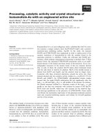
RP-HPLC as a major peak (peak 1) and a minor peak (peak
2) (Fig. 1). The major peak was purified by rechromato-
graphy on RP-HPLC. We could not obtain an adequate
amount of protein from peak 2 to continue analysis on it.
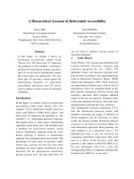
The purified FGSF (peak 1) gave a single band at 13.4 kDa
under reducing condition and at 26 kDa under nonreducing
condition, respectively (Fig. 2). The N-terminal amino acid
sequence of the purified FGSF could not be successfully
performed, suggesting that the N-terminal amino acid is
blocked. Therefore, FGSF was carboxymethylated and
treated with cyanogen bromide and proteinase V8; 4 (CN-1
to CN-4) and 12 (V-1 to V-12) peptides were then isolated
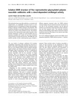
by RP-HPLC. Although we could not determine any amino
acid residues from peptides CN-4 and V-5, other peptides
show a significant sequence similarity to rat S100A9
(Fig. 3).
Fig. 1. RP-HPLC separation of fibroblast growth-stimulating factors
from the inflammatory exudate. Proteins were eluted by a linear gra-
dient of acetonitrile from 0 to 50.4% (v/v) in 0.05% (v/v) trifluoro-
acetic acid. Growth-stimulating activity of each fraction for BALB/c
3T3 cells was assayed at a concentration of 1 lgÆmL
)1
.Eachcolumn
represents the mean ± standard errors of six determinations.
Fig. 2. SDS/PAGE analyses of a fibroblast growth-stimulating factor (FGSF) and recombinant S100A9. FGSF purified by rechromatography of
peak 1 indicated in Fig. 1, recombinant S100A9 and oxidized S100A9 (A9ox) were analyzed by SDS/PAGE in the absence (–) or presence (+) of
2-mercaptoethanol (final 10%) and stained with Coomassie brilliant blue.
Ó FEBS 2004 Fibroblast growth-stimulating activity of S100A9 (Eur. J. Biochem. 271) 2139
Production of recombinant S100A9
The coding region of cDNA for rat S100A9 was amplified
from macrophage RNA by RT-PCR. The nucleotide
sequence of S100A9 cDNA was identical with that regis-
tered in GenBank/EMBL/DDBJ (T. Imamichi; accession
number L18948) except for the replacement of G with C
that resulted in a change from arginine to serine at position
106. Nucleotide sequence data is available in the DDBJ/
EMBL/GenBank databases under the accession number
AB118215. Raftery et al. [35] pointed out a cDNA sequen-
cing error due to the fact that mass spectrometry of S100A9
isolated from rat spleen found serine instead of arginine at
position 106.
Recombinant S100A9 was produced using glutathione
S-transferase (GST) expression plasmid in Escherichia coli,
purified, and analyzed on SDS/PAGE (Fig. 2). A single
band had a molecular mass of 13.6 kDa and was not altered
by reduction. The sequence of N-terminal 10 residues of
recombinant S100A9 was identical to that of rat S100A9
(T. Imamichi; accession number NP_446039) except for
two extra amino acids (Gly-Ser) at the N-terminus and the
lack of the initiator methionine. After oxidation, most of
S100A9 existed as the disulfide-linked homodimer (Fig. 2).
Growth-stimulating activity of FGSF and S100A9
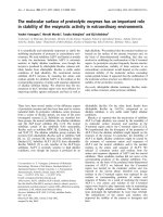
As shown in Fig. 4A, addition of FGSF purified from
exudate to the cultures of NRK-49F cells resulted in dose-
dependent stimulation of proliferation. FGSF stimulated
proliferation of BALB/c 3T3 cells more efficiently (data not
shown). S100A9 and its disulfide-linked homodimer stimu-
lated proliferation of NRK-49F cells at concentrations
higher than 390 ngÆmL
)1
(30 n
M
) and 260 ngÆmL
)1
(10 n
M
), respectively (Fig. 4B).
The concentration of S100A9 in exudate
Inflammation was induced by carrageenan on the back
of rats and granulation tissues and exudates were
collected (Fig. 5). The volume of exudate continued to
increase even 7 days after carrageenan injection. How-
ever, the wet weight of granulation tissue increased
rapidly until 4 days following the initial injection,
suggesting that granulation tissue formed 4 days after
carrageenan injection.
To determine the concentration of S100A9 in the exudate,
polyclonal antiserum was raised in rabbits against GST-
S100A9 fusion protein. The exudates collected (Fig. 5) were
diluted, electrophoresed and immunoblotted for S100A9
(Fig. 6A) with three different amounts of recombinant
Fig. 3. Comparison of the amino acid sequence
of rat S100A9 and peptides isolated from
FGSF. The identified amino acid residues of a
cyanogen bromide-cleaved peptide (CN-3)
and proteinase V8-digested peptides (V-2 to
V-10) are aligned with the amino acid
sequence of rat S100A9 (accession number
NP_446039). Asterisks indicate amino acid
residues identical with those of rat S100A9.
Fig. 4. Growth-stimulating activity of FGSF and S100A9. NRK-49F
cells were incubated with varying concentrations of FGSF purified
from exudate (A) or S100A9 (B) in the presence of 1% (v/v) calf serum
for 96 h (A) or 0.5% (v/v) fetal bovine serum for 48 h (B). Cell
numbers were measured by crystal violet staining (A) or XTT staining
(B). Each point represents the mean ± standard errors of six deter-
minations. Asterisks indicate significant differences (P<0.01) from
control.
2140 F. Shibata et al. (Eur. J. Biochem. 271) Ó FEBS 2004
S100A9 as standards. A band with a molecular mass of
13.6 kDa was detected under reducing condition. A faint
band of 60 kDa appears to be BSA added in SDS buffer,
because the same band was detected without exudates. The
concentration of S100A9 was estimated by quantification of
chemiluminescence of immunoblots of exudates and recom-
binant S100A9 (Fig. 6B). Protein concentration in the
exudate reached a maximum 4 days after carrageenan
injection and then slightly decreased, while the concentra-
tion of S100A9 reached a maximum at day 3 and then
decreased rapidly, indicating that the transient increase of
S100A9 was specific, and not leakage from serum.
Discussion
In the present study, a fibroblast growth-stimulating factor
(FGSF) was purified from the exudate of carrageenan-
induced inflammation in rats (Fig. 1). Amino acid sequence
analyses and SDS/PAGE indicated that the major protein
in FGSF was S100A9 homodimer (Figs 2 and 3). A
relatively higher concentration (100 ngÆmL
)1
)ofFGSFwas
required to stimulate proliferation of fibroblasts (Fig. 4A),
whereas fibroblast growth factor [36], epidermal growth
factor [37], and connective tissue growth factor [38] were
active at lower concentrations (0.4–3 ngÆmL
)1
). Recombin-
ant S100A9 also stimulated the proliferation at a similar
concentration to FGSF (Fig. 4B), suggesting that the major
active protein in FGSF was S100A9. Yui et al. [28] reported
that calprotectin, a complexed form of S100A8 and
S100A9, inhibited the growth of human dermal fibroblasts
presumably by the chelation of zinc ions. This conflict may
come from the difference of subunit composition. Indeed,
Newton and Hogg reported that S100A9 and S100A8/
S100A9 heterodimer showed a different biological activity
[22].
S100B, another member of S100 family protein, stimu-
lated proliferation of rat astroglial cells [39] and activated
extracellular signal regulated kinase (ERK) in astrocytes
[40]. S100A12 and S100B bound a receptor for advanced
glycation end products (RAGE) [41], which was reported to
activate ERK [42]. The receptor and signal transduction
pathways leading to growth-stimulating activity of S100A9
have yet to be elucidated.
S100A9 isolated from rat spleen was acetylated at the
N-terminus after removal of the initiator methionine [35].
Because we could not detect the N-terminal amino acid
sequence of FGSF purified from the exudate, a similar
modification may exist. This N-terminal modification does
not appear to affect growth-stimulating activity, as
recombinant S100A9 also showed activity (Fig. 4B).
Although FGSF existed as a disulfide-linked homodimer
(Fig. 2), this oxidation may occur during purification
steps. However, both monomer and disulfide-linked
homodimer forms of S100A9 stimulated the proliferation
of fibroblasts (Fig. 4B).
The concentration of S100A9 in carrageenan-elicited
exudates was very high (> 1 mgÆmL
)1
) during the forma-
tion of granulation tissue (Fig. 6), and these high concen-
trations of S100A9 were enough to stimulate fibroblast
proliferation (Fig. 4B). Our observations have led to the
hypothesis that S100A9 contributes to the formation of
granulation tissue by stimulating growth of fibroblasts.
Fig. 6. The concentration of S100A9 in the exudate of carrageenan-
induced inflammation in rats. (A) Exudates (Fig. 5) were analyzed by
immunoblotting for S100A9 in the presence of 2-mercaptoethanol
(final 10%). M: recombinant S100A9. (B) The concentration of
S100A9 was estimated by quantification of chemiluminescence
of immunoblots using a luminoimage analyzer. Protein concentration
of the exudate was also determined. Each point represents the
mean ± standard errors of 4–6 rats.
Fig. 5. Formation of granulation tissue and retention of exudate.
Granulation tissue and exudate were collected on day 1–7 after car-
rageenaninjectionintoapreformedairpouchonthebackofrats.
Each point represents the mean ± standard errors of 4–6 rats.
Ó FEBS 2004 Fibroblast growth-stimulating activity of S100A9 (Eur. J. Biochem. 271) 2141
Acknowledgements
The valuable technical assistance of Mariko Kitahara, Kumi Ichinose,
Noriko Mannen and Manabu Kumakura is acknowledged.
References
1. Warren, K.S. (1976) A functional classification of granulomatous
inflammation. Ann. NY Acad. Sci. 278, 7–18.
2. Pierce, G.F., Mustoe, T.A., Altrock, B.W., Deuel, T.F. &
Thomason, A. (1991) Role of platelet-derived growth factor in
wound healing. J. Cell. Biochem. 45, 319–326.
3. O’Kane, S. & Ferguson, M.W. (1997) Transforming growth factor
bs and wound healing. Int. J. Biochem. Cell Biol. 29, 63–78.
4. Buntrock,P.,Buntrock,M.,Marx,I.,Kranz,D.,Jentzsch,K.D.
& Heder, G. (1984) Stimulation of wound healing, using brain
extract with fibroblast growth factor (FGF) activity. III. Electron
microscopy, autoradiography, and ultrastructural auto-
radiography of granulation tissue. Exp. Pathol. 26, 247–254.
5. Buntrock, P., Jentzsch, K.D. & Heder, G. (1982) Stimulation of
wound healing, using brain extract with fibroblast growth factor
(FGF) activity. II. Histological and morphometric examination of
cells and capillaries. Exp. Pathol. 21, 62–67.
6. Buntrock, P., Jentzsch, K.D. & Heder, G. (1982) Stimulation of
wound healing, using brain extract with fibroblast growth factor
(FGF) activity. I. Quantitative and biochemical studies into
formation of granulation tissue. Exp. Pathol. 21, 46–53.
7. Frazier, K., Williams, S., Kothapalli, D., Klapper, H. & Gro-
tendorst, G.R. (1996) Stimulation of fibroblast cell growth, matrix
production, and granulation tissue formation by connective tissue
growth factor. J. Invest. Dermatol. 107, 404–411.
8. Schafer, B.W., Wicki, R., Engelkamp, D., Mattei, M.G. & Heiz-
mann, C.W. (1995) Isolation of a YAC clone covering a cluster of
nine S100 genes on human chromosome 1q21: rationale for a new
nomenclature of the S100 calcium-binding protein family. Geno-
mics 25, 638–643.
9. Wilkinson, M.M., Busuttil, A., Hayward, C., Brock, D.J., Dorin,
J.R. & Van, H.V. (1988) Expression pattern of two related cystic
fibrosis-associated calcium-binding proteins in normal and
abnormal tissues. J. Cell Sci. 91, 221–230.
10. Odink, K., Cerletti, N., Bruggen, J., Clerc, R.G., Tarcsay, L.,
Zwadlo, G., Gerhards, G., Schlegel, R. & Sorg, C. (1987) Two
calcium-binding proteins in infiltrate macrophages of rheumatoid
arthritis. Nature 330, 80–82.
11. Zwadlo, G., Bruggen, J., Gerhards, G., Schlegel, R. & Sorg, C.
(1988) Two calcium-binding proteins associated with specific
stages of myeloid cell differentiation are expressed by subsets of
macrophages in inflammatory tissues. Clin.Exp.Immunol.72,
510–515.
12. Kelly, S.E., Jones, D.B. & Fleming, S. (1989) Calgranulin
expression in inflammatory dermatoses. J. Pathol. 159, 17–21.
13. Mork, G., Schjerven, H., Mangschau, L., Soyland, E. & Brandt-
zaeg, P. (2003) Proinflammatory cytokines upregulate expression
of calprotectin (L1 protein, MRP-8/MRP-14) in cultured human
keratinocytes. Br. J. Dermatol. 149, 484–491.
14. Aguiar-Passeti, T., Postol, E., Sorg, C. & Mariano, M. (1997)
Epithelioid cells from foreign-body granuloma selectively
express the calcium-binding protein MRP-14, a novel down-
regulatory molecule of macrophage activation. J. Leukoc. Biol. 62,
852–858.
15. Delabie, J., de Wolf-Peeters, C., van den Oord, J.J. & Desmet, V.J.
(1990) Differential expression of the calcium-binding proteins
MRP8 and MRP14 in granulomatous conditions: an
immunohistochemical study. Clin. Exp. Immunol. 81, 123–126.
16. Kuruto,R.,Nozawa,R.,Takeishi,K.,Arai,K.,Yokota,T.&
Takasaki, Y. (1990) Myeloid calcium binding proteins: expression
in the differentiated HL-60 cells and detection in sera of patients
with connective tissue diseases. J. Biochem. 108, 650–653.
17. Lugering, N., Stoll, R., Kucharzik, T., Schmid, K.W., Rohlmann,
G., Burmeister, G., Sorg, C. & Domschke, W. (1995)
Immunohistochemical distribution and serum levels of the Ca
2+
-
binding proteins MRP8, MRP14 and their heterodimeric form
MRP8/14 in Crohn’s disease. Digestion 56, 406–414.
18. Stulik, J., Kovarova, H., Macela, A., Bures, J., Jandik, P., Langr,
F., Otto, A., Thiede, B. & Jungblut, P. (1997) Overexpression of
calcium-binding protein calgranulin B in colonic mucosal diseases.
Clin. Chim. Acta 265, 41–55.
19. Bogumil, T., Rieckmann, P., Kubuschok, B., Felgenhauer, K. &
Bruck, W. (1998) Serum levels of macrophage-derived protein
MRP-8/14 are elevated in active multiple sclerosis. Neurosci. Lett.
247, 195–197.
20. Raftery, M.J., Harrison, C.A., Alewood, P., Jones, A. & Geczy,
C.L. (1996) Isolation of the murine S100 protein MRP14 (14 kDa
migration-inhibitory-factor-related protein) from activated spleen
cells: characterization of post-translational modifications and zinc
binding. Biochem. J. 316, 285–293.
21. Robinson, M.J., Tessier, P., Poulsom, R. & Hogg, N. (2002) The
S100 family heterodimer, MRP-8/14, binds with high affinity to
heparin and heparan sulfate glycosaminoglycans on endothelial
cells. J. Biol. Chem. 277, 3658–3665.
22. Newton, R.A. & Hogg, N. (1998) The human S100 protein MRP-
14 is a novel activator of the b2 integrin Mac-1 on neutrophils.
J. Immunol. 160, 1427–1435.
23. Manitz, M.P., Horst, B., Seeliger, S., Strey, A., Skryabin, B.V.,
Gunzer, M., Frings, W., Schonlau, F., Roth, J., Sorg, C. &
Nacken, W. (2003) Loss of S100A9 (MRP14) results in reduced
interleukin-8-induced CD11b surface expression, a polarized
microfilament system, and diminished responsiveness to chemo-
attractants in vitro. Mol. Cell Biol. 23, 1034–1043.
24. Giorgi, R., Pagano, R.L., Dias, M.A., Aguiar-Passeti, T., Sorg, C.
& Mariano, M. (1998) Antinociceptive effect of the calcium-
binding protein MRP-14 and the role played by neutrophils on the
control of inflammatory pain. J. Leukoc. Biol. 64, 214–220.
25. Ryckman,C.,Vandal,K.,Rouleau,P.,Talbot,M.&Tessier,P.A.
(2003) Proinflammatory activities of S100: proteins S100A8,
S100A9, and S100A8/A9 induce neutrophil chemotaxis and
adhesion. J. Immunol. 170, 3233–3242.
26. Sohnle, P.G., Collins-Lech, C. & Wiessner, J.H. (1991) The zinc-
reversible antimicrobial activity of neutrophil lysates and abscess
fluid supernatants. J. Infect. Dis. 164, 137–142.
27. Steinbakk, M., Naess-Andresen, C.F., Lingaas, E., Dale, I.,
Brandtzaeg, P. & Fagerhol, M.K. (1990) Antimicrobial actions of
calcium binding leucocyte L1 protein, calprotectin. Lancet 336,
763–765.
28. Yui, S., Mikami, M., Tsurumaki, K. & Yamazaki, M. (1997)
Growth-inhibitory and apoptosis-inducing activities of calpro-
tectin derived from inflammatory exudate cells on normal fibro-
blasts: regulation by metal ions. J. Leukoc. Biol. 61, 50–57.
29. Kueng, W., Silber, E. & Eppenberger, U. (1989) Quantification of
cells cultured on 96-well plates. Anal. Biochem. 182, 16–19.
30. Scudiero, D.A., Shoemaker, R.H., Paull, K.D., Monks, A.,
Tierney, S., Nofziger, T.H., Currens, M.J., Seniff, D. & Boyd,
M.R. (1988) Evaluation of a soluble tetrazolium/formazan assay
for cell growth and drug sensitivity in culture using human and
other tumor cell lines. Cancer Res. 48, 4827–4833.
31. Fukuhara, M. & Tsurufuji, S. (1969) The effect of locally injected
anti-inflammatory drugs on the carrageenin granuloma in rats.
Biochem. Pharmacol. 18, 475–484.
32. Imamichi, T., Uchida, I., Wahl, S.M. & McCartney-Francis, N.
(1993) Expression and cloning of migration inhibitory factor-
related protein (MRP) 8 and MRP14 in arthritis-susceptible rats.
Biochem. Biophys. Res. Commun. 194, 819–825.
2142 F. Shibata et al. (Eur. J. Biochem. 271) Ó FEBS 2004
33. Kobashi, K. (1968) Catalytic oxidation of sulfhydryl groups by
o-phenanthroline copper complex. Biochim. Biophys. Acta 158,
239–245.
34. Laemmli, U.K. (1970) Cleavage of structural proteins during
the assembly of the head of bacteriophage T4. Nature 227, 680–
685.
35. Raftery, M.J. & Geczy, C.L. (1998) Identification of posttransla-
tional modifications and cDNA sequencing errors in the rat S100
proteins MRP8 and 14 using electrospray ionization mass spec-
trometry. Anal. Biochem. 258, 285–292.
36. Gospodarowicz, D. (1974) Localisation of a fibroblast growth
factor and its effect alone and with hydrocortisone on 3T3 cell
growth. Nature 249, 123–127.
37. Hollenberg, M.D. & Cuatrecasas, P. (1973) Epidermal growth
factor: receptors in human fibroblasts and modulation of action
by cholera toxin. Proc.NatlAcad.Sci.USA70, 2964–2968.
38. Bradham, D.M., Igarashi, A., Potter, R.L. & Grotendorst, G.R.
(1991) Connective tissue growth factor: a cysteine-rich mitogen
secreted by human vascular endothelial cells is related to the
SRC-induced immediate early gene product CEF-10. J. Cell Biol.
114, 1285–1294.
39. Selinfreund, R.H., Barger, S.W., Pledger, W.J. & Van E.L.J.
(1991) Neurotrophic protein S100b stimulates glial cell prolifera-
tion. Proc. Natl Acad. Sci. USA 88, 3554–3558.
40. Goncalves,D.S.,Lenz,G.,Karl,J.,Goncalves,C.A.&Rodnight,
R. (2000) Extracellular S100B protein modulates ERK in astro-
cyte cultures. Neuroreport 11, 807–809.
41. Hofmann,M.A.,Drury,S.,Fu,C.,Qu,W.,Taguchi,A.,Lu,Y.,
Avila, C., Kambham, N., Bierhaus, A., Nawroth, P., Neurath,
M.F., Slattery, T., Beach, D., McClary, J., Nagashima, M.,
Morser, J., Stern, D. & Schmidt, A.M. (1999) RAGE mediates a
novel proinflammatory axis: a central cell surface receptor for
S100/calgranulin polypeptides. Cell 97, 889–901.
42. Lander, H.M., Tauras, J.M., Ogiste, J.S., Hori, O., Moss, R.A. &
Schmidt, A.M. (1997) Activation of the receptor for advanced
glycation end products triggers a p21
ras
-dependent mitogen-
activated protein kinase pathway regulated by oxidant stress.
J. Biol. Chem. 272, 17810–17814.
Ó FEBS 2004 Fibroblast growth-stimulating activity of S100A9 (Eur. J. Biochem. 271) 2143