Transcriptome dynamics in early in vivo developing and in vitro produced porcine embryos
Bạn đang xem bản rút gọn của tài liệu. Xem và tải ngay bản đầy đủ của tài liệu tại đây (1.36 MB, 7 trang )
van der Weijden et al. BMC Genomics
(2021) 22:139
/>
RESEARCH ARTICLE
Open Access
Transcriptome dynamics in early in vivo
developing and in vitro produced porcine
embryos
Vera A. van der Weijden1, Meret Schmidhauser1, Mayuko Kurome2, Johannes Knubben3, Veronika L. Flöter1,3,
Eckhard Wolf2 and Susanne E. Ulbrich1*
Abstract
Background: The transcriptional changes around the time of embryonic genome activation in pre-implantation
embryos indicate that this process is highly dynamic. In vitro produced porcine blastocysts are known to be less
competent than in vivo developed blastocysts. To understand the conditions that compromise developmental
competence of in vitro embryos, it is crucial to evaluate the transcriptional profile of porcine embryos during preimplantation stages. In this study, we investigated the transcriptome dynamics in in vivo developed and in vitro
produced 4-cell embryos, morulae and hatched blastocysts.
Results: In vivo developed and in vitro produced embryos displayed largely similar transcriptome profiles during
development. Enriched canonical pathways from the 4-cell to the morula transition that were shared between in vivo
developed and in vitro produced embryos included oxidative phosphorylation and EIF2 signaling. The shared canonical
pathways from the morula to the hatched blastocyst transition were 14–3-3-mediated signaling, xenobiotic metabolism
general signaling pathway, and NRF2-mediated oxidative stress response. The in vivo developed and in vitro produced
hatched blastocysts further were compared to identify molecular signaling pathways indicative of lower developmental
competence of in vitro produced hatched blastocysts. A higher metabolic rate and expression of the arginine
transporter SLC7A1 were found in in vitro produced hatched blastocysts.
Conclusions: Our findings suggest that embryos with compromised developmental potential are arrested at an early
stage of development, while embryos developing to the hatched blastocyst stage display largely similar transcriptome
profiles, irrespective of the embryo source. The hatched blastocysts derived from the in vitro fertilization-pipeline
showed an enrichment in molecular signaling pathways associated with lower developmental competence, compared
to the in vivo developed embryos.
Keywords: Transcriptomics, Porcine, Embryo development, In vivo embryo development, in vitro fertilization
* Correspondence:
1
ETH Zurich, Animal Physiology, Institute of Agricultural Sciences,
Universitätstrasse 2, CH-8092 Zurich, Switzerland
Full list of author information is available at the end of the article
© The Author(s). 2021 Open Access This article is licensed under a Creative Commons Attribution 4.0 International License,
which permits use, sharing, adaptation, distribution and reproduction in any medium or format, as long as you give
appropriate credit to the original author(s) and the source, provide a link to the Creative Commons licence, and indicate if
changes were made. The images or other third party material in this article are included in the article's Creative Commons
licence, unless indicated otherwise in a credit line to the material. If material is not included in the article's Creative Commons
licence and your intended use is not permitted by statutory regulation or exceeds the permitted use, you will need to obtain
permission directly from the copyright holder. To view a copy of this licence, visit />The Creative Commons Public Domain Dedication waiver ( applies to the
data made available in this article, unless otherwise stated in a credit line to the data.
van der Weijden et al. BMC Genomics
(2021) 22:139
Background
In pigs and humans, embryo development is under maternal control until the 4-cell stage [1, 2]. Until this
stage, proteins and RNA, stored in the oocyte, control
embryo development. The embryonic cells contain
inactive nucleolus precursor bodies [3]. After embryonic
genome activation (EGA), embryonic control commences at around day 3 post fertilization [1]. The
inactive nucleolus precursor bodies transform into functional nucleoli [3]. These nucleoli exhibit functional
components including fibrillar centers containing rRNA
genes and enzymes facilitating transcription, dense fibrillary components containing nascent rRNA and enzymes
required for its processing, and granular components
containing large ribosomal subunits and enzymes required for packaging [3]. Compaction is initiated in the
oviduct by the 8- to 16-cell stage, and by day 4, the morula is formed [1, 3]. Blastulation takes place in the uterus
and during this process, the outer embryonic cells connect by tight junctions and desmosomes, thereby sealing
the expanding blastocoel [3]. The blastocyst is formed
by day 5 after fertilization and consists of lipid containing inner cell mass and trophectoderm cells [1, 3]. At
day 7 of development, the embryo hatches from the zona
pellucida and increases in size until day 10 of development [4]. Up to the blastocyst stage, embryos can be
produced and cultured in vitro. Despite ongoing efforts
to improve the quality of in vitro produced blastocysts,
these embryos are less competent than in vivo developed
blastocysts [5]. Therefore, it is important to understand
which molecular pathways are affected by the in vitro
embryo production pipelines. In vivo, the embryo starts
to rapidly elongate by day 11 of development and secretes
estradiol-17β (E2) as primary recognition of pregnancy
signal [6]. The secretion of embryonic E2 coincides with
the endometrial expression of E2-regulated genes [7]. The
transition of the hatched blastocyst to an elongated embryo takes place rapidly [8].
A dynamic and embryonic developmental stagespecific mRNA expression has been shown in various
species [9, 10]. Single-cell RNA sequencing of murine
and bovine embryos revealed a transcriptional variation
of single blastomeres [10, 11]. Single murine blastomeres
showed an increasing transcriptional variation with
developmental progression [10]. Similar findings have
been reported for stem cell differentiation. Stem cells
had a more uniform transcriptome profile compared to
differentiated cells [12]. The single cell reconstruction of
murine preimplantation development showed distinct
developmental stage-dependent clusters, i.e., 2-cell, 4cell, 8-cell and 16-cell stage embryos, while single cells
from the early, mid and late blastocyst clustered together
[10]. In pigs, the transcriptional changes of embryos
around the time of EGA (2- and 4-cell stage embryos)
Page 2 of 13
have been investigated in both in vivo developed and
in vitro produced whole embryos, aiming at gaining
insights into the mechanisms that lead to reduced developmental potential of in vitro produced embryos [13]. In
vitro produced embryos displayed altered transcript
levels for apoptotic factors, cell cycle regulation factors
and spindle components, as well as transcription factors,
collectively contributing to reduced developmental competence of in vitro produced embryos [13]. To understand the species-specific regulatory networks involved
in EGA, the first lineage commitment and the primitive
endoderm differentiation, Cao et al. (2014) evaluated the
expression of putative inner cell mass (ICM) and trophectoderm (TE) markers in oocytes, 1-cell, 2-cell, 4-cell,
8-cell embryos, morulae, early blastocysts, and expanded
blastocysts [14]. By comparing the transcriptome
changes with those of mouse and human preimplantation embryos, a unique pattern was found in
pig embryos [14]. In addition, the global gene expression
pattern was different in somatic cell nuclear transfer
(SCNT) embryos compared to in vivo developed embryos [14]. The pig EGA was confirmed to take place at
the 4-cell stage, while this only appeared at the 8-cell
stage in SCNT embryos [14]. The differentially
expressed genes from the hatched blastocyst to tubular
and filamentous embryos included glycolytic enzymes
that are potentially regulated by estrogen [15, 16].
To date, the developmental competence, as well as
pregnancy rates after transferring in vitro produced
porcine embryos remain low [17]. This can, in part, be
attributed to aberrant chromatin dynamics [18]. Compared to in vivo produced embryos, in vitro produced
embryos showed developmental stage-dependent altered
chromatin dynamics. Already at the two-cell stage, they
displayed aberrant chromatin-nuclear envelope interactions [18]. In vitro produced embryos showed global
chromatin remodeling imperfections and failed to establish a proper first lineage segregation at the blastocyst
stage [18]. To improve the developmental competence
of in vitro embryos, it is crucial to elucidate their transcriptional profile during pre-implantation development.
In this study, we aimed at furthering the understanding
of early embryo development, and to identify molecular
pathways that could explain lower developmental competence of in vitro produced hatched blastocysts.
Results
Samples and RNA sequencing
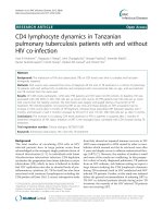
RNA sequencing was performed using 50 single embryos
(Fig. 1).
A total of 1405 million raw reads was obtained after
RNA sequencing, with a duplication rate of 63 ± 7%
(mean ± SD) and a GC content of 45 ± 1% (mean ± SD).
The mapping rate after quality filtering was 84 ± 6%
van der Weijden et al. BMC Genomics
(2021) 22:139
Page 3 of 13
Fig. 1 Experimental set-up for single embryo RNA-sequencing. The arrows indicate the between-group analyses
(mean ± SD). The number of detected transcripts, defined as any transcript with at CPM > 0.1, increased with
developmental progression for the in vivo produced embryos, while it decreased for the in vitro produced embryos (Additional file 1). The low number of detected
transcripts for the 4-cell in vivo embryos might be the
consequence of analyzing 4-cell embryos with a reduced
RNA quality, relatively low input and cDNA yield during
library preparation (Additional file 2). Given the differences in RNA quality as assessed by the cDNA profile,
library smear analyses, and read alignment at the 4-cell,
as well as at the morula stage (Additional file 2 and 3),
the in vivo developed and in vitro produced embryos
were analyzed separately and were not compared to each
other. To identify in vitro fertilization pipeline-induced
transcriptome differences, the hatched blastocysts were
used for an in vivo developed versus in vitro produced
comparison.
Developmental transcriptome dynamics
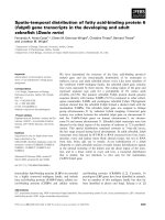
To provide a developmental stage-specific overview, global developmental transcriptome dynamics were investigated. Principal component analyses (PCA) were
performed separately for the in vivo developed and
in vitro produced embryos and showed a clear developmental stage-specific clustering of the embryos (Fig. 2a
and b). For the in vivo developed embryos, PC1 and PC2
explained 77.8 and 11.4% of the variance in transcript
levels. For the in vitro produced embryos, PC1 and PC2
explained 71.8 and 17.3% of the variance. The in vivo 4cell embryos displayed a larger degree of transcriptional
heterogeneity than the in vitro 4-cell embryos. The morulae and hatched blastocysts were sexed based on the
expression of Y-chromosome specific transcripts. At the
morula stage, male and female embryos clustered together, yet the clusters were not fully overlapping. At the
blastocyst stages, the male and female clusters were fully
overlapping.
In vivo and in vitro embryonic developmental dynamics
The developmental transcriptome dynamics were further
analyzed by identifying differentially expressed genes
(DEGs) between the 4-cell and morula stage, and the
morula and hatched blastocyst stage for both the in vivo
developed and in vitro produced embryos. The number
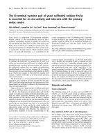
of DEGs was higher between the 4-cell to morula stage,
than for the morula to hatched blastocyst stage (Fig. 3).
For the in vivo embryos, 10089 and 2347 DEGs were
identified between the 4-cell to the morula stage and the
morula stage to the hatched blastocyst stage, respectively
(Fig. 3a). For the in vitro embryos, 8152 and 4023 DEGs
were identified between the 4-cell to the morula stage
and the morula stage to the hatched blastocyst stage, respectively (Fig. 3b).
The developmental dynamics were assessed with a
self-organizing tree algorithm (Fig. 4a and b). For both
the in vivo and in vitro produced embryos, the detected
transcript expression changed from the 4-cell to the
morula stage. The transcripts in cluster 1 decreased
from the 4-cell to the morula stage, and remained low at
the hatched blastocyst stage. The transcripts in cluster 2
displayed a gradual increase with developmental progression. The transcripts in cluster 3 were increased at
the morula stage, while remaining low at the 4-cell and
the hatched blastocyst stage.
Biological functions of embryonic developmental
dynamics
To gain insight into the biological functions of the
DEGs, a canonical pathway enrichment analysis was
conducted (Fig. 5). In both the in vivo and the in vitro
produced 4-cell to morula stage embryos, there was a
significant enrichment of oxidative phosphorylation and
EIF2 signaling. From the morula to the hatched blastocyst stage, the DEGs in the pathways 14–3-3-mediated
signaling, xenobiotic metabolism general signaling pathways, and NRF2-mediated oxidative stress response were
van der Weijden et al. BMC Genomics
(2021) 22:139
Page 4 of 13
Fig. 2 Between-group analyses of the 4-cell stage embryos, morulae and hatched blastocysts of a. In vivo developed embryos, and b. In vitro
produced embryos
all higher expressed at the hatched blastocyst stage for
both the in vivo and in vitro produced embryos.
In vivo and in vitro differences at the hatched blastocyst
stage
The in vivo and in vitro hatched blastocysts were compared, as the embryos displayed similar cDNA profiles,
library smears and alignment coverages for the most
abundant transcripts at this developmental stage
(Additional file 2 and 3). Embryos at this stage of development are thought to be more alike than at earlier
stages, as time differences related to fertilization at earlier stages contribute more substantially to the actual developmental stage.
At the hatched blastocyst stage, the selection of developmentally competent embryos has already taken
place. Yet, we unraveled in vitro fertilization pipelineinduced sex-specific differences. The in vivo
van der Weijden et al. BMC Genomics
(2021) 22:139
Page 5 of 13
Fig. 3 Upset plot displaying the differentially expressed genes during embryo development from the 4-cell to the morula stage and the morula
to the hatched blastocyst stage in a. In vivo developed embryos, and b. In vitro produced embryos
developed female and male hatched blastocysts clustered largely together (Fig. 6a). They were separated
from the in vitro hatched blastocyst in a sex-specific
manner by principal component 1. While 33 DEGs
were identified between the female in vivo and
in vitro produced embryos, 241 DEGs were identified
between the male in vivo and in vitro produced embryos. Figure 6b displays the difference between
in vivo developed and in vitro produced embryos in a
sex-independent manner. There were no DEGs when
van der Weijden et al. BMC Genomics
(2021) 22:139
Page 6 of 13
Fig. 4 Transcriptome dynamics during development displayed by a self-organizing tree algorithm for a. In vivo developed embryos, and b. In vitro
produced embryos. The number of genes per cluster and the embryonic sex of the morulae and hatched blastocysts are indicated
comparing male and female embryos for either
in vivo developed or in vitro produced embryos. By
comparing the female in vivo developed versus
in vitro produced embryos, the DEGs inositol polyphosphate multikinase (IPMK) and Rac family small
GTPase 1 (RAC1) were specific to this comparison.
The other 31 DEGs were also discovered by comparing the in vivo and in vitro male hatched blastocysts.
These genes were involved in amino acids transport,
synthesis and metabolism, and similarly expressed in
both female and male embryos (Fig. 6c). Both male
and female in vivo derived embryos had a lower expression of genes involved in amino acid transport,
synthesis and metabolism compared to the male and
female in vitro produced embryos.
When disregarding the sex of the embryos and emphasizing on the embryo source, a total of 398 DEGs
were identified. The persistent difference between
in vivo developed and in vitro produced embryos at
the hatched blastocyst stage were illustrated by an enrichment of four canonical pathways (Fig. 6d). Except
for a higher expression in in vivo versus in vitro
hatched blastocysts of DEGs involved in cyclins and
cell cycle regulation and LXR/RXR activation, the
DEGs involved in tRNA charging and xenobiotic metabolism AHR signaling pathways were higher
expressed in in vitro than in in vivo hatched
blastocysts.
Discussion
Transcriptome dynamics during early embryo
development
Early developing porcine embryos displayed a great
adaptive capacity towards their environment, evidenced
by largely similar transcriptome dynamics observed in
both in vivo developed and in vitro produced embryos.
in vitro produced embryos offer the opportunity to study
molecular pathways of interest in a developmental-stage
specific manner, as there is a higher degree of certainty regarding the time of fertilization compared to in vivo developed embryos. However, developmental rates and embryo
competence of in vitro produced embryos are still lower
compared to their in vivo developed counterparts [5]. A
number of factors are known to contribute to embryo development. The presence of cumulus cells during maturation facilitates full oocyte maturation [19]. In pigs, the
presence of cumulus cells during oocyte maturation is essential for oocyte maturation, fertilization and subsequent
embryo development [20]. The discrepancy in embryo development between in vivo developed and in vitro produced embryos at early post-fertilization developmental
stages might be explained by the use of a pool of nonselected oocytes of overall lower competence for in vitro
maturation, compared to those selected for ovulation, and
the effects of in vitro maturation on oocyte quality. A
higher blastocyst rate has previously been shown after oocyte maturation under a 20% oxygen atmosphere [21].
van der Weijden et al. BMC Genomics
(2021) 22:139
Page 7 of 13
Fig. 5 Enriched canonical pathways. Red (−) dots represent canonical pathways of which genes were significantly lower expressed in the 4-cell
versus morula and morula versus hatched blastocysts, and blue (+) represent canonical pathways of which genes were significantly higher
expressed in the 4-cell versus morula or morula versus hatched blastocysts. The GeneRatio indicates the proportion of DEGs that were identified
in an enriched canonical pathway. Shared enriched canonical pathways in both in vivo developed and in vitro produced embryos at the 4-cell
versus morula or morula versus hatched blastocyst stage are indicated in purple
However, blastocyst quality assessed by the expression of
genes related to metabolism (GLUT1 and LDHA), antioxidant response (SOD2 and GPX1), growth factors and
apoptosis (IGF2R, BCL2 and BAX), methylation
(DNMT3B), and blastocyst quality (AKR1B1, POU5F1 and
CDX2) were not affected [21]. In addition, the blastocyst
rates of in vivo and in vitro matured rabbit oocytes did
not significantly differ, while at earlier developmental
stages, the in vivo embryo development rates were significantly higher than observed for embryos produced with
in vitro matured oocytes [22]. Thus, while oocyte quality
and competence, and subsequent embryo development
are affected by the maturation conditions, only minor
transcriptional differences have been reported at the
hatched blastocyst stage [23]. In line with previous findings, we found more similar transcriptome profiles at later
developmental stages. At the hatched blastocyst stage,
only limited transcriptional differences persisted. Additionally, the developmental-stage specific differences were
more pronounced than the sex-specific differences, as previously described by Zeng et al. (2019), studying the transcriptome dynamics in in vivo developed day 8, 10, and 12
porcine embryos [16].
Early porcine embryo development
The early embryo development was studied at the 4-cell,
morula and hatched blastocyst stage for both in vivo developed and in vitro produced embryos. Previously, porcine embryos after EGA have been shown to display an
increased abundance of transcripts involved in, among
others, transcription [13]. Both the in vivo developed
and in vitro produced 4-cell to morula transition was
characterized by an enrichment of oxidative phosphorylation and EIF2 signaling. An increase in oxidative