Báo cáo khoa học: A single mutation in Escherichia coli ribonuclease II inactivates the enzyme without affecting RNA binding pot
Bạn đang xem bản rút gọn của tài liệu. Xem và tải ngay bản đầy đủ của tài liệu tại đây (361.4 KB, 12 trang )
A single mutation in Escherichia coli ribonuclease II
inactivates the enzyme without affecting RNA binding
Mo
´
nica Amblar and Cecı
´lia
M. Arraiano
Instituto de Tecnologia Quı
´
mica e Biolo
´
gica ⁄ Universidade Nova de Lisboa, Oeiras, Portugal
The balance between mRNA synthesis and decay is an
important aspect of gene expression in all organisms.
The RNases are involved in many functions such as
RNA processing, stability and degradation, and their
concerted action allows strict regulation of the RNA
metabolism [1–5]. The mRNA decay in Escherichia coli
is normally initiated by a series of endonucleolytic
cleavages catalyzed by RNase E [6–8] or RNase III
[9,10]. The breakdown products are subsequently degra-
ded by the processive 3¢ to 5¢ exoribonucleases PNPase
and ⁄ or RNase II [11–14]. RNase II and PNPase are the
major 3¢ to 5¢ exoribonucleases present in E. coli cells.
RNase II accounts for 90% of the exoribonucleolytic
activity of E. coli crude extracts, while PNPase is
responsible for the remaining 10% [15,16]. It is known
that the presence of secondary structures in the RNA is
an important determinant for mRNA stability. The
processive degradation activity of RNase II is easily
blocked by stem–loop structures, while PNPase is able
to overcome many of the stem–loop structures that it
encounters [17–19]. In vivo, RNase II can rapidly
degrade some polyadenylated stretches necessary for
degradation by PNPase and possibly other exoribonuc-
leases [20,21]. As a consequence, RNase II can paradox-
ically act as a protector of some RNAs from
degradation by blocking the access of other 3¢ to 5¢ exo-
ribonucleases [19–24].
The use of E. coli strains harboring a defective
RNase II has been very useful in the study of the cellu-
lar function of this protein, as well as in determining
Keywords
RNase II; exoribonuclease; RNA
degradation; RNA binding; RNR family
Correspondence
C. M. Arraiano, Instituto de Tecnologia
Quı
´
mica e Biolo
´
gica ⁄ Universidade Nova de
Lisboa, Apartado 127, 2781–901 Oeiras,
Portugal
Fax: +351 21 4411277
Tel: +351 21 4469547
E-mail:
(Received 30 July 2004, revised 29 October
2004, accepted 11 November 2004)
doi:10.1111/j.1742-4658.2004.04477.x
Exoribonuclease II (RNase II), encoded by the rnb gene, is a ubiquitous
enzyme that is responsible for 90% of the hydrolytic activity in
Escherichia coli crude extracts. The E. coli strain SK4803, carrying the
mutant allele rnb296, has been widely used in the study of the role of
RNase II. We determined the DNA sequence of rnb296 and cloned this
mutant gene in an expression vector. Only a point mutation in the coding
sequence of the gene was detected, which results in the single substitution
of aspartate 209 for asparagine. The mutant and the wild-type RNase II
enzymes were purified, and their 3¢ to 5¢ exoribonucleolytic activity, as well
as their RNA binding capability, were characterized. We also studied the
metal dependency of the exoribonuclease activity of RNase II. The results
obtained demonstrated that aspartate 209 is absolutely essential for RNA
hydrolysis, but is not required for substrate binding. This is the first evi-
dence of an acidic residue that is essential for the activity of RNase II-like
enzymes. The possible involvement of this residue in metal binding at the
active site of the enzyme is discussed. These results are particularly relevant
at this time given that no structural or mutational analysis has been per-
formed for any protein of the RNR family of exoribonucleases.
Abbreviations
His(6)-RNase II, RNase II with a six-His-Tag fused at the N-terminal end; His(6)-RNase IID209N, His(6)-RNase II with the amino acid
substitution D209N; IPTG, isopropyl thio-b-d-galactoside; nt, nucleotides; pol, polymerase; K
D
, equilibrium dissociation constant; ss, single
strand; PAA, polyacrylamide; UE, units of enzymatic activity, namely the amount of protein required for the release of 10 nmol of [
3
H]AMP
in 15 min at 30 °C.
FEBS Journal 272 (2005) 363–374 ª 2004 FEBS 363
the role of other ribonucleases [11,21,24–29]. For
instance, the rnb500 temperature-sensitive mutant
strain demonstrates that the absence of both RNase II
and PNPase activity leads to cell death [25]. Despite
the wide use of these mutant strains during the last
20 years, nothing is known about the mutations
responsible for such phenotypes. The study of the
molecular basis of the absence of RNase II activity in
these strains will highlight some important information
in the knowledge of RNase II proteins. To date, there
are no structural or mutational data available from any
other proteins of the family. The SK4803 strain is par-
ticularly interesting since the mutant gene (rnb296)
encodes an inactive RNase II enzyme [25]. In this
report we demonstrate that the single amino acid sub-
stitution Asp209fiAsn in RNase II is able to cause the
total inactivation of the enzyme without affecting its
RNA binding capability. In addition, metal ions seem
to be required for activity but not for substrate bind-
ing, suggesting the involvement of Asp209 in metal
binding at the active site of the enzyme.
Results
Cloning of the rnb296 mutant gene and
overexpression of the mutant protein
The E. coli SK4803 strain deficient in RNase II acti-
vity, which carries the rnb296 allele, has been previ-
ously described [25]. Crude extracts from this strain
were totally inactive in the degradation of polyadenylic
acid [poly(A)] but this seemed to have no detrimental
effect on cell viability or mRNA degradation rate [25].
In order to identify the mutation(s) responsible for this
phenotype, we sequenced the gene encoding the RNase
II protein of this mutant strain. To achieve this, the
chromosomal DNA of E. coli SK4803 was used as a
source in the PCR amplification of the rnb296 gene and
the DNA sequence of the PCR product was deter-
mined. The results revealed that the rnb296 gene differs
only in one base from the wild-type rnb gene: G1148
was replaced with A in the rnb296 mutant gene and this
single mutation leads to the substitution of Asp209
with Asn in the RNase II sequence. RNase II is the
prototype of the widely distributed RNR family of
ribonucleases. It has been hypothesized that the cata-
lytic activity of the RNR proteins resides in their cen-
tral domain, named RNB. Asp209 of RNase II lies in
this RNB domain, more specifically in the highly con-
served motif I of this domain (Fig. 1), and is present in
almost all members of the RNR family of exoribonuc-
leases [30,31]. All of these data suggest that Asp209
might have a key role in the RNase II enzyme. In order
to analyze the function of this residue, the rnb296
mutation was cloned into the previously described
pFCT6.9 plasmid [29]. This plasmid contains the wild-
type rnb gene from E. coli cloned into the pET15 fusion
vector (Novagen). Under isopropyl thio-b-d-galactoside
(IPTG) induction, the pFCT6.9 plasmid directs the
expression of the six-histidine-tagged RNase II protein
[His(6)-RNase II] that was previously shown to be act-
ive [29]. The 996-bp NheI fragment from the rnb296
gene containing the corresponding mutation (G1148A)
was cloned into plasmid pFCT6.9 obtaining pMAA
(see Experimental procedures). The presence of the cor-
rect 296 mutation was confirmed by DNA sequencing,
and the pMAA plasmid was further transferred to
E. coli BL21(DE3) to overproduce the corresponding
six-histidine-tagged RNase IID209N mutant protein
[His(6)-RNase IID209N].
The suitability of E. coli BL21(DE3)[pMAA] as a
source of His(6)-RNase IID209N was tested by induc-
tion of the corresponding cells with IPTG at 37 °C.
Samples were taken at different induction times and
the protein content was analyzed by SDS ⁄ PAGE
(Fig. 2). The results revealed that, after 2 h of IPTG
treatment, His(6)-RNase IID209N was the major pro-
tein in cell extracts, corresponding to 14% of the
total protein content (Fig. 2A). The solubility of
the protein upon induction was also analyzed and the
results revealed that more than 70% of the His
(6)-RNase IID209N was soluble (Fig. 2B), similar to
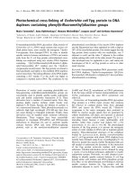
Fig. 1. Schematic representation of the structure of RNase II. Three
different domains can be proposed for RNase II: the N-terminal
cold shock domain (CSD), the central RNB domain (RNB), and the
C-terminal S1 domain (S1). The four sequence motifs of the RNB
domains are depicted (I–IV) and the sequence pattern of the motif I
is shown. The syntax of the pattern follows that used in
PHI-BLAST searches ( />phiblast.html#pattern). Residues conserved in > 80% of the ana-
lyzed sequences are shown as bold letters. The position corres-
ponding to the D209N mutation is indicated with an arrow.
RNase II mutant with RNA binding but no activity M. Amblar and C. M. Arraiano
364 FEBS Journal 272 (2005) 363–374 ª 2004 FEBS
that obtained with His(6)-RNase II protein from
BL21(DE3) cells containing pFCT6.9 (data not
shown). These results revealed that, as with the
wild-type enzyme [29], the maximum induction of
His(6)-RNase IID209N is reached after 2 h of IPTG
treatment, and these were the conditions used for fur-
ther purification of the protein.
The exoribonucleolytic activity of the crude extracts,
before and after induction with IPTG, was also ana-
lyzed using poly[8-
3
H]adenylic acid as a substrate and
the results obtained are summarized in Table 1. The
activity levels of the extracts from cells overexpressing
His(6)-RNase IID209N protein (BL21(DE3)[pMAA])
after 2 h of induction were similar to those obtained
with the BL21(DE3) without plasmid. In contrast, the
presence of plasmid pFCT6.9, encoding the wild-type
His(6)-RNase II enzyme, produced a 60-fold induction
of RNase II activity after 2 h of IPTG treatment.
Therefore, the His(6)-RNaseII-D209N protein pro-
duced upon IPTG induction was totally inactive, and
this inactivation was exclusively caused by the substitu-
tion of Asp209 with Asn.
Purification and properties of His(6)-RNase
IID209N
A purification procedure for the His(6)-RNase II pro-
tein has been previously described [29]. Based on this
initial protocol, a new purification procedure for His
(6)-RNase IID209N protein and the wild-type enzyme
was standardized by the introduction of several modifi-
cations. Cultures of 100 mL of BL21(DE3) cells con-
taining the pMAA or pFCT6.9 plasmids, respectively,
were grown at 37 °C and induced with IPTG for 2 h.
Then, the cells were disrupted and treated as described
in the Experimental procedures, and the extracts
obtained were applied to chelating sepharose high per-
formance columns previously charged with Ni
2+
ions.
Different concentrations of imidazol-eluting agent were
tested to develop the purification procedure described in
the Experimental procedures. Following this procedure,
His(6)-RNase IID209N and His(6)-RNase II proteins
were obtained with > 90% purity.
The exoribonucleolytic activity of purified enzymes
was first tested by using the linear substrate,
poly[8-
3
H]adenylic acid. The D209N mutant protein
was unable to degrade the polyribonucleotide and had
Table 1. Specific exoribonucleolytic activity in crude extracts from
BL21(DE3) overproducer strains on poly(A) substrate. The exoribo-
nuclease activity was measured before and after 2 h of isopropyl
thio-b-
D-galactoside (IPTG) induction of BL21(DE3) containing the
indicated plasmid. BL21(DE3) cells without plasmid were used as a
control. Each value is the mean of at least three independent
experiments. UE, the amount of protein required for the release of
10 nmol of [
3
H]AMP in 15 min at 30 °C.
Specific exoribonuclease activity
(UEÆlg
)1
of protein)
BL21(DE3)
BL21(DE3)
[pFCT6.9]
BL21(DE3)
[pMAA]
Before IPTG induction 0.12 0.37 0.03
After 2 h of
IPTG induction
0.22 22.42 0.17
AB
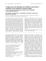
Fig. 2. Overexpression of RNase IID209N by induction with isopropyl thio-b-D-galactoside (IPTG). (A) Crude extracts from BL21(DE3) cells
harboring the pMAA plasmid were induced by IPTG. Samples were withdrawn at the time-points indicated in the figure, after the addition of
IPTG, and the total protein content was analyzed by electrophoresis in a 0.1% SDS, 10% acrylamide gel. (B) The soluble (S) and insoluble (I)
protein fraction from cultures induced for 2 h with IPTG were analyzed in a 0.1% SDS, 10% polyacrylamide gel. The His(6)-RNase IID209N
(RNase IID209N) protein is indicated with an arrow. St, molecular mass maker.
M. Amblar and C. M. Arraiano RNase II mutant with RNA binding but no activity
FEBS Journal 272 (2005) 363–374 ª 2004 FEBS 365
no detectable activity, even when higher amounts of
protein were used (23 lg per reaction). By contrast, the
purified His(6)-RNase II was highly active in the degra-
dation of poly[8-
3
H]adenylic acid, with an activity of
> 325 UEÆlg
)1
of protein (results not shown) (UE:
units of enzymatic activity, namely the amount of pro-
tein required for the release of 10 nmol of [
3
H]AMP in
15 min at 30 °C).
Previous studies on the RNase II enzyme revealed
that its activity is blocked by double-stranded structures
on the RNA molecule [12,13,18,19]. Various mRNA
transcripts harboring stem–loop structures have been
tested as RNase II substrates [13,18,19,32–34] and in all
cases the enzyme catalyzed the degradation of the sin-
gle-stranded (ss) portion of the RNA molecule from its
3¢ end until it reached the double-stranded region. In
order to analyze the effect of the D209N mutation on
the exoribonucleolytic activity of RNase II on struc-
tured substrates, we tested the degradation ability of
both His(6)-RNase II and the D209N mutant enzyme
by using two different mRNAs, namely SL9A [13] and
malE-malF [18]. The SL9A substrate is a small RNA
molecule of 83 nucleotides (nt), consisting of an ss 3¢-
extension (of 41 nucleotides), which mimics a typical
bacterial poly(A) tail, plus a stem–loop structure (9 bp
stem and four-residue loop) and a short 5¢-single stran-
ded arm (of 19nt). The exonuclease assays performed
revealed that His(6)-RNase II, similarly to that previ-
ously reported for RNase II [13], degrades the SL9A
RNA substrate in a two-step process (Fig. 3A). The
enzyme initially catalyzes a rapid shortening of the
RNA molecule from its 3¢ end, generating a set of inter-
mediates, followed by the further degradation of these
intermediates at a slower rate. As shown in Fig. 3A, the
SL9A substrate was totally converted into shorter inter-
mediate products in only 30 s by 2 nm purified His(6)-
RNase II. These intermediates, partially resistant to
degradation, presumably correspond to the stem–loop
structure with a 3¢ ss extension of 6–9 nucleotides
[13]. Longer reaction times (up to 30 min) resulted in
the diminution of the intermediate length as a result of
limited digestion by the enzyme. By contrast, the
D209N mutant enzyme was unable to degrade the
SL9A RNA. As shown in Fig. 3A, 100% of the full-
length starting material remained intact, even after
30 min of incubation with 540 nm of the mutant
enzyme. Similar results were obtained with the longer
mRNA transcript corresponding to the intergenic
region of the malE-malF operon. This substrate consists
of a 375 nucleotides RNA molecule containing two
stem–loop structures: a large secondary structure
formed by the two inverted palyndromic REP
sequences; and a smaller and weaker secondary
structure at the 3¢ end of the mRNA [18,35]. As with
the other substrates tested, no exoribonuclease activity
was detected with the D209N mutant on the malE-malF
transcript, even after 30 min of incubation (Fig. 3B),
confirming the complete inactivation of the enzyme
caused by the mutation. However, His(6)-RNase II was
highly active in the degradation of this substrate and in
only 30 s 70% of the full-length product disappeared
(Fig. 3B). In agreement with data previously reported
for the wild-type RNase II enzyme [18], digestion of the
malE-malF transcript by the fusion derivative His(6)-
RNase II rendered two main intermediate products: P1
and P2 (Fig. 3). Such intermediates presumably corres-
pond to the stalling of the enzyme in the vicinity of the
two secondary structures of the mRNA.
Metal dependency of the exoribonuclease activity
of RNase II
The above results pointed out the importance of
Asp209 for RNase II, as its substitution with Asn
leads to a total inactivation of the enzyme. RNase II,
like other exoribonucleases, requires the presence of
Mg
2+
in the reaction to catalyze the degradation of
RNA. The acidic nature of Asp209, together with its
conservation in RNase II-like enzymes, suggests that
this residue is one of the metal ligands at the active site
of RNase II. If this assumption is correct, a reduced
affinity for the metal ion should be expected in the
D209N mutant protein. Consequently, the use of a
higher Mg
2+
concentration might allow us to detect
some nuclease activity with the mutant enzyme. To test
this hypothesis, the exoribonuclease activity of His(6)-
RNase II and the D209N mutant protein was tested in
the presence of different concentrations of the metal
ion. MgCl
2
concentrations ranging from 0.5 lm to
10 mm were assayed by using the malE-malF tran-
script (Fig. 4A). Quantification of the reaction prod-
ucts revealed that His(6)-RNase II was able to degrade
the mRNA within a wide range of Mg
2+
concentra-
tions (Fig. 4B). The enzyme was highly active at all
MgCl
2
concentrations tested, although the maximum
activity was obtained between 5 lm and 1 m m. Inter-
estingly, the rate between the P1 and P2 products var-
ied depending on the metal ion concentration. The P2
intermediate was the main product obtained at lower
metal concentrations (from 0 to 100 lm), while P1 was
only observed at MgCl
2
concentrations of ‡ 500 lm,
being the major product at ‡ 1mm MgCl
2
. These
results suggest different properties of the exoribo-
nucleolytic activity of RNase II depending on the
metal ion concentration. Surprisingly, activity assays
performed without adding MgCl
2
to the reaction
RNase II mutant with RNA binding but no activity M. Amblar and C. M. Arraiano
366 FEBS Journal 272 (2005) 363–374 ª 2004 FEBS
mixture revealed a residual activity of His(6)-RNase II
that only disappeared after the addition of 10 mm
EDTA. This fact indicates that some essential Mg
2+
atoms are bound to the protein and that they cannot
be easily removed from the protein structure simply by
buffer-exchange. In the case of the D209N mutant pro-
tein, no exoribonuclease activity was detected at any
metal ion concentration tested (data not shown), indi-
cating that the loss of activity in the mutant protein
cannot be restored by increasing the Mg
2+
concentra-
tion.
RNA binding ability of RNase II and the D209N
mutant
The data presented above demonstrate that the D209N
amino acid substitution leads to a loss in the exoribo-
nucleolytic activity of RNase II. Such inactivation can
be caused by a defect in the catalytic reaction, by a
decrease in substrate affinity, or both. To investigate
whether the D209N mutation impairs the RNA bind-
ing, band-shift assays were performed with the radio-
actively labeled transcripts SL9A and malE-malF. In
order to reduce the degradation of the substrate upon
binding of the wild-type enzyme, the incubation was
performed at different temperatures (from 15 °Cto
37 °C). The results obtained showed that, even at
15 °C, the incubation of His(6)-RNase II with either
SL9A or malE-malF transcripts resulted in bands with
higher gel mobility than the free substrate (Fig. 5).
Such bands corresponded to degradation products,
and the intensity increases with the protein concentra-
tion. By contrast, the D209N protein generated only
retardation bands with both transcripts. These bands
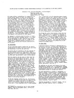
Fig. 3. Exoribonuclease activity of RNase II
and the D209N mutant enzyme on mRNA
transcripts. The exoribonuclease activity of
His(6)-RNase II (wild-type, WT) and His(6)-R-
Nase IID209N (D209N) enzymes was assay-
ed by using SL9A mRNA (A) or malE-malF
mRNA (B) transcripts. Reactions were per-
formed as described in the Experimental
procedures using 2 n
M WT enzyme or 540
n
M D209N mutant protein. Samples were
taken at the time-points indicated in the
figure, and the reaction products were
analyzed in 8% polyacrylamide (PAA) (A) or
6% PAA (B), 7
M urea gels. A schematic
representation of the substrates and reac-
tion products is depicted.
M. Amblar and C. M. Arraiano RNase II mutant with RNA binding but no activity
FEBS Journal 272 (2005) 363–374 ª 2004 FEBS 367
correspond to RNA–protein complexes, indicating that,
despite the mutation, the D209N protein is able to
bind RNA.
To investigate the role of MgCl
2
in formation of the
RNA–protein complex, binding assays were performed
with the malE-malF substrate at 37 °C in the presence
or in the absence of EDTA. As shown in Fig. 6A,
incubation of the wild-type protein with the RNA sub-
strate in the absence of EDTA resulted in the expected
degradation bands. However, when 10 mm EDTA was
added to the binding reaction, the degradation of the
substrate was inhibited and only retarded bands cor-
responding to RNA–protein complexes were detected.
These results indicate that RNase II requires Mg
2+
ions for catalysis but not for substrate binding. RNA–
protein complexes were detected from 5 nm of wild-
type enzyme when the incubation was performed in
the presence of EDTA.With the D209N mutant
enzyme, retarded bands were observed either in the
presence or absence of EDTA, and in both conditions
RNA–protein complexes were also observed from 5 to
10 nm of protein (Fig. 6B). The equilibrium dissoci-
ation constant (K
D
) values of both wild-type and
mutant proteins were estimated from gel-shift assays.
The values obtained in the presence of EDTA were
382 nm for the wild-type protein and 344 nm for the
D209N mutant. In the absence of EDTA, the K
D
value
for the mutant protein was 330 nm. The K
D
of the two
proteins was analogous, showing that both enzymes
have similar affinity for this substrate. These results
clearly indicate that the D209N mutation does not
affect the ability of RNase II to form stable RNA–
protein complexes and that the presence or the absence
of Mg
2+
does not influence the substrate binding of
the mutant protein.
Discussion
Eight different 3¢ to 5¢ exoribonucleases have been char-
acterized in E. coli and this group of enzymes accounts
for all the exoribonucleolytic activities present in an
E. coli cell [36]. These enzymes have been grouped into
six superfamilies and various subfamilies based on
extensive sequence analysis and catalytic properties
[31]. The RNase II belongs to the RNR family of exo-
ribonucleases and, together with RNase R, has been
considered as the prototype of the RNR-like enzymes.
This family is widely distributed among all organisms,
and RNase II homologs are found in almost all pro-
karyotes and eukaryotes [30,31]. Many in vitro studies
of the 3¢ to 5¢ exoribonucleolytic activity of E. coli
RNase II have been performed [12,18,19,37] and its
implications in prokaryotic mRNA decay in vivo have
been well characterized [11,20,21,24,27,28]. However,
to date no structural or mutational analysis have been
performed for E. coli RNase II or for any other RNR
family member.
An E. coli strain deficient in RNase II activity [25]
has been widely used for many years in the study of
RNase II. This strain (SK4803) carries the rnb296
allele and it was previously demonstrated that the
crude extracts were unable to degrade the polyadenylic
acid [11,25]. Although this strain has been extensively
used, nothing is known about the mutation responsible
for the synthesis of an inactive RNase II. In this
report, we determined the DNA sequence of the
rnb296 gene and we demonstrated that the single
substitution of Asp209 by Asn in RNase II (D209N) is
responsible for the loss of RNase II activity. Our stud-
ies on the purified His(6)-RNase IID209N mutant
B
A
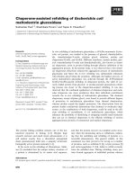
Fig. 4. Metal dependence of the exoribonuclease activity of
RNase II. Exoribonuclease activity of His(6)-RNase II in the presence
of different concentrations of MgCl
2
. Assays were performed as
described in the Experimental procedures using 1 n
M enzyme and
the metal ion concentration indicated in the figure. (A) The reaction
products were analyzed in a 6% PAA ⁄ 7
M urea gel. (B) The percent-
age of exonuclease activity was estimated from the gel by quanti-
fication of the band intensities. The RNA degradation was
determined by calculating the ratio of the reaction products (P1 and
P2) and the substrate (S) on the respective lane. Each value is the
mean of three independent experiments.
RNase II mutant with RNA binding but no activity M. Amblar and C. M. Arraiano
368 FEBS Journal 272 (2005) 363–374 ª 2004 FEBS
protein demonstrated that Asp209 is absolutely essen-
tial for the exoribonucleolytic activity of RNase II but
does not seem to be involved in substrate binding. No
cleavage activity was detected either with the linear
polyadenylic acid substrate or with stem–loop contain-
ing RNAs (SL9A or malE-malF RNAs) in the pres-
ence of 540 nm His(6)-RNase IID209N. However,
5nm of the mutant protein was sufficient to form sta-
ble RNA–protein complexes. Moreover, we also dem-
onstrate that RNase II requires metal ions for catalysis
but not for substrate binding. These findings provide
the first identification of a key residue for catalysis in
RNase II and support the hypothesis of the organiza-
tion in independent functional domains for these
enzymes. It has been previously proposed that the cat-
alytic ability of RNR proteins resides in a central
region of 400 residues, termed the RNB domain [31].
Multiple sequence alignments of this catalytic domain
revealed the presence of four highly conserved
sequence motifs (I–IV) containing some invariant carb-
oxylate residues [30,38]. Figure 1 shows the sequence
pattern of motif I inferred from a multiple sequence
alignment that includes 27 prokaryotic and eukaryotic
RNR-like enzymes ( />Pfam/getacc?PF00773). Asp209 of RNase II lies in
motif I of the RNB domain. This position is occupied
by an acidic residue (aspartate or glutamate) in 86%
of the RNR proteins aligned (Fig. 1), and in 75% of
the enzymes corresponds to an aspartate. The presence
of carboxylate residues is common in proteins that cat-
alyze phosphoryl transfer reactions, such as nucleic
acid polymerases or nucleases, and they are required
for the co-ordination of divalent metal ions that are
essential for catalysis [39]. Given its high degree of
conservation, Asp209 seems to be a good candidate
for being one of the metal ligands at the active site of
A
B
Fig. 5. Gel retardation assay of mRNA transcripts with His(6)-RNase II and His(6)-RNase IID209N. SL9A (2 fmol) (A) or malE-malF (2 fmol)
(B) mRNA transcripts were incubated at 15 °C with the His(6)-RNase II (wild-type; WT) and His(6)-RNase IID209N (D209) mutant enzyme
under the conditions described in the Experimental procedures. The enzyme concentration used is indicated in the figure. After electrophor-
esis, the mobility of free RNA as well as the RNA–protein complexes was detected by autoradiography.
M. Amblar and C. M. Arraiano RNase II mutant with RNA binding but no activity
FEBS Journal 272 (2005) 363–374 ª 2004 FEBS 369
the enzyme. Under this hypothesis, its replacement
with Asn would lead to a loss of the metal co-ordina-
tion with the subsequent loss in activity, without
affecting substrate binding. Our band-shift experiments
demonstrate that the RNA binding of the D209N
mutant is not reduced and not influenced by the pres-
ence or absence of a metal binding inhibitor (10 mm
EDTA). In some cases, these metal ligands can be sub-
stituted with a water molecule or by another acidic
residue in the vicinity, allowing the metal binding even
in the absence of these residues. This normally results
in a decrease in metal affinity, indicating that the activ-
ity of the mutant protein could be restored, at least in
part, by increasing the metal concentration. Despite
our efforts to detect any exoribonucleolytic activity at
different concentrations of Mg
2+
, we were unable to
observe digestion by the D209N mutant protein. Nev-
ertheless, these results do not rule out that Asp209 is
involved in the binding of metal ions at the active site.
The substitution of this aspartate with asparagine can
induce conformational changes in the metal binding
pocket that totally prevent the co-ordination of Mg
2+
to the active site. However, another hypothesis must
be taken into account. Instead of a metal ligand,
Asp209 may act as a general base during the reaction,
generating the nucleophilic hydroxide group that will
attack the scissile phosphate of the RNA. Under this
hypothesis, Asp209 would be directly responsible for
the catalytic event in RNase II. Based on the results
presented here we cannot exclude the possibility that
inactivation of the enzyme is caused by subtle con-
formational changes owing to the amino acid replace-
ment. However, the dramatic effect on RNase II
catalysis caused by the substitution of only one residue
strongly suggests a crucial role of this amino acid in
the RNase II enzyme.
The analysis of the exoribonuclease activity with dif-
ferent Mg
2+
concentrations revealed that the wild-type
enzyme is active within a wide range of the metal ion.
However, different kinds of products are released
depending on the concentration of this metal ion. The
malE-malF RNA molecule contains two stem–loop
Fig. 6. Effect of EDTA in RNA binding. malE-malF mRNA transcripts (2 fmol) were incubated at 37 °C with His(6)-RNase II (A) or His(6)-
RNase IID209N (B) mutant enzyme in the absence of EDTA (– EDTA) or in the presence of 10 m
M EDTA (+ EDTA). The reaction was per-
formed as described in the Experimental procedures. The enzyme concentration used is indicated in the figure. RNA–protein complexes
were detected and quantified by using the PhosphorImager system from Molecular Dynamics.
RNase II mutant with RNA binding but no activity M. Amblar and C. M. Arraiano
370 FEBS Journal 272 (2005) 363–374 ª 2004 FEBS
structures. RNase II may catalyze the nibbling of this
RNA from the 3¢ end until it reaches the first secon-
dary structure, rendering the P1 product, or the degra-
dation may continue until the second stem–loop (a
more stable secondary structure) is reached, generating
the P2 product. At lower concentrations of MgCl
2
,
RNase II seems to be able to easily overcome the first
stem–loop and the degradation only stops in the vicin-
ity of the second structure (P2 is the major product).
However, at higher concentrations of Mg
2+
, the deg-
radation is blocked by the first stem–loop structure.
These phenomena may respond to alterations in the
strength of the stem–loop structure caused by the
increase of MgCl
2
concentration, becoming more
resistant to degradation, or to different cleavage
properties of RNase II, depending on the metal ion
concentration. The hypothesis that RNase II activity
can be regulated by the Mg
2+
concentration is very
interesting. Aspects such as processivity of the RNase
II enzyme or its ability to overcome weak secondary
structures during degradation could be adequate to the
physiological requirements by changes in concentra-
tions of free divalent metal ions.
Experimental procedures
Materials
Restriction enzymes and T4 DNA ligase were purchased from
New England Biolabs (Hertfordshire, UK), T7 RNA polym-
erase was obtained from Promega (Charbonnie
`
res-les-Bains,
France), and Pfu DNA polymerase was obtained from Fer-
mentas (Vilnius, Lithuania). Oligonucleotide primers were
synthesized by Sigma Genosys (Cambridge, UK).
Bacterial strains, plasmids, and RNA substrates
The E. coli strains used were JM109 [F¢ (traD36 proA
+-
B
+
lacI
q
D(lacZ)M15 ⁄D(lac-proAB) glnV44 e 14
–
gyrA96
recA1 relA1 endA1 thi hsdR17] [40] for cloning experiments
and BL21(DE3) (F
–
r
B
–
m
B
–
gal ompT (int::P
lacUV5
T7 -
gen1 imm21 nin5) [41] for expression and purification of
enzymes. The rnb296 mutant gene was obtained from the
SK4803 E. coli strain deficient in RNase II activity [25].
The plasmids used for in vitro transcription reactions were
pCH77 [18] and pSL9A [13].
Cloning of the rnb296 mutation
The rnb296 mutant gene was amplified from the chromoso-
mal DNA of E. coli SK4803 by using a standard PCR reac-
tion with Pfu DNA polymerase. The primers used for the
amplification matched perfectly 45 nucleotides upstream of
the initiation codon (5¢-GCGTAAAACTGTCAGCCGCT-
3¢) and 47 nucleotides downstream of the stop codon
(5¢-CTGGATATAACGAAGGTAGAGC-3¢) of RNase II,
respectively. The DNA sequence of the 2048 bp PCR prod-
uct was determined (STABvida, Oeiras, Portugal). To
ensure that the mutation(s) detected were not introduced
during amplification, three independent PCR reactions were
performed and both strands of each PCR product were
sequenced. A point mutation (G fi A) at the 1148 position
of the rnb gene was detected and further inserted into the
previously described pFCT6.9 plasmid [29] that contains
the wild-type rnb gene cloned into the pET15 vector
(Novagen, Lisbon, Portugal). The insertion was performed
by digestion of the rnb296 PCR product and pFCT6.9 with
NheI followed by ligation with T4 DNA ligase. The result-
ing plasmid carrying the 296 mutation, named pMAA, was
transferred to the E. coli BL21(DE3) host strain.
Overexpression and protein purification
The expression of His(6)-RNase II and its mutant deriv-
ative carrying the substitution of Asp209 with Asn [His(6)-
RNase IID209N] was achieved by IPTG induction of
BL21(DE3) [42] containing either pFCT6.9 or pMAA plas-
mids, respectively. Cells were grown in LB (Luria–Bertani)
medium, supplemented with 150 lgÆmL
)1
of ampicillin, at
37 °C. After reaching an attenuance (D) of 0.4 at 600 nm,
the cultures were induced by adding 1 mm IPTG. Samples
were withdrawn at different induction times, and crude
extracts were prepared as previously described [43] to ana-
lyze the exoribonucleolytic activity and the total protein
content. The solubility of both wild-type and mutant pro-
teins during induction was tested by separation of the sol-
uble and insoluble protein fractions as previously described
[44], followed by fractionation in SDS ⁄ PAGE.
The purification of His(6)-RNase II and His(6)-RNase
IID209N proteins was performed by histidine affinity
chromatography using the HiTrap Chelating HP system
(Amersham Biosciences, Buckinghamshire, UK). For this
purpose, 100 mL of IPTG-induced cultures were harvested
by centrifugation, washed with 20 mL of buffer A (20 mm
Na
2
HPO
4,
0.5 m NaCl), and suspended in 4 mL of lysis
buffer (20 mm imidazol, 1 mm phenylmethanesulfonyl
fluoride, 1 mg mL
)1
of lysozyme in buffer A). Cell lysis
was performed as previously described [43] and the clarified
extracts were added to a HiTrap Chelating Sepharose 1 mL
column equilibrated in buffer A plus 20 mm imidazol. After
a washing step with 70 mm imidazol in buffer A, the pro-
tein was eluted from the column with buffer A containing
0.5 m imidazol. The sample buffer was changed by 20 mm
Tris pH 8 and 100 mm KCl through ion-exchange chroma-
tography, and 50% (v ⁄ v) glycerol was added prior to stor-
age at )20 °C. The protein concentration was determined
by using the Lowry method [45].
M. Amblar and C. M. Arraiano RNase II mutant with RNA binding but no activity
FEBS Journal 272 (2005) 363–374 ª 2004 FEBS 371
In vitro transcription of RNAs
SL9A and malE-malF RNA molecules were obtained by
in vitro transcription using the pSL9A plasmid linearized
with XbaI [13] or the pCH77 plasmid linearized with EcoRI
[18] as templates, respectively. The transcription reactions
were performed by using the Riboprobe kit from Promega
following the instructions given by the manufacturers, in a
20 lL volume, containing 20 lCi of [
32
P]dUTP[aP] (Amer-
sham Biosciences). Radioactively labeled RNA transcripts
were purified on a 6% polyacrylamide ⁄ 7 m urea gel, as pre-
viously described [46].
Activity assays
The exoribonucleolytic activity on poly[8-
3
H]adenylic acid
(Amersham Biosciences) was assayed essentially as des-
cribed previously [11] except for the introduction of some
modifications in order to make the experiment quantitative.
RNase II activity was determined by measuring the release
of acid-soluble radioactivity from 6 nmol of substrate. The
reactions were performed in a 60 lL volume of activity buf-
fer (100 mm KCl, 20 mm Tris pH 8, 0.5 mgÆmL
)1
BSA)
containing 1 mm MgCl
2
and the protein concentration
(crude extracts or purified proteins) indicated above. The
mixtures were incubated at 30 °C for 5 min and the reac-
tions were stopped by cooling at 4 °C. Trichloroacetic acid
(10%, v ⁄ v) was added to the mixture to precipitate the
undegraded substrate and, after centrifugation (15 min,
20 000 g,4°C), the soluble [
3
H]AMP was measured in a
scintillation counter. One UE is defined as the amount of
protein required for the release of 10 nmol of [
3
H]AMP in
15 min at 30 °C.
The exonucleolytic activity of purified proteins was also
assayed on the in vitro-transcribed mRNAs SL9A [13] and
malE-malF [18]. Cleavage assays were performed at 37 °C
in 15 lL of cleavage buffer containing 20 mm Tris ⁄ HCl,
pH 8, 2 mm dithiothreitol, 100 mm KCl, and 0.5 mgÆmL
)1
BSA. The concentration of MgCl
2
in the reaction mixture
was varied in order to determine its influence on cleavage
activity (see figure legends). The RNA substrate (10 000
counts per minute per reaction) was denatured for 10 min
at 90 °C in the Tris component of the assay buffer and
allowed to reanneal at 37 °C for 20 min prior to the addi-
tion of the other buffer components. The reaction was initi-
ated by the addition of 2 nm His(6)-RNase II or 540 nm
His(6)-RNase IID209N purified proteins. Samples were
withdrawn at the time-points indicated in the figure legends
and quenched in 3 volumes of formamide-containing dye.
Reaction products were incubated at 90 °C for 5 min and
analyzed on a 6% (w ⁄ w) or an 8% (w ⁄ w) polyacryl-
amide ⁄ 7 m urea gel (for SL9A or malE-malF substrates,
respectively). Bands were detected by autoradiography and
the exonucleolytic activity was calculated by quantification
of the relative intensities of the bands.
Band-shift assays
The RNA binding ability of purified enzymes was analyzed
through band-shift experiments by using the in vitro tran-
scribed mRNAs SL9A and malE-malF. The reaction mixture
(10 lL) contained 2 fmol of the mRNA substrate (10 000
counts per minute), 100 mm KCl, 2 mm dithiothreitol,
20 mm Tris ⁄ HCl, pH 8, and 10% (v ⁄ v) glycerol. Appropriate
amounts of BSA were added to the reaction in order to
obtain a final concentration of 0.1 lg of protein per assay.
No divalent metal ions were added to the mixture and, when
indicated, 10 mm EDTA was used. The mRNA substrates
were denatured ⁄ renatured prior adding to the mixture, as
described for the activity assays. Protein was added last to
the final concentration specified in the figure legends, and
incubations were performed at different temperatures for
10 min. The reactions were stopped by adding 2 lL of load-
ing buffer containing 30% (v ⁄ v) glycerol, 0.25% (v ⁄ v) xylene
cyanol, and 0.25% (v ⁄ v) bromophenol blue, and analyzed in
a5%(w⁄ v) nondenaturing polyacrylamide gel. Electrophor-
esis was performed with 89 mm Tris ⁄ borate, 8 mm EDTA,
pH 8.5 (Tris ⁄ borate ⁄ EDTA) buffer at 20 mA and 4 °C.
After 5 h of electrophoresis the gel was fixed by incubation in
7% (v ⁄ v) acetic acid for 5 min and further dried. The
RNA–protein complexes were detected by using the phos-
phorImager system from Molecular Dynamics.
The K
D
value of wild-type– and D209N–RNA complex
formation was estimated from the gel by quantification of
the bands using the imagequant software (Molecular
Dynamics).
The values obtained for the RNA–protein complex [C],
free RNA [R] and protein concentration [P], were plotted
using the Hill representation (log([C] ⁄ ([R]–[C])) vs. log[P]).
The Hill coefficient n for the protein tested ranged from
1.02 to 1.06. The apparent K
D
(K) of wild-type and of
D209N proteins was calculated from the equation log[K] ¼
n log[P] – log ([C] ⁄ ([R]–[C])), as described previously [43].
Acknowledgements
We thank Dr G. Rivas and Dr P. Go
´
mez-Puertas for
helpful discussions, and Dr P. Lo
´
pez for critical read-
ing of the manuscript. M. Amblar was a recipient of a
FCT Postdoctoral fellowship. The work at the ITQB
was supported by FCT-Fundac¸ a
˜
o para a Cieˆ ncia e
Tecnologia, Portugal.
References
1 Arraiano CM & Maquat LE (2003) Post-transcriptional
control of gene expression: effectors of mRNA decay.
Mol Microbiol 49, 267–276.
2 Deutscher MP (1993) Ribonuclease multiplicity, diver-
sity, and complexity. J Biol Chem 268, 13011–13014.
RNase II mutant with RNA binding but no activity M. Amblar and C. M. Arraiano
372 FEBS Journal 272 (2005) 363–374 ª 2004 FEBS
3 Apirion D & Miczak A (1993) RNA processing in pro-
karyotic cells. Bioessays 15, 113–120.
4 Tollervey D & Caceres JF (2000) RNA processing
marches on. Cell 103, 703–709.
5Re
´
gnier P & Arraiano CM (2000) Degradation of
mRNA in bacteria: emergence of ubiquitous features.
Bioessays 22, 235–244.
6 Coburn GA & Mackie GA (1999) Degradation of
mRNA in Escherichia coli: an old problem with some
new twists. Prog Nucleic Acids Res Mol Biol 62,
55–108.
7 Grunberg-Manago M (1999) Messenger RNA stability
and its role in control of gene expression in bacteria
and phages. Annu Rev Genet 33, 193–227.
8 Ehretsmann CP, Carpousis AJ & Krisch HM (1992)
mRNA degradation in procaryotes. FASEB J 6, 3186–
3192.
9 Portier C, Dondon L, Grunberg-Manago M &
Re
´
gnier P (1987) The first step in the functional inacti-
vation of the Escherichia coli polynucleotide phosphory-
lase messenger is a ribonuclease III processing at the
5¢ end. EMBO J 6, 2165–2170.
10 Re
´
gnier P & Grunberg-Manago M (1990) RNase III
cleavages in non-coding leaders of Escherichia coli tran-
scripts control mRNA stability and genetic expression.
Biochimie 72, 825–834.
11 Donovan WP & Kushner SR (1986) Polynucleotide
phosphorylase and ribonuclease II are required for cell
viability and mRNA turnover in Escherichia coli K-12.
Proc Natl Acad Sci USA 83, 120–124.
12 Cannistraro VJ & Kennell D (1999) The reaction
mechanism of ribonuclease II and its interaction with
nucleic acid secondary structures. Biochem Biophys Acta
1433, 170–187.
13 Spickler C & Mackie A (2000) Action of RNases II and
polynucleotide phosphorylase against RNAs containing
stem-loops of defined structure. J Bacteriol 182, 2422–
2427.
14 Ghosh S & Deutscher MP (1999) Oligoribonuclease is
an essential component of the mRNA decay pathway.
Proc Natl Acad Sci USA 96, 4372–4377.
15 Deutscher MP & Reuven NB (1991) Enzymatic basis
for hydrolytic versus phosphorolytic mRNA degrada-
tion in Escherichia coli and Bacillus subtilis. Proc Natl
Acad Sci USA 88, 3277–3280.
16 Deutscher MP (1993) Promiscuous exoribonucleases of
Escherichia coli. J Bacteriol 175, 4577–4583.
17 Guarneros G & Portier C (1990) Different specificities
of ribonuclease II and polynucleotide phosphorylase in
3¢ mRNA decay. Biochimie 72, 771–777.
18 McLaren RS, Newbury SF, Dance GSC, Causton HC
& Higgins CF (1991) mRNA degradation by processive
3¢)5¢ exoribonucleases in vitro and the implications for
prokaryotic mRNA decay in vivo. J Mol Biol 221,
81–95.
19 Coburn GA & Mackie GA (1996) Overexpression,
purification and properties of Escherichia coli ribonu-
clease II. J Biol Chem 271, 1048–1053.
20 Marujo PE, Hajnsdorf E, Le Derout J, Andrade R,
Arraiano CM & Re
´
gnier P (2000) RNases II removes
the oligo (A) tails that destabilizes the rpsO mRNA of
E. coli. RNA 6, 1185–1193.
21 Mohanty BK & Kushner SR (2000) Polynucleotide
phosphorylase, RNase II and RNase E play different
roles in the in vivo modulation of polyadenylation in
Escherichia coli. Mol Microbiol 36, 982–994.
22 Hajnsdorf E, Steier O, Coscoy L, Teysset L &
Re
´
gnier P (1994) Roles of RNase E, RNase II and
PNPase in the degradation of the rpsO transcripts of
Escherichia coli: stabilising function of RNase II and
evidence for efficient degradation in an ams pnp rnb
mutant. EMBO J 13, 3368–3377.
23 Pepe CM, Maslesa-Galic S & Simons RW (1994) Decay
of the IS10 antisense RNA by 3¢ exoribonucleases: evi-
dence that RNase II stabilises RNA-OUT against
PNPase attack. Mol Microbiol 13, 1133–1142.
24 Cruz AA, Marujo PE, Newbury SF & Arraiano CM
(1996) A new role for RNase II in mRNA decay: strik-
ing differences between RNase II mutants and similari-
ties with a strain deficient in RNase E. FEMS Microbiol
Lett 145, 315–324.
25 Donovan WP & Kushner SR (1983) Amplification of
ribonuclease II (rnb) activity in Escherichia coli K-12.
Nucleic Acids Res 11, 265–275.
26 Zilha
˜
o R, Camelo L & Arraiano CM (1993) DNA
sequencing and expression of the gene rnb encoding
Escherichia coli ribonuclease II. Mol Microbiol 8, 43–51.
27 Piedade J, Zilha
˜
o R & Arraiano CM (1995) Construc-
tion and characterization of an absolute deletion mutant
of Escherichia coli ribonuclease II. FEMS Microbiol Lett
127, 187–194.
28 Zilha
˜
o R, Cairra
˜
oF,Re
´
gnier P & Arraiano CM (1996)
PNPase modulates RNase II expression in Escherichia
coli: implications for mRNA decay and cell metabolism.
Mol Microbiol 20, 1033–1042.
29 Cairra
˜
o F, Chora A, Zilha
˜
o R, Carpousis J & Arraiano
CM (2001) RNase II levels change according to the
growth conditions: characterization of gmr, a new
Escherichia coli gene involved in the modulation of
RNase II. J Biol Chem 276, 19172–19181.
30 Mian IS (1997) Comparative sequence analysis of ribo-
nucleases HII III, II, PH and D. Nucleic Acids Res 25,
3187–3195.
31 Zuo Y & Deutscher MP (2001) Survey and summary.
Exoribonuclease superfamilies: structural analysis and
phylogenetic distribution. Nucleic Acids Res 209, 1017–
1026.
32 Coburn GA & Mackie GA (1996) Differential sensitiv-
ities of portions of the mRNA for ribosomal protein
S20–3¢-exonucleases dependent on oligoadenylation and
M. Amblar and C. M. Arraiano RNase II mutant with RNA binding but no activity
FEBS Journal 272 (2005) 363–374 ª 2004 FEBS 373
RNA secondary structure. J Biol Chem 271, 15776–
15781.
33 Coburn GA & Mackie GA (1998) Reconstitution of the
degradation of mRNA for ribosomal protein S20 with
purified enzymes. J Mol Biol 279, 1061–1074.
34 Li Z & Deutscher MP (1996) Maturation pathways for
E. coli tRNA precursors: a random multienzyme process
in vivo. Cell 86, 503–512.
35 Newbury SF, Smith NH & Higgins CF (1987) Differen-
tial mRNA stability controls relative gene expression
with a polycistronic operon. Cell 51, 1131–1143.
36 Deutscher MP & Li Z (2000) Exoribonucleases and
their multiple roles in RNA metabolism. Prog Nucleic
Acids Res Mol Biol 66, 67–105.
37 Cannistraro VJ & Kennell D (1994) The processive
reaction mechanism of ribonuclease II. J Mol Biol 243,
930–943.
38 Uesono Y, Tho-e A & Kikuchi Y (1997) Ssd1p of
Saccharomyces cerevisiae associates with RNA. J Biol
Chem 272, 16103–16109.
39 Joyce CM & Steitz TA (1994) Function and structure
relationships in DNA polymerases. Annu Rev Biochem
63, 777–822.
40 Yanish- Perron C, Vieira J & Messing J (1985)
Improved M13 phage cloning vectors and host strains:
nucleotide sequence of the M13mp18 and pUC19 vec-
tors. Gene 33, 103–119.
41 Studier FW & Moffatt BA (1986) Selective expression
of cloned genes directed by T7 RNA polymease. J Mol
Biol 189, 113–130.
42 Studier FW, Rosenberg AH, Dunn JJ & Dubendorff JW
(1990) Use of T7 RNA polymerase to direct expression
of cloned genes. Methods Enzymol 185, 60–89.
43 Amblar M, Lacoba MGD, Corrales MA & Lo
´
pez P
(2001) Biochemical analysis of point mutations in the
5¢)3¢ exonuclease of DNA polymerase I of Streptococ-
cus pneumoniae. Functional and structural implications.
J Biol Chem 276, 19172–19181.
44 Amblar M & Lo
´
pez P (1998) Purification and properties
of the 5¢-3¢ exonuclease D190 fi A mutant of DNA
polymerase I from Streptococcus pneumoniae. Eur J Bio-
chem 252, 124–132.
45 Lowry OH, Rosebrough NJ, Farr AL & Randall RJ
(1951) Protein measurement with the Folin phenol
reagent. J Biol Chem 193, 265–275.
46 Conrad C, Rauhut R & Klug G (1998) Different cleavage
specificities of RNases III from Rhodobacter capsulatus
and Escherichia coli. Nucleic Acids Res 26, 4446–4453.
RNase II mutant with RNA binding but no activity M. Amblar and C. M. Arraiano
374 FEBS Journal 272 (2005) 363–374 ª 2004 FEBS