Báo cáo khoa học: An NMR study of the interaction between the human copper(I) chaperone and the second and fifth metal-binding domains of the Menkes protein pot
Bạn đang xem bản rút gọn của tài liệu. Xem và tải ngay bản đầy đủ của tài liệu tại đây (274.11 KB, 7 trang )
An NMR study of the interaction between the human
copper(I) chaperone and the second and fifth
metal-binding domains of the Menkes protein
Lucia Banci
1,2
, Ivano Bertini
1,2
, Simone Ciofi-Baffoni
1,2
, Christos T Chasapis
1,3
, Nick Hadjiliadis
3
and Antonio Rosato
1,2
1 Magnetic Resonance Center (CERM), University of Florence, Italy
2 Department of Chemistry, University of Florence, Italy
3 Section of Inorganic and Analytical Chemistry, Department of Chemistry, University of Ioannina, Greece
Copper, an essential trace metal, is utilized as a cofac-
tor in a variety of redox and hydrolytic proteins,
which, in eukaryotes, are found in various cellular
locations [1]. However, the amount of copper is pre-
sumably strictly controlled and a complex machinery
of proteins that bind the metal ion strictly controls the
uptake, transport, sequestration and efflux of copper
in vivo [2–4]. In particular, so-called metallochaperones
deliver copper to specific intracellular targets, acting
like enzymes to lower the activation barrier for copper
transfer to their specific partners [5]. A fast kinetics of
metal transfer may circumvent the significant thermo-
dynamic overcapacity for copper chelation of cyto-
plasm components [6].
One of the pathways of copper transfer present in
humans involves HAH1 (also known as Atox1), a
small soluble metallochaperone [7,8], which is capable
of delivering copper(I) both to the Menkes and the
Wilson disease proteins (ATP7A and ATP7B, respect-
ively; EC 3.6.3.4) [2–4]. The latter two proteins are
membrane-bound P-type ATPases that translocate
copper in the trans-Golgi network or across the plasma
membrane [2–4], depending on environmental condi-
tions [9]. In fact, both proteins experience copper-
regulated trafficking between the Golgi and plasma
membranes [9]. ATP7A and ATP7B have a long
N-terminal cytosolic tail containing six putative
metal-binding domains. Homologues of HAH1 and
Keywords
copper(I); metal homeostasis;
metallochaperone; protein–protein
interaction
Correspondence
I. Bertini, Magnetic Resonance Center,
University of Florence, Via L. Sacconi, 6,
50019 Sesto Fiorentino, Italy
Fax: +39 055-457-4271
Tel: +39 055-457-4272
E-mail: fi.it
(Received 30 September 2004, revised 30
November 2004, accepted 13 December
2004)
doi:10.1111/j.1742-4658.2004.04526.x
The interaction between the human copper(I) chaperone, HAH1, and one
of its two physiological partners, the Menkes disease protein (ATP7A), was
investigated in solution using heteronuclear NMR. The study was carried
out through titrations involving HAH1 and either the second or the fifth
soluble domains of ATP7A (MNK2 and MNK5, respectively), in the pres-
ence of copper(I). The copper-transfer properties of MNK2 and MNK5
are similar, and differ significantly from those previously observed for the
yeast homologous system. In particular, no stable adduct is formed
between either of the MNK domains and HAH1. The copper(I) transfer
reaction is slow on the time scale of the NMR chemical shift, and the
equilibrium is significantly shifted towards the formation of copper(I)–
MNK2 ⁄ MNK5. The solution structures of both apo- and copper(I)-
MNK5, which were not available, are also reported. The results are
discussed in comparison with the data available in the literature for the
interaction between HAH1 and its partners from other spectroscopic tech-
niques.
Abbreviations
HSQC, heteronuclear single quantum coherence; MNK2, second metal binding domain of the human Menkes protein (ATP7A); MNK5, fifth
metal binding domain of the human Menkes protein (ATP7A); RMSD, root mean square deviation.
FEBS Journal 272 (2005) 865–871 ª 2005 FEBS 865
ATP7A ⁄ ATP7B are found in a large number of pro-
karyotic and eukaryotic organisms. The number of
metal-binding domains in ATP7A ⁄ ATP7B homologues
is variable, ranging from one to six, with proteins from
higher eukaryotic organisms, e.g. mammals, having a
higher number of such domains than prokaryotic (typ-
ically one or two) or yeast (two) homologues [10,11].
The reasons why higher organisms have as many as six
metal-binding domains are still unclear. Available
studies on ATP7A or ATP7B trying to address this
matter indicate some functional differentiation between
the first four (counting from the N-terminus) and the
last two domains, and suggest that the last two
domains are sufficient for function [12–14]. In addi-
tion, the mechanism of copper(I) transfer from HAH1
to either human ATPase is not completely elucidated.
In this respect, it is noteworthy that homology model-
ling of the ATP7A metal-binding domains shows signi-
ficant variations among the various domains in the
electrostatic surface implicated in partner recognition,
potentially making it possible for them to interact with
one another [11].
At present, high-resolution data mapping the
regions of interaction between an HAH1 homologue
and a soluble metal-binding domain from an ATPase
are available only for the yeast [15] and the Bacillus
subtilis [16] systems. The data obtained on the yeast
proteins have been used to determine a three-dimen-
sional structure for the protein adduct [17]. Even
though the sequence similarity between yeast Atx1
and HAH1, as well as between the domains of yeast
Ccc2 and human ATP7A ⁄ ATP7B, is remarkable,
there are several well-documented structural differ-
ences that warrant direct investigation of the human
proteins. In particular, human HAH1 has been shown
to bind copper(I) in a linear bidentate fashion [18,19],
whereas in Atx1 the copper(I) ion is tricoordinate
[20], with two ligands provided by the protein and a
third by a reductant molecule recruited from the solu-
tion. Also the extent of structural variation upon
copper(I) binding observed in Atx1 is different and
significantly larger than for HAH1 [19]. The electro-
static potential at the surface of Atx1 and HAH1 is
quite similar, but that of the metal binding domains
of Ccc2 is somewhat different from ATP7A ⁄ ATP7B
[11]. In addition, although the two metal-binding
domains of Ccc2 are very similar as far as electro-
static features are concerned, the six domains of
ATP7A ⁄ ATP7B differ widely in this same respect,
even showing charge reversals. There seems also to be
some differentiation among the ATP7A domains with
respect to the structural and dynamic effects of cop-
per(I) binding [21].
In this study we investigated using high-resolution
NMR the interaction between HAH1 and two differ-
ent soluble domains of ATP7A: the second (MNK2
hereafter) and the fifth (MNK5 hereafter). The solu-
tion structure of both the apo- and copper(I)-form of
MNK2 was already available [21]. No NMR assign-
ment or structural data were instead available for
MNK5, which has been expressed in Escherichia coli,
and structurally characterized by NMR in this study.
Particular interest in the study of the interaction
between HAH1 and MNK2 is due to the recent pro-
position that the second soluble domain of ATP7B,
which has a pI quite close to that of MNK2, is the
first entry point for delivery of copper(I) ions by
HAH1 to the ATPase [22].
Results
NMR spectra assignment and structural
calculations
Backbone assignments for MNK5 were obtained using
standard strategies based on triple resonance experi-
ments [23]. In
15
N-heteronuclear single quantum coher-
ence (HSQC) spectra the resonances of the backbone
amide moieties of residues 13–17 were not detectable
nor were those of the residues in the C-terminal tag.
As in the case of MNK2, where only two residues
escaped detection [21], the lack of signals from residues
in the metal-binding loop is likely to originate from
conformational exchange processes. Variations in the
chemical shifts between apo- and copper(I)–MNK5 are
observed for residues close (in sequence) to the binding
loop, as reported previously for similar systems
[21,24,25], and, to a small extent, for residue 65. NMR
assignments have been deposited in the BMRB
1
.
One thousand two hundred and twenty-seven and
1121 meaningful upper distance limits were used for
structure calculations of apo–MNK5 and copper(I)–
MNK5, respectively. In addition, 37 / and 37 w tor-
sion angles were constrained in each protein form. The
structures obtained and the constraints used for calcu-
lations have been deposited in the PDB (codes 1Y3K
and 1Y3J). The final (after REM refinement) apo–
MNK5 and copper(I)–MNK5 families have an average
total target function of 0.30 A
˚
2
(CYANA units),
and an average backbone root mean square deviation
(RMSD) values (over residues 2–73) of 0.70 A
˚
; the
all heavy atoms RMSD value instead was instead
1.20 A
˚
.
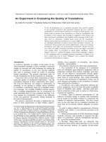
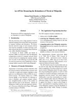
Figure 1 shows a comparison of the structures of
apo–MNK5 and copper(I)–MNK5, highlighting the
metal site structure in the latter. Both structures adopt
Interaction between HAH1 and ATP7A L. Banci et al.
866 FEBS Journal 272 (2005) 865–871 ª 2005 FEBS
the ferredoxin-like babbab fold. The RMSD between
the backbone atoms for the mean structures of the two
families of conformers, excluding the metal-binding
loop region and the poorly defined C-terminal tail is
1.1 A
˚
.
Interaction between MNK2 and HAH1
To investigate the interaction of MNK2 with HAH1,
we titrated
15
N-enriched copper(I)–MNK2 with unla-
belled apo–HAH1, and followed the process via
1
H-
15
N HSQC spectra. No variation in the chemical
shifts of the amide signals in copper(I)–MNK2 could
be observed at any stage of the titration. Instead, the
intensities of signals decreased with increasing HAH1
concentration. Concomitantly, signals corresponding
to apo–MNK2 appeared and increased in intensities
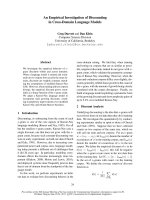
along the titration (Fig. 2). No additional signals from
a possible (transiently populated) intermediate could
be detected at any point of the titration.
The above data thus indicate that an adduct
between MNK2 and HAH1 does not form at detect-
able concentration, even if an interaction between the
two proteins does occur, resulting in copper(I) transfer.
The latter process is slow on the chemical shift time
scale, setting an upper limit for the equilibration rate
of 10
2
)10
3
s
)1
(determined by the smallest chemical
shift difference between apo–MNK2 and copper(I)–
MNK2 that can be detected, i.e. 0.1 p.p.m). The
profiles of signal intensity as a function of the
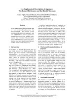
MNK2 ⁄ HAH1 molar ratio can be fitted with an equi-
librium constant for the transfer of copper(I) from
HAH1 to MNK2 between 5.0 and 10 (Fig. 3). The
relatively high spread of the data in Fig. 3 is due to
the fact that during the titration some broadening of
the signals occurs, to a different extent at different
HAH1 ⁄ MNK2 ratios. This contributes to scattering
the values of the signal integrals.
Interaction between MNK5 and HAH1
The interaction of MNK5 and HAH1 was studied by
titrating
15
N-enriched apo–MNK5 into
15
N-enriched
copper(I)–HAH1. As observed for MNK2, there is no
detectable formation of a protein ⁄ protein adduct, and
the copper(I) transfer equilibrium is slow on the chem-
ical shift time scales. Already at the first addition of
apo–MNK5 (MNK5 ⁄ HAH1 ratio 1 : 5), signals due
to copper(I)–MNK5 appeared, with an intensity signi-
ficantly higher than those of apo–MNK5. Only after
an excess of apo–MNK5 with respect to copper(I)–
HAH1 is reached, was a steady increase of the intensi-
ties of apo–MNK5 signals observed, although the sig-
nals of copper(I)–MNK5 did not increase significantly.
These data are consistent with the copper(I) transfer
process favouring the formation of copper(I)–MNK5.
The titration data can be fit to an equilibrium constant
similar to that observed in the case of HAH1. In par-
allel, the intensity of the signals of copper(I)–HAH1 in
the HSQC spectra decreased steadily along all the
titration, and apo(I)–HAH1 was formed.
Discussion
As expected, in solution MNK5 adopts the classical
babbab ferredoxin fold regardless of the presence of
the metal ion. As observed for other proteins of this
class [26,27], in copper(I)–MNK5 the copper ion is
close to the protein surface and solvent exposed.
Chemical shift variations observed between apo–
MNK5 and copper(I)–MNK5 indicate that perturba-
tions due to copper(I) binding affect mainly the Cys-
containing loop (loop 1). Indeed, the comparison of
the two structures highlights that this is the region
where structural rearrangement occurs upon metal
binding, while the remainder of the polypeptide chain
does not experience significant conformational changes
(Fig. 1). For the two copper(I)-binding cysteines, it is
difficult to appreciate the extent of conformational
rearrangement as their conformation in the two famil-
ies is not very precisely defined. Overall, the behaviour
of MNK5 upon copper(I) binding is similar to what
observed for MNK2 [21].
The behaviour observed for the interaction of
HAH1 with MNK2 and MNK5 is somewhat different
from that observed for the yeast homologues [15], and
from that observed for Bacillus subtilis CopZ and
CopA [16]. In the latter two systems an adduct is
formed in fast (with respect to the time scale of NMR
chemical shifts) equilibrium with the two separate pro-
teins. This was evident from the fact that in a mixture
of two partners in the presence of only one equivalent
Fig. 1. Comparison of the solution structures of apo–MNK5 (left)
and copper(I)–MNK5 (right). The side chains of Cys14 and Cys17
are shown as sticks; the copper(I) ion is shown as a sphere. This
figure was prepared with
MOLMOL [31].
L. Banci et al. Interaction between HAH1 and ATP7A
FEBS Journal 272 (2005) 865–871 ª 2005 FEBS 867
of copper, only a single set of signals from each pro-
tein was detected, as a result of fast averaging between
the apo- and copper(I)-loaded forms [15,16]. Forma-
tion of an adduct in solution was apparent from the
measurement of protein tumbling rates in solution
[15,16]. Instead, in the present case of the interaction
of HAH1 with MNK2 and MNK5 a slow equilibrium
is observed. The absence of additional signals, besides
those of the apo- and copper(I)-loaded proteins, indi-
cates that there is no accumulation of a protein⁄
protein adduct in solution. However, copper(I) transfer
between HAH1 and MNK2 ⁄ MNK5 is clearly
observed, indicating that an interaction does occur.
Indeed, formation of an adduct can be detected
through surface plasmon resonance measurements,
with a k
on
for formation of the adduct of the order of
10
2
)10
3
m
)1
Æs
)1
[28].
HAH1 has a distribution of electrostatic charges
at the protein surface in the region of putative
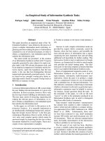
Fig. 2. HSQC spectra of copper(I)–MNK2 (blue) and copper(I)–MNK2 in the presence of apo-HAH1 at a 1 : 3 molar ratio (red), showing the
simultaneous presence of signals of copper(I)–MNK2 and apo–MNK2.
Fig. 3. Fit of the molar fraction of apo–MNK2 as a function of the
HAH1 ⁄ MNK2 molar ratio to the equilibrium Cu(I)–MNK2 + HAH1 )
*
MNK2 + Cu(I)–HAH1. The signals of residues 18, 20 and 26 have
been selected to independently evaluate the molar fraction.
Interaction between HAH1 and ATP7A L. Banci et al.
868 FEBS Journal 272 (2005) 865–871 ª 2005 FEBS
interaction with the partner that is quite similar to
that of yeast Atx1, in spite of its lower pI (6.7 vs.
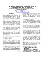
8.6). Figure 4 (upper) shows a comparison of the
electrostatic surface of HAH1 and Atx1, highlighting
the strong positive potential at the putative interac-
tion region. By contrast, MNK2 is possibly the
metal-binding domain in ATP7A most different from
either of the two domains of yeast Ccc2 with respect
to electrostatic properties. Indeed, MNK2 has a pI of
8.7 vs. 4.3–4.4 for the two domains of Ccc2. The pI
of MNK5 is instead 6.4. As can be seen from Fig. 4
(lower), there is little similarity between the electro-
static potential at the surface of MNK2, MNK5 and
Ccc2. The poor energetics of electrostatic interaction
between MNK2 ⁄ MNK5 and HAH1 is such that
the formation of a long-lived Cu(I)MNK2 ⁄ (MNK5 ⁄
HAH1) adduct is unfavourable, as indicated by the
behaviour of the NMR signals along titrations. Con-
sequently, we can observe experimentally only the
copper(I) exchange process. The thermodynamic con-
tribution to the formation of the adduct resulting
from the formation of copper(I)-bridged heterodimers
is not sufficient to stabilize the adduct. In this respect
it is worth noting that reversal of the charge of
amino acids at the Atx1 ⁄ Ccc2 interface is known to
be able to abolish their interaction altogether [29] as
does mutation of the metal-binding cysteines to
serines [28]. The data are thus consistent with a
mechanism in which HAH1 and any of the ATP7A
metal-binding domains interact via an unstable
bi-molecular intermediate (transition state), whose
concentration in solution at equilibrium is too low to
allow detection by NMR. The intermediate could
form through a copper(I) bridge, with the metal ion
coordinated by one or two cysteines of both mole-
cules. It is possible to speculate that the formation of
the bridged intermediate should logically constitute
the slow step in the copper(I) transfer reaction, while
dissociation of the intermediate immediately after
copper(I) transfer should be fast due to the poor
energetics of interaction between the two proteins
(Fig. 4). In the yeast system, attraction between resi-
dues of opposite charge at the surface of the two
partners stabilizes the intermediate, which becomes
detectable by NMR (and can be structurally charac-
terized) [15,17]. Note that key residues of Ccc2
involved in the formation of the latter adduct [17] are
indeed nonconservatively replaced in the two MNK
domains studied here.
The copper(I) transfer process has an equilibration
constant of the order of 5–10, with the soluble domains
of ATP7A being better ligands for copper(I) than
HAH1. In other words, our data are consistent with
the copper(I)-binding constant of MNK2 and MNK5
being 5–10 times that of HAH1. The same ratio is close
to one for yeast Atx1⁄ Ccc2 [5]. This result is in agree-
ment with competition experiments performed on
HAH1 and the second metal-binding domain of the
ATP7B (Wilson) protein (WND2 hereafter), which
showed that WND2 has a higher affinity for copper(I)
than HAH1 [22]. In contrast, isothermal titration calor-
imetry performed on HAH1 and various constructs of
ATP7B present a relatively complex picture in which
the number of metal-binding domains contained in
each specific construct appeared to affect significantly
copper(I) capabilities [30]. In fact, the binding constant
of a given domain could differ by a 10-fold in a two-
domain construct with respect to the entire six-domain
construct, thereby possibly making HAH1 a copper(I)
ligand as good as the ATP7B domains [30]. If the
above data are relevant also to the ATP7A protein
studied here, it should be concluded that the affinity of
each domain for copper(I) is dependent on the context
within which it is located. Long-range interactions
between different metal-binding domains should then
reduce the affinity for copper(I) of the individual
metal-binding domains with respect to the ‘intrinsic’
affinity of the isolated domain, which, as shown here, is
higher than that of the chaperone.
Fig. 4. Electrostatic potential at the surface in the putative inter-
molecular interaction region of yeast Atx1 and human HAH1
(upper), and of the various metal binding domains of yeast Ccc2
and human ATP7A (lower). Positively charged areas are blue, nega-
tively charged areas are in red. This figure was generated with
MOLMOL [31].
L. Banci et al. Interaction between HAH1 and ATP7A
FEBS Journal 272 (2005) 865–871 ª 2005 FEBS 869
It has been proposed for the ATP7B protein that
the second metal-binding domain constitutes the pre-
ferred one for the uptake of the first metal ion by the
ATPase from the chaperone, as a result of the specific-
ity of protein–protein interactions between WND2 and
HAH1 [22]. Our data suggest that a preferential (with
respect to the other metal-binding domains of ATP7A)
protein–protein interaction between MNK2 and
HAH1 is unlikely. Given that the surface charges of
MNK2 and WND2 are fairly similar, a preferential
interaction of the chaperone with the second domain
seems unlikely also in the case of ATP7B. The selectiv-
ity, if any, for the interaction of one of the six metal-
binding domains with the chaperone should thus result
from the global conformation of the entire soluble
portion of the ATPases.
Materials and methods
HAH1 and MNK2 samples were produced as described
previously [19,21]. The protocol adopted to clone, express
and purify MNK5 was essentially the same as that used for
MNK2 [21]. The main exception was that samples retaining
the poly(His) tag were used to record the spectra for NMR
frequency assignments as they showed a markedly longer
lifetime. Comparison of two-dimensional HSQC and
NOESY spectra of MNK5 with and without the poly(His)
tag shows that there is no detectable interaction between
the tag and the remainder of the protein and that the solu-
tion structure of MNK5 is not sensitive to the presence of
the tag. Recombinant protein characterization, NMR fre-
quency assignments and solution structure determination of
MNK5 in both the apo- and copper(I) forms were carried
out following the same approach used for NMK2 [21], and
showed MNK5 to be monomeric in solution in both forms.
Copper(I)–MNK5 was found from atomic absorption
measurements to bind one copper(I) ion per protein mole-
cule.
The procedure used for NMR titrations was the same as
described in a previous study from our laboratory reporting
on the interaction between yeast Atx1 and Ccc2 [15]. Pro-
tein concentrations were typically around 0.3–0.5 mm; titra-
tions were carried out up to protein ratios of 4 : 1.
Acknowledgements
We thank Fiorenza Cramaro for MNK5 frequency
assignments and initial structure calculations. Manuele
Migliardi is thanked for help in HAH1 protein prepa-
rations. This work was supported by MIUR-COFIN
2003, Ente Cassa di Risparmio di Firenze and the
European Commission (contract-no. QLG2-CT-2002-
00988).
References
1 Linder MC (1991) Biochemistry of Copper. Plenum
Press, New York.
2 O’Halloran TV & Culotta VC (2000) Metallochaper-
ones: an intracellular shuttle service for metal ions.
J Biol Chem 275, 25057–25060.
3 Harrison MD, Jones CE, Solioz M & Dameron CT
(2000) Intracellular copper routing: the role of copper
chaperones. Trends Biochem Sci 25, 29–32.
4 Puig S & Thiele DJ (2002) Molecular mechanisms of
copper uptake and distribution. Curr Opin Chem Biol 6,
171–180.
5 Huffman DL & O’Halloran TV (2000) Energetics of
copper trafficking between the Atx1 metallochaperone
and the intracellular copper transporter, Ccc2. J Biol
Chem 275, 18611–18614.
6 Rae T, Schmidt PJ, Pufahl RA, Culotta VC & O’Hallo-
ran TV (1999) Undetectable intracellular free copper:
the requirement of a copper chaperone for superoxide
dismutase. Science 284, 805–808.
7 Klomp LW, Lin SJ, Yuan D, Klausner RD, Culotta
VC & Gitlin JD (1997) Identification and functional
expression of HAH1, a novel human gene involved in
copper homeostasis. J Biol Chem 272, 9221–9226.
8 Pufahl RA, Singer CP, Peariso KL, Lin S-J, Schmidt
PJ, Fahrni CJ, Cizewski Culotta V, Penner-Hahn JE &
O’Halloran TV (1997) Metal ion chaperone function of
the soluble Cu (I) receptor Atx1. Science 278, 853–856.
9 Petris MJ, Mercer JF, Culvenor JG, Lockhart P &
Camakaris J (1996) Ligand-regulated transport of the
Menkes copper P-type ATPase efflux pump from the
Golgi apparatus to the plasma membrane: a novel
mechanism of regulated trafficking. EMBO J 15, 6084–
6095.
10 Bull PC & Cox DW (1994) Wilson disease and Menkes
disease: new handles on heavy-metal transport. Trends
Genet 10, 246–252.
11 Arnesano F, Banci L, Bertini I, Ciofi-Baffoni S, Molteni
E, Huffman DL & O’Halloran TV (2002) Metallochap-
erones and metal transporting ATPases. A comparative
analysis of sequences and structures. Genome Res 12,
255–271.
12 Goodyer ID, Jones EE, Monaco AP & Francis MJ
(1999) Characterization of the Menkes protein copper-
binding domains and their role in copper-induced pro-
tein relocalization. Hum Mol Genet 8, 1473–1478 doi:
9208958.
13 Voskoboinik I, Strausak D, Greenough M, Brooks H,
Petris M, Smith S, Mercer JF & Camakaris J (1999)
Functional analysis of the N-terminal CXXC metal-
binding motifs in the human Menkes copper-transport-
ing P-type ATPase expressed in cultured mammalian
cells. J Biol Chem 274, 22008–22012.
Interaction between HAH1 and ATP7A L. Banci et al.
870 FEBS Journal 272 (2005) 865–871 ª 2005 FEBS
14 Huster D & Lutsenko S (2003) The distinct roles of the
N-terminal copper-binding sites in regulation of cataly-
tic activity of the Wilson’s disease protein. J Biol Chem
2789, 32212–32218.
15 Arnesano F, Banci L, Bertini I, Cantini F, Ciofi-Baffoni
S, Huffman DL & O’Halloran TV (2001) Characteriza-
tion of the binding interface between the copper chaper-
one Atx1 and the first cytosolic domain of Ccc2
ATPase. J Biol Chem 276, 41365–41376.
16 Banci L, Bertini I, Ciofi-Baffoni S, Del Conte R &
Gonnelli L (2003) Understanding copper trafficking in
bacteria: interaction between the copper transport pro-
tein CopZ and the N-terminal domain of the copper
ATPase CopA from Bacillus subtilis. Biochemistry 42,
1939–1949.
17 Arnesano F, Banci L, Bertini I & Bonvin AMJJ (2004)
A docking approach to the study of copper trafficking
proteins: interaction between metallochaperones and
soluble domains of copper ATPases. Structure 12 , 669–
676.
18 Ralle M, Lutsenko S & Blackburn NJ (2003) X-Ray
absorption spectroscopy of the copper chaperone
HAH1 reveals a linear two-coordinate Cu (I) center
capable of adduct formation with exogenous thiols and
phosphines. J Biol Chem 278, 23163–23170.
19 Anastassopoulou J, Banci L, Bertini I, Cantini F,
Katsari E & Rosato A (2004) Solution structure of the
apo- and copper (I) loaded human metallo-chaperone
HAH1. Biochemistry 43, 13046–13053.
20 Arnesano F, Banci L, Bertini I, Huffman DL &
O’Halloran TV (2001) Solution structure of the Cu (I)
and Apo forms of the yeast metallochaperone, Atx1.
Biochemistry 40, 1528–1539.
21 Banci L, Bertini I, Del Conte R, D’Onofrio M &
Rosato A (2004) Solution structure and backbone
dynamics of the Cu (I) and apo-forms of the second
metal-binding domain of the Menkes protein ATP7A.
Biochemistry 43, 3396–3403.
22 Walker JM, Huster D, Ralle M, Morgan CT, Blackburn
NJ & Lutsenko S (2004) The N-terminal metal-binding
site 2 of the Wilson’s disease protein plays a key role in
the transfer of copper from Atox1. J Biol Chem 279,
15376–15384.
23 Cavanagh J, Fairbrother WJ, Palmer AG III & Skelton
N (1996) Protein NMR Spectroscopy. Principles and
Practice. Academic Press, San Diego.
24 Banci L, Bertini I, Ciofi-Baffoni S, Huffman DL &
O’Halloran TV (2001) Solution structure of the yeast
copper transporter domain Ccc2a in the apo and Cu
(I)-loaded states. J Biol Chem 276, 8415–8426.
25 Jones CE, Daly NL, Cobine PA, Craik DJ & Dameron
CT (2003) Structure and metal binding studies of the
second copper binding domain of the Menkes ATPase.
J Struct Biol 143, 209–218.
26 Rosenzweig AC (2001) Copper delivery by metallochap-
erone proteins. Acc Chem Res 34, 119–128.
27 Banci L & Rosato A (2003) Structural genomics of
proteins involved in copper homeostasis. Acc Chem Res
36, 215–221.
28 Strausak D, Howie MK, Firth SD, Schlicksupp A,
Pipkorn R, Multhaup G & Mercer JF (2003) Kinetic
analysis of the interaction of the copper chaperone
Atox1 with the metal binding sites of the Menkes
protein. J Biol Chem 278, 20821–20827.
29 Portnoy ME, Rosenzweig AC, Rae T, Huffman DL,
O’Halloran TV & Cizewski Culotta V (1999) Structure–
function analyses of the ATX1 metallochaperone. J Biol
Chem 274, 15041–15045.
30 Wernimont AK, Yatsunyk LA & Rosenzweig AC
(2004) Binding of copper (I) by the Wilson disease
protein and its copper chaperone. J Biol Chem 279,
12269–12276.
31 Koradi R, Billeter M & Wu
¨
thrich K (1996) molmol:a
program for display and analysis of macromolecular
structure. J Mol Graphics 14, 51–55.
L. Banci et al. Interaction between HAH1 and ATP7A
FEBS Journal 272 (2005) 865–871 ª 2005 FEBS 871