Báo cáo khoa học: Protein dissection enhances the amyloidogenic properties of a-lactalbumin potx
Bạn đang xem bản rút gọn của tài liệu. Xem và tải ngay bản đầy đủ của tài liệu tại đây (596.78 KB, 13 trang )
Protein dissection enhances the amyloidogenic properties
of a-lactalbumin
Patrizia Polverino de Laureto
1
, Erica Frare
1
, Francesca Battaglia
1
, Maria F. Mossuto
1
,
Vladimir N. Uversky
2,3,4
and Angelo Fontana
1
1 CRIBI Biotechnology Centre, University of Padua, Italy
2 Institute for Biological Instrumentation, Russian Academy of Sciences, Pushchino, Russia
3 Department of Biochemistry and Molecular Biology, Medical School, Indiana University, Indianapolis, IN, USA
4 Molecular Kinetics Inc., Indianapolis, IN, USA
Keywords
amyloid; a-lactalbumin; circular dichroism;
infrared spectroscopy; limited proteolysis;
molten globule; protein aggregation
Correspondence
A. Fontana, CRIBI Biotechnology Centre,
University of Padua, Viale G. Colombo 3,
35121 Padua, Italy
Fax: +39 49 827 6159
Tel: +39 49 827 6156
E-mail:
(Received 21 December 2004, revised 23
February 2005, accepted 2 March 2005)
doi:10.1111/j.1742-4658.2005.04638.x
a-lactalbumin (LA) in its molten globule (MG) state at low pH forms
amyloid fibrils. Here, we have studied the aggregation propensities of LA
derivatives characterized by a single peptide bond fission (1–40 ⁄ 41–123,
named Th1-LA) or a deletion of a chain segment of 12 amino acid resi-
dues located at the level of the b-subdomain of the native protein (1–
40 ⁄ 53–123, named desb-LA). We have also compared the early stages of
the aggregation process of these LA derivatives with those of intact LA.
Th1-LA and desb-LA aggregate at pH 2.0 much faster than the intact
protein and form long and well-ordered fibrils. Furthermore, in contrast
to intact LA, the LA derivatives form regular fibrils also at neutral pH,
even if at much reduced rate. In acidic solution, Th1-LA and desb-LA
adopt a MG state which appears to be similar to that of intact LA, as
given by spectroscopic criteria. At neutral pH, both Th1-LA and desb-LA
are able to bind the hydrophobic dye 1-anilinonaphtalene-8-sulfonate, thus
indicating the presence of exposed hydrophobic patches. It is concluded
that nicked Th1-LA and gapped desb-LA are more relaxed and expanded
than intact LA and, consequently, that they are more suitable protein spe-
cies to allow the large conformational transitions required for the poly-
peptide chain to form the amyloid cross-b structure. As a matter of fact,
the MG of LA attains an even more flexible conformational state dur-
ing the early phases of the aggregation process at acidic pH, as deduced
from the enhancement of its susceptibility to proteolysis by pepsin. Our
data indicate that deletion of the b-subdomain in LA does not alter the
ability of the protein to assemble into well-ordered fibrils, implying that
this chain region is not essential for the amyloid formation. It is proposed
that a proteolytic hydrolysis of a protein molecule at the cellular level can
trigger an easier formation of amyloid precipitates and therefore that lim-
ited proteolysis of proteins can be a causative mechanism of protein
aggregation and fibrillogenesis. Indeed, a vast majority of protein deposits
in amyloid diseases are given by protein fragments derived from larger
protein precursors.
Abbreviations
[h], mean residue ellipticity; ANS, 1-anilinonaphtalene-8-sulfonate; apo-LA, calcium-depleted form of LA; desb-LA, protein species with
excision of the 41–52 chain segment; E ⁄ S, enzyme to substrate ratio; LA, a-lactalbumin; MG, molten globule; RP, reverse-phase; TFA,
trifluoroacetic acid; Th1-LA, disulfide-crosslinked, nicked protein species of LA with the peptide bond 40–41 cleaved; ThT, thioflavin T.
2176 FEBS Journal 272 (2005) 2176–2188 ª 2005 FEBS
The propensity to form amyloid fibrils appears to be a
general property of polypeptide chains, as under speci-
fic experimental conditions a variety of proteins can be
induced to aggregate into highly ordered protein aggre-
gates [1–10]. Considering that all protein fibrils, inde-
pendently of the native structure of the given
amyloidogenic protein, adopt the common cross-b
structure motif, a large conformational rearrangement
has to occur prior to fibrillation [10]. Such changes
cannot develop within a tightly packed native protein
and thus destabilization of the protein structure and
formation of a partly folded state is required. In fact,
experimental conditions that favor the partial unfold-
ing of the protein are known to trigger the fibrillation
process [5–7,10]. It has been proposed that specific
interactions between amino acid residues or chain
regions of a polypeptide with a high propensity to
acquire a b-sheet secondary structure lead to the kernel
that determines aggregation of other molecules into
the final, well-ordered fibrils [1–3]. However, as even
small unfolded peptides are able to form amyloid
fibrils, protein conformational parameters are likely to
be insufficient to explain the molecular mechanism(s)
of fibrillation [11,12]. In a number of cases, a proteo-
lytic event of a precursor protein appears to be
required for amyloidogenesis [13–16].
Lysozyme and a-lactalbumin (LA) belong to the
same protein superfamily, being homologous proteins
displaying similarity in their amino acid sequence and
overall 3D structure ([17] and references cited therein).
Human lysozyme has been shown to form amyloid
fibrils in individuals having point mutations in the lyso-
zyme gene [18]. Recently, in order to understand better
the mechanism of lysozyme fibrillation, the aggregation
processes of lysozymes from different sources have been
investigated in detail (see [19] for references]. Overall, it
seems appropriate to study the aggregation phenomena
of proteins belonging to the same protein superfamily.
Probably, by studying similar proteins that form fibrils
in vitro it will be possible to highlight some peculiar
structural features that dictate the overall process of
protein fibrillogenesis. Indeed, bovine LA is also able
to form amyloid fibrils if the protein is induced to
adopt the molten globule (MG) state at low pH [20].
However, fibril formation at pH 2.0 is much more
rapid if the protein, upon partial reduction oif its four
disulfide bridges, adopts a more open conformation
than that of the classical MG in acid solution [21,22].
This finding correlates with the observation that the
conformational features of apomyoglobin leading to
amyloid precipitates are more expanded than those of a
well defined and compact partly folded state of the pro-
tein [23]. Similarly, the aggregation process of a SH3
domain at low pH implies significant structural rear-
rangements, requiring a more flexible protein species
that subsequently forms well-ordered fibrillar structures
[24]. Also a partly folded state has been shown to be
involved in the fibrillogenesis of a-synuclein, a ‘natively
unfolded’ protein [25–27] known to be involved in the
pathogenesis of several neurodegenerative disorders
[28]. It has been found that a-synuclein undergoes
fibrillation under experimental conditions that stabilize
a partly folded state, named premolten globule [29].
These various observations can be interpreted as indi-
cating that partly folded, but substantially open and
dynamic states of proteins are those required for trig-
gering the process of fibrillogenesis [10,20].
In this work, we analyse the effects of the dissection
of the LA molecule on its conformational features and
aggregation processes. Two protein species are herewith
examinated, those given by the N-terminal fragment
1–40 covalently linked by the disulfide bridges of the
protein to the C-terminal fragment 41–123 (Th1-LA) or
fragment 53–123 (desb-LA) (Fig. 1). These species have
been prepared by limited proteolysis of LA by thermo-
lysin (E.C. 3.4.24.27) in 50% trifluoroethanol at neutral
pH or by pepsin (E.C. 3.4.23.1) in acid solution
[30–33]. LA is not associated to any specific disease,
but several observations have suggested that LA can
evoke a variety of physiological effects [34]. Besides its
role in modulating the activity of the lactose synthase,
new intriguing properties have been evidenced, such as
the apoptotic activity in tumor cells of a partly folded
variant of LA bound to oleic acid [35,36], the ability of
the protein to bind histones [37,38] and the bactericidal
activity of some of its chymotryptic peptides [39]. Here,
we show that nicking of the 123-residue chain of LA at
a single peptide bond (Th1-LA) or removing a 12-resi-
due segment (desb-LA) leads to protein species that
easily form amyloid fibrils at pH 2.0. It is concluded
that the inherent flexibility of these LA derivatives
allows the large conformational changes required to
form the cross-b-structure of the amyloid fibrils. Of
interest, it is also shown that the initial stages of fibril-
lation of intact LA at low pH involve protein inter-
mediate(s) characterized by enhanced chain flexibility.
This study emphasizes that the precursor structures of
amyloid fibrils require a more unfolded and ⁄ or flexible
state than that of the MG [21,22].
Results
Molecular features of Th1-LA and desb-LA
Far-UV CD measurements indicate that, in acidic
solution at pH 2.0 or at neutral pH in the presence of
P. Polverino de Laureto et al. Proteolysis of proteins enhances fibrillogenesis
FEBS Journal 272 (2005) 2176–2188 ª 2005 FEBS 2177
EDTA, the LA variants Th1-LA and desb-LA adopt
a conformation similar to the MG state displayed by
intact LA at low pH, retaining a native-like a-helical
content (Fig. 2A,B). Furthermore, Th1-LA and desb-
LA bind the fluorescent dye 1-anilinonaphtalene-8-sulf-
onic acid (ANS), which is considered diagnostic of a
protein MG state [40]. While at pH 2.0 the three LA
species bind ANS in a similar way, at pH 7.4 the
nicked or gapped LA species bind ANS more signifi-
cantly than the intact protein, indicating that a larger
hydrophobic surface is exposed to the solvent in the
LA derivatives (Fig. 2C,D) (see also [32]).
The molecular features of LA, Th1-LA and desb-LA
at pH 2.0 have been further analysed by limited proteo-
lysis, urea-induced unfolding and hydrogen ⁄ deuterium
(H ⁄ D) exchange measurements. Limited proteolysis has
been conducted at pH 2.0 using pepsin as protease and
LA and Th1-LA as substrates [41]. Desb-LA was not
analysed, because this species is a product of proteo-
lysis of LA at pH 2.0 [30–32]. The comparative suscep-
tibility to proteolytic attack by pepsin of LA and
Th1-LA is shown in Fig. 3A. The extent of proteolysis
was calculated from the amount of residual protein
determined by reverse phase (RP)-HPLC analysis of
aliquots of the reaction mixtures at various times of
proteolysis. After 10 min’ incubation of the proteins
with pepsin at pH 2.0, Th1-LA has been 80%
cleaved, while LA only 50%. After 20 min’ reaction,
the proteolysis is complete for Th1-LA, whereas some
intact LA is still present in the proteolysis mixture even
after 50 min’ reaction (Fig. 3A).
To evaluate further the molecular differences between
LA and its derivatives, the effect of urea on the protein
tertiary structure was analysed by near-UV CD spectros-
copy (Fig. 3B). Earlier it was shown [32] that these LA
derivatives retain some tertiary interactions at pH 2.0, as
given by a near-UV CD spectrum characterized by a
band centered at about 290 nm due to the contribution
of tryptophan residues [42]. As shown in Fig. 3B, the
increase in urea concentration at pH 2.0 was accompan-
ied by a gradual reduction of the CD signal at 291 nm
for the three LA species, implying that the urea-medi-
ated denaturation is not a cooperative process. Even if
the overall processes are quite similar for the three pro-
tein species, it seems that intact LA retains some residual
structure in the presence of 4 m urea, whereas desb-LA
appears to be completely unfolded under the same
conditions. In the case of Th1-LA, a complete disappear-
ance of the CD signal at 291 nm occurs in 6 m urea.
The flexibility of the polypeptide chain of LA and
its gapped and nicked species was also analysed by
H ⁄ D measurements, i.e. monitoring by ESI-MS the
change in the protein mass that results from the
replacement of labile protons by deuterium. The fea-
tures of the H ⁄ D exchange of the low-pH MG of LA
have been already examined [43–46], showing that only
the amides in the a-domain are most protected from
H ⁄ D exchange [43]. The protein species herewith inves-
tigated are characterized by a nicking or a removal of
the b-subdomain (Fig. 1) and therefore a significant
N
C
Ala40-Ile41
Leu52-Phe53
1 123
H1
5-11
h1b
23-34
S1 S2 S3
H2
h2
86-98 105-110
h3c
H3 H4
Gln-Ala-Ile-Val-Gln-Asn-Asn-Asp-Ser-Thr-Glu-Tyr-Gly-Leu-Phe-Gln
40 45 50
1
40
53
123
1
40 123
41
Th1-LA
desβ-LA
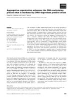
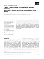
Fig. 1. (Top) Schematic representation of the 3D structure of LA.
The diagram was drawn using the PDB file 1HFZ using the program
WEBLAB VIEWER PRO 4.0 (Molecular Simulations Inc., San Diego, CA).
The chain segment 41–52 encompassing the antiparallel b-sheet is
colored in red. The four disulfide bonds are represented by yellow
sticks and the calcium atom by a solid sphere in green. (Middle)
Scheme of the secondary structure of the 123-residue chain of LA
[58]. The four a-helices (H1–H4) along the protein chain are indica-
ted by major boxes and below them the corresponding chain seg-
ments are given. The three b-strands (S1, 41–44; S2, 47–50; S3,
55–56) and the 3
10
helices (h1b, 18–20; h2, 77–80, h3c, 115–118)
are indicated by small boxes. The amino acid sequence of the chain
region 39–54 of LA is explicitly shown and the sites of proteolysis
by pepsin and thermolysin are indicated by arrows. (Bottom) Sche-
matic structure of the LA derivatives Th1-LA and desb-LA. These
protein species are given by the N-terminal fragment 1–40 cova-
lently linked by the four disulfide bridges of the protein to the C-ter-
minal fragment 41–123 (Th1-LA) or fragment 53–123 (desb-LA).
The connectivities of the four disulfide bridges (thin lines) of LA are
6–120, 28–111, 61–77 and 73–91.
Proteolysis of proteins enhances fibrillogenesis P. Polverino de Laureto et al.
2178 FEBS Journal 272 (2005) 2176–2188 ª 2005 FEBS
difference in their H ⁄ D behavior is not expected. In
fact, Fig. 3C shows that the time-courses of H ⁄ D
exchange for the three LA species are only slightly dif-
ferent. However, the lower and the higher extent of
exchanged protons by LA and desb-LA, respectively,
are consistent with a more compact and a more flex-
ible structure in LA and desb-LA, respectively.
Aggregation properties of LA derivatives
To induce amyloid formation, Th1-LA and desb-LA
were dissolved (5 mgÆmL
)1
)in10mm HCl pH 2.0, or
in 50 mm Tris ⁄ HCl buffer pH 7.4, containing 0.1 m
NaCl, and incubated at 35 °C for up to 20 days. The
aggregation process was followed by thioflavin T (ThT)
binding assay [47,48] and electron microscopy (EM).
CD and FTIR measurements have been used to mon-
itor the conformational changes during aggregation.
The nicked or gapped LA derivatives aggregate very
fast (Fig. 4) and form long and well-defined fibrils
(Fig. 5). The sigmoid curve of the ThT fluorescence
emission at 485 nm vs. time of aggregation (Fig. 4)
observed with intact LA is characterized by a lag time
and is consistent with a nucleation-dependent elonga-
tion model of fibrillogenesis [20]. On the other hand,
the lag time is almost not observed with both Th1-LA
and desb-LA incubated at pH 2.0 (Fig. 4). Further-
more, with the LA derivatives a larger increase in ThT
fluorescence is observed upon prolonged incubation at
low pH and the intensity of fluorescence reaches a plat-
eau after about 70–80 h. Assuming that the ThT fluor-
escence enhancement is proportional to the population
of well-ordered protein aggregates [47,48], we can con-
clude that the amount of fibrils formed by Th1-LA and
desb-LA is decidedly larger than that formed by intact
LA under similar experimental conditions. EM reveals
that the fibrils formed by Th1-LA and desb-LA after
70 h incubation at pH 2.0 are typical amyloid, with a
filamentous aspect, unbranched and with a diameter of
10 nm (Fig. 5), quite similar to those formed by
intact LA under the same experimental conditions [20].
Aggregation experiments of the LA derivatives have
been conducted also at pH 7.4. While intact LA does
not form fibrils at neutral pH [20], both Th1-LA and
desb-LA after 230 h of incubation do form ordered
aggregates. In fact, EM reveals the presence of spher-
ical aggregates with dimensions of 4–8 nm (Fig. 5,
right panels) and some of them show the typical mor-
phology of protofibrils [49]. In the case of Th1-LA,
the initial aggregates are rare, if compared to those
250240230220210200190
[
θ
01 x ]
3-
ged(
.
mc
2
.
lomd
1-
)
-15
-10
-5
0
5
pH 2.0
pH 7.4
fibrils
Wavelength (nm)
250240230220210200190
-15
-10
-5
0
5
pH 2.0
pH 7.4
fibrils
400 450 500 550 600 650
ecnecseroulF e
vitaleR
0
200
400
600
800
1000
LA
Th1-LA
desβ-LA
pH 2.0
400 450 500 550 600 650
0
10
20
30
40
50
LA
Th1-LA
desβ-LA
pH 7.4
Th1-LA
des
β
-LA
AB
CD
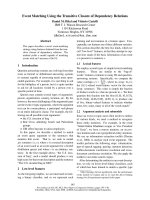
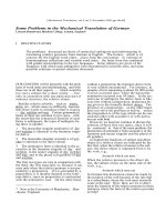
Fig. 2. Conformational characterization of
Th1-LA and desb-LA by CD (A, B) and ANS
binding (C, D). Far-UV CD spectra of Th1-LA
and desb-LA were recorded in 10 m
M
Tris ⁄ HCl ⁄ 0.1 M NaCl buffer pH 7.4, contain-
ing 1 m
M EDTA or in 0.01 M HCl ⁄ 0.1 M NaCl
pH 2.0, at a protein concentration of 0.05–
0.1 mgÆmL
)1
. The far-UV CD spectra of
fibrils of Th1-LA and desb-LA refer to those
of samples obtained by aggregation of the
protein at 35 °C pH 2.0, for 48 h and 65 h,
respectively. Fluorescence emission spectra
(C, D) of 20 l
M ANS in the presence of
10 l
M of Th1-LA or desb-LA. The spectra
are recorded at 20–22 °C from 390 to
650 nm after excitation at 370 nm in 10 m
M
HCl pH 2.0, or in 10 mM Tris ⁄ HCl pH 7.4.
P. Polverino de Laureto et al. Proteolysis of proteins enhances fibrillogenesis
FEBS Journal 272 (2005) 2176–2188 ª 2005 FEBS 2179
observed with desb-LA. Moreover, after a prolonged
time of incubation, fibrils with a more defined mor-
phology develop in the case of desb-LA, but more
slowly in the case of Th1-LA.
Analysis of the protein secondary structure has been
carried out by far-UV CD measurements [50] on aliqu-
ots taken at intervals (40–70 h) from a solution of
Th1-LA and desb-LA incubated at 35 °C pH 2.0. An
extensive conformational rearrangement takes place
during the aggregation process, as given by the disap-
pearance of the a-helix bands at 208 and at 220 nm
and by the development of the band near 217 nm, typ-
ical of b-secondary structure (Fig. 2A,B). On the other
hand, a sample of intact LA incubated under similar
solvent conditions does not develop significant changes
of CD spectra (data not shown), even after prolonged
time of incubation and thus even if aggregated
(Fig. 4). This could be explained by considering that
the helical secondary structure, even if present in mod-
erate percentages in respect to other types of secondary
structures (random coil, b-sheet), shows a CD signal
that prevails on the others [50].
To verify if under the acid conditions used to induce
fibril formation the protein samples are chemically
modified or hydrolyzed, the fibrillar material produced
after 10 days of incubation has been analysed by MS,
following essentially procedures previously described
Time (min)
0 30 60 90 120 150
xe D/Hch )%( egna
0
20
40
60
80
100
Urea concentration (M)
012345678
]θ[
[/
]θ
0
mn192
0.0
0.2
0.4
0.6
0.8
1.0
Time (min)
0 102030405060
idnUg ietorP detse)%( n
0
20
40
60
80
100
A
B
C
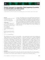
Fig. 3. Molecular features at pH 2.0 of LA (d), Th1-LA (s) and
desb-LA (.) probed by limited proteolysis (A), urea-induced unfold-
ing (B) and H ⁄ D exchange rates (C). (A) Susceptibility to proteolysis
by pepsin at pH 2.0 of LA and Th1-LA. The percent of undigested
protein was calculated from the area of the protein peaks in the
RP-HPLC chromatograms of protein samples analysed at different
time intervals. (B) Urea-induced unfolding of the tertiary structure
of LA, Th1-LA and desb-LA monitored by CD measurements at
291 nm at 20–22 °C pH 2.0. (C) H ⁄ D exchange profiles of LA,
Th1-LA and desb-LA at pH 2.0 as monitored by MS.
Time of Incubation (h)
0 50 100 150 200 250 300
aleRT evithTF loure ecsn 4 ta ec85n
m
0
20
40
60
80
100
desβ-LA
Th1-LA
LA
Fig. 4. Time course analysis of aggregation at 35 °C pH 2.0, of LA,
Th1-LA and desb-LA monitored by thioflavin T (ThT) binding. Aliqu-
ots (7 lL) were taken from the protein solution after the indicated
time and added to a 25 l
M solution (500 lL) of ThT in 25 mM phos-
phate buffer pH 6.0. The excitation wavelength was at 440 nm and
the emission was measured at 485 nm. The protein concentration
of each protein sample was 0.4 m
M.
Proteolysis of proteins enhances fibrillogenesis P. Polverino de Laureto et al.
2180 FEBS Journal 272 (2005) 2176–2188 ª 2005 FEBS
[19]. In the case of LA, some protein degradation
( 10%) has been actually observed (data not shown).
Instead, the desb-LA and Th1-LA fibrils are composed
by intact protein molecules. This could be due to the
more rapid aggregation process of the LA derivatives
in respect to that of the intact protein, likely rendering
the aggregated desb-LA and Th1-LA somewhat pro-
tected from degradation.
Analysis of fibrillogenesis of intact, nicked and
gapped LA by FTIR
To evaluate the conformational rearrangements of
LA and its derivatives during the aggregation process,
FTIR measurements [51–53] were conducted on mono-
meric and aggregated proteins. Aggregation was moni-
tored by analysing the second derivative of the FTIR
spectra of protein samples incubated for varying length
of time at 35 °C pH 2.0. The second derivative of the
FTIR spectrum of LA at pH 2.0 before aggregation
(Fig. 6) shows three major bands, one centered at
1648 cm
)1
characteristic of a-helix and ⁄ or random
structure and two centered at 1632 and 1675 cm
)1
related to the antiparallel b-structure [51–53]. These
spectral characteristics are in agreement with those
reported for intact LA exposed to low pH [20]. After
6–8 h incubation, the main band at 1648 cm
)1
shifts to
a lower wavenumber (1643 cm
)1
), indicating a preval-
ence of random coil and therefore a further structural
unfolding of the protein. After 48 h, the bands due to
the b-sheet structure (1632 and 1675 cm
)1
) are evident
and, upon prolonged incubation (300 h), a band cen-
tered at 1616 cm
)1
develops, together with a shift of
the band at 1675 cm
)1
to higher wavelengths. These
last bands have been related to fibril formation, as the
association of b-sheets cause a splitting of the antipar-
allel b-sheet bands [20].
Figure 6 (middle panel) shows the evolution of the
FTIR spectrum of Th1-LA during aggregation at
pH 2.0. Initially, the spectrum of Th1-LA is quite
similar to that of intact LA under the same solvent
conditions. A structural rearrangment of Th1-LA
appears to take place before initiation of the aggrega-
tion process (Fig. 6, 1h). In fact, the FTIR spectrum
recorded after 1 h shows a main band at 1645 cm
)1
,
which is indicative of an enhanced content of dis-
ordered structure. The spectrum remains unmodified
up to 8–10 h of incubation. After 24 h of Th1-LA
incubation, the band at 1643 cm
)1
is no more present
and a new band at 1616 cm
)1
(aggregation band)
appears. Upon prolonged incubation, there is a preval-
ence of the bands characteristic of protein fibrils (1616
200 nm
200 nm
200 nm
200 nm
200 nm
200 nm
des -LA, pH 7.4, 235 h
Th1-LA, pH 7.4, 235 h
desbb b-LA, pH 2.0, 70 h
Th1-LA, pH 2.0, 70 h
des -LA, pH 7.4, 720 h
Th1-LA, pH 7.4, 800 h
200 nm
200 nm
200 nm
200 nm
200 nm
200 nm
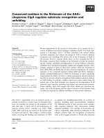
Fig. 5. Electron micrographs of Th1-LA and desb-LA fibrils obtained after 70 h incubation of the LA variants at 35 °C pH 2.0. The morphology
of the initial aggregates produced after 235 h of incubation of the protein samples at 35 °C pH 7.4, and of those produced after a much pro-
longed incubation time are also reported.
P. Polverino de Laureto et al. Proteolysis of proteins enhances fibrillogenesis
FEBS Journal 272 (2005) 2176–2188 ª 2005 FEBS 2181
and 1680 cm
)1
), whereas those of the regular structure
reduce their intensity. Overall, the FTIR data indicate
that the aggregation processes of LA and Th1-LA are
similar, but that Th1-LA aggregates significantly fas-
ter. The aggregation process of desb-LA, followed by
FTIR analysis, shows a similar behavior to that dis-
played by Th1-LA both in terms of rate of appearance
and type of FTIR bands (Fig. 6, right panel).
Early stages of LA aggregation followed
by limited proteolysis
In previous studies we have shown that proteolytic
probes can be used to analyse structure and dynamics
of proteins in their partly folded or MG states [41]
and during their aggregation process [24]. Here, we
have applied this technique for analysing the early sta-
ges in the aggregation process of intact LA. To this
end, aliquots of the protein sample of LA incubated at
35 °C pH 2.0, were withdrawn from the reaction mix-
ture at various times (from 0 to 200 h) and treated
with pepsin in order to test the susceptibility to proteo-
lysis of LA during the aggregation process. Fig. 7
shows the reverse phase–HPLC analyses of the proteo-
lysis reactions conducted on LA after 0, 8, 24 and 72 h
incubation. The pattern of LA proteolysis with pepsin
(panel A) is similar to that reported previously [30–32],
with the major products being the fragment species
1–40 ⁄ 53–123 (desb-LA), 1–40 ⁄ 104–123 and 53–103.
The protein sample left to aggregate for 8 h at pH 2.0
appears to be more sensitive to the protease action,
because more fragments and less intact protein are
found in the mixture (Fig. 7B). The analysis of the
proteolysis of a sample of LA incubated for a pro-
longed time (panels C and D) reveals that the protein
becomes more and more resistant to proteolysis. In
Fig. 7 (bottom), the percent hydrolysis of LA by pep-
sin as a function of aggregation time is reported. It is
seen that LA at the initial stages of the aggregation
process is more easily digested by proteolysis and
thus is more unfolded and ⁄ or flexible, a feature that
appears to render the polypeptide chain more prone to
fibril formation [24].
Discussion
Here, we have studied the aggregation processes of LA
species with a nick (Th1-LA) or a chain deletion
(desb-LA) at the level of the b-subdomain of the 123-
residue chain of the protein (Fig. 1). We have also
characterized the early stages of protein aggregation in
order to get insights into the molecular mechanisms of
fibrillogenesis. At pH 2.0, the overall features of the
partly folded or MG states formed by Th1-LA and
desb-LA appear to be quite similar to that formed by
intact LA at low pH under equilibrium conditions, as
judged from CD measurements (see also [32]). How-
ever, there are some differences in stability and ⁄ or
Absorbance
h 0
h 6
h
8
4
Wa mc( rebmun
e
v
1
-
)
00610261
0
461066108610071
h 003
57
6
1
8
4
61
2361
61
6
1
3
4
61
08
61
5
7
6
1
5
7
61
8
4
61
236
1
Absorbance
h
0
h
1
h
42
a
W
e
vnu bm
er (
cm
1-
)
006102610461066108610071
h 69
2361
9
4
6
1
57
6
1
54
6
1
94
6
1
2361
6161
0861
5
7
61
57
61
sed
β
-AL
Absorbance
h
0
h1
h 42
Wa
v
emunbre( cm
1-
)
0
0
610261
0
461
0
661
0
86
1
00
7
1
h
69
5
761
8461
236
1
6
161
5461
0861
5
7
6
1
5
7
61
8461
2361
Th1-LA
tc
at
nI
Fig. 6. Aggregation process of LA, Th1-LA and desb-LA monitored by FTIR. The panels show the time-evolution of the FTIR spectra of the
amide I region (continuous line) of LA (left panel), Th1-LA (middle panel) and desb-LA (right panel) during fibril formation at 35 °C pH 2.0.
The second derivative spectra (dashed lines) are also shown.
Proteolysis of proteins enhances fibrillogenesis P. Polverino de Laureto et al.
2182 FEBS Journal 272 (2005) 2176–2188 ª 2005 FEBS
flexibility between the three protein species, as inferred
from proteolysis experiments, urea-mediated denatura-
tion and H ⁄ D exchange measurements (Fig. 3). The
major conclusion of this study is that the conforma-
tional features of Th1-LA and desb-LA, which are
more relaxed and expanded in comparison to intact
LA at low pH, are more suitable for triggering the
protein fibrillation process. Importantly, the deletion
of the b-subdomain in LA does not alter the ability of
the protein to assemble into well-ordered fibrils. In this
context, it is interesting to recall that in previous
studies it has been proposed that the b-domain is of
particular significance in triggering the fibril formation
in the case of lysozyme ([54] and references cited
therein), a protein belonging to the same structural
superfamily of LA. Instead, here it is shown that a LA
derivative such as desb-LA, lacking the three b-strands
of the protein (Fig. 1), is able to aggregate even more
readily than the intact protein, implying that the
b-sheet region of LA is not required for fibrillogenesis.
It may be proposed that the exposure of the hydro-
phobic interior of the protein at low pH is sufficient
for promoting the aggregation phenomenon, leading
ultimately to well-ordered fibrils.
An important aspect of this study is the analysis of
the molecular features of LA and its derivatives at the
initial stages of protein aggregation at pH 2.0. During
the lag time, intact LA undergoes significant conform-
ational changes, as evidenced from FTIR (Fig. 6) and
limited proteolysis experiments (Fig. 7), leading to an
increase in the amount of disordered structure in LA
at the early stages of fibrillation (after 6–10 h incuba-
tion). In fact, less intact LA remains in the proteolysis
mixture with pepsin (Fig. 7), indicating that the pro-
teolytic degradation increases after incubation of LA
at pH 2.0 for 6–10 h. We may infer that LA, at the
initial stages of aggregation, exists in a more expanded
and flexible conformational state and, for this reason,
is more sensitive to proteolytic attack [41]. Import-
antly, also Th1-LA and desb-LA appear to initially
reach a more unfolded ⁄ flexible state (from FTIR data,
see Fig. 6), but faster and in about 1 h only. This is
likely due to the fact that Th1-LA and desb-LA are
more open and flexible protein species than intact LA.
By analogy, similar arguments can be used also to
explain why, at variance from the LA derivatives, the
intact protein does not aggregate at neutral pH [20].
Clearly, the intact protein is native and rigid at neutral
pH and does not populate the partly folded state
required for fibrillation.
An interesting question here is the apparent discrep-
ancy between FTIR data and far-UV CD measure-
ments. FTIR data provide evidence of an initial phase
in which the three proteins herewith studied develop
an increase in the disordered structure and, at longer
incubation times, the band characteristic of the b-sheet
structure of the amyloid is clearly observed (Fig. 6).
The FTIR data are in agreement with the fact that
aggregated Th1-LA and desb-LA show a far-UV CD
spectrum which is typical of the b-sheet secondary
structure (see Fig. 2A,B). On the other hand, far-UV
leRative sbA ocnabre mn 622 ta
Retention Time (min)
0 5 10 15 20 25 30
0 h 8 h
24 h
41-52
1-40/104-123
LA
1-40/53-123
1-40/53-123
LA
1-40/104-123
41-52
1-40/53-123
LA
1-40/104-123
41-52
53-103
53-103
0 5 10 15 20 25 30
72 h
LA
Time of Incubation (h)
0 1020304050607080
Per egavaelC tneC
0
20
40
60
80
100
AB
CD
Fig. 7. Early stages of aggregation of intact LA at pH 2.0 as monit-
ored by limited proteolysis. LA was allowed to aggregate at 35 °C
pH 2.0. Aliquots (50 lL) were taken at intervals from the LA solu-
tion and mixed with a pepsin solution for 45 min at 20–22 °C. (Top)
A sample of the proteolysis mixture was analysed by RP-HPLC
using a Vydac C18 column (150 · 4.6 mm), eluted with a linear gra-
dient of acetonitrile ⁄ 0.1% TFA from 5% to 34% in 4 min and from
34% to 50% in 18 min (Bottom) Time-course of the susceptibility
of LA to proteolysis by pepsin during aggregation at 35 °C pH 2.0.
The extent of cleavage was calculated by integration of the area of
the peaks in the RP-HPLC chromatograms of protein samples ana-
lysed at different time intervals.
P. Polverino de Laureto et al. Proteolysis of proteins enhances fibrillogenesis
FEBS Journal 272 (2005) 2176–2188 ª 2005 FEBS 2183
CD spectra measured for intact LA during the entire
aggregation process show minor changes in their shape
and characteristics mostly of a-helical structure (data
not shown). This can be explained by considering that
it is likely that residual a-helical structure is still pre-
sent in the aggregated mixture of intact LA and that
the intensity of the CD signal of the helical structure is
much higher than that of other secondary structures,
including b-sheet [50].
The key result of this study is that limited proteo-
lysis of LA leads to protein species that are much
more prone to fibrillogenesis than the intact protein.
Therefore, it is shown here, with the model protein
LA, that amyloid fibril formation can require proteoly-
sis. This is in line with the fact that a large proportion
of protein deposits associated with amyloid diseases is
made by protein fragments derived from proteolysis of
larger protein precursors [2,11,12]. First of all, we may
mention here that the prototypic fibril-forming frag-
ment Ab in Alzheimer’s diseases is derived from the
amyloid precursor protein by a combination of pro-
teolytic cleavages given by b- and c-secretase [55].
Caspase-cleavage of the cytoskeletal tau protein is an
early event in Alzheimer’s diseases tangle pathology
[16]. Human gelsolin is expressed as an 81-kDa intra-
cellular protein and its limited proteolysis at the level
of peptide bonds 172–173 and 243–244 leads to a
71-residue fragment that is found in protein deposits
in individuals with familial amyloidosis [13]. The
34-residue ABri peptide is derived from a putative
transmembrane precursor and is found in plaques of
familial British dementia [56]. These and other obser-
vations [2,11,12], in line with the results of this study,
provide an evidence that proteolysis can be a critical
prefibrillogenic event.
Protein fragments, originating by proteolysis of pro-
tein precursors, are particularly vulnerable to aggrega-
tion, because they can usually adopt, at most, partly
folded states and cannot establish the long-range inter-
actions that stabilize the native intact protein. In par-
ticular, protein fragments may contain hydrophobic
clusters of residues that can trigger protein aggrega-
tion. In the case of the LA derivatives Th1-LA and
desb-LA, it may be well that the nicking of the 123-
residue chain polypeptide chain causes an untighting
of the structural domains of LA (see Fig. 1), determin-
ing exposure of hydrophobic patches. Furthermore,
the fact that the LA derivatives aggregate much more
easily at low pH can be explained by considering that
a minimization in acid solution of the negative charges
of the carboxylates of the Asp and Glu residues in the
calcium-binding region of LA can produce a marked
decrease in the charge-to-charge repulsions and a
concomitant enhancement of the protein association
process by favoring intermolecular hydrophobic inter-
actions.
In recent years, numerous studies have been conduc-
ted on the low pH MG state of LA, nowadays consid-
ered a prototype MG in protein folding studies [21,22].
A consensus view is that the MG of LA retains signifi-
cant native-like structure in the a-domain, while the
b-domain (approximately region 50–90) is disordered.
From this and previous [32] study, it seems that the
MG state at pH 2.0 retains substantial native-like
structure that does not allow the protein to form the
amyloid precipitate. On the other hand, the low pH
MGs adopted by Th1-LA and desb-LA are similar to
the MG of intact LA, but they are more open and
flexible and thus more prone to aggregation. There-
fore, a limited proteolysis phenomenon of a protein
can lead to an enhanced population of a rather unfol-
ded and ⁄ or flexible protein species that triggers the
fibrillation process.
Conclusions
Substantial data are available to support a model for
the amyloid formation in which intermolecular inter-
actions between hydrophobic patches in partly folded
or MG states of proteins are responsible for protein
aggregation [6–10]. The intermediates are more prone
to aggregate than the unfolded state because they
retain clusters of hydrophobic side chains, which have
a strong propensity for aggregation [6]. However, a
conformation that is more expanded than typical MG
appears to be required before protein assembly into an
ordered amyloid-like structure [10,20]. Our observa-
tions are in agreement with a general view that a more
flexible conformation or a moderately unfolded struc-
ture could be the key species triggering fibril forma-
tion. In particular, the results of this study highlight
the possible role of limited proteolysis as a causative
event of fibrillogenesis. It seems possible to propose
that a proteolytic attack of a protein at the cellular
level can shift the equilibrium between the different
protein conformational states towards a species that is
more prone to aggregate.
Experimental procedures
Materials
Bovine a-lactalbumin, porcine pepsin, thermolysin and ThT
were from Sigma (St. Louis, MO, USA). All other chemi-
cals were of analytical reagent grade and were obtained
from Sigma or Fluka (Basel, Switzerland).
Proteolysis of proteins enhances fibrillogenesis P. Polverino de Laureto et al.
2184 FEBS Journal 272 (2005) 2176–2188 ª 2005 FEBS
Isolation of nicked and gapped LA
The LA derivatives were obtained by limited proteolysis of
the protein, as described previously [30–33]. Proteolysis of
bovine LA with thermolysin was performed in a 1 : 1 (v ⁄ v)
mixture of 50 mm Tris ⁄ HCl pH 7.0, containing 5 mm CaCl
2
and trifluoroethanol using an enzyme ⁄ substrate (E ⁄ S) ratio
of 1 : 20 (w ⁄ w) [30,32]. The reaction was conducted at 20–
22 °C for 6 h and then proteolysis was quenched by acidifi-
cation. The proteolysis of LA with pepsin was performed in
0.01 m HCl ⁄ 0.1 m NaCl pH 2.0, for 45 min at 20–22 °C
and at an E ⁄ S ratio of 1 : 750 (w ⁄ w) [32]. All digestions
of LA were carried out a protein concentration of
1mgÆmL
)1
. Proteolysis was stopped by alkalinization
of the solution with aqueous ammonia (pepsin) or by acidifi-
cation with 4% (v ⁄ v) trifluoroacetic acid (TFA) in water
(thermolysin). The LA derivatives (Th1-LA and desb-LA)
were purified by micropreparative RP-HPLC on a Vydac
C
18
column (150 · 4.6 mm, 5 lm; The Separations Group,
Hesperia, CA), eluted at a flow rate 0.6 mLÆmin
)1
with a
gradient of acetonitrile containing 0.1% TFA and monitor-
ing the effluent by absorbance measurements at 226 nm.
The proteolysis experiments by pepsin conducted in parallel
on LA and its derivative Th1-LA were performed under the
same conditions as above described for LA.
Fibril formation and characterization
The amyloid fibrils of LA and its fragments were prepared
by incubating protein samples (5 mgÆmL
)1
)in10mm HCl
pH 2.0, and in 10 mm Tris ⁄ HCl pH 7.4, at 35 ° C for up to
30 days. To confirm the presence of protein aggre-
gates ⁄ fibrils, aliquots of the samples were examined by the
ThT fluorescence assay [47,48] and by EM. The ThT bind-
ing assay was performed using a freshly prepared 25 lm
ThT solution in 25 mm sodium phosphate pH 6.0. Protein
samples from suspensions containing aggregates were dilu-
ted into the ThT buffer (final volume 500 lL). Fluorescence
emission measurements were conducted at 25 °C, using an
excitation wavelength of 440 nm and recording the ThT
fluorescence emission at 485 nm. EM pictures were taken
on a JEOL model JEM-1010 instrument operating at
80 kV. Samples were diluted 20-fold and a drop of the
solution was placed on a Formvar-coated nickel grid
(400-square mesh, Agar Scientific, Stansted, UK). A drop
of uranyl acetate solution (2%, w ⁄ v) was placed on the grid
and after a few seconds the grid was washed with deionized
water (MilliQ, Millipore, Billerica, MA, USA).
Spectroscopic measurements
Protein concentrations were determined by absorption
measurements at 280 nm on a double-beam Lamda-20 spec-
trophotometer from Perkin Elmer (Norwalk, CT, USA).
Extinction coefficients (e mgÆmL
)1
) at 280 nm for LA and
its derivatives were evaluated on the basis of their amino
acid composition [57] and were 2.01 for LA and Th1-LA
(1–40 ⁄ 41–123) and 2.23 for desb-LA (1–40 ⁄ 53–123). CD
spectra were recorded on a Jasco J-710 (Tokyo, Japan)
spectropolarimeter equipped with a thermostated cell
holder. Far-UV CD spectra were recorded using a 1 mm
pathlength quartz cell and a protein concentration of 0.05–
0.1 mgÆmL
)1
. The mean residue ellipticity [h] (degÆcm
2
Æ
dmol
)1
) was calculated from the formula [h] ¼
(h
obs
⁄ 10)Æ(MRW lc
)1
), where h
obs
is the observed ellipticity
in deg, MRW is the mean residue molecular weight
(molecular weight of the protein divided by the number of
amino acids), l the optical pathlength in cm and c the pro-
tein concentration in mgÆmL
)1
. The urea-mediated unfold-
ing at pH 2.0 of LA, Th1-LA and desb-LA was monitored
by following the near-UV CD signal at 291 nm at 20–
22 °C. The measurements were made after equilibrating the
protein samples for 10 min. A protein concentration of
25 lm and a cuvette of 0.5 cm pathlength were used.
Fluorescence measurements were performed using a
Perkin-Elmer model LS-50 spectrofluorimeter, utilizing a
cuvette with 0.1-cm pathlength. An excitation wavelength
of 370 nm was used for ANS binding experiments and the
emission spectra scanned from 390 to 650 nm [40]. All spec-
tra were recorded at 20–22 °C using a 20 lm solution of
ANS and a 10 lm solution of protein. ANS-binding experi-
ments were conducted with protein derivatives dissolved
in 0.01 m HCl ⁄ 0.1 m NaCl pH 2.0, or in 10 mm TrisÆHCl ⁄
0.1 m NaCl buffer pH 7.4. The concentration of the ANS
stock solution was determined using a molar absorption
coefficient of 5 · 10
3
m
)1
Æcm
)1
at 350 nm.
FTIR spectra were recorded at 20–22 °C using a Perkin
Elmer 1720X spectrometer, purged with a continuous flow
of N
2
gas. Solutions of 0.35 mm LA in D
2
O were acidified
to the desired pH using DCl. Protein solutions were placed
between a pair of CaF
2
windows separated by a 50 lm
Mylar spacer. For each protein sample, 50 interferograms
were accumulated at a spectral resolution of 2 cm
)1
. Buffer
spectra were recorded under identical conditions to those of
the protein samples and subtracted from the protein spec-
tra. The second derivative of the amide I band was used to
identify the different spectral components.
H/D exchange measurements
H ⁄ D exchange measurements [43–46] were conducted
by recording the spectra on a Q-Tof Micro (Micromass,
Manchester, UK) at a capillary voltage of 3 KV and a cone
voltage of 40 V. To perform the H ⁄ D exchange, lyophilized
samples of LA, Th1-LA and desb-LA were dissolved in
10 mm HCl pH 2.0, and diluted 35-fold in D
2
O at the same
pH to give a final concentration of 10 lm. The deuterium
content was deduced from the increase in molecular mass of
P. Polverino de Laureto et al. Proteolysis of proteins enhances fibrillogenesis
FEBS Journal 272 (2005) 2176–2188 ª 2005 FEBS 2185
the protein samples estimated using the mass-lynx software
4.0 (Micromass) with a tolerance of 2 Da. The percentage
of hydrogen exchange was calculated by the ratio between
the observed mass of the protein at each time-point and the
measured mass of the fully deuterated protein. Fully deuter-
ated LA, Th1-LA and desb-LA were prepared by incubation
of the proteins in D
2
O acidified with DCl to pD 2.0, for
30 min at 38 °C for LA and 25 °C for the other species,
followed by lyophilization. Then, the samples were incuba-
ted in D
2
O for 6 h at 65 °C for LA and 38 °C for both
Th1-LA and desb-LA and then lyophilized.
Acknowledgements
We thank Mr Vittorio Moretto for assistance in
recording FTIR spectra and Dr Marco Crisma for
help in the interpretation of the data. The assistance of
Mr Giuseppe Tognon in the use of transmission elec-
tron microscope is also gratefully acknowledged. This
work was supported by the Italian Ministry of Univer-
sity and Research (FIRB Project on Protein Folding,
PRIN-2003 and PRIN-2004).
References
1 Wetzel R (1999) Amyloid, Prion and Other Protein
Aggregates. Academic Press, San Diego, CA.
2 Rochet JC & Lansbury PT (2000) Amyloid fibrillogen-
esis: Themes and variation. Curr Opin Struct Biol 10,
60–68.
3 Selkoe DJ (2003) Folding proteins in fatal ways. Nature
426, 900–904.
4 Carrell RW & Lomas DA (1997) Conformational dis-
ease. Lancet 350, 134–138.
5 Kelly JW (1996) Alternative conformations of amyloid-
ogenic proteins govern their behaviour. Curr Opin
Struct Biol 6, 11–17.
6 Fink AL (1998) Protein aggregation: Folding aggre-
gates, inclusion bodies and amyloid. Folding Des 3,
R9–R23.
7 Dobson CM (1999) Protein misfolding, evolution and
disease. Trends Biochem Sci 24, 329–332.
8 Dobson CM (2003) Protein folding and disease: a view
from the first Horizon Symposium. Nat Rev Drug Dis-
cov 2, 154–160.
9 Uversky VN (2003) Protein folding revisited: a polypep-
tide chain at the folding–misfolding–non-folding cross-
roads: Which way to go? Cell Mol Life Sci 60, 1852–
1871.
10 Uversky VN & Fink AL (2004) Conformational con-
straints for the amyloid fibrillation: The importance of
being unfolded. Biochim Biophys Acta 1698, 131–153.
11 Cohen FE & Kelly JW (2003) Therapeutic approaches
to protein-misfolding diseases. Nature 426, 905–909.
12 Stefani M & Dobson CM (2003) Protein aggregation
and aggregate toxicity: New insights into protein fold-
ing, misfolding diseases and biological evolution. J Mol
Med 81, 678–699.
13 Ratnaswamy G, Koepf E, Bekele H, Yin H & Kelly JW
(1999) The amyloidogenicity of gelsolin is controlled by
proteolysis and pH. Chem Biol 6, 293–304.
14 Bellotti V, Gallieni M, Giorgetti S & Brancaccio D
(2001) Dynamics of beta
2
-microglobulin fibril formation
and reabsorption: The role of proteolysis. Seminars Dia-
lysis 14, 117–122.
15 Goldberg A (2003) Protein degradation and protection
against misfolded or damaged proteins. Nature 426,
895–899.
16 Rissman RA, Poon WW, Blurton-Jones M, Oddo S,
Torp R, Vitek MP, LaFerla FM, Rohn TT & Cotman
CW (2004) Caspase-cleavage of tau is an early event in
Alzheimer disease tangle pathology. J Clin Invest 114,
121–130.
17 Polverino de Laureto P, Frare E, Gottardo R.,
van Dael H & Fontana A (2002) Partly folded
states of members of the lysozyme ⁄ lactalbumin super-
family: A comparative study by circular dichroism
spectroscopy and limited proteolysis. Protein Sci 11,
2932–2946.
18 Booth DR, Sunde M, Bellotti V, Robinson CV, Hut-
chinson WL, Fraser PE, Hawkins PN, Dobson CM,
Radford SE, Blake CC & Pepys MB (1997) Instability,
unfolding and aggregation of human lysozyme variants
underlying amyloid fibrillogenesis. Nature 385, 787–797.
19 Frare E, Polverino de Laureto P, Zurdo J. Dobson CM
& Fontana A (2004) A highly amyloidogenic region of
hen lysozyme. J Mol Biol 340, 1153–1165.
20 Go
¨
ers J, Permyakov SE, Permyakov EA, Uversky VN
& Fink AL (2002) Conformational prerequisites for
a-lactalbumin fibrillation. Biochemistry 41, 12546–
12551.
21 Kuwajima K (1996) The molten globule state of alpha-
lactalbumin. FASEB J 10, 102–109.
22 Arai M & Kuwajima K (2000) Role of the molten
globule state in protein folding. Adv Protein Chem 53,
209–282.
23 Fa
¨
ndrich M, Forge V, Buder K, Kittler M, Dobson
CM & Dickmann S (2003) Myoglobin forms amyloid
fibrils by association of unfolded polypeptide segments.
Proc Natl Acad Sci USA 100, 15463–15468.
24 Polverino de Laureto P, Taddei N, Frare E, Capanni C,
Costantini S, Zurdo J. Chiti F, Dobson CM & Fontana
A (2003) Protein aggregation and amyloid fibril forma-
tion by an SH3 domain probed by limited proteolysis.
J Mol Biol 334, 129–141.
25 Wright PE & Dyson HJ (1999) Intrinsically unstruc-
tured proteins: Re-assessing the protein structure-func-
tion paradigm. J Mol Biol 293, 321–331.
Proteolysis of proteins enhances fibrillogenesis P. Polverino de Laureto et al.
2186 FEBS Journal 272 (2005) 2176–2188 ª 2005 FEBS
26 Uversky VN, Gillespie JR & Fink AL (2000) Why are
‘natively unfolded’ proteins unstructured under physio-
logical conditions? Proteins 41, 415–427.
27 Uversky VN (2002) Natively unfolded proteins: a point
where biology waits for physics. Protein Sci 11, 739–
756.
28 Uversky VN (2003) A protein-chameleon: Conforma-
tional plasticity of a-synuclein, a disordered protein
involved in neurodegenerative disorders. J Biomol Struct
Dyn 1, 211–234.
29 Uversky VN, Li J & Fink AL (2001) Evidence for a
partially folded intermediate in alpha-synuclein fibril
formation. J Biol Chem 276, 10737–10744.
30 Polverino de Laureto P, De Filippis V, Di Bello M.
Zambonin M & Fontana A (1995) Probing the molten
globule state of alpha-lactalbumin by limited proteoly-
sis. Biochemistry 3, 12596–12604.
31 Polverino de Laureto P, Scaramella E, Frigo M, Gefter-
Wondrich F, De Filippis V, Zambonin M & Fontana A
(1999) Limited proteolysis of bovine a-lactalbumin:
Isolation and characterization of protein domains.
Protein Sci 8, 2290–2303.
32 Polverino de Laureto P, Vinante D, Scaramella E. Frare
E & Fontana A (2001) Stepwise proteolytic removal of
the b-subdomain in a-lactalbumin: The protein remains
folded and can form the molten globule in acid solution.
Eur J Biochem 268, 4324–4333.
33 Polverino de Laureto P, Frare E. Gottardo R & Fon-
tana A (2002) The molten globule of bovine a-lactalbu-
min at neutral pH induced by heat, trifluoroethanol and
oleic acid: A comparative analysis by circular dichroism
spectroscopy and limited proteolysis. Proteins 49, 385–
397.
34 Permyakov EA & Berliner LJ (2000) a-Lactalbumin:
Structure and function. FEBS Lett 473, 269–274.
35 Ha
¨
kansson A, Zhivotovsky B, Orrenius S, Sabharwal H
& Svanborg C (1995) Apoptosis induced by a human
milk protein. Proc Natl Acad Sci USA 92, 8064–8068.
36 Svensson M, Sabharwal H, Hakansson A, Mossberg
AK, Lipniunas P, Leffler H, Svanborg C & Linse S
(1999) Molecular characterization of alpha-lactalbumin
folding variants that induce apoptosis in tumor cells.
J Biol Chem 274, 6388–6396.
37 Duringer C, Hamiche A, Gustafsson L, Rimura H &
Svanborg C (2003) HAMLET interacts with histones
and chromatin in tumor cell nuclei. J Biol Chem 278,
42131–42135.
38 Permyakov SE, Pershikova IV, Khokhlova TI, Uversky
VN & Permyakov EA (2004) No need to be HAMLET
or BAMLET to interact with histones: Binding of
monomeric alpha-lactalbumin to histones and basic
poly-amino acids. Biochemistry 43, 5575–5582.
39 Pellegrini A, Thomas U, Bramaz N, Hunziker P & von
Fellenberg R (1999) Isolation and identification of three
bactericidal domains in the bovine alpha-lactalbumin
molecule. Biochim Biophys Acta 1426, 439–448.
40 Semisotnov GV, Rodionova NA, Razgulyaev OI, Uver-
sky VN, Gripas AF & Gilmanshin RI (1991) Study of
the ‘molten globule’ intermediate state in protein folding
by a hydrophobic fluorescent probe. Biopolymers 13,
119–128.
41 Fontana A, Polverino de Laureto P, De Filippis V,
Scaramella E & Zambonin M (1997) Probing the partly
folded states of proteins by limited proteolysis. Folding
Des 2, R17–R28.
42 Strickland EH (1974) Aromatic contributions to circular
dichroism spectra of proteins. CRC Crit Rev Biochem 2,
113–175.
43 Schulman BA, Redfield C, Peng ZY, Dobson CM &
Kim PS (1995) Different subdomains are most protected
from hydrogen exchange in the molten globule and
native states of human alpha-lactalbumin. J Mol Biol
253, 651–657.
44 Robinson CV, Gross M, Eyles SJ, Ewbank JJ, Mayhew
M, Hartl FU, Dobson CM & Radford SE (1994) Con-
formation of GroEL-bound alpha-lactalbumin probed
by mass spectrometry. Nature 372, 646–651.
45 Chung EW, Nettleton EJ, Morgan CJ, Gross M, Mir-
anker A, Radford SE, Dobson CM & Robinson CV
(1997) Hydrogen exchange properties of proteins in
native and denatured states monitored by mass spectro-
metry and NMR. Protein Sci 6, 1316–1324.
46 Last AM, Schulman BA, Robinson CV & Redfield C
(2001) Probing subtle differences in the hydrogen
exchange behaviour of variants of the human alpha-lac-
talbumin molten globule using mass spectrometry.
J Mol Biol 311, 909–919.
47 LeVine H (1993) Thioflavine-T interaction with syn-
thetic Alzheimer’s disease b-amyloid peptides: Detection
of amyloid aggregation in solution. Protein Sci 2, 404–
410.
48 LeVine H (1999) Quantification of b-sheet amyloid fibril
structures with thioflavin-T. Methods Enzymol 309,
275–285.
49 Walsh DM, Hartley DM, Kusumoto Y, Fezoui Y, Con-
dron MM, Lomakin A, Benedek GB, Selkoe DJ &
Teplow DB (1999) Amyloid beta-protein fibrillogenesis:
Structure and biological activity of protofibrillar inter-
mediates. J Biol Chem 274, 25945–25952.
50 Woody RW (1995) Circular dichroism. Methods Enzy-
mol 246, 34–71.
51 Krimm S & Bandekar J (1986) Vibrational spectroscopy
and conformation of peptides, polypeptides and pro-
teins. Adv Protein Chem 38, 181–364.
52 Arrendo JL, Muga A, Castresana J & Goni FM (1993)
Quantitative studies of the structure of proteins in solu-
tion by Fourier-transform infrared spectroscopy. Prog
Biophys Mol Biol 59, 23–56.
P. Polverino de Laureto et al. Proteolysis of proteins enhances fibrillogenesis
FEBS Journal 272 (2005) 2176–2188 ª 2005 FEBS 2187
53 Fabian H, Schultz C, Naumann D, Landt O, Hahn U
& Saenger W (1993) Secondary structure and tempera-
ture-induced unfolding and refolding of ribonuclease T1
in aqueous solution: a Fourier transform infrared spec-
troscopic study. J Mol Biol 232, 967–981.
54 Krebs MR, Wilkins DK, Chung EW, Pitkeathly MC,
Chamberlain AK, Zurdo J, Robinson C & Dobson CM
(2000) Formation and seeding of amyloid fibrils from
wild-type hen lysozyme and a peptide fragment from
the b-domain. J Mol Biol 300, 541–549.
55 Teplow DB (1998) Structural and kinetic features of
amyloid b-protein fibrillogenesis. Amyloid: Int J Exp
Clin Invest 5, 121–142.
56 Vidal R, Frangione B, Rostagno A, Mead S, Revesz T,
Plant G & Ghiso J (1999) A stop-codon mutation in the
BRI gene associated with familial British dementia.
Nature 399, 776–781.
57 Gill SC & von Hippel PH (1989) Calculation of protein
extinction coefficients from amino acid sequence data.
Anal Biochem 182, 319–326.
58 Pike ACW, Brew K & Acharya KR (1996) Crystal
structure of guinea-pig, goat and bovine a-lactalbumin
highlight the enhanced conformational flexibility of
regions that are significant for its action in lactose
synthase. Structure 4, 691–703.
Proteolysis of proteins enhances fibrillogenesis P. Polverino de Laureto et al.
2188 FEBS Journal 272 (2005) 2176–2188 ª 2005 FEBS