Báo cáo khoa học: Development of an HSV-tk transgenic mouse model for study of liver damage pptx
Bạn đang xem bản rút gọn của tài liệu. Xem và tải ngay bản đầy đủ của tài liệu tại đây (454.02 KB, 9 trang )
Development of an HSV-tk transgenic mouse model
for study of liver damage
Yan Zhang, Shu-Zhen Huang, Shu Wang and Yi-Tao Zeng
Shanghai Institute of Medical Genetics, Shanghai Children’s Hospital, Shanghai Jiao Tong University, People’s Republic of China
The morbidity of severe liver disease is usually very
high, seriously threatening the patient’s health. Availab-
ility of animal models expressing related hepatic disor-
ders should provide a means of studying the pathogenic
mechanism of such diseases. Among these are the gen-
etically engineered animal models. Unfortunately, cur-
rently available transgenic models are unsatisfactory for
experimental use, because those transgenic mice expres-
sing toxic protein often die too early due to the over-
expression of toxic protein in such a vital organ. Others,
for example the alb-uPA transgenic mouse and FAH
–
knockout mouse developed in the 1990s can be main-
tained only with constant medical treatment [1,2].
In 1989, Heyman et al. [3] developed a new trans-
genic mouse in which ablation of a specific cell type is
TK-dependent. In such a transgenic mouse, the inser-
ted herpes simplex virus thymidine kinase (HSV-tk)
gene products can phosphorylate certain nucleoside
analogues such as ganciclovir (GCV) that are not
metabolized by conventional cellular enzymes. Phos-
phorylated nucleoside analogues such as GCV triphos-
phate are potent toxic metabolites for cells.
Nevertheless, neither GCV nor the HSV-tk alone is
harmful to cells. Hence, this conditional cell-depleting
effect is achieved by expressing HSV-tk with a cell-
specific promoter. It has been used for depletion of
Keywords
albumin promoter ⁄ enhancer; animal model;
ganciclovir; herpes simplex virus thymidine
kinase; inducible liver-specific disease
Correspondence
Y T. Zeng, Shanghai Institute of Medical
Genetics, Shanghai Children’s Hospital,
Shanghai Jiao Tong University, Shanghai
200040, People’s Republic of China
Fax: +86 21 6247 5476
Tel: +86 21 6247 2308
E-mail:
(Received 13 January 2005, revised 23
February 2005, accepted 7 March 2005)
doi:10.1111/j.1742-4658.2005.04644.x
The herpes simplex virus thymidine kinase ⁄ ganciclovir (HSV-tk ⁄ GCV) sys-
tem that selectively depletes cells expressing HSV-tk upon treatment with
GCV has provided a valuable tool for developing a new animal model
expressing the desired tissue damage. In this paper, an HSV-tk vector with
an albumin promoter ⁄ enhancer was constructed. Based on the favourable
killing effect on Hep-G2 cells by the recombinant construct, the HSV-tk
transgenic mouse strains were developed. One strain of the TK transgenic
mouse (TK5) was studied intensively. Integration of the target gene was
confirmed primarily by PCR. Fluorescence in situ hybridization following
G-banding analysis demonstrated that the insertion site was located at
2F1-G3. The hepatocyte-specific transcription and expression of HSV-tk
was verified by reverse transcription (RT)–PCR as well as by immunohisto-
chemical staining. When two second-generation mice (TK5-F1 and TK5-
F2) were injected with GCV, the pathogenic alterations in the liver were
readily identified, including the appearance of vaculation in the hepatocytes
with inflammatory infiltration in the liver, and diffuse proliferation of
hepatocytes. In addition, the blood test demonstrates a significant increase
of serum alanine aminotransferase, aspartate aminotransferase and total
bilirubin. In conclusion, the transgenic mouse model with hepatocyte-speci-
fic expressed HSV-tk developed hepatitis with administration of GCV, had
morphological and clinical chemical characteristics indicative of hepato-
cellular disease and should be useful for the the study of inducible liver-
specific diseases.
Abbreviations
ALT, alanine aminotransferase; AST, aspartate aminotransferase; FISH, fluorescence in situ hybridization; GCV, ganciclovir; HSV-tk, herpes
simplex virus thymidine kinase; RT, reverse transcription.
FEBS Journal 272 (2005) 2207–2215 ª 2005 FEBS 2207
lymphoid cells, growth hormone-secreting cells, inter-
leukin-2 and interleukin-4-expressing cells, dendritic
cells or fibroblasts under the control of a cell-specific
promoter [4–9]. Such a system is used in the transgenic
rats of Kawasaki et al. [10], in which the rats develop
experimental hepatitis on administration of GCV. The
genome of the mouse is much better characterized than
that of the rat and the cost of producing and maintain-
ing transgenic mice is less than for rats. The high
conservation and strong liver-specific regulatory
machinery of the mouse serum albumin cluster makes
it appropriate for use as a promoter for hepatic-speci-
fic expression [11,12].
In this study, HSV-tk transgenic mice were produced,
in which the inserted gene is regulated by an albumin
enhancer ⁄ promoter; liver injury is readily induced in this
model. Among five founder transgenic mice generated,
only one (TK5$) transmitted the transgene to progeny
through the germ line by mating with male – ⁄ – wild-type
KM mice. Therefore, the F1 and F2 generations of TK5
were used for the inducible hepatic injury. In addition,
the founder TK3# was also used for preliminary ana-
lysis of the relationship between expression level and
histopathological changes.
Results
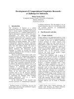
Liver damage in transfected Hep-G2 cells after
treatment with GCV
The pCMV-TK vectors were transfected into Hep-G2
cells, which were then induced with GCV. The trans-
fected cells started to detach on third day after a single
exposure to 40 lmolÆL
)1
GCV. Hep-G2 cells transfected
with pLLTK started to detach on day 5, and maximal
expression was achieved on day 7 after GCV treatment.
Cell apoptosis was recognized in both of the two groups
mentioned above; sick or damaged cells were seen to
swell and burst. By contrast, the control cells transfected
with vector pcDNA 3.1 ⁄ zeo(+) grew and proliferated
normally. The Hep-G2 transfected pLLTK showed
completely different morphology as compared to the
control cells (Fig. 1A). Such increased cell death could
also be assessed by 3-(4,5-dimethylthiazol-2-yl)-2,5-
diphenyl tetrazolium bromide assay. The survival rate
of Hep-G2 cells was reduced significantly after trans-
fection with HSV-tk (Fig. 1B).
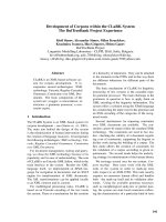
Detection of inserted transgene and monitoring
of the pedigree of mouse family TK5 by PCR
A total of 182 eggs were microinjected and subse-
quently reimplanted into eight pseudopregnant foster
mothers, of which five became pregnant and gave birth
to 36 mice. Among them, six mice showed the inser-
tion of the HSV-tk gene as detected by PCR. The
integration rate was 16.7% (6 ⁄ 36). One line of trans-
genic mice is female; the transgene is transmitted to
the offspring at a rate of about 50% according to
Mendel’s laws (Fig. 2A).
Chromosomal localization of transgene
integration as demonstrated by fluorescence
in situ hybridization (FISH)
More than 50 metaphases were analysed for each
transgenic mouse. All of the metaphase cells showed
one positive hybrid signal. According to the standard
idiograms of mouse chromosomes, the integration site
is located at 2F1-G3 in TK5-F1-455 (Fig. 2B). The
A
B
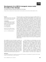
Fig. 1. Comparison of different HSV-tk transfected Hep-G2 cells
post-treatment with GCV and the negative control group. (A) Mor-
phology of the Hep-G2 cells transfected with pLLTK after adminis-
tration of GCV. Left, 40 lmolÆL
)1
GCV; right, no GCV (original
magnification, · 200). (B) Comparison of cellular survival rates
among HSV-tk transfected Hep-G2 cells post-treatment with GCV
and the negative control group. *P < 0.05. Results are expressed
as mean ± SD of three separate experiments. Bar1, cells trans-
fected with pCMV-TK (positive control group); Bar2, cells trans-
fected with pLLTK (experimental group); Bar3, cells transfected
with pcDNA3.1(+) ⁄ zeo (negative control group). Cellular survival
rates are assessed by 3-(4, 5-dimethylthiazol-2-y)-2, 5-diphenyl tetra-
zolium bromide staining.
Development of an HSV-tk transgenic mouse model Y. Zhang et al.
2208 FEBS Journal 272 (2005) 2207–2215 ª 2005 FEBS
integration site of TK5-F2-327 was similar to that of
TK5-F1-455 (data not shown).
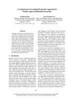
Reverse transcription (RT)–PCR of tissue-specific
expression of HSV-tk in transgenic mice
RT-PCR showed that the 390 bp specific band of
HSV-tk was detectable only in the transfected cells,
liver and testicle. It was not detectable in the cells used
as negative control, or in blood, kidney, pancreas,
intestine, brain, skin or heart, even though an internal
control band of 190 bp b-actin was present in all of
the samples (Fig. 3).
HSV-tk protein expression in the liver of
transgenic mice
Immunohistochemical staining was performed using a
polyclonal rabbit-(anti-HSV-tk) Ig. The yellowish-
brown staining sites were located mainly in the nucleus
of hepatocytes integrated by the HSV-tk gene, and the
HSV-tk-positive cells were distributed scattered or
clustered in liver lobules, located mainly around the
central vein, and occasionally in the periportal areas
(Fig. 4A), while there was no staining in the liver of the
wild-type mice (Fig. 4B). Simultaneously, the positive
signal can be observed in both the nucleus and the
cytoplasm after GCV treatment (Fig. 4C). The staining
cells account for 20–30% of the total hepatocytes of
TK5-F1-455 (Fig. 4A), whereas in mouse TK3, there
were approximately 60–70% HSV-tk staining hepato-
cytes, with visible slight yellowish-brown signals in the
focal necrosis, but several regenerative foci (regener-
ating parenchyma hepatocytes) displayed reduced or no
staining (Fig. 4D). The percentage of HSV-tk-positive
hepatocytes in the F2 mice (TK5-F2-327) of TK5 was
similar to those of TK5-F1-455 (data not shown).
Hematoxylin and eosin staining for histological
analysis
Microscopic analysis of the livers of GCV-treated
HSV-tk mice (F1 and F2) showed that the diseased liv-
ers display a number of abnormalities, including the
appearance of apoptosis bodies, hepatocyte vaculation,
lymphocyte infiltration, hepatocyte megalocytosis, and
diffused proliferation of hepatocytes (Fig. 5A). In
transgenic mouse TK3, mutifocal coagulation necrosis
was evident in the liver (Fig. 5B). Histological analysis
of the kidney showed no apparent abnormity in the
GCV-treated transgenic mice and wild-type mice (data
not shown).
Biochemical analysis of the blood
Twenty-one days after the injection of GCV, the values
of alanine aminotransferase (ALT), aspartate amino-
transferase (AST) and total bilirubin were significantly
increased in the TK5-F1 transgenic mice (P<0.05),
whereas there were no significant increase in the wild-
type control mice. The value of creatinine was not
altered significantly in either group (Fig. 6). The chan-
ges of the four serum values in the F2 generation of
the TK5 family and mouse TK3 showed similar results
(data not shown).
Fig. 3. RT-PCR of HSV-tk expression in the transgenic mouse TK5-
F1-455. M, 100-bp marker; 1, positive control (Hep-G2 cells trans-
fected with plasmid of pCMV-TK); 2, negative control (Hep-G2
cells); 3, blank control; 4, testis; 5, liver; 6, blood; 7, kidney; 8, pan-
creas; 9, intestines; 10, brain; 11, skin; 12, heart.
A
B
Fig. 2. Analysis of integration of transgene in mouse family TK5.
(A) PCR analysis of the transgenic mice in TK5-F1. 1, Hep-G2 cells
transfected pCMV-TK as a positive control; 2, Hep-G2 cells as a
negative control; 3, blank control; 4, founder mouse; 5,7,9,12, neg-
ative offspring; 6,8,10,11,13, positive offspring; M, 100 bp marker.
(B) FISH and G-banding metaphase of the transgenic mouse (TK5-
F1-455). The arrow indicates the integration site of the transgene
located at 2F1-G3. The right panel shows the mouse chromosome
ideogram.
Y. Zhang et al. Development of an HSV-tk transgenic mouse model
FEBS Journal 272 (2005) 2207–2215 ª 2005 FEBS 2209
Discussion
We have generated a number of transgenic mice for
liver damage, in which the HSV-tk gene was regulated
by an albumin promoter ⁄ enhancer. The excellence of
this mouse model is that liver damage and its extent in
HSV-tk mice can be induced and controlled by GCV
treatment. When the mice are injected with GCV, the
pathologic changes and biochemical abnormalities,
including vaculation of the hepatocytes, inflammatory
infiltration, diffuse proliferation of hepatocytes as well
as a significant increase of serum ALT, AST and total
bilirubin, can be easily recognized. However, renal
function is not affected by GCV treatment. This indi-
cates that GCV at the dosage used in this study is
associated with toxin-mediated hepatocyte damage in
our HSV-tk mice.
We used FISH and RT-PCR to investigate trans-
gene integration and HSV-tk expression in various
tissues of the transgenic mice. FISH indicated that the
Fig. 5. Histology of liver tissue (hematoxylin
and eosin stain). (A) GCV-treated TK5-F1 tra-
nsgenic mouse showed several apoptotic
bodies, variably severe cytoplasmic vacuoli-
zation, lymphocyte infiltration, hepatocyte
megalocytosis, and proliferation of hepato-
cytes. (B) GCV-treated TK3 mouse, showed
apoptosis bodies, mutifocal coagulation
necrosis with lymphocyte infiltration,
hepatocyte megalocytosis, and proliferation
of hepatocytes. (C) GCV-treated nontrans-
genic mouse. (D) Untreated transgenic
mouse. (Original magnification, · 200.)
Fig. 4. Immunohistochemical staining
observation of the HSV-tk expression.
(A) Liver of transgenic mouse TK5-F1-455.
HSV-tk-positive cells are clustered around
the central vein, scattered in the liver lobule
tissue, or clustered in the periportal areas.
(B) Liver of the wild-type mouse showed no
staining. (C) Liver of transgenic mouse
TK5-F1-452 after 21-days of GCV treatment,
the positive signal appeared in the nucleus
and the cytoplasm after GCV treatment.
(D) Liver of TK3 mouse, several regenerative
foci (regenerating parenchyma hepatocytes)
displayed reduced or no staining (slight
yellowish-brown signal in focal necrosis.
(Original magnification, · 400.)
Development of an HSV-tk transgenic mouse model Y. Zhang et al.
2210 FEBS Journal 272 (2005) 2207–2215 ª 2005 FEBS
HSV-tk gene was stably integrated in the genomes of
the mouse family (TK5), and the expression of HSV-tk
was readily detectd in the liver and the testis of TK5
family, but was not detectable in other tissues, such as
blood, kidney, pancreas, intestines, brain, skin and
heart. It indicated that the recombinant construct dri-
ven by the albumin ⁄ enhancer that we used in this study
was highly tissue specific. Immunohistochemical analy-
sis confirmed that the HSV-tk protein was expressed
specifically in the liver. In TK3 mice, approximately
60–70% of the total hepatocytes showed HSV-tk
expression, and 20–30% of the liver cells in the TK5
family gave positive results. The discrepancies of levels
of HSV-tk expression between these two mouse strains
may be due to differences in HSV-tk gene integration
sites, that may be caused by random integration of the
transgene.
Recent studies showed that HSV-tk converts the
nontoxic prodrug GCV into GCV-triphosphate, which
can cause chain termination and single-strand breakage
upon incorporation into DNA. Although blocking of
DNA synthesis of GCV is especially toxic for dividing
cells, it can also cause damage of nondividing cells,
such as hepatocytes, and liver toxicity of HSV-tk
[13,14]. This provides the basis for selected hepatocyte
killing using a hepatocyte-specific promoter in vivo.
Hepatocyte replication was not a prerequisite for
this effect, indicating that interference with DNA syn-
thesis during S phase of the cell cycle is not the only
mechanism of toxicity of phosphorylated GCV.
Furthermore, although the exact mechanism by which
suicide genes kill the HSV-tk-expressing cells is not yet
clear, apoptosis has been considered to be a major
contributor to GCV killing [15–18]. Song et al. [19]
have shown that GCV induced HSV-tk expressing cells
into apoptosis, thus inhibiting the growth of ovarian
cancer cells. Shibata et al. [20] injected the HSV-tk vec-
tor into rats with bladder cancer and observed apopto-
sis of bladder cancer cells. Kawasaki et al. [10] created
an AL-HSV-tk transgenic rat that expressed HSV-tk in
hepatocytes, in which apoptosis was demonstrated
after treatment with GCV. The administration of GCV
elicited leukocyte infiltration and induced chronic
hepatitis [21,22]. Although in the hepatitis model the
precise role of Kupffer cells is unclear, it is possible
that they are involved in inflammation [10]. Activated
Kupffer cells release cytokines and chemokines that
activate and transport T cells [23–25]. It seems that
hepatitis in the rat is primed by hepatocyte apoptosis
[10]. To examine the immunological mechanisms
involved in cell killing using the HSV-tk ⁄ GCV system,
HSV-tk-transduced human hepatocellular carcinoma
(HCC) cells were implanted subcutaneously into im-
munocompetent syngeneic mice. After GCV treatment,
marked infiltration by lymphocytes including CD4+
and CD8+ T cells, apoptosis of cells was induced, and
significant reduction or even complete regression of
tumours was achieved. Conversely, no significant
inhibitory effects on tumour formation were observed
in athymic nude mice. The results indicate that T cell-
mediated immune responses may be a critical factor
for achieving successful cell killing using the HSV-
tk ⁄ GCV system [26]. Administration of GCV to mice
and rats injected with adenovirus encoding HSV-tk
Fig. 6. Comparison of serum parameters of
TK5-F1 mice with the wild-type mice (n ¼ 3).
Results are expressed as mean ± SD of
three mice. After 21 days of GCV treatment
the values of ALT, AST and total bilirubin
were significantly increased in the TK5-F1
transgenic mice group (P<0.05), whereas
there was no significant increase in the
wild-type control group. Creatinine was not
significantly altered in either group.
Y. Zhang et al. Development of an HSV-tk transgenic mouse model
FEBS Journal 272 (2005) 2207–2215 ª 2005 FEBS 2211
caused extensive signs of liver degradation with negli-
gible survival rate [14]. Microscopic analysis of the
GCV-treated HSV-tk rat model of Kawasaki et al. [10]
revealed moderate hepatocyte vacuolation and an
increased number of inflammatory cells. In this study,
we found that there was more severe focal necrosis of
the liver tissue in TK3 than in TK5 mice. Moreover,
the liver regenerating focus was more evident in TK3,
in which clones of transgene expression-deficient cells
were formed as detected by immunohistochemistry.
The reasons for the different pathological changes
between these two mouse strains are not clear. We sug-
gest that these differences may be associated with the
quantities of HSV-tk expressing cells. In addition, the
patchy focal necrosis and regeneration in the TK3 liver
could be explained by the possibility that this founder
mouse may be a mosaic as approximately 5–10% of
founder mice showed mosaicism of some sort (either
multiple integration sites or patchy cellular distribu-
tion). Boucher et al. [27] compared the efficacy of the
HSV-tk ⁄ GCV system in two human carcinoma cell
lines after exposure to GCV and found that the killing
effect depended on the concentration of the tk enzyme,
the number of cells expressing HSV-tk, different cell
types and the overall confluence of the HSV-tk expres-
sing-cells. These results emphasise the importance of
cell-specific metabolism in HSV-tk ⁄ GCV-mediated
cytotoxicity. In conclusion, the killing of cells with
HSV-tk ⁄ GCV is a complex interactive sequence of
biochemical and cellular events involving incorporation
and accumulation of the monophosphorylate deriv-
ative of GCV into DNA, disruption and inhibition of
the cell cycle, gap junction metabolite transfer, and
apoptosis. Thus, we conclude that the exact mecha-
nisms may differ in: (a) different cell types; (b) differ-
ent species; (c) the concentration of GCV used; (d) the
quantity of cells expressing HSV-tk; and (e) the distri-
bution of cells expressing HSV-tk.
It is of interest that male HSV-tk mice (including
the founder TK3 and male offspring of TK5) gener-
ated in this study were not able to reproduce when
mated with wild-type female mice. RT-PCR showed
that HSV-tk was expressed both in the liver tissue and
ectopically in the testis even though a heptocyte-speci-
fic promoter was used in generating HSV-tk transgenic
mouse. The results suggest that male infertility may
result from ectopic expression of HSV-tk in the testis.
It has been reported that the natural HSV-tk gene con-
tains a cryptic internal testis-specific promoter [28–31],
and pathology has revealed that testicular development
was immature and almost no sperm was produced in
these mice (data not shown). When we treated female
transgenic mice with GCV liver damage was induced
showing that the liver damage in the HSV-tk trans-
genic mice may be independent of the HSV-tk expres-
sion in testis (data not shown).
In summary, transgenic mice specifically expressing
HSV-tk in the liver were generated. When transgenic
mice were treated with GCV, morphological, clinical,
and biochemical characteristics indicative of hepato-
cellular disease developed. These HSV-tk mice could
be an alternative model for the study of inducible
liver-specific disease, and may be useful in the study
of the pathogenesis of liver diseases and potential
therapies.
Experimental procedures
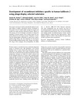
Plasmid construction and generation of
transgenic mice
Total DNA was extracted from KM mouse blood for PCR
amplification of murine albumin promoter ()310 bp to
+25 bp) and enhancer ()9192 bp to )11 250 bp). The
primers for albumin promoter and enhancer are: pro1,
5¢-CTTAGGTACCTCCATGCCAAGGCCCACA-3¢; pro2,
5¢-CTTGCTCACCATGGTGGCGACCGGTAGTGGGGT
TGATAGGAAAGG-3¢; en1, 5¢-ACGAGTCTAGAGTG
GAGCTTACTTCTTTGATTTGA-3¢; en2, 5¢-CCGCGTC
GACGGAAAAGCGCCTCCCCTAC-3¢; The 1800 bp of
the HSV-tk coding sequence were also amplified by PCR
from the pTK-neo plasmid and the consensus Kozak
sequence GCCACC was introduced in front of the transla-
tion start codon ATG by the primers tk1, 5¢-CGTA
TACCGGTGCCACCATGGCTTCGTACCCCGGC-3 ¢ and
tk2, 5¢-CCGCGTCGACGGAAAAGCGCCTCCCCTAC-3¢)
[32]. Recombinant plasmid pLLTK was obtained by insert-
ing all three of the amplified fragments into the multiple
cloning sites of pcDNA3.1(+) ⁄ zeo (Invitrogen, Carlsbad,
CA, USA) by cohesive-blunt end ligation. Then pLLTK
was digested with HindIII and the 4200 bp fragment of
LLTK (Fig. 7) was obtained with the QIAquick gel extrac-
tion kit (Qiagen, Valencia, CA, USA). After purification
with S & S Elutip minicolumns (Schleicher & Schuell,
Keen, NH, USA), the DNA fragment was microinjected
into the male pronuclei of the KM mouse fertilized eggs
and transgenic mice were generated.
Fig. 7. Schematic illustration of recombinant construction. Ealb and
Palb represent mouse albumin enhancer and mouse albumin
cDNA, respectively.
Development of an HSV-tk transgenic mouse model Y. Zhang et al.
2212 FEBS Journal 272 (2005) 2207–2215 ª 2005 FEBS
GCV-induced cytotoxicity in cultured transfected
cells
Human hepatic cell line Hep-G2 and mouse breast epithelia
cell line HC-11 cells were seeded in 24-well plates and grown
in Dulbecco’s modified Eagle’s medium supplemented with
10% (v ⁄ v) fetal bovine serum and penicillium ⁄ streptomycin.
The cells were then transfected with pLLTK by using Lipo-
fectamine
TM
2000 (Invitrogen). In a parallel setting, control
cells were transfected with the positive pCMV-TK and
pcDNA3.1(+) ⁄ zeo. Hep-G2 cells were treated with
40 lmolÆL
)1
GCV (Roche, Indianapolis, IN, USA) 24 h
after transfection. The morphology of the cells was exam-
ined by using an Olympus IX 70 inverted microscope
(Hamburg, Germany) each day; cell survival rates were
measured by 3-(4,5-dimethylthiazol-2-yl)-2,5-diphenyl tetra-
zolium bromide (Sigma Aldrich, St Louis, MO, USA) stain-
ing at 7 days later. The units of absorption were measured
by an Elx800 plate reader (Bio-Tek Instruments, Winooski,
VT, USA) at 570 nm. Differences in survival rates among
different transfected Hep-G2 cells post-GCV treatment were
analysed statistically using Student’s t-test (SAS Software).
A P-value < 0.05 was considered significant.
PCR analysis for the integration of the transgene
The transgene in the founder animals and their progeny
was identified by PCR analysis of genomic DNA obtained
from tail biopsies. PCR analysis was performed in 25 lL
reaction mixtures. The primers (stk1, 5¢-GTATACCGG
TATGCCCACGCTACTGCGG-3¢; SH552: 5¢-GCACTC
GAGACCCGTGCGTTTTATTCTGTCT-3¢) for HSV-tk
were designed to amplify a 390 bp region. Amplification
was performed on a thermocycler for 30 cycles of: 1 min at
94 °C, 1 min at 59 °C and 30 s at 72 °C. PCR products
were then separated electrophoretically on 2% agarose gel
and visualized after ethidium bromide staining.
Chromosomal localization of transgene integration
sites by using FISH following G-banding
Chromosome preparation was performed following the
reported methods with modifications [33–35]. FISH was
carried out according to the previous study with some
modifications [36,37]. The DNA fragment LLTK was used
as probe and labelled with the DIG-Nick translation mix
(Roche) according to the manufacturer’s protocol. Finally,
slides were counter-stained with propidium iodide anti-
fading solution (Sigma-Aldrich), and examined on a
fluorescent microscope (Leica DM RXA2, Wetzlar,
Germany). The nuclei were red and the hybridization sig-
nals were yellow-green. Previously photographed G-ban-
ded metaphases were relocated, and re-photographed.
Chromosomal localization of the transgene integration site
was determined by combining FISH hybridization signals
and G-banding results on the same metaphases. The mice
examined included TK5-F1-455#, TK5-F2-325$. The
standard idiograms of mouse chromosomes were obtained
from the web site: />guide/mouse/.
RT-PCR of HSV-tk transcription
The 60-day-old transgenic mouse TK5-F1-455 was killed
under pentobarbital anaesthesia (60 mgÆkg
)1
) with all possi-
ble measures taken to ensure minimum pain and discomfort.
Animal experiments were performed according to the
National Institute of Health Guidelines for Care and Use of
Laboratory Animals. The tissues of heart, liver, spleen, kid-
ney, brain, intestine, pancreas, skin, testis and blood were
powdered over an ice bath, total RNA was extracted using
the Trizol reagent (Gibco ⁄ BRL). Primers stk1 and SH552
were used to amplify the 390-bp region HSV-tk. The 190 bp
b-actin fragment amplified using primers MA2 (5¢-CCAC
AGGCATTGTGATGGA-3¢) and MA3 (5¢-GCTGTGGT
GGTGAAGCTGTA-3¢) was used as an internal control.
Immunohistochemical analysis of HSV-tk
expression
Immunohistochemical staining was performed to detect
HSV-tk expression in hepatocytes. Tissues fixed in 4%
(v ⁄ v) phosphate-buffered formalin were embedded in paraf-
fin and 5 lm-thick sections were stained. Briefly, paraffin-
embedded tissue sections were dewaxed, rehydrated, and
permeated before blotting, the slides were then incubated
with the rabbit polyclonal anti-(HSV-tk) Ig diluted 1 : 250
in NaCl ⁄ Tris for 1 h at 37 °C in a humidified chamber.
The slides were then washed three times and incubated with
biotinylated goat anti-rabbit immunoglobulins (DAKO) at
a dilution of 1 : 300 in NaCl ⁄ Tris for 1 h at room tempera-
ture with protection against light. After washing three
times with NaCl ⁄ Tris, peroxidase-conjugated streptavidin
(DAKO) diluted 1 : 300 was added for 1 h at room tem-
perature and washed three times with NaCl ⁄ Tris. Finally,
the signal was visualized by incubating the slides with 3–3¢-
diaminobenzidine (DAKO). The examined mice included
TK5-F1-455, TK5-F2-325 and TK3.
Induction of liver damage
For the present study, mice were housed individually at
22 °C using a 12 h light ⁄ 12 h dark photoperiod. Three trans-
genic mice of TK5-F1 (including three #), TK5-F2 (including
two # and one $) and TK3 aged 8–14 weeks, received tail
vein injections of sodium GCV (10 mgÆkg
)1
) at 48 h intervals
through a 29-gauge needle on 10 occasions, meanwhile three
nontransgenic mice served as the control. GCV was dissolved
in NaCl ⁄ P
i
and filter-sterilized before administration.
Y. Zhang et al. Development of an HSV-tk transgenic mouse model
FEBS Journal 272 (2005) 2207–2215 ª 2005 FEBS 2213
Pathological examination
Twenty-one days after GCV treatment the mice were killed
under pentobarbital anaesthesia. The tissues were fixed with
4% (v ⁄ v) phosphate-buffered formalin and paraffin-embed-
ded sections were stained using hematoxylin and eosin as
described previously [38,39].
Biochemical analysis of blood
The physiological function of the liver was examined by
determining biochemical serum values. Before the experi-
ment, physiological function of the liver was determined to
be normal by examination of serum ALT, AST, total biliru-
bin, albumin, globulin creatinine, total protein, lactate
dehydrogenase and others. Peripheral blood was extracted
from the tail vein once a week for 3 weeks for analysis of
ALT, AST, total bilirubin and creatinine. Student’s t-test
(SAS software) was performed to compare the differences
in serum values in transgenic mice and controls after GCV
administration. P < 0.05 was considered significant.
Acknowledgements
We thank Drs Yi-Ping Hu (Department of Cell Bio-
logy, Second Military Medical University of PLA,
Shanghai, China), Willams Summers (Yale University,
USA), Zheng-Hong Yuan (Key Laboratory of Medical
Molecular Virology, Fudan University, Shanghai,
China) and Hynes (Friedrich Miescher Institute of
Switzerland, Basel, Switzerland) for pTK-neo plasmid,
polyclonal rabbit anti-(HSV-tk), Hep-G2 and HC-11
cell lines, respectively. We would like to thank Drs
Zhao-Rui Ren, Jing-Zhi Zhang (Institute of Medical
Genetics, School of Medicine, Shanghai Jiao Tong
University, China) and Dr Xiao-Kang Li (the National
Institute for Child and Development, Japan) for their
very helpful discussion in this work. We also thank
Yi-Wen Zhu, Wen-Ying Huang, Xiu-Li Gong (Insti-
tute of Medical Genetics, School of Medicine, Shang-
hai Jiao Tong University, China) for their performing
the FISH and microinjections. The study was supported
by: the Chinese National Program for High Technology
Research and Development (No.2002AA216091).
References
1 Grompe M, Lindstedt S, al-Dhalimy M, Kennaway NG,
Papaconstantinou J, Torres-Ramos CA, Ou CN &
Finegold M (1995) Pharmacological correction of
neonatal lethal hepatic dysfunction in a murine model of
hereditary tyrosinaemia type I. Nat Genet 10, 453–460.
2 Braun KM, Thompson AW & Sandgren EP (2003)
Hepatic microenvironment affects oval cell localization
in albumin-urokinase-type plasminogen activator trans-
genic mice. Am J Pathol 162, 195–202.
3 Heyman RA, Borrelli E, Lesley J, Anderson D, Rich-
man DD, Baird SM, Hyman R & Evans RM (1989)
Thymidine kinase obliteration: creation of transgenic
mice with controlled immune deficiency. Proc Natl Acad
Sci USA 86, 2698–2702.
4 Borrelli E, Heyman RA, Arias C, Sawchenko PE &
Evans RM (1989) Transgenic mice with inducible dwarf-
ism. Nature 339, 538–541.
5 Dzierzak E, Daly B, Fraser P, Larsson L & Muller A
(1993) Thy-1 tk transgenic mice with a conditional lym-
phocyte deficiency. Int Immunol 5, 975–984.
6 Kamogawa Y, Minasi LA, Carding SR, Bottomly K &
Flavell RA (1993) The relationship of IL-4- and IFN
gamma-producing T cells studied by lineage ablation of
IL-4-producing cells. Cell 75, 985–995.
7 Salomon B, Lores P, Pioche C, Racz P, Jami J & Klatz-
mann D (1994) Conditional ablation of dendritic cells in
transgenic mice. J Immunol 152, 537–548.
8 Tian B, Han L, Kleidon J & Henke C (2003) An HSV-
TK transgenic mouse model to evaluate elimination of
fibroblasts for fibrosis therapy. Am J Pathol 163, 789–
801.
9 Minasi LE, Kamogawa Y, Carding S, Bottomly K &
Flavell RA (1993) The selective ablation of interleukin
2-producing cells isolated from transgenic mice. J Exp
Med 177, 1451–1459.
10 Kawasaki M, Fujino M, Li XK, Kitazawa Y, Funeshi-
ma N, Takahashi R, Ueda M, Amano T, Hakamata Y
& Kobayashi E (2003) Inducible liver injury in the
transgenic rat by expressing liver-specific suicide gene.
Biochem Biophys Res Commun 311, 920–928.
11 Kramer MG, Barajas M, Razquin N, Berraondo P,
Rodrigo M, Wu C, Qian C, Fortes P & Prieto J (2003)
In vitro and in vivo comparative study of chimeric liver-
specific promoters. Mol Ther 7, 375–385.
12 Zhang Y, Huang SZ & Zeng YT (2004) The effect of
HSV-tk ⁄ GCV on hepatic specific damage driven by
murine ALB promoter ⁄ enhancer, Acta Genetica Sinica
31, 1053–1060.
13 van der Eb MM, Cramer SJ, Vergouwe Y, Schagen FH,
van Krieken JH, van der Eb AJ, Rinkes IH, van de
Velde CJ & Hoeben RC (1998) Severe hepatic dys-
function after adenovirus-mediated transfer of the
herpes simplex virus thymidine kinase gene and
ganciclovir administration. Gene Ther 5 , 451–458.
14 Brand K, Arnold W, Bartels T, Lieber A, Kay MA,
Strauss M & Dorken B (1997) Liver-associated toxicity
of the HSV-tk ⁄ GCV approach and adenoviral vectors.
Cancer Gene Ther 4, 9–16.
15 Elshami AA, Saavedra A, Zhang H, Kucharczuk JC,
Spray DC, Fishman GI, Amin KM, Kaiser LR &
Albelda SM (1996) Gap junctions play a role in the
Development of an HSV-tk transgenic mouse model Y. Zhang et al.
2214 FEBS Journal 272 (2005) 2207–2215 ª 2005 FEBS
‘bystander effect’ of the herpes simplex virus thymidine
kinase ⁄ ganciclovir system in vitro. Gene Ther 3, 85–92.
16 Krohne TU, Shankara S, Geissler M, Roberts BL,
Wands JR, Blum HE & Mohr L (2001) Mechanisms of
cell death induced by suicide genes encoding purine
nucleoside phosphorylase and thymidine kinase in
human hepatocellular carcinoma cells in vitro. Hepatol-
ogy 34, 511–518.
17 Beltinger C, Fulda S, Kammertoens T, Meyer E, Uckert
W & Debatin KM (1999) Herpes simplex virus thymidine
kinase ⁄ ganciclovir-induced apoptosis involves ligand-
independent death receptor aggregation and activation
of caspases. Proc Natl Acad Sci USA 96, 8699–8704.
18 Freeman SM, Abboud CN, Whartenby KA, Packman
CH, Koeplin DS, Moolten FL & Abraham GN (1993)
The ‘bystander effect’: tumor regression when a fraction
of the tumor mass is genetically modified. Cancer Res
53, 5274–5283.
19 Song J, Kim H, Yoon W, Lee K, Kim M, Kim K, Kim
H & Kim Y (2003) Adenovirus-mediated suicide gene
therapy using the human telomerase catalytic subunit
(hTERT) gene promoter induced apoptosis of ovarian
cancer cell line. Bioscience Biotechnol Biochem 67, 2344–
2350.
20 Shibata MA, Horiguchi T, Morimoto J & Otsuki Y
(2003) Massive apoptotic cell death in chemically
induced rat urinary bladder carcinomas following in situ
HSVtk electrogene transfer. J Gene Med 5, 219–231.
21 Freedman AR, Sharma RJ, Nabel GJ, Emerson SG &
Griffin GE (1992) Cellular distribution of nuclear factor
kappa B binding activity in rat liver. Biochem J 287,
645–649.
22 Komatsu Y, Shiratori Y, Kawase T, Hashimoto N,
Han K, Shiina S, Matsumura M, Niwa Y, Kato N,
Tada M, et al. (1994) Role of polymorphonuclear leuko-
cytes in galactosamine hepatitis: mechanism of adherence
to hepatic endothelial cells. Hepatology 20, 1548–1556.
23 Sass G, Heinlein S, Agli A, Bang R, Schumann J &
Tiegs G (2002) Cytokine expression in three mouse
models of experimental hepatitis. Cytokine 19, 115–120.
24 Tamaru M, Nishioji K, Kobayashi Y, Watanabe Y,
Itoh Y, Okanoue T, Murai M, Matsushima K &
Narumi S (2000) Liver-infiltrating T lymphocytes are
attracted selectively by IFN-inducible protein-10. Cyto-
kine 12, 299–308.
25 Yoneyama H, Harada A, Imai T, Baba M, Yoshie O,
Zhang Y, Higashi H, Murai M, Asakura H & Matsush-
ima K (1998) Pivotal role of TARC, a CC chemokine,
in bacteria-induced fulminant hepatic failure in mice.
J Clin Invest 102, 1933–1941.
26 Kuriyama S, Kikukawa M, Masui K, Okuda H,
Nakatani T, Akahane T, Mitoro A, Tominaga K, Tsuji-
noue H, Yoshiji H, Okamoto S, Fukui H & Ikenaka K
(1999) Cancer gene therapy with HSV-tk ⁄ GCV system
depends on T-cell-mediated immune responses and
causes apoptotic death of tumor cells in vivo. Int J
Cancer 83, 374–380.
27 Boucher PD, Ruch RJ & Shewach DS (1998) Differen-
tial ganciclovir-mediated cytotoxicity and bystander kill-
ing in human colon carcinoma cell lines expressing
herpes simplex virus thymidine kinase. Hum Gene Ther
9, 801–814.
28 Ellison AR, Wallace H, al-Shawi R & Bishop JO (1995)
Different transmission rates of herpesvirus thymidine
kinase reporter transgenes from founder male parents
and male parents of subsequent generations. Mol
Reprod Dev 41, 425–434.
29 Huttner KM, Pudney J, Milstone DS, Ladd D & Seid-
man JG (1993) Flagellar and acrosomal abnormalities
associated with testicular HSV-tk expression in the
mouse. Biol Reprod 49, 251–261.
30 Al-Shawi R, Burke J, Jones CT, Simons JP & Bishop
JO (1988) A Mup promoter-thymidine kinase reporter
gene shows relaxed tissue-specific expression and confers
male sterility upon transgenic mice. Mol Cell Biol 8,
4821–4828.
31 Braun RE, Lo D, Pinkert CA, Widera G, Flavell RA,
Palmiter RD & Brinster RL (1990) Infertility in male
transgenic mice: disruption of sperm development by
HSV-tk expression in postmeiotic germ cells. Biol
Reprod 43, 684–693.
32 Kozak M (1987) At least six nucleotides preceding the
AUG initiator codon enhance translation in mammalian
cells. J Mol Biol 196, 947–950.
33 Sawyer JR, Moore MM & Hozier JC (1987) High reso-
lution G-banded chromosomes of the mouse. Chromo-
soma 95, 350–358.
34 Matsuda Y, Harada YN, Natsuume-Sakai S, Lee K,
Shiomi T & Chapman VM (1992) Location of the mouse
complement factor H gene (cfh) by FISH analysis and
replication R-banding. Cytogenet Cell Genet 61, 282–285.
35 Miao CX, Lu GX, Wang YJ & Xie Y (1998) Mapping
BNLF-1 transgene on transgenic mouse progeny chro-
mosome by fluorescence in situ hybridization. Acta
Genetica Sinica 25, 422–426.
36 Xiao Y, Jiang X & Wang R (2003) Screening for
DMD ⁄ BMD deletion carriers by fluorescence in situ
hybridization. Genet Test 7, 195–201.
37 Xiao YP, Xi Y, Huang WY & Huang Y (2002) Detec-
tion of the integration of human FIX (hFIX) on chro-
mosome of transgenic mice by fluorescene in situ
hybridization. Hereditas (China) 24, 232–236.
38 Tian B, Carlyle WC, Weigold WG, McDonald KM, Judd
DL, Toher CA, Homans DC & Cohn JN (1999) Locali-
zation of changes in beta-actin expression in remodeled
canine myocardium. J Mol Cell Cardiol 31, 751–760.
39 Tian B, Liu J, Bitterman PB & Bache RJ (2002)
Mechanisms of cytokine induced NO-mediated cardiac
fibroblast apoptosis. Am J Physiol Heart Circ Physiol
283, H1958–H1967.
Y. Zhang et al. Development of an HSV-tk transgenic mouse model
FEBS Journal 272 (2005) 2207–2215 ª 2005 FEBS 2215