Báo cáo khoa học: Formation of nucleoprotein RecA filament on single-stranded DNA Analysis by stepwise increase in ligand complexity potx
Bạn đang xem bản rút gọn của tài liệu. Xem và tải ngay bản đầy đủ của tài liệu tại đây (245.65 KB, 12 trang )
Formation of nucleoprotein RecA filament on
single-stranded DNA
Analysis by stepwise increase in ligand complexity
Irina P. Bugreeva, Dmitry V. Bugreev and Georgy A. Nevinsky
Institute of Chemical Biology and Fundamental Medicine, Siberian Division of Russian Academy of Sciences, Novosibirsk, Russia
Homologous recombination, required for the mainten-
ance of genetic diversity and DNA repair, is one of the
most important molecular genetic processes. In Escheri-
chia coli, a pivotal role in homologous recombination is
played by RecA protein, which is responsible for search
for homologous DNA sequences and strand transfer
between them [1]. RecA is an ATP-dependent DNA-
binding protein consisting of 352 amino acids
(37.8 kDa) [1]. Binding of RecA to DNA occurs in three
stages: first, the presynapsis, when RecA is polymerized
on ssDNA forming a right-handed nucleoprotein
filament; second, the synapsis, when the presynaptic
complex binds dsDNA and actively searches for homo-
logy with the ssDNA; and third strand exchange, when
a new DNA duplex is formed and one of the strands
formerly in dsDNA is released as ssDNA. Thus, a RecA
filament assembles on DNA at the first stage; this
process is more efficient with ssDNA. Binding of RecA
to ssDNA must be nonspecific, but the protein displays
some preferences for binding poly(dT) and GT-rich
sequences [2–5].
In the presence of ATP or its nonhydrolysable thio
analog (ATPcS), RecA forms a right-handed filament
of 100 A
˚
diameter and 95 A
˚
pitch [6]. The filament
is assembled cooperatively in the 5¢)3¢ direction (in
respect to the ssDNA) [7]. DNA in such complex is
stretched by % 50%, with the internucleotide distance
increasing to 5.1 A
˚
[8]. If RecA binds to dsDNA, the
Keywords
RecA; DNA recognition mechanism
Correspondence
G. A. Nevinsky, Laboratory of repair
enzymes, Institute of Chemical Biology and
Fundamental Medicine, 8, Lavrentieva Ave.,
630090, Novosibirsk, Russia
Fax: +7 3832 333677
Tel: +7 3832 396226
E-mail:
(Received 8 January 2005, revised 24
February 2005, accepted 31 March 2005)
doi:10.1111/j.1742-4658.2005.04693.x
RecA protein plays a pivotal role in homologous recombination in Escheri-
chia coli. RecA polymerizes on single-stranded (ss) DNA forming a nucleo-
protein filament. Then double-stranded (ds) DNA is bound and searched
for segments homologous to the ssDNA. Finally, homologous strands are
exchanged, a new DNA duplex is formed, and ssDNA is displaced. We
report a quantitative analysis of RecA interactions with ss d(pN)
n
of var-
ious structures and lengths using these oligonucleotides as inhibitors of
RecA filamentation on d(pT)
20
. DNA recognition appears to be mediated
by weak interactions between its structural elements and RecA monomers
within a filament. Orthophosphate and dNMP are minimal inhibitors of
RecA filamentation (I
50
¼ 12–20 mm). An increase in homo-d(pN)
2)40
length by one unit improves their affinity for RecA (f factor) approximately
twofold through electrostatic contacts of RecA with internucleoside phos-
phate DNA moieties (f % 1.56) and specific interactions with T or C bases
(f % 1.32); interactions with adenine bases are negligible. RecA affinity for
d(pN)
n
containing normal or modified nucleobases depends on the nature
of the base, features of the DNA structure. The affinity considerably increa-
ses if exocyclic hydrogen bond acceptor moieties are present in the bases. We
analyze possible reasons underlying RecA preferences for DNA sequence
and length and propose a model for recognition of ssDNA by RecA.
Abbreviations
EMSA, electrophoretic mobility shift assay; ODN, deoxyribooligonucleotide(s); SILC, stepwise increase in ligand complexity; ss-,
single-stranded; ds-, double-stranded.
2734 FEBS Journal 272 (2005) 2734–2745 ª 2005 FEBS
parameters of the resulting filament are the same as
for ssDNA, and the DNA duplex in the filament is
unwound as compared with B-DNA [9,10]. In the
absence of ATP RecA forms a more compact inactive
filament of 64 A
˚
pitch and 2.1 A
˚
internucleotide dis-
tance [11].
After the filament is formed, the second DNA bind-
ing site of RecA can bind dsDNA. In addition, ssDNA
can be bound there, even more efficiently than
dsDNA. After strand exchange, the second RecA
DNA binding site binds the displaced strand following
new DNA duplex formation [12].
Binding of dsDNA to a RecA filament is followed
by search of homology between the appropriate strands
and then by strand exchange. The mechanism of this
process is still unclear. It was hypothesized that ssDNA
could invade through the minor or major groove of the
duplex and displace the respective strand [1]; if the inva-
sion occurs through the major groove, formation of a
peculiar DNA triplex (R-form DNA) was proposed
[13,14]. An alternative mechanism (melting–annealing
model) for the homology search involves only formation
of canonical Watson–Crick pairs after melting of the
duplex and annealing of its appropriate strand to the
incoming strand [15]. As DNA in the filament is consid-
erably stretched and unwound, the bases could be easily
extruded from the helix to be ‘examined’ for homology
with the incoming strand.
Howard-Flanders proposed a triple helix as a tran-
sient, or even a stable, intermediate in the reaction [16].
However, all recent efforts have failed to detect such
a structure as a stable intermediate. Instead, several
groups have described a stable synaptic complex con-
sisting of three strands and RecA, in which strand
exchange has already taken place [17,18]. In this com-
plex, the incoming ssDNA is part of the new duplex and
the leaving strand has not yet been released. Leaving
aside such an early triplex, one can jump forward and ask
what are the steps leading to such a poststrand exchange
intermediate? One can envision several slower confor-
mational changes, such as homology recognition via
base flipping (melting) and switching (annealing) [19].
DNA binding by RecA is thought to be mediated by
amino-acid residues from two protein loops, L1 (resi-
dues 157–164) and L2 (residues 195–209) [1,20]. Both
these regions are rather conserved among bacterial
RecA proteins but not between bacterial, archaean and
eukaryotic RecA homologs. In addition, DNA could
interact with several RecA tyrosine residues (Tyr65,
Tyr103, Tyr264) [21–23], as well as with Lys183 [23,24],
Arg243 [22] and residues 233–242 [24]. This list all
but exhausts the available information regarding
RecA–DNA interactions. To our knowledge, there have
been no quantitative studies on general parameters of
and individual contacts within the forming nucleo-
protein filament.
Our laboratory has designed a novel approach to
analysis of protein ⁄ nucleic acid interactions, based
on stepwise increase in ligand complexity (SILC
approach). SILC produces quantitative estimates of
the contributions of individual structural elements of
DNA or RNA molecules into the affinity of enzymes
to such extended ligands [25–27]. We have applied
SILC to analyze DNA binding by a number of
DNA polymerases [25–27], DNA repair enzymes [28–
31], EcoRI restriction endonuclease [32], HIV-1 integ-
rase [33], and type I DNA topoisomerases [34,35]. In
all these instances, virtually every nucleotide unit
within the DNA binding cleft (10–20 base pairs cov-
ered by the protein globule) interacts with the
enzyme through weak additive electrostatic, hydro-
phobic or van der Waals contacts to various struc-
tural elements of the ligands, with electrostatic
interactions of internucleoside phosphate moieties
contributing most to the affinity (reviewed in [25–
27]). These nonspecific contacts provide high affinity
(K
d
¼ 10
)5
)10
)8
m) of all enzymes for specific and
nonspecific DNA. A transition from nonspecific to spe-
cific DNA usually leads to formation of specific contacts
and increase of the affinity by 1–2 orders of magnitude
(up to K
d
¼ 10
)8
)10
)10
m), while the reaction rate
(k
cat
) is enhanced by 5–8 orders. Thus, specificity of
DNA-dependent enzymes is not of thermodynamic
nature (the enzyme-substrate complex formation) but
mostly originates from the following stage of enzyme-
induced adjustment of DNA conformation and from
chemical steps (k
cat
) of catalysis [25–27].
Quantitative studies concerning the efficiency of
interactions between a RecA filament and DNA are
a prerequisite for understanding the nature of RecA
filamentation; however, no such information is available
so far. SILC is a very promising approach for obtaining
the appropriate data. Here we present a SILC analysis
of RecA interactions with ssODN of different structures
and lengths and estimate the contribution of individual
DNA elements in its affinity for a RecA filament.
Results
Filamentation of RecA on ssDNA
and its inhibition
In the presence of ATP or ATPcS RecA is polymer-
ized on DNA forming a nucleoprotein filament. We
have studied the stability of a RecA filament formed
with different individual 5¢-[
32
P]d(pN)
n
(n ¼ 2–20) by
I. P. Bugreeva et al. RecA filament interaction with DNA
FEBS Journal 272 (2005) 2734–2745 ª 2005 FEBS 2735
electrophoretic mobility shift assay (EMSA). The com-
plexes between RecA and short individual d(pN)
n
(n ¼ 2–15) were easily disassembled, confirming the lit-
erature data on their low stability [36,37]. Individual
5¢-[
32
P]d(pN)
16)20
formed detectable complexes with
RecA under the condition used (data not shown), and
the best of them d(pT)
20
was used for the rest of the
study. At the RecA monomer: ODN ratio of 10 : 1,
almost all d(pT)
20
was in the filament, in agreement
with the known RecA monomer interaction with three
nucleotide units of ssDNA [1].
As the interactions with short ODNs are of low affin-
ity, they are undetectable by EMSA and many other
widely used physicochemical techniques [27]. However,
interactions of enzymes with low-affinity ligands can be
easily followed by observing inhibition of appropriate
enzymatic activity by these ligands (reviewed in [25–27]).
In the case of short ODN interacting with RecA mono-
mers or forming short unstable filaments, the respective
ODN ligands should inhibit RecA filamentation on
d(pT)
20
. In addition, at high concentration short ODN
can compete with d(pT)
20
for the filament formed on this
substrate. We have shown that the addition of any short
ODN causes a decrease in the amount of 5¢-[
32
P]d(pT)
20
detectable in the RecA filament complex. Concentration
dependencies of RecA-d(pT)
20
complex formation on
the inhibitor concentration had regular hyperbolic
shapes (Fig. 1), indicating that RecA filamentation on
d(pT)
20
and its inhibition by short ODN, including
orthophosphate (I
50
¼ 0.5 m) and various dNMPs
(I
50
¼ 12–20 mm) as minimal ligands, obey formally
canonical steady-state equations of complex formation.
The apparent values of I
50
(Fig. 1) were used to charac-
terize the relative efficiency of RecA interactions with
various ODN; these data are summarized in Table 1.
The Gibbs free energy characterizing enzyme-ligand
complex formation can be presented as a sum of DG°
values for each individual contact:
DG
0
¼ DG
0
1
þ DG
0
2
þ ::: þ DG
0
n
with DG
0
i
¼ÀRT ln K
di
ð1Þ
where K
di
is the contribution of an individual contact
to the overall affinity [38]. It follows from the additi-
vity of Gibbs free energies that the overall K
d
(K
d
¼ K
I
)
value characterizing complex formation is the product
of the K
d
values for individual contacts:
DG
0
¼ÀRT ln K
d
¼ÀRT ln½K
d1
K
d2
:::K
dn
; and
K
d
¼ K
d1
K
d2
:::K
dn
ð2Þ
To assess possible additivity of the interactions of
ODN with RecA filament, the data from Table 1 were
analyzed as logarithmic dependencies of I
50
for d(pN)
n
on the number (n) of mononucleotide units
(0 £ n £ 20, n ¼ 0 corresponds to orthophosphate, P
i
).
Affinity of d(pN)
n
ligands to RecA increased mono-
tonously in the d(pN)
2
–d(pN)
20
interval, d(pT)
n
and
d(pC)
n
producing nearly identical results (Fig. 2).
Dependencies of lgI
50
on n were linear at 2 £ n £ 20
(Fig. 2), indicating that the affinity of RecA to each of
the nucleotide units of d(pN)
20
is additive.
Interestingly, experimentally estimated affinities of
dNMP (I
50
¼ 12–20 mm) were somewhat higher than
that for corresponding d(pN)
2
(40–47 mm, Table 1).
This phenomenon, also observed for some other
enzymes, arises from greater conformational freedom
of individual dNMP (or short ODN) compared with
the same ligands as elements of long DNA [25–27].
Considerable stretching and unwinding of DNA in a
RecA filament is associated with energetic costs
required for sugar-phosphate backbone deformation
and stacking disruption [6]. Mononucleotides are not
subject to such restrictions and thus can bind RecA
more efficiently. Extrapolation of the log dependencies
for d(pT)
n
and d(pC)
n
to n ¼ 1 (Fig. 2) gives lower
A
B
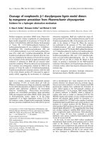
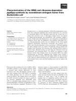
Fig. 1. Dependence of the relative level of inhibition of RecA filam-
entation on [
32
P]d(pT)
20
on the concentration of d(pT)
10
inhibitor. (A)
Reaction products separated by EMSA in polyacrylamide gel. (B)
Band intensities in (A) quantified by Cherenkov counting and plotted
against inhibitor concentrations. Lane 1, filamentation without the
inhibitor; lane 10, reaction mixture without RecA; d(pT)
10
inhibitor
added at 0.05 m
M (lane 2), 0.1 mM (lane 3), 0.2 mM (lane 4),
0.3 m
M (lane 5), 0.5 mM (lane 6), 0.6 mM (lane 7), 0.8 mM (lane 8)
and 1 m
M (lane 9). The upward shift in free oligonucleotide position
appears due to a time lag in loading different reaction mixtures
onto a running gel.
RecA filament interaction with DNA I. P. Bugreeva et al.
2736 FEBS Journal 272 (2005) 2734–2745 ª 2005 FEBS
affinity values for RecA binding single d(pT) and
d(pC) units (I
50
¼%63 mm) within longer d(pN)
n
(Fig. 2; Table 1). Thus, this value of I
50
¼%63 mm is
a better parameter to characterize RecA affinity for
the higher-affinity nucleotide unit of d(pN)
n
in com-
parison with the remaining (n–1) nucleotide units
within d(pT)
n
or d(pC)
n
, which have lower affinity for
the filament (490 mm, see below).
I
50
values are usually related to the K
I
values [38].
For example, in the case of competitive inhibition, they
are related through the equation I
50
¼ aK
I
(a ¼
1 + [S]⁄ K
S
; K
S
is K
M
or K
d
for substrate), where the
coefficient a depends on the affinity and concentration
of a substrate, d(pT)
20
in our case. Therefore, the ratio
of K
I
values for two different inhibitors, K
I
(2) ⁄ K
I
(1), is
equal to the ratio of apparent I
50
values for these
inhibitors, I
50
(2) ⁄ I
50
(1), and the ratio of these values
gives the K
d
value characterizing a difference of the
enzyme contacts between the first and the second
inhibitors (Eqns 1 and 2) [38].
From the slope of the lgI
50
vs n dependency
(Fig. 2) the factor (f) reflecting an increase in affin-
ity of the enzyme for d(pN)
n
upon a one-unit
increase in the ligand length can be calculated as:
f ¼ 10
–[lgI
50
(n ¼ 20) ⁄ –lgI
50
(n ¼ 2)] ⁄ 18
(exact average values
of lgI
50
were calculated using the log curves). From
the slopes of the curves for d(pT)
n
and d(pC)
n
(Fig. 2), the value f ¼ 2.04 was calculated for the
f factor. As 1 ⁄ f(n) ¼ I
50
(n) ⁄ I
50
(n +1)¼ K
I
(n) ⁄
K
I
(n +1)¼ K
d
(n) ⁄ K
d
(n + 1), interaction of a RecA
filament with any of the 19 units of d(pT)
20
or
d(pC)
20
is characterized by K
d
¼ K
I
¼ 1 ⁄ f ¼ 0.49 m.
Comparison of this value with I
50
for free dNMP
determined experimentally I
50(experimental)
¼ 12–20 mm,
Table 1 or for a dNMP unit within d(pN)
n
by extra-
polation to n ¼ 1 I
50(extrapolated)
¼%63 mm; Fig. 2
shows that the affinity of RecA for one of the units
or d(pT)
20
or d(pC)
20
is 8–41-fold higher than for
any of the remaining 19 units. Extrapolation of the
log dependencies for d(pT)
n
and d(pC)
n
to n ¼ 0
gives I
50(extrapolated)
¼ 15 mm for a single internucleo-
side phosphate group of d(pN)
n
, approximately
3.3-fold lower than the experimental I
50
¼ 0.5 m for
free orthophosphate. Overall, the affinity of a RecA
filament for d(pT)
n
and d(pC)
n
at 2 £ n £ 20 may
be described as I
50
[d(pN)
n
] ¼ I
50
(d(pN)
2
) · (1 ⁄ f)
n)2
¼
Table 1. I
50
values for interactions of different ligands with the high-affinity DNA-binding center of E. coli RecA filament.
Ligand (inhibition of d(pT)
20
) I
50
, M* –lgI
50
Ligand (inhibition of d(pT)
20
) I
50
, M –lgI
50
PO
4
3–
0.5 0.30 d(pR)§ 0.6 0.22
One internucleoside phosphate
within d(pC)
n
and d(pT)
n
**
0.15 0.63 One internucleoside phosphate
within d(pA)
n
**
0.23 0.82
dTMP 2.0 · 10
)2
1.70 dCMP 1.3 · 10
)2
1.89
One (pT)-unit of d(pT)
n
** % 6.3 · 10
)2
1.20 One (pC)-unit of d(pC)
n
** % 6.3 · 10
)2
1.20
d(pT)
2
4.0 · 10
)2
1.40 d(pC)
2
4.7 · 10
)2
1.33
d(pT)
3
1.75 · 10
)2
1.76 d(pC)
4
1.1 · 10
)2
1.96
d(pT)
4
1.0 · 10
)2
2.00 d(pC)
6
2.5 · 10
)3
2.60
d(pT)
5
5.0 · 10
)3
2.30 d(pC)
8
5.7 · 10
)4
3.24
d(pT)
6
2.5 · 10
)3
2.60 d(pC)
10
5.0 · 10
)4
3.30
d(pT)
8
5.0 · 10
)4
3.30 d(pC)
12
4.3 · 10
)5
4.37
d(pT)
10
2.0 · 10
)4
3.70 d(pC)
16
1.8 · 10
)6
5.74
d(pT)
12
3.5 · 10
)5
4.45 d(pC)
20
1.6 · 10
)7
6.80
d(pT)
14
8.3 · 10
)6
5.08 dAMP 1.24 · 10
)2
1.90
d(pT)
16
1.5 · 10
)6
5.82 One (pA)-unit of d(pA)
n
** % 10.0 · 10
)2
1.20
d(pT)
20
1.0 · 10
)7
7.00 d(pA)
2
4.5 · 10
)2
1.35
d(Tp)
7
T 4.8 · 10
)3
2.32 d(pA)
4
7.0 · 10
)3
2.15
d(Tp)
8
2.5 · 10
)3
2.60 d(pA)
6
1.04 · 10
)3
2.98
d(pTT(pR)
17
T)*** 9.5 · 10
)6
5.02 d(pA)
8
5.2 · 10
)4
3.28
d[p(ethyl)T]
10
5.0 · 10
)3
2.30 d(pA)
10
2.5 · 10
)4
3.60
d(pR)
20
** 2.1 · 10
)5
4.68 d(pA)
12
1.3 · 10
)4
3.89
I
50
determined using d(pT)
40
as substrate
d(pA)
14
8.0 · 10
)5
4.10
d(pT)
20
1.0 · 10
)7
7.00 d(pA)
16
5.4 · 10
)5
4.27
d(pT)
30
2.3 · 10
)8
7.64 d(pA)
18
3.2 · 10
)5
4.50
d(pT)
40
7.0 · 10
)9
8.15 d(pA)
20
2.4 · 10
)5
4.62
*Error in I
50
values was 10–30%; means of 3–4 measurements are given; **The values of I
50
determined by extrapolation of lg-curves to
n ¼ 0 for Pi and n ¼ 1 for dNMPs (Fig. 2); §d(pR), deoxyribosephosphate; ***R is a tetrahydrofuran analog of abasic deoxyribose.
I. P. Bugreeva et al. RecA filament interaction with DNA
FEBS Journal 272 (2005) 2734–2745 ª 2005 FEBS 2737
I
50
(d(pN)
2
) · ( f)
2–n
, when at 1 £ n £ 20 as
I
50
[d(pN)
n
] ¼ I
50
(dNMP, extrapolated) · (1⁄ f)
n)1
,
where I
50
(dNMP, extrapolated) ¼%63 mm reflects
the contribution of the high-affinity nucleotide init
within longer d(pN)
n
, and f (2.04) describes an
increase in affinity due to a one-unit increase in
d(pN)
n
length.
The logarithmic dependence for d(pA)
n
(Fig. 2) can
be broken in two nearly linear segments with different
slopes at 2 £ n £ 6–7 and 6–7 £ n £ 20. For the first
segment, f ¼ 2.12 (K
d
% 0.47 m), and for the second,
f ¼ 1.32 (K
d
% 0.76 m). Interestingly, the affinity of
RecA for d(pA)
20
is % 240-fold lower than for d(pT)
20
(Table 1, Fig. 2). This observation agrees well with
lower stability of a RecA ⁄ d(pA)
20
complex during
EMSA. Extrapolation of the logarithmic dependency
for d(pA)
n
towards higher n suggests that only for
d(pA)
40)45
the I
50
value will be comparable with that
for d(pT)
20
; empirically, complexes of RecA with
d(pA)
n
are stable during electrophoresis from this
length onward (data not shown).
It can be clearly seen in Fig. 2 that the nature of
protein–DNA interactions was nearly the same for
different d(pN)
n
at 1 £ n £ 10. The next 10 DNA units
were bound better in pyrimidine ODN. A decrease in
the interaction efficiency at n > 7–8 for d(pA)
n
could
mean that the structure of DNA complex with the first
three RecA monomers may be important for the
assembly of the next monomers.
The data shown in Fig. 2 suggest that the further
elongation of d(pT)
n
(n > 20) should also be accom-
panied with a monotonous increase in their affinity.
To investigate this possibility, we used a 5¢-[
32
P]d(pT)
40
substrate and analyzed inhibition of RecA filamenta-
tion by d(pT)
20)40
(Table 1). The apparent I
50
values
for d(pT)
20
determined with [
32
P]d(pT)
20
and
[
32
P]d(pT)
40
as substrates were nearly the same
(Table 1). Figure 2 (inset) shows that the lgI
50
values
for d(pT)
20
, d(pT)
30
, and d(pT)
40
apparently fall on a
straight line. This is consistent with an increase in
RecA filament affinity with increasing ssDNA length.
The shallowing of the log dependence slope at n ¼
20–40 can be due to two reasons. First, it cannot be
excluded that correct determination of I
50
values for
d(pN)
30)40
may be unreliable and the observed I
50
val-
ues are higher than real I
50
values. On the other hand,
the change in the slope of the log dependencies may
reflect a decrease in the efficiency of RecA filament
interaction with very long DNA due to ‘polymeric
effect’ usually associated with increased mobility and
flexibility of long polymeric structures with high con-
formational freedom.
Nature of RecA interactions with nucleic acids
It has been shown for many DNA-depending enzymes
that strongest contacts they form with ssDNA are
those with the internucleoside phosphate moieties;
some enzymes also can interact with nucleobases [25–
27]. Introduction of 5¢-or3¢-terminal phosphate moiet-
ies in ODN increased their affinity for RecA. For
instance, the apparent I
50
value for d(Tp)
7
T
(4.8 · 10
)3
m) was about an order of magnitude higher
than that for d(pT)
8
(5.0 · 10
)4
m) and twofold higher
than for d(Tp)
8
(2.5 · 10
)3
m). Whereas the introduct-
ion of a 3¢-phosphate moiety had an effect similar to
that of the f factor nature (f ¼ 2.04) for pyrimidine
ODN, the effect of a 5¢ phosphate was much more
pronounced. Although the negative charge at the ter-
minal phosphates is one negative charge higher than at
internucleotide phosphate moieties, this increase seems
to influence the filament affinity for the 5¢-terminal
ODN phosphate to a larger extent than to the 3¢-ter-
minal phosphate. It is possible that the 5¢-terminal
phosphate of ODN has more conformational freedom
and can form additional contacts with the filament. As
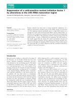
Fig. 2. Affinity of RecA (logarithmic dependencies of apparent I
50
)
to homo-ODN of different lengths (n) determined using inhibition
of RecA filamentation on [
32
P]d(pT)
20
. The I
50
values for different
d(pN)
n
(1 £ n £ 20) and oligonucleotides containing abasic units
or ethylated internucleoside phosphates are obtained using
[
32
P]d(pT)
20
and for d(pT)
20)40
[
32
P]d(pT)
40
(20 £ n £ 40, see the
inset): d(pT)
n
(open sircules; including the inset), d(pA)
n
(cross),
d(pC)
n
(triangles), Positions of –lgI
50
values for ethylated d[(pEt)T]
10
and d[(pT)
2
(pR)
17
(pT)] (pR is a tetrahydrofuran analog of abasic
deoxyribose) are shown.
RecA filament interaction with DNA I. P. Bugreeva et al.
2738 FEBS Journal 272 (2005) 2734–2745 ª 2005 FEBS
the filament assembly on ssDNA occurs cooperatively
in the 5 ¢)3¢ direction [8], the increased affinity of RecA
to the 5¢-terminal phosphate of ODN may be import-
ant for better anchoring of ODN on the first RecA
monomer during the initiation of filamentation.
Ethylation of internucleoside phosphate moieties
neutralizes their charges. The affinity of a RecA fila-
ment for d(pT)
10
(I
50
¼ 2.0 · 10
)4
m) was % 25-fold
lower than for ethylated d[p(Et)T]
10
(I
50
¼ 5.0 ·
10
)3
m) (Table 1), indicative of an important role of
negative charges of internucleoside phosphates for
RecA complexation with DNA.
The affinity of RecA to d(pT)
20
(I
50
¼ 1.0 · 10
)7
m)
was % 100-fold higher than to d[(pT)
2
(pR)
17
pT] (I
50
¼
9.5 · 10
)6
m), a 20-mer lacking 17 out of 20 nucleo-
bases (R is a tetrahydrofuran analog of abasic deoxy-
ribose). As was shown earlier [25–27], deoxyribose
moieties of DNA have little effect on its affinity for
proteins, while internucleoside phosphate groups make
the main contribution. Taking this into account and
assuming that the lack of the bases did not influence
the filament interactions with the backbone, the
increase in affinity due to a single internucleoside
phosphate residue (electrostatic factor e) can be esti-
mated as e ¼ (I
50
¼ 1.75 · 10
)2
m for d(pT)
3
) ⁄ (I
50
¼
9.5 · 10
)6
m for d[(pT)
2
(pR)
17
pT])
1 ⁄ 17
¼ 1842
1 ⁄ 17
¼
1.56 (K
d
¼ 0.64 m). As an increase in the affinity for
one (pT) unit (f ¼ 2.04) is a product of its increase
due to an internucleoside phosphate group (factor e ¼
1.56) and a T base (factor f
T
), f
T
can be calculated as
a ratio f ⁄ e ¼ 1.31 K
d
(T base) ¼ 1 ⁄ 1.31 ¼ 0.76 (m).
The same value of f
T
can be calculated directly: f
T
¼
(I
50
¼ 9.5 · 10
)6
m for d[(pT)
2
(pR)
17
pT]) ⁄ (I
50
¼
1.0 · 10
)7
m for d[(pT)
20
])
1 ⁄ 17
¼ 95
1 ⁄ 17
¼ 1.31. The
affinity increases due to one C base (f
C
¼ 1.31) and
one T base (f
T
¼ 1.31) are the same. Thus, RecA in
the filament forms weak additive contacts with each
internucleoside phosphate moiety and each base of
pyrimidine ODN, with the phosphates contribution
into the affinity being % 1.2-fold more than that of
C or T bases.
As the filament affinity for d(pA)
20
(I
50
¼
2.4 · 10
)5
m) was very similar to that for
d[(pT)
2
(pR)
17
pT] (I
50
¼ 9.5 · 10
)6
m) or for the affin-
ity calculated for a totally abasic oligomer d(pR)
20
(I
50
% 2.1 · 10
)5
m), the filament probably interacts with
adenine bases in DNA very weakly if at all.
RecA interactions with nucleobases
To evaluate the importance of exocyclic acceptor moi-
eties, we have compared the efficiency of RecA filam-
entation on d(pA)
20
and d(pI)
20
, where in the latter,
the O6 acceptor moiety of hypoxanthine base substi-
tutes for the exocyclic amino group of adenine. The
amount of d(pI)
20
incorporated in the filament was less
than with d(pT)
20
but d(pI)
20
formed a stronger com-
plex with RecA than did d(pA)
20
(Fig. 3).
RecA is a DNA-dependent ATPase, with the effi-
ciency of ATP hydrolysis correlating with the stability
and length of the RecA filament [36]. Figure 4 shows
that the extent of ATP hydrolysis correlates well
with the efficiency of RecA filamentation on various
d(pN)
20
, allowing us to use ATP hydrolysis to estimate
the RecA filamentation efficiency and the stability of
the resulting nucleoprotein filaments for a variety of
DNA substrates.
The highest values of ATP hydrolysis rate (expressed
as percentage of initial ATP) in the presence of differ-
ent polynucleotides are summarized in Table 2. The
results show that DNA substrates can be divided into
three classes according to the efficiency of ATP hydro-
lysis stimulation (Table 2). Although both guanine and
hypoxanthine have an acceptor O6 and a donor NH1
moiety, poly(dG) was similar to poly(dA) in poor sti-
mulation of ATP hydrolysis. Deamination of poly(dG)
and poly(dA) significantly increased the rate of ATP
hydrolysis and the efficiency of filamentation. Similar
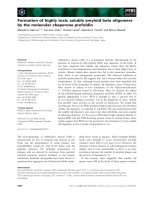
A
B
Fig. 3. Efficiency of RecA filamentation on
32
P-labeled d(pT)
20
,
d(pA)
20
, and d(pI)
20
: electrophoretic mobility shift after 5 min of
incubation (A) and time course of filamentation (B). d(pT)
20
(m),
d(pI)
20
(d), d(pA)
20
(j).
I. P. Bugreeva et al. RecA filament interaction with DNA
FEBS Journal 272 (2005) 2734–2745 ª 2005 FEBS 2739
increase in ATP hydrolysis accompanied a switch from
poly(dAG) to a mixed deoxy(inosine ⁄ xanthine) poly-
mer. DNA containing both purines and pyrimidines
displayed wide variations in its interactions with
RecA. For instance, poly(dAC) and poly(dTG) were
efficiently bound by RecA, poly(dAT) fell between
poly(dA) and poly(dT) ligands, and poly(dCG) promo-
ted very little ATP hydrolysis. The data in Table 2
indicate that purine poly(dN), even those containing
exocyclic hydrogen bond acceptors, generally interacts
with RecA and stimulates ATP hydrolysis less effi-
ciently than pyrimidine polymers. Perhaps the reason
is larger size of purine bases compared with pyrimi-
dines, hindering binding of the former by RecA. In
addition, contacts formed by RecA could be important
not only for the complex formation but also for con-
formational changes in individual RecA monomers
and their ATPase activity.
Deamination of mixed polynucleotides with forma-
tion of dI from dA, dX from dG, and dU from dC,
caused an increase in the efficiency of interactions with
RecA, especially for the poly(dCG) fi poly(dUX)
transition. Interestingly, RecA interaction with purine
ligands was also improved by replacement of adenine
exocyclic amino group with a halogen atom, also a
hydrogen bond acceptor due to its lone electron pairs.
Discussion
We have previously shown that the interaction of dif-
ferent sequence-specific DNA enzymes (repair, topo-
isomerization, restriction, integration enzymes) with
each nucleotide unit of nonspecific ss- or ds-ODNs is
usually a superposition of weak electrostatic and
hydrophobic or van der Waals interactions with the
individual structural elements [25–27]. The interaction
can be described by the power law:
K
d
½dðpNÞ
n
¼K
d
½ðP
i
ÞðeÞ
Àn
ðh
C
Þ
Àc
ðh
T
Þ
Àt
ðh
G
Þ
Àg
ðh
A
Þ
Àa
;
where K
d
[(P
i
)] is the K
d
for the minimal orthophos-
phate ligand (or sometimes dNMPs), e is a factor
reflecting an increase of affinity due to one internucleo-
side phosphate group; h
N
are coefficients of increase in
affinity due to hydrophobic and ⁄ or van der Waals
interactions of the enzyme with one of the bases: C, T,
G and A, the numbers of which in d(pN)
n
are equal to
c, t, g and a, respectively. In addition, factor f reflect-
ing increase in affinity due to one (pN)-unit is equal to
(h
N
· e). When passing from one enzyme to another
only the values of e (1.35–2.0) and h
N
(1.0–1.4) factors
and K
d
for orthophosphate (10
-3
-10
-1
m) or dNMP as
minimal ligands are changed [25–27]. As shown above,
a similar algorithm I
50
[d(pN)
n
] ¼ I
50
(dNMP) · f
1–n
can
be used for description of RecA filament interaction
with ssODNs.
Protein globules of various enzymes usually cover
from 10 to 20 nucleotides of DNA and the affinity of
the enzyme active center (or its specific site) for one
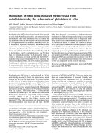
Fig. 4. Time course of RecA filamentation on
32
P-labeled d(pT)
20
,
d(pC)
20
, and d(pA)
20
(A) and RecA-dependent [
32
P]ATP[cP] hydro-
lysis stimulated by the same ODN (B). d(pT)
20
(m), d(pC)
20
(d),
d(pA)
20
(j).
Table 2. Highest levels of RecA-catalyzed ATP hydrolysis in the
presence of various poly(dN).
DNA
ATP
hydrolyzed (%) DNA
ATP
hydrolyzed (%)
poly(dA) 1.6 poly(dG) 3.5
poly(dAT) 33.2 poly(dIT) 63.4
poly(dAC) 59.4 poly(dIX) 43.4
poly(dAG) 1.3 poly(dTX) 63.6
poly(dC) 63.1 poly(dXU) 62.1
poly(dGC) 1.5 poly(dU) 63.0
poly(dT) 61.0 poly(dI) 24.3
poly(dTG) 58.5 poly(dX) 28.4
RecA filament interaction with DNA I. P. Bugreeva et al.
2740 FEBS Journal 272 (2005) 2734–2745 ª 2005 FEBS
nucleotide of d(pN)
10)20
is usually significantly higher
(K
d
¼ 10
)3
)10
)1
m) th an for the remaining 9–19 nucleo-
tides of DNA (K
d
¼ 0.5–0.8 m) [25–27]. RecA was no
exception, accepting free orthophosphate (I
50
¼ 0.5 m)
and various dNMPs (I
50
¼ 12–20 mm) as minimal lig-
ands (Table 1). These experimental I
50
values for free
minimal ligands of RecA do not coincide with K
d
val-
ues reflecting the affinity of a single internucleoside
phosphate (I
50
¼ 0.15–0.23 m ) or a single d(pN) unit
(I
50
¼ 0.063–0.1 m) when they are structural elements
of longer d(pN)
n
(Table 1). Similarly to some other
enzymes [25–27], the latter I
50
values were determined
by extrapolation of lg dependencies to n ¼ 0orn ¼ 1,
respectively (Fig. 2; Table 1). Interestingly, the affinity
of a single internucleoside phosphate or a single d(pN)
unit of d(pN)
n
for RecA is comparable with the
affinity of these DNA structural elements in the case
of other enzymes [25–27].
Usually interactions of various enzymes with mono-
nucleotides of d(pC)
n
, d(pT)
n
, d(pG)
n
and d(pA)
n
are
additive and elongation of these d(pN)
n
by one nucleo-
tide unit results in an increase in the affinity by a factor
f of 1.4–2.0 [25–27]. In principle, similar results were
observed for RecA in the case of all d(pN)
n
(see above).
The affinity of some enzymes for d(pN)
n
does not
always depend on the relative hydrophobicity of the
bases (f ¼ 1). However, if the enzyme interacts with the
bases, the increase in affinity for such ODNs usually
follows the same order as the increase in the relative
hydrophobicity of the bases: C < T < G <A(h
N
¼
1.1–1.4) [25–27]. The likely reason for this correlation
is the formation of very weak hydrophobic and ⁄ or van
der Waals contacts of different efficiency and different
free energy gain upon transfer of these bases from
water to more hydrophobic DNA-binding sites of the
enzymes. In a deviation from this empirical rule, RecA
binds more hydrophobic d(pA)
20
approximately 240-
fold less efficiently than d(pT)
20
and d(pC)
20
(Table 1).
A % 25-fold decrease in the affinity of d[p(Et)T]
10
as
compared with d(pT)
10
(Table 1) has shown that inter-
nucleoside phosphate groups are important for RecA
filament interaction with ssDNA. From the comparison
of I
50
for d[(pT)
2
(pR)
17
pT] and d(pT)
20
(% 100-fold)
the increase in affinity due to a single internucleoside
phosphate residue was estimated as the factor e ¼ 1.56.
The calculated I
50
for totally abasic oligomer d(pR)
20
(% 2.1 · 10
)5
m) was found practically the same as I
50
for d(pA)
20
(2.4 · 10
)5
m) (Fig. 2, Table 1). This data
indicate that the filament probably does not or interact
very weakly with poly(dA) adenine bases and contacts
mostly with its phosphate groups. The factor e (1.56) for
RecA is comparable with e factors for other enzymes:
uracil-DNA glycosylase (1.35), AP endonuclease (1.51),
DNA polymerases (1.52), Fpg (1.54), RNA helicase
(1.61), topoisomerase I (1.67), EcoRI (2.0) and DNA
ligase (2.14) [25–27]. DG° % )0.4 kcalÆmol
)1
corres-
ponding to factor e ¼ 1.56 is significantly lower than
would be expected for strong electrostatic contacts (up
to )1.0 kcalÆmol
)1
), but comparable with the values for
weak ion-dipole and dipole–dipole interactions [38].
Thus, as in the case of the above-mentioned enzymes,
the interaction of negatively charged internucleoside
groups of ODNs with the RecA filament likely relies on
dipolar electrostatic interactions rather than on electro-
static interactions of immediately contacting groups.
From the ratio of factor f ¼ 2.04 reflecting the
increase in the affinity due to one (pN) unit of d(pT)
n
and d(pC)
n
and factor e ¼ 1.56 showing the increase
in affinity due to a single internucleoside phosphate of
these ODNs, the increases in affinity due to RecA
interactions with a single T or C base were estimated
as the factors f
T
¼ f
C
¼ 1.31. Thus, RecA in the fila-
ment forms weak additive contacts with each inter-
nucleoside phosphate mo iety and each base of pyrimidine
ODN, with the phosphates contribution into the affin-
ity being % 1.2-fold more than that of C or T bases.
As the relative affinity of RecA for d(pC)
n
, d(pT)
n
,
and d(pA)
n
does not correlate with the relative hydro-
phobicity of their bases and RecA does not interact
with the bases of d(pA)
n
, it is reasonable to suggest
that RecA could interact with C and T bases by form-
ing specific bonds with appropriate amino acids rather
than through nonspecific hydrophobic contacts.
Wittung et al. reported that entalpy of Rec A bind-
ing to ssDNA in the presence of [
35
S]ATP[cS] depends
on the base sequence with a clear preference to T than
to A and C bases [39]. Similar results concerning
higher affinity of RecA to poly(dT) than to poly(dA)
and poly(dC) were demonstrated in the absence of
cofactor [3]. Thus, our data are in agreement with the
preferential interaction of RecA with d(pT)
n
in com-
parison with d(pA)
n
, but not with the data concerning
d(pC)
n
. However, data about interactions of RecA
with poly(dC) reported in the literature are quite con-
tradictory. Amarahung et al. observed that poly(dC) is
a very bad effector of ATPase activity of RecA [2]. In
contrast, McEttee and Weistock reported poly(dC) to
be the most efficient effector of the RecA ATPase
activity [40]. Binding of RecA to poly(dC) under a var-
ity of conditions has been found to be worse than to
other DNA sequences [40]. Thus, the observed differ-
ences for poly(dC) interaction with RecA cannot be
easily explained.
Unlike C and T bases, adenine possesses no exo-
cyclic acceptors suitable for hydrogen bonding with
RecA amino-acid residues. Deamination of homo- and
I. P. Bugreeva et al. RecA filament interaction with DNA
FEBS Journal 272 (2005) 2734–2745 ª 2005 FEBS 2741
mixed polynucleotides with formation of dI from dA,
dX from dG or and dU from dC containing C ¼ O
exocyclic hydrogen bond acceptors also promote for-
mation of more stable RecA ⁄ ssDNA filament com-
plexes (Table 2). In addition, RecA had high affinity
to poly(dN) containing exocyclic acceptor halogen
atom instead amino group and to d(eA)
n
(eA, 1,N
6
-
ethenoadenine), in which a hydrogen bond donor moi-
ety at C6 is also replaced with an acceptor group (data
not shown). Thus, it can be suggested that the RecA
filament monomers possess in special positions of sites
for binding nucleobases hydrogen bond-donating
groups, which can form contacts with C ¼ O exocyclic
acceptor groups at C6 of purines and C4 of pyrimi-
dines. Figure 5 demonstrates schematically possible
hydrogen bonds of RecA with different DNA bases.
Thus, NH
2
groups of G bases of ss poly(dG) can form
hydrogen bonds with an appropriate group in RecA
(for example, OH groups of Ser, Thr, Tyr, or acidic
amino acids). Oxygen atoms of G bases (Fig. 5) can
interact, for example, with hydrogen atoms of guanidi-
nium groups of Arg residues (or NH
2
groups of Lys
residues). Similar hydrogen bonds can be formed by C
and T bases, but there is no possibility for A bases to
form such bonds (Fig. 5) which may be one reason for
the low affinity of RecA for d(A)
20
(Fig. 2).
As mentioned above, specific interaction of RecA
with one C or T base leads to the increase in d(pN)
n
affinity by a factor of 1.31 (DG° ¼ )0.16 kcalÆmol
)1
).
Interestingly, this DG° value is significantly lower than
DG° values (from )1 kcalÆmol
)1
up to )6 kcalÆmol
)1
)
for strong hydrogen bonds which were observed
between enzymes and different small ligands [38].
However, a formation of very weak hydrogen bonds is
a common situation at recognition of lengthy DNA by
various enzymes [25–27]. During formation of a speci-
fic complex of dsDNA with EcoRI, 12 specific hydro-
gen bonds are formed, providing in total only about
two orders of affinity [32]. This means that the energy
of every of these 12 bonds is rather low (DG°
% )0.23 kcalÆmol
)1
) and comparable with the energy
of weak additive nonspecific interactions (see above).
DG° % )0.28 kcalÆmol
)1
is characterized each of five
pseudo-Watson–Crick hydrogen bonds formed by a
uracil residue with uracil DNA glycosylase [28]. Sim-
ilar weak specific contacts with nucleotides of DNA
were observed for all other investigated sequence speci-
fic enzymes [25–35].
Altogether, the efficiency of RecA filament inter-
action with any individual nucleotide unit (I
50
¼ 0.5–
0.76 m) except one (I
50
% 63–100 mm) is very low.
Nevertheless, the additivity of RecA filament inter-
actions should provide extremely high affinity of the
filament to long ssDNA. It is reasonable to suggest
that the presence of exocyclic acceptor groups capable
of hydrogen bonding to the protein can be a critical
factor accounting for the efficiency of ssDNA binding
by RecA. Depending on the type of the nucleobase
(purine or pyrimidine), the nature of RecA interaction
with the bases and the conformation of RecA monomers
may differ, which could play a key role in the search for
homologous DNA. One cannot exclude that interaction
of complex of RecA filament and ssDNA with dsDNA
can lead to reorganization of firstly formed hydrogen
bonds between protein and bases (Fig. 5) and assist
formation of new hydrogen bonds between C and G
or T and A bases of new DNA duplex.
Experimental procedures
Materials
ATP, ATPcS, poly(N), and poly(dN) were purchased from
Sigma-Aldrich (St. Louis, MO, USA), and [
32
P]ATP[cP]
Fig. 5. Proposed RecA amino-acid residue interactions with G, A, C
and T bases of poly(dN). Impossibility of hydrogen bond formation
is marked (filled star).
RecA filament interaction with DNA I. P. Bugreeva et al.
2742 FEBS Journal 272 (2005) 2734–2745 ª 2005 FEBS
(2000 CiÆmmol
)1
), from Amersham Biosciences (Piscataway,
NJ, USA). Deaminated oligo- and polynucleotides were
synthesized as described in [41,42]. To substitute amino
groups of different nucleobases in polynucleotides with
halogen atoms, the deamination reactions were performed
in the presence of 1 m of respective sodium halides.
ODN were synthesized, purified and characterized as
described [43]. All ODN were proven homogeneous by ion-
exchange and reverse-phase chromatography. Concentra-
tions of the ODNs were determined from their absorption
at 260 nm using molar extinction coefficients calculated
according to [44] ODN were 5¢-labelled using bacteriophage
T4 polynucleotide kinase and [
32
P]ATP[cP]. Electrophoreti-
cally homogeneous E. coli RecA protein was prepared as
described [45].
RecA filamentation
The reaction of RecA filamentation was carried out with
5¢-[
32
P]d(pT)
20
or 5¢-[
32
P]d(pT)
40
at 30 °C for 5 min. The
standard reaction mixture (10 lL) included 50 mm
Tris ⁄ HCl (pH 7.5), 10 mm MgCl
2
,2mm DTT, 1 mm
[
35
S]ATP[cS], 0.1 lm 5¢-[
32
P]d(pT)
20
, and 1 lm RecA.
dNMP, d(pN)
2
or other individual homogeneous d(pN)
n
(n ¼ 3–20), and their modified analogs used as filamenta-
tion inhibitors were added in various concentrations
depending on their affinity. Apparent I
50
values for
d(pT)
20)40
were obtained using 5¢-[
32
P]d(pT)
40
(0.04 lm)as
a filamentation substrate. The reactions were initiated by
adding RecA into the mixture containing 5¢-[
32
P]d(pT)
20,40
and one of the inhibitors. Free 5¢-[
32
P]d(pT)
20,40
was separ-
ated from 5¢-[
32
P]d(pT)
20,40
incorporated in the filament by
electrophoresis in 10–20% nondenaturing polyacrylamide
gel [12] in TBE buffer. The results were visualized by auto-
radiography, the bands were cut out from the gel and their
radioactivity determined by Cherenkov counting. Affinity
of various ligands for RecA was estimated from their I
50
values (inhibitor concentration producing a 50% decrease
in filamentation).
DNA-dependent ATPase activity of RecA
The efficiency of ATP hydrolysis by RecA in the presence
of ssDNA was followed by the decrease in [
32
P]ATP[cP]
and accumulation of
32
P-labelled orthophosphate ([
32
P]P
i
)
using TLC on PEI-cellulose plates in 0.3 m KH
2
PO
4
(pH 7.5). The standard reaction mixture (20 lL) included
20 mm Tris ⁄ HCl (pH 8.0), 10 mm MgCl
2
,30mm NaCl,
1mm DTT, 1 mm ATP, 4 lm RecA, and poly(dN) or
poly(N) in the concentration 0.1 mm nucleotides, or 70 lm
d(pN)
20
or other individual d(pN)
n
(n ¼ 2–40). The mix-
tures were incubated at 30 °C, 2 lL aliquots were with-
drawn and spotted on a TLC plate. Vertical development
of the plate was performed in the ascending mode using 0.3
m potassium phosphate (pH 7.5). The plates were auto-
radiographed, the spots corresponding to [
32
P]ATP[cP] and
[
32
P]P
i
were cut out and their radioactivity determined by
Cherenkov counting.
Acknowledgements
The research was made possible in part by grants from
the Program of Basic research of the Presidium of
RAS ‘Presidium of the Russian Academy of Sciences
(Molecular and Cell Biology Program 10.5)’, from the
Russian Foundation for Basic Research, and from the
Siberian Division of the Russian Academy of Sciences.
References
1 Lusetti SL & Cox MM (2002) The bacterial RecA pro-
tein and the recombinational DNA repair of stalled
replication forks. Annu Rev Biochem 71, 71–100.
2 Amaratunga M & Benight AS (1988) DNA sequence
dependence of ATP hydrolysis by RecA protein. Bio-
chem Biophys Res Commun 157, 127–133.
3 Cazenave C, Chabbert M, Toulme JJ & Helene C
(1984) Absorption and fluorescence studies of the bind-
ing of the recA gene product from E. coli to single-
stranded and double-stranded DNA. Ionic strength
dependence. Biochim Biophys Acta 781, 7–13.
4 McEntee K, Weinstock GM & Lehman IR (1981) Bind-
ing of the recA protein of Escherichia coli to single- and
double-stranded DNA. J Biol Chem 256, 8835–8844.
5 Tracy RB & Kowalczykowski SC (1996) In vitro selec-
tion of preferred DNA pairing sequences by the Escheri-
chia coli RecA protein. Genes Dev 10, 1890–1903.
6 Yu, X & Egelman EH (1992) Structural data suggest
that the active and inactive forms of the RecA filament
are not simply interconvertible. J Mol Biol 227, 334–346.
7 Register JC, 3rd & Griffith J (1985) The direction of
RecA protein assembly onto single strand DNA is the
same as the direction of strand assimilation during
strand exchange. J Biol Chem 260, 12308–12312.
8 Bork JM, Cox MM & Inman RB (2001) RecA protein
filaments disassemble in the 5¢-to 3¢ direction on single-
stranded DNA. J Biol Chem 276, 45740–45743.
9 Stasiak A & Di Capua E (1982) The helicity of DNA in
complexes with recA protein. Nature 299, 185–186.
10 Pugh BF, Schutte BC & Cox MM (1989) Extent of
duplex DNA underwinding induced by RecA protein
binding in the presence of ATP. J Mol Biol 205, 487–
492.
11 Heuser J & Griffith J (1989) Visualization of RecA pro-
tein and its complexes with DNA by quick-freeze ⁄ deep-
etch electron microscopy. J Mol Biol 210, 473–484.
12 Mazin AV & Kowalczykowski SC (1998) The function
of the secondary DNA-binding site of RecA protein
during DNA strand exchange. EMBO J 17, 1161–1168.
I. P. Bugreeva et al. RecA filament interaction with DNA
FEBS Journal 272 (2005) 2734–2745 ª 2005 FEBS 2743
13 Zhurkin VB, Raghunathan G, Ulyanov NB, Camerini-
Otero RD & Jernigan RL (1994) A parallel DNA tri-
plex as a model for the intermediate in homologous
recombination. J Mol Biol 239, 181–200.
14 Kim MG, Zhurkin VB, Jernigan RL & Camerini-Otero
RD (1995) Probing the structure of a putative inter-
mediate in homologous recombination: the third strand
in the parallel DNA triplex is in contact with the major
groove of the duplex. J Mol Biol 247, 874–889.
15 Zhou X & Adzuma K (1997) DNA strand exchange
mediated by the Escherichia coli RecA protein initiates
in the minor groove of double-stranded DNA. Biochem-
istry 36, 4650–4661.
16 Howard-Flanders P, West SC & Stasiak A (1984) Role
of RecA protein spiral filaments in genetic recombina-
tion. Nature 309, 215–219.
17 Roca AI & Cox MM (1997) RecA protein: structure,
function, and role in recombinational DNA repair. Prog
Nucleic Acid Res Mol Biol 56, 129–223.
18 Folta-Stogniew E, O’Malley S, Gupta R, Anderson KS
& Radding CM (2004) Exchange of DNA Base Pairs
that Coincides with Recognition of Homology Pro-
moted by E. coli RecA Protein. Mol Cell 15, 965–975.
19 Voloshin ON & Camerini-Otero RD (2004) Synaptic
complex revisited; a homologous recombinase flips and
switches bases. Mol Cell 15, 846–847.
20 Malkov VA & Camerini-Otero RD (1995) Photocross-
links between single-stranded DNA and Escherichia coli
RecA protein map to loops L1 (amino-acid residues
157–164) and L2 (amino-acid residues 195–209). J Biol
Chem 270, 30230–30233.
21 Morimatsu K, Horii T & Takahashi M (1995) Interac-
tion of Tyr103 and Tyr264 of the RecA protein with
DNA and nucleotide cofactors. Fluorescence study of
engineered proteins. Eur J Biochem 228, 779–785.
22 Morimatsu K & Horii T (1995) Analysis of the DNA
binding site of Escherichia coli RecA protein. Adv Bio-
phys 31, 23–48.
23 Morimatsu K, Funakoshi T, Horii T & Takahashi M
(2001) Interaction of tyrosine 65 of RecA protein with the
first and second DNA strands. J Mol Biol 306, 189–199.
24 Rehrauer WM & Kowalczykowski SC (1996) The DNA
binding site (s) of the Escherichia coli RecA protein.
J Biol Chem 271, 11996–12002.
25 Bugreev DV & Nevinsky GA (1999) Possibilities of the
method of step-by-step complication of ligand structure
in studies of protein–nucleic acid interactions: mecha-
nisms of functioning of some replication, repair, topo-
isomerization, and restriction enzymes. Biochemistry
(Mosc) 64, 237–249.
26 Nevinsky GA (2004) The role of specific and nonspecific
interactions in enzymatic recognition and conversion of
long DNA. Mol Biol (Mosc) 38 , 636–662.
27 Nevinsky GA (2003) Structural, thermodynamic, and
kinetic basis of DNA- and RNA-dependent enzymes
functioning. Important role of weak nonspecific additive
interactions between enzymes and long nucleic acids for
their recognition and transformation. In Protein Struc-
tures. Kaleidoscope of Structural Properties and Func-
tions, (Uversky VN, ed), pp. 133–222. Research
Signpost, Fort P.O., India.
28 Vinogradova NL, Bulychev NV, Maksakova GA, John-
son F & Nevinskii GA (1998) Uracil DNA glycosylase:
interpretation of X-ray data in the light of kinetic and
thermodynamic studies. Mol Biol (Mosc) 32, 489–499.
29 Ishchenko AA, Vasilenko NL, Sinitsina OI, Yamkovoi
VI, Fedorova OS, Douglas KT & Nevinsky GA (2002)
Thermodynamic, kinetic, and structural basis for recog-
nition and repair of 8-oxoguanine in DNA by Fpg pro-
tein from Escherichia coli. Biochemistry 41, 7540–7548.
30 Fedorova OS, Nevinsky GA, Koval VV, Ishchenko AA,
Vasilenko NL & Douglas KT (2002) Stopped-flow
kinetic studies of the interaction between Escherichia
coli Fpg protein and DNA substrates. Biochemistry 41,
1520–1528.
31 Beloglazova NG, Kirpota OO, Starostin KV, Ishchenko
AA, Yamkovoy VI, Zharkov DO, Douglas KT &
Nevinsky GA (2004) Thermodynamic, kinetic and struc-
tural basis for recognition and repair of abasic sites in
DNA by apurinic ⁄ apyrimidinic endonuclease from
human placenta. Nucleic Acids Res 32, 5134–5146.
32 Kolocheva TI, Maksakova GA, Bugreev DV &
Nevinsky GA (2001) Interaction of endonuclease EcoRI
with short specific and nonspecific oligonucleotides.
IUBMB Life 51, 189–195.
33 Bugreev DV, Baranova S, Zakharova OD, Parissi V,
Desjobert C, Sottofattori E, Balbi A, Litvak S, Tarrago-
Litvak L & Nevinsky GA (2003) Dynamic, thermo-
dynamic, and kinetic basis for recognition and
transformation of DNA by human immunodeficiency
virus type 1 integrase. Biochemistry 42, 9235–9247.
34 Bugreev DV, Buneva VN, Sinitsyna OI & Nevinskii GA
(2003) The mechanism of the supercoiled DNA recogni-
tion by the eukaryotic type I topoisomerases. I. The
enzyme interaction with nonspecific oligonucleotides.
Bioorg Khim (Mosc) 29, 143–154.
35 Bugreev DV, Sinitsyna OI, Buneva VN & Nevinskii GA
(2003) The mechanism of supercoiled DNA recognition
by eukaryotic type I topoisomerases: II. A comparison
of the enzyme interaction with specific and nonspecific
oligonucleotides. Bioorg Khim (Mosc) 29, 249–262.
36 Bianco PR & Weinstock GM (1996) Interaction of the
RecA protein of Escherichia coli with single-stranded
oligodeoxyribonucleotides. Nucleic Acids Res 24, 4933–
4939.
37 Leahy MC & Radding CM (1986) Topography of the
interaction of recA protein with single-stranded deoxy-
oligonucleotides. J Biol Chem 261, 6954–6960.
38 Fersht A (1984) Enzyme Structure and Mechanism.
W.H. Freeman, New York.
RecA filament interaction with DNA I. P. Bugreeva et al.
2744 FEBS Journal 272 (2005) 2734–2745 ª 2005 FEBS
39 Wittung P, Ellouze C, Maraboeuf F, Takahashi M &
Norden B (1997) Thermochemical and kinetic evidence
for nucleotide-sequence-dependent RecA–DNA inter-
actions. Eur J Biochem 245, 715–719.
40 McEntee K & Weinstock GM (1981) The RecA enzyme
of Escherichia coli and recombination assays. Enzymes
14, 445–470.
41 Schuster H (1960) The method of reaction of desoxyri-
bonucleic acid with nitrous acid [in German]. Z Natur-
forsch 15B, 298–304.
42 Vielmetter W & Schuster H (1960) Base specificity in
the induction of mutations by nitrous acid in phage T-2
[in German]. Z Naturforsch 15B, 304–311.
43 Ishchenko AA, Bulychev NV, Zharkov DO, Maksakova
GA, Johnson F & Nevinskii GA (1997) Isolation of
8-oxoguanine-DNA-glycosylase from Escherichia coli
and substrate specificity of the enzyme. Mol Biol (Mosc)
31, 332–337.
44 Fasman GD, ed. (1975) Handbook of Biochemistry and
Molecular Biology: Nucleic Acids. CRC Press, Boca
Raton, Florida.
45 Cox MM, McEntee K & Lehman IR (1981) A simple
and rapid procedure for the large scale purification of
the recA protein of Escherichia coli. J Biol Chem 256,
4676–4467.
I. P. Bugreeva et al. RecA filament interaction with DNA
FEBS Journal 272 (2005) 2734–2745 ª 2005 FEBS 2745