Activation of metabolic and stress responses during subtoxic expression of the type i toxin hok in erwinia amylovora
Bạn đang xem bản rút gọn của tài liệu. Xem và tải ngay bản đầy đủ của tài liệu tại đây (2.06 MB, 7 trang )
Peng et al. BMC Genomics
(2021) 22:74
/>
RESEARCH ARTICLE
Open Access
Activation of metabolic and stress
responses during subtoxic expression of
the type I toxin hok in Erwinia amylovora
Jingyu Peng1, Lindsay R. Triplett2 and George W. Sundin1*
Abstract
Background: Toxin-antitoxin (TA) systems, abundant in prokaryotes, are composed of a toxin gene and its cognate
antitoxin. Several toxins are implied to affect the physiological state and stress tolerance of bacteria in a population.
We previously identified a chromosomally encoded hok-sok type I TA system in Erwinia amylovora, the causative
agent of fire blight disease on pome fruit trees. A high-level induction of the hok gene was lethal to E. amylovora
cells through unknown mechanisms. The molecular targets or regulatory roles of Hok were unknown.
Results: Here, we examined the physiological and transcriptomic changes of Erwinia amylovora cells expressing hok
at subtoxic levels that were confirmed to confer no cell death, and at toxic levels that resulted in killing of cells. In
both conditions, hok caused membrane rupture and collapse of the proton motive force in a subpopulation of E.
amylovora cells. We demonstrated that induction of hok resulted in upregulation of ATP biosynthesis genes, and
caused leakage of ATP from cells only at toxic levels. We showed that overexpression of the phage shock protein
gene pspA largely reversed the cell death phenotype caused by high levels of hok induction. We also showed that
induction of hok at a subtoxic level rendered a greater proportion of stationary phase E. amylovora cells tolerant to
the antibiotic streptomycin.
Conclusions: We characterized the molecular mechanism of toxicity by high-level of hok induction and
demonstrated that low-level expression of hok primes the stress responses of E. amylovora against further
membrane and antibiotic stressors.
Keywords: Toxin:antitoxin, Fire blight, Phage shock protein, Transcriptome, Antibiotic tolerance
Background
Toxin-antitoxin (TA) systems are simple genetic loci
that encode a stable proteinaceous toxin and an unstable
counteracting antitoxin. TA systems are widely found
throughout the chromosomes and plasmids of free-living
prokaryotes [1]. In type I TA systems, the antitoxins are
small RNAs that inhibit the translation of or facilitate
the degradation of the transcript encoding the corresponding toxin (reviewed in [2, 3]). Type I toxins, such
* Correspondence:
1
Department of Plant, Soil, and Microbial Sciences, Michigan State University,
East Lansing, MI, USA
Full list of author information is available at the end of the article
as Hok, HokB, and TisB, tend to be small (≤60 amino
acids) hydrophobic proteins containing one transmembrane domain [4–6]. A high induction level of the toxin
genes hok or tisB causes drastic cell death of E. coli cells,
accompanied by collapse of the proton motive force
(PMF) [7–9]. The gene products of both hokB and tisB
form membrane pores in Escherichia coli [8, 10] and
lead to leakage of cellular ATP during moderate [10] or
high-level [7] induction of the toxin genes. The PMF,
the proton gradient generated via oxidation of NADH
and FADH2, is required to generate ATP through ATP
synthase, as well as to power membrane-localized cell
machinery, such as the flagellum [11, 12]. The hok/sok
© The Author(s). 2021 Open Access This article is licensed under a Creative Commons Attribution 4.0 International License,
which permits use, sharing, adaptation, distribution and reproduction in any medium or format, as long as you give
appropriate credit to the original author(s) and the source, provide a link to the Creative Commons licence, and indicate if
changes were made. The images or other third party material in this article are included in the article's Creative Commons
licence, unless indicated otherwise in a credit line to the material. If material is not included in the article's Creative Commons
licence and your intended use is not permitted by statutory regulation or exceeds the permitted use, you will need to obtain
permission directly from the copyright holder. To view a copy of this licence, visit />The Creative Commons Public Domain Dedication waiver ( applies to the
data made available in this article, unless otherwise stated in a credit line to the data.
Peng et al. BMC Genomics
(2021) 22:74
TA system in E. coli has been suggested as a target for
killing host bacterial cells [13, 14]. Through sequestering
the sRNA sok from interacting with hok mRNA by
addition of anti-Sok peptide nucleic acid (PNA) oligomers [13] or doxycycline that inhibits RNase III degradation of the hok-sok dsRNA complex, hok mRNA is
released and consequently causes cell death [14].
The molecular targets and regulatory roles of many
TA systems are still enigmatic. Although inactivation of
a single type I TA system does not frequently result in a
phenotype [15], studies using low-level ectopic expression have revealed that a few membrane-associated TA
systems can affect the physiological state and stress tolerance of bacteria in a population. In E. coli, expression
of hokB or tisB at sub-toxic levels increased the proportion of persister cells with tolerance to multiple antibiotics, which was hypothesized to result from growth
retardation following ATP leakage and the loss of the
PMF [7, 8, 15–17]. Plasmid expression of the hok-sok
locus also increased T4 bacteriophage exclusion in E.
coli [18]. Interestingly, despite its role in compromising
membrane integrity, moderate hokB expression was observed to increase metabolic activity in E. coli, determined via a fluorescent redox sensor [10].
Through transcriptomics and in vitro RNA degradation analyzes, Wang et al. demonstrated that the type V
antitoxin GhoS cleaves the membrane-associated toxin
ghoT mRNA [19]. However, the global transcriptional effects of a type I membrane-associated TA, to the best of
our knowledge, have not been previously examined. It
has been hypothesized that induction of hokB may activate phage shock protein (psp) genes, based on the protective effects of Psp proteins in mitigating various
membrane stresses in E. coli [20, 21]. Though the effects
vary in different bacteria, perturbation of the cell membrane seems to cause shared consequences in activating
stress responses and downregulating genes that encode
energy consuming machinery [22–26]. Addition of polymyxin, an antibiotic that causes formation of membrane
pores and cell death in bacteria, caused increased expression of genes associated with vancomycin resistance
and decreased expression of virulence factor-related
genes in Staphylococcus aureus [22]; exposure of Klebsiella pneumoniae to 1-(1-Naphthylmethyl)-piperazine
depolarized the membrane PMF yet upregulated many
envelope stress response genes [26]. Still, it is not known
whether endogenous pore-forming toxins also trigger
stress response or influence the expression of virulence
genes.
Recently, we identified a chromosomally encoded hoksok type I TA system in Erwinia amylovora [27], a model
enterobacterial plant-pathogenic bacterium that causes
the destructive fire blight disease of pome fruit trees including apple (Malus sp.) and pear (Pyrus sp.) [28, 29].
Page 2 of 15
Episomal overexpression of the hok gene caused massive
killing of E. amylovora cells and arrested cell division
after septa were formed [27]. We proposed that cell
death due to hok induction at toxic levels in E. amylovora is likely to be associated with the disturbance of essential functions of the cell membrane. Although
upregulation of toxin genes occurs under a variety of different stress conditions [30–34], natively expressed toxin
genes are not known to be induced to cell-killing levels
in any environmental context, to the best of our knowledge. Therefore, we hypothesized that hok might actually confer a selective advantage to E. amylovora at
moderate (subtoxic) levels of induction, when no cell
death is observed. In this study, we compared the transcriptome profiles of E. amylovora cultures expressing
hok at toxic, subtoxic, and wild-type levels. We found
that Hok plays important roles in activating ATP biosynthesis and priming the tolerance of E. amylovora cells
against membrane and antibiotic damage.
Results
Moderate overexpression of hok does not suppress
bacterial growth
A hok overexpression construct, pOE-hok, was previously generated by cloning the E. amylovora Ea1189 hok
gene into the lac promoter-containing plasmid pEVS143
[27]. The lac promoter allows low levels of transcription
in the absence of the inducer isopropyl β-D-1-thiogalactopyranoside (IPTG) [35]. We did not observe any
growth defect in E. amylovora Ea1189 cells transformed
with pOE-hok (Fig. S1), suggesting that E. amylovora is
able to tolerate leaky hok expression without inhibiting
growth. Therefore, we hypothesized that Ea1189(pOEhok) grown in the absence of IPTG induction may provide a useful system to identify the physiological roles of
Hok separate from those caused by its toxicity. We used
quantitative real-time PCR (qRT-PCR) to measure the
expression levels of hok in Ea1189(pEVS143) and
Ea1189(pOE-hok) without IPTG and in four progressively increasing doses of IPTG, and monitored the
growth of the cultures in the same conditions. In the
absence of IPTG, expression of hok was approximately
40-fold higher in Ea1189(pOE-hok) compared to
Ea1189(pEVS143), and expression of hok increased by
another ~ 130-fold when 1 mM IPTG was added to the
Ea1189(pOE-hok) culture (Fig. 1a). The expression levels
of the small RNA antitoxin sok remained almost unchanged in these conditions (Fig. 1a). Induction of hok
did not result in cell death until expression reached
about 60-fold induction or greater, induced by the
addition of 0.01 mM IPTG (Fig. 1b). Henceforth, we will
define hok expression from the lac promoter with 0.01
mM, 0.1 mM or 1 mM IPTG as the “toxic” expression
conditions for this study, while expression from the lac
Peng et al. BMC Genomics
(2021) 22:74
Page 3 of 15
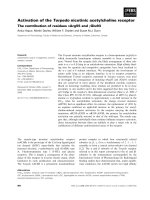
Fig. 1 Induction of hok and its effect on cell survival in E. amylovora. a Expression levels of hok induced with four progressive doses (0.001 mM,
0.01 mM, 0.1 mM, and 1 mM) of isopropyl β-D-1-thiogalactopyranoside (IPTG) or with water. b The effect hok induction on survival rate of E.
amylovora. The concentrations of IPTG supplemented are indicated in parentheses. After IPTG or water addition, cultures were incubated at 28 °C
with 200 rpm shaking for 1 h. Expression levels of hok were measured using quantitative real-time PCR (qRT-PCR), and fold changes were
calculated using the 2-ΔΔCT formula. The recA gene was used as an endogenous control. Survival rate was determined as the ratio of colony
forming units (CFU)/ml in Ea1189(pOE-hok) after and before the addition of IPTG. Results represent the means of three replications, and error bars
indicate the standard deviation. Different letters indicate significant differences (P < 0.05) using Tukey’s HSD (honestly significant difference) test.
The experiments were conducted three times with similar results
promoter with 0.001 mM or no IPTG will be defined as
the “subtoxic” expression conditions.
Induction of hok causes PMF collapse and membrane
rupture
Membrane-associated type I toxins of E. coli, including HokB and TisB, form membrane pores [8,
10, 19], and cause collapse of the PMF [9, 10, 17].
We therefore wondered if the transmembrane
domain-containing E. amylovora Hok, sharing 48
and 14% amino acid identity to HokB and TisB, respectively, also causes membrane depolarization and
rupture. To assess this possibility, we measured
membrane potential using DiBAC 4 [3] (bis-(1,3dibutylbarbituric acid) trimethine oxonol), a membrane potential-sensitive fluorescent dye. Fluorescence level negatively correlates to membrane
potential, meaning that higher fluorescence indicates a greater level of PMF collapse. Carbonyl
cyanide-m-chlorophenylhydrazone (CCCP), a protonophore that uncouples the PMF, was used as a
positive control for the DiBAC 4 [3] staining (Fig.
S2). Propidium iodide (PI) was used as an indicator
of membrane rupture, which binds to nucleic acid
and generates fluorescence in membrane integrity
compromised cells. Ethanol disturbs the physical
structure of cell membranes and was used as a positive control for the PI staining (Fig. S2). Fluorescence
was measured in single cells using a flow cytometer.
We found that induction of hok to subtoxic levels
caused membrane depolarization and rupture in a
subpopulation of cells, though many cells remained
unchanged in their membrane states (Fig. 2a). More
drastic membrane depolarization and rupture was
observed when hok was induced to toxic levels (Fig.
2a). At the highest level of hok induction, almost
the entire population was shifted to the membrane
depolarization state, with varied levels of membrane
rupture. We next asked whether mannitol, a bacterial metabolite that feeds into glycolysis and was
shown to stimulate the PMF in E. coli [36], was
able to restore the collapsed PMF and rupture of
cell membrane due to the toxicity of Hok in E.
amylovora. In cells expressing hok with 0.1 mM
IPTG induction, mannitol partially relieved the
membrane stress (Fig. 2a). Similarly, addition of
mannitol significantly alleviated the inhibitory effect
of bacterial growth during 0.01 or 0.1 mM induction
of hok (Fig. 2b). However, when 1 mM IPTG was
supplemented, the protective effect of mannitol was
not observed in any of these phenotypes (Fig. 2a
and Fig. 2b). Arabinose, which does not contribute
to the PMF [36], was used a negative control for
the assays (Fig. S3).
Transcriptomic analysis reveals that hok overexpression
affects genes involved in stress responses and energy
generation/consumption
While overexpression of E. amylovora hok causes extreme disturbance of essential membrane functions, it is
not clear how the membrane disruption capacity of these
toxins may affect bacterial physiology when hok is
expressed in subtoxic or native expression conditions.
To distinguish potential downstream effects of E. amylovora Hok from those resulting from toxicity, we compared the transcriptomes of E. amylovora cultures
Peng et al. BMC Genomics
(2021) 22:74
Page 4 of 15
Fig. 2 hok induction disturbs essential membrane functions of E. amylovora. Effect of hok induction on the proton motive force (PMF) and
membrane integrity without (panel labelled as “None”) or with the addition of 10 mM mannitol (panel labelled as “Mannitol”) immediately before
IPTG supplementation (a), and effect of mannitol in reversing the toxicity of Hok (b). E. amylovora cultures grown overnight for 20 h in LB broth
were washed twice and diluted to OD600 = 0.2 in fresh LB broth. The concentrations of IPTG supplemented are indicated in parentheses. After
incubation at 28 °C with 200 rpm shaking for 1 h, the PMF of cultures was examined using bis-(1,3-dibutylbarbituric acid) trimethine oxonol
(DiBAC4 [3]), and membrane integrity was determined via propidium iodide (PI). Fluorescence was measured using a BD LSR II flow cytometer.
Ten thousand events were examined with a 488 nm laser and a 530/30 emission filter (DiBAC4 [3]) staining and a 561 nm laser and a 620/15
emission filter (PI). Subsequent analyses were conducted on Flowing Software 2.5.1 and R v3.4.0. Increased fluorescence after treatment with
DiBAC4 [3] or PI indicates greater collapse of the PMF or compromised membrane integrity, respectively. To test the effect of bacterial metabolites
on the toxicity of Hok, 10 mM mannitol or 10 mM arabinose was added to the cultures (OD600 = 0.2) immediately before IPTG was added, and the
OD600 was measured 4 h after the incubation using a Tecan spectrophotometer. Bacterial growth was monitored by measuring OD600 of the cultures
using a Tecan spectrophotometer. Results represent the means of three biological replicates and error bars indicate the standard deviation. Different
letters indicate significant differences (P < 0.05) using Student’s t-test. The assays were done three times with similar results
expressing hok at wild-type levels (i.e., wild-type strains
carrying the empty vector) with cultures expressing hok
at subtoxic (IPTG untreated) and toxic (1 mM IPTG
treated) levels. Expression of each gene was quantified as
counts per million reads (CPM), and differentiallyexpressed genes (DEGs) were defined as those having
greater than 2-fold change of CPM values and less than
0.05 of the corresponding false discovery rate (FDR)
values (Fig. S4 and Table S1).
Compared with Ea1189(pEVS143), which was also untreated with IPTG, 321 DEGs were identified in IPTGuntreated Ea1189(pOE-hok), of which 234 had increased
expression and 87 had decreased expression (Fig. 3a).
After 1 mM IPTG treatment of Ea1189(pOE-hok), a
much larger set of 541 and 560 genes were up- and
down-regulated, respectively (Fig. 3a). Approximately
83% of the DEGs identified in the subtoxic condition
were differentially expressed in the same direction and
Peng et al. BMC Genomics
(2021) 22:74
Page 5 of 15
Fig. 3 Comparative transcriptomic analysis of E. amylovora cells expressing hok at wild-type, subtoxic and toxic levels, respectively. a Venn
diagram of the differentially-expressed genes (DEGs) in E. amylovora cells expressing hok at subtoxic or toxic level. b Expression of representative
DEGs in subtoxic or toxic condition examined using qRT-PCR. Fold changes were calculated using the 2-ΔΔCT formula. The recA gene was used as
an endogenous control. The error bars indicate standard deviation. The concentrations of IPTG supplemented are indicated in parentheses
to a greater extent in the toxic condition. Expression
of representative genes in Ea1189(pOE-hok) in subtoxic and toxic conditions was validated through
qRT-PCR (Fig. 3b). The housekeeping gene recA was
used as an endogenous control, that had negligible
differences in expression among E. amylovora cultures expressing wild-type, subtoxic, or toxic levels
of hok in our transcriptomic analysis. Based on the
read count, the ratio of hok to sok was approximately 18 in the wild-type condition, that increased
to ~ 200 in the subtoxic condition and ~ 6000 in the
toxic condition (Fig. S5). Gene ontology (GO) enrichment analysis of the DEGs further revealed that
hok exerts substantial effects in the essential metabolism of E. amylovora (Fig. 4 and Table S2). Oxidative phosphorylation-related genes (GO:0006119),
that include NADH-coenzyme Q oxidoreductase
(complex I), Succinate-Q oxidoreductase (complex
II), Cytochrome c oxidase (complex IV) and F1FoATPase (complex V), were enriched among the
higher expressed genes in both toxic and subtoxic
conditions. Specifically, in the toxic condition, higher
expressed genes were also significantly associated
with the “tricarboxylic acid cycle” GO term (GO:
0006099).
Several genes with demonstrated importance to bacterial plant pathogenesis were negatively affected by
elevated hok expression. Specifically, hrpA and flhD,
encoding a T3SS protein and a flagellar transcriptional activator, respectively, decreased in expression
at both levels of hok induction. In toxic but not subtoxic conditions, down-regulated genes were primarily
comprised of flagellar genes and “protein secretion”
(GO:0009306) genes, which included type II secretion
system (T2SS) and type III secretion system (T3SS)related genes.
Induction of hok also activated multiple genes involved in stress responses. Several genes with known
roles in antibiotic persistence and other stress responses, i.e. groS, groL, dnaK, dnaJ, skp, surA, sucB
and lon [37–42], were consistently more highly
expressed in both hok induction conditions. Also upregulated were genes in the “response to virus” ontology (GO:0009615), including genes encoding phage
shock proteins, i.e. pspABCD, and CRISPR-associated
proteins. The catalase gene katA showed increased
expression in the subtoxic condition, consistent with
our previous observation that catalase activity is significantly compromised in a hok-sok deletion mutant
[27]. The stress-induced ATP-dependent chaperone
gene clpB was also more highly expressed in the subtoxic but not the toxic condition. Together, these results show that different hok expression levels exert
diverse and overlapping effects on the E. amylovora
transcriptome, enhancing expression of metabolic and
stress-related traits while suppressing genes required
for infection.
hok positively affects ATP biosynthesis
Membrane-associated type I toxins have been shown to
cause leakage of cellular ATP as indicated by either decrease level of intracellular ATP or increase level of
extracellular ATP [7, 10, 19]. In this study, we found
that genes associated with oxidative phosphorylation, the
process of ATP generation through electron transfer,
were higher expressed in the subtoxic condition and
were higher expressed to a greater extent in the toxic
condition (Fig. 5). We hypothesized that the upregulation of ATP biogenesis-related genes could be part of a
response to compensate for the possible leakage of intracellular ATP through increased ATP synthesis in
Ea1189(pOE-hok) cultures in both subtoxic and toxic
conditions. To determine whether ATP leakage was occurring, we performed simultaneous measurements of
both the intracellular and the extracellular levels of ATP
in both subtoxic and toxic conditions. When induced
Peng et al. BMC Genomics
(2021) 22:74
Page 6 of 15
Fig. 4 Overrepresented Gene Ontology (GO) terms enriched in the GO enrichment analysis with a cutoff FDR of 0.01. Scale bar indicates the
color key of log2 fold-change values
with 0.1 or 1 mM IPTG, conditions causing more than
70% dieoff (Fig. 1b), E. amylovora Hok caused dramatic
leakage of ATP from the cells, indicated by the decreased level of intracellular ATP and increased level of
extracellular ATP (Fig. 5 and Fig. S6). In contrast, a significant increase in intracellular ATP was measured after
induction with 0.01 mM or less IPTG (Fig. 5 and Fig.
S6), expression conditions that were associated with
minimal or no cell death of E. amylovora (Fig. 1b). No
ATP leakage was observed in these subtoxic conditions.
Combining intercellular and extracellular ATP measurements allowed us to assess the total ATP concentration under each expression condition. In the absence of
IPTG, total ATP was greater in Ea1189(pOE-hok) cultures than Ea1189(pEVS143). Total ATP in
Ea1189(pOE-hok) increased with IPTG addition at concentrations up to 0.1 mM (Fig. 5). At the highest
concentration of IPTG tested, 1 mM, the total ATP in
Ea1189(pOE-hok) cultures started to decrease compared
with lower levels of inducer, likely due to the massive
kill-off of ATP-generating cells at this induction level.
Taken together, our results suggest that hok positively
affects the biosynthesis of ATP, and leakage of ATP only
occurs when hok was induced at toxic levels.
Overexpression of the ATP synthase gene atpB is toxic
to E. coli cells; it allows leakage of protons through the
F0 sector of F1Fo-ATPase [43–46]. Given that hok positively affects ATP synthase gene expression and ATP
biosynthesis in subtoxic conditions, we wondered if the
toxicity of Hok was increased by the upregulation in
ATP synthase genes. To test this hypothesis, we generated
ATP synthase gene deletion mutants, Ea1189ΔatpB and
Ea1189ΔatpBEFHAGDC. The growth of Ea1189ΔatpB
and Ea1189ΔatpBEFHAGDC mutants was severely
Peng et al. BMC Genomics
(2021) 22:74
Page 7 of 15
Fig. 5 Effect of hok induction on ATP biosynthesis in E. amylovora. Both extracellular and intracellular levels of ATP were simultaneously
quantified using a luciferase reporter system. Results represent the means of three biological replications and error bars indicate the standard
deviation. Different letters indicate significant differences (P < 0.05) using Tukey’s HSD test. The assays were done twice with similar results
reduced, as overnight cultures only reached OD600 ≈ 0.3
compared with OD600 ≈ 1.5 in the wild-type Ea1189 strain
(data not shown). pOE-hok was transformed into the ATP
synthase mutants to generate Ea1189ΔatpB(pOE-hok) and
Ea1189ΔatpBEFHAGDC(pOE-hok), respectively. Hok expression was induced in the wild-type and ATPase mutant
backgrounds with 1 mM IPTG, and survival rates were
measured. Hok killing efficiency was not changed between
the wild-type and the mutants (Fig. S7), suggesting that
the toxicity of Hok is not affected by the increased expression of ATP biosynthesis genes.
Expression of pspA is induced in known PMF dissipation
conditions and relieves the toxicity of Hok
Our transcriptome results indicated that psp genes were
upregulated in both expression conditions. The psp
genes are induced on exposure to conditions that dissipate the PMF, such as bacteriophage infection, alkaline
pH, and addition of uncoupling agents, in both Gramnegative and -positive bacteria (reviewed in [47]). The
protective roles of PspA in managing membrane stresses
have been validated in E. coli and Salmonella enterica
serovar Typhimurium [48–50]. As the functions of psp
genes have not been previously investigated in E. amylovora, we constructed a transcriptional fusion of the promoter region of the pspABCD operon to a green
fluorescence protein (gfp) reporter. As expected, the promoter activity of the pspABCD operon was significantly
increased in E. amylovora cells after exposure to bacteriophage, and was increased to a lesser extent in the
presence of CCCP, ethanol, or Triton X-100 (Fig. S8).
To examine the possible protective role of pspA under
the condition of membrane stress in E. amylovora, we
generated the pspA-overexpression construct, pBAD33pspA, through cloning the pspA gene into the pBAD33
plasmid, containing the arabinose-inducible PBAD promoter. Compared with Ea1189(pBAD33), Ea1189(pBAD33-pspA) cultures were ~ 100 times more tolerant
to CCCP (Fig. 6a). Interestingly, without supplementing
any IPTG, Ea1189(pOE-hok) cultures survived at significantly higher rates than Ea1189(pEVS143) (Fig. 6a), suggesting that induction of hok at subtoxic levels protect E.
amylovora cells from further membrane damage by activating the expression of pspA. Interestingly, pspA overexpression significantly alleviated the toxicity due to
high levels of hok induction (Fig. 6b), further validating
the defensive role of pspA in response to membrane
stress in E. amylovora.
Subtoxic expression of hok increases tolerance of
stationary-phase E. amylovora cells to the aminoglycoside
antibiotic streptomycin
Transcriptome results showed that hok expression upregulated several genes previously associated with antibiotic persistence, so we next asked whether hok has a
role in antibiotic tolerance during stationary phase.
Without addition of IPTG, stationary phase E. amylovora cultures expressing hok had 10 times the number of
survivors to streptomycin exposure than the vector control strain (Fig. 7). concentration that is routinely used
for management of fire blight and screening of
streptomycin-resistant E. amylovora isolates [51–53]. Of