Báo cáo khoa học: Nuclear import of mPER3 in Xenopus oocytes and HeLa cells requires complex formation with mPER1 pdf
Bạn đang xem bản rút gọn của tài liệu. Xem và tải ngay bản đầy đủ của tài liệu tại đây (565.79 KB, 11 trang )
Nuclear import of mPER3 in Xenopus oocytes and HeLa
cells requires complex formation with mPER1
Susanne Loop and Tomas Pieler
Abteilung Entwicklungsbiochemie, Zentrum fu
¨
r Biochemie und Molekulare Zellbiologie, Georg-August Universita
¨
t, Go
¨
ttingen, Germany
The genetic control of circadian rhythmicity was first
analysed in Drosophila. A central autoregulatory feed-
back loop that involves different transcriptional regula-
tors was uncovered. The bHLH transcription factors
CLOCK (CLK) and CYCLE (CYC) drive expression
of the period (per) and timeless (tim) genes. Conversely,
Period and Timeless proteins (PER and TIM) inhibit
CLK ⁄ CYC-mediated transcription of their own genes,
resulting in a gradual loss of PER and TIM proteins.
At a critically reduced level of PER and TIM protein
activity, CLK ⁄ CYC repression is relieved and per ⁄ tim
gene expression returns [1–5].
A similar mechanism seems to operate in verte-
brates. In mammals, CLOCK–BMAL1 heterodimers
activate transcription of Period (mPer) and Crypto-
chrome ( mCry) genes. mPER and mCRY proteins act
as negative regulators of their own expression by
directly interacting with and thereby inhibiting
CLOCK–BMAL1 [6,7]. Gene duplications have gener-
ated three different mPER proteins (mPER1, mPER2
and mPER3) and two different mCRY proteins
(mCRY1 and mCRY2). Functional diversity among
the individual members of each of these clock protein
subfamilies has been reported [8–15].
Post-translational control constitutes a further
important level of regulation in both vertebrate and
invertebrate systems. In Drosophila, phoshorylation of
both PER and TIM affects stability and ⁄ or nuclear
transport [16–21]. Phosphorylated forms of the two
proteins are targeted for degradation by the ubiquitin–
proteasome pathway [22–24]. It has also been pro-
posed that PER⁄ TIM phosphorylation may promote
nuclear transfer, but more recent studies argue in
favour of phosphorylation positively regulating their
transcriptional repressor activity [25]. Conversely, the
regulated rhythmic dephosphorylation of PER by pro-
tein phosphatase 2A stabilizes PER, thereby contribu-
ting to the rhythmicity of PER protein concentrations
[26].
Mammalian PER proteins have also been found to
become phosphorylated; mPER1, mPER2 and mPER3
are subjected to rhythmical phosphorylation mediated
Keywords
circadian rhythm; mCRY, mPER; nuclear
import; Xenopus oocytes
Correspondence
T. Pieler, Abteilung Entwicklungsbiochemie,
Zentrum fu
¨
r Biochemie und Molekulare
Zellbiologie, Georg-August Universita
¨
t,
Justus von Liebig Weg 11, D-37077
Go
¨
ttingen, Germany
Fax: +49 551 3914614
Tel: +49 551 395683
E-mail:
(Received 14 March 2005, revised 27 May
2005, accepted 31 May 2005)
doi:10.1111/j.1742-4658.2005.04798.x
Several transcription factors with the function of setting the biological
clock in vertebrates have been described. A detailed understanding of their
nucleocytolasmic transport properties may uncover novel aspects of the
regulation of the circadian rhythm. This assumption led us to perform a
systematic analysis of the nuclear import characteristics of the different
murine PER and CRY proteins, using Xenopus oocytes and HeLa cells as
experimental systems. Our major finding is that nuclear import of mPER3
requires complex formation with mPER1. We further show that the nuclear
localization signal (NLS) function of mPER1 and not activation of a
masked NLS in mPER3 is critical for the import of the mPER1–mPER3
complex. Finally, and as previously described in other cell systems, nuclear
import of mPER proteins in Xenopus oocytes correlates positively with
their phosphorylation.
Abbreviations
CK, casein kinase; NLS, nuclear localization signal.
3714 FEBS Journal 272 (2005) 3714–3724 ª 2005 FEBS
by casein kinases (CKIe and CKId) [27–30]. The phos-
phorylation status of murine PER proteins, similar to
that reported for Drosophila PER, influences stability
and nuclear transport; phosphorylated forms of
mPER1 and mPER3 are rapidly degraded [31,32].
Furthermore, mPER1 mutant mice have been used to
demonstrate that mPER1 is required for phosphoryla-
tion and nuclear transfer of mPER3 [33]. Phosphoryla-
tion of mPER1 itself correlates with nuclear transport
[34]. Earlier studies had already indicated that nuclear
translocation of mPER3 is promoted by mPER1 in
NIH3T3 cells [14].
Other studies, using different cell systems, had come
to additional and sometimes apparently contradictory
conclusions. Yagita et al. [35], using COS7 cells, repor-
ted that mPER3 by itself is predominantly cytoplas-
mic, and nuclear accumulation is obtained by serum
shock-induced formation of mPER1 ⁄ 3 or mPER2 ⁄ 3
heterodimers. Furthermore, Vielhaber et al. [36] had
observed that mPER1 is predominantly nuclear,
whereas mPER2 is predominantly cytoplasmic in
HEK293 cells; CKIe-mediated phosphorylation of
mPER1 was reported to lead to masking of the nuclear
localization signal (NLS) and coexpression of mPER1
with mPER2 and cytoplasmic localization of the
heterodimer. Finally, Miyazaki et al. [37], using COS1
cells, had observed that mammalian PER2 has a posit-
ive regulatory function with respect to the nuclear
import of mCRY1.
What all these studies have in common is the idea
that dimerization of the different mammalian PER and
CRY proteins modulates their nucleocytoplasmic dis-
tribution and thereby probably also their function as
transcriptional repressors. In the work presented here,
we systematically analyzed nuclear import of the dif-
ferent murine PER and CRY proteins, either individu-
ally or in all possible heterodimeric combinations,
primarily using Xenopus oocytes as an experimental
system. We found that interaction with mPER1 is
required for the nuclear import of mPER3, and we
observed a positive correlation between nuclear import
of mPER proteins and their phosphorylation.
Results
Positive correlation between phosphorylation
and nuclear import of mPER proteins in Xenopus
oocytes
The different individual murine PER and CRY pro-
teins were generated by in vitro translation and injected
into the cytoplasm of Xenopus oocytes. The kinetics of
nuclear import were analysed after manual separation
of cytoplasmic and nuclear fractions by gel electro-
phoresis (Fig. 1A). The data obtained reveal that,
whereas the mCRY proteins, as well as mPER1 and
mPER2, are readily imported into the nucleus of Xeno-
pus oocytes, mPER3 is not. We also observed reduced
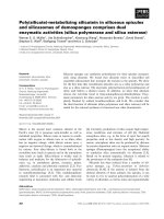
A
B
Fig. 1. Nuclear import of murine PER and CRY proteins in Xenopus oocytes. mPER1 and mPER2, but not mPER3, are phosphorylated and
imported into the nucleus of Xenopus oocytes. (A)
35
S-labelled mPER1, mPER2, mPER3 and derived protein fragments fused to six copies
of the myc tag in tandem repeat were translated in vitro and injected into the cytoplasm of Xenopus oocytes. The nucleus and cytoplasm
were separated manually at the time points indicated. Proteins were immunoprecipitated from 10 pooled nuclear and cytoplasmic fractions
using the myc antibody and analysed by SDS ⁄ PAGE and phosphorimaging. To test for phosphorylation, immunoprecipitated nuclear or cyto-
plasmic fractions were incubated with lambda protein phosphatase (k PPase) before gel electrophoresis. (B) mCRY1 and mCRY2 are impor-
ted into the nucleus of Xenopus oocytes.
S. Loop and T. Pieler Nuclear import of circadian clock proteins
FEBS Journal 272 (2005) 3714–3724 ª 2005 FEBS 3715
electrophoretic mobility of mPER1 and mPER2, but
not of mPER3, which increases with the time of incu-
bation after microinjection. It also seems that the
relative amount of the phosphorylated forms of the
proteins is higher in the nucleus than in the cytoplasm.
Reduced electrophoretic mobility suggests chemical
modification events, such as phosphorylation. Phos-
phatase treatment of cytoplasmic and nuclear protein
fractions isolated from microinjected oocytes equalizes
the electrophoretic mobility of all samples tested,
revealing that mPER1 and mPER2 are indeed phos-
phorylated after injection into Xenopus oocytes. Thus,
we found a positive correlation between phosphoryla-
tion and nuclear import for mPER1 and mPER2,
whereas mPER3, which is not imported into the nuc-
leus, is also not phosphorylated. On the other hand,
there is no evidence for phosphorylation of mCRY1
and mCRY2, which are readily imported into the nuc-
leus of injected oocytes (Fig. 1B).
The absence of nuclear import of mPER3 injected
into Xenopus oocytes suggests that the protein is
devoid of a nuclear import signal that is functional in
this experimental system. To address this question, all
three murine PER proteins were broken down into
four fragments, and each one tested for nuclear
import activity in Xenopus oocytes (Fig. 2). In agree-
ment with earlier NLS-mapping experiments in other
experimental systems [31,35–37], the corresponding
region (fragment 3) of all three PER proteins har-
bours a functional NLS. Mutation of the putative
NLS in mPER3 abrogates import activity (data not
shown). In extension of previous studies, we further
detected a novel, additional NLS located in the C-ter-
minal portion (fragment 4) of mPER1 within the 186
C-terminal amino acids (Fig. 3, fragment 4b). We
also noted faint nuclear signals for the corresponding
C-terminal fragments derived from mPER2 and
mPER3 (Fig. 2A). However, nuclear import rates
A
B
Fig. 2. mPER1 contains an additional NLS in its C-terminal domain. (A) Mapping of NLS function in murine Per proteins. Fragments corres-
ponding to different portions of mPER1, mPER2 and mPER3 (as indicated) were assayed for nuclear import in Xenopus oocytes. MPER2
Frag2 was rapidly degraded in Xenopus oocytes. mPER1: myc Frag 1, aa 1–323; myc Frag 2, aa 324–645; myc Frag 3, aa 646–972; myc Frag
4, aa 973–1291. mPER2: myc Frag 1, 1–314; myc Frag 2, aa 314–628; myc Frag 3, 629–942; myc Frag 4, aa 943–1257. mPER3: myc Frag 1,
aa 1–280; myc Frag 2, 281–559; myc Frag 3, 560–835; myc Frag 4, 836–1113. (B) Schematic representation of the fragments used for map-
ping experiments and percentage of nuclear import in multiple independent experiments. The grey boxes define the location of the NLSs in
the Per proteins (in bold the newly identified NLS2 in mPER1).
Nuclear import of circadian clock proteins S. Loop and T. Pieler
3716 FEBS Journal 272 (2005) 3714–3724 ª 2005 FEBS
below 10% (Fig. 2B) are considered nonsignificant. A
primary sequence comparison of the three murine
PER proteins revealed a high degree of structural
diversity in the C-terminal domain (data not shown),
correlating with functional diversity with respect to
NLS activities. Mutation or deletion of one of the
two NLSs in mPER1 led to reduced nuclear import.
A complete block occurred only after mutation ⁄ dele-
tion of both NLSs (Fig. 3, myc- mPER1mutNLSDC);
phosphorylation was not affected in these mutants
(data not shown).
Alternative explanations exist for the observed
absence of mPER3 nuclear import when injected by
itself; either it is rapidly degraded in the nucleus or
rapid export prevents its nuclear accumulation. How-
ever, in a separate study on the nuclear export of clock
proteins in Xenopus oocytes [38], we observed that,
after nuclear injection of mPER3, the protein is only
slowly exported and there is no indication of protein
degradation in the nucleus.
Thus, in summary, microinjection of individual iso-
lated murine PER proteins reveals that mPER1 and
mPER2 become phosphorylated and are imported into
the nucleus of Xenopus oocytes. In contrast, mPER3 is
not phosphorylated and not transferred to the nucleus,
even though it contains an NLS that is functional in
this system. Furthermore, deletion analysis uncovered
a novel NLS (NLS2) that is specific to the C-terminal
region of mPER1.
Complex formation with mPER1 promotes
nuclear import of mPER3 in Xenopus oocytes
As heterodimerization of clock proteins is known to
modulate nuclear import activity, we tested whether
complex formation with either mPER1 or mPER2
would enable transfer of mPER3 into the nucleus of
Xenopus oocytes. For this purpose, mPER dimers were
formed in vitro (Fig. 4A); we found that cotranslation
of different combinations of mPER proteins allowed
heterodimerization, whereas coincubation after in vitro
translation did not. In good agreement with earlier
studies [35,39], we also found that the entire PAS
domain in mPER1 was required for complex forma-
tion with mPER3 (data not shown), and the NLS-defi-
cient mPER1 mutant was not impaired with respect to
its ability to interact with mPER3 (Fig. 4A).
Microinjection of complexes formed with different
combinations of mPER proteins into the cytoplasm of
Xenopus oocytes revealed that, whereas mPER3 by
itself (Fig. 1A) or in complex with mPER2 was not
imported, it was readily transferred to the nucleus in
complex with mPER1 (Fig. 4B). As expected, a com-
plex of mPER1 and mPER2 was also imported. Thus,
A
B
Fig. 3. Mutation of the NLS function in
mPER1 blocks nuclear import activity. (A)
Different mutants of mPER1 (as indicated)
were assayed for nuclear import activity in
Xenopus oocytes. To mutate mPER1-NLS1,
three of the basic amino acids were chan-
ged to alanine (RRHHCRSKAKRSR). In
mPER1DC and mPER1mutNLS1DC, the 186
C-terminal amino-acid sequence containing
NLS2 was deleted. myc mPER1 Frag 4a,
aa 973–1104; myc mPER1 Frag 4b,
1105–1291; myc mPER1mutNLS1, aa
1–1291; myc mPER1DC, aa 1–1104; myc
mPER1mutNLS1DC, aa 1–1291. (B) Percent-
age of nuclear import of multiple independ-
ent experiments.
S. Loop and T. Pieler Nuclear import of circadian clock proteins
FEBS Journal 272 (2005) 3714–3724 ª 2005 FEBS 3717
mPER1 seems to serve as an adaptor for the nuclear
import of mPER3 in Xenopus oocytes.
As both mPER1 and mPER3 contain functional
NLSs (as described above), we tested whether complex
formation with mPER1 would unmask the NLS activity
in full-length mPER3. We constructed a mutant version
of mPER1 that had lost both of its two NLSs but
retained its ability to form a heterodimer with mPER3
(mPER1mutNLS1DC; Figs 3 and 4A). In complex with
this mutant mPER1 variant, mPER3 was no longer
transferred to the nucleus (Fig. 4C). Conversely, upon
mutation of the NLS in mPER3, the mPER1 ⁄
mPER3mutNLS heterodimer was still imported into the
nucleus of Xenopus oocytes (Fig. 4C), suggesting that it
A
BC
Fig. 4. mPER3 is imported into the nucleus of Xenopus oocytes in complex with mPER1. (A) Homodimer and heterodimer formation of
mPER proteins. Flag-tagged mPER3 was cotranslated in vitro with myc-tagged versions of full-length mPER1, mPER2, mPER3 and
mPER1mutNLS1DC. Complex formation was detected by coimmunoprecipitation using a flag antibody (bottom panel). As a negative control,
myc tagged period proteins were translated without flag mPER3 and immunprecipitated by using the flag antibody (left hand panel). 50% of
the input was loaded on the SDS ⁄ polyacrylamide gel. (B) Complexes formed by cotranslation of different combinations of myc-tagged
mPER1, mPER2 and mPER3 (as indicated) were injected into the cytoplasm of Xenopus oocytes and assayed for nuclear import after 3 and
6 h incubation at 18 °C as described in Fig. 1. (C) The NLS function in mPER1 is required for mPER3 import. The heterodimer of myc
mPER3 and flag mPER1mutNLS1DC was injected into the cytoplasm of Xenopus oocytes; nuclear and cytoplasmic fractions were immuno-
precipitated by using the myc and flag antibodies at the time points indicated. Myc-tagged, cotranslated mPER1 and mPER3mutNLS were
analysed for nuclear import. All proteins were treated with lambda protein phosphatase before electrophoresis.
Fig. 5. The NLS functions of mPER proteins are also active in HeLa cells. (A) Schematic representation of mPER proteins and derived
fragments used for transient transfection into HeLa cells and their nucleocytoplasmic distribution. (B) HeLa cells were transfected with the
indicated myc-tagged mPER proteins. The intracellular localization of these proteins was detected by immunofluorescence staining using
Cy3-coupled myc antibodies (red). The nuclei were visualized by DAPI DNA staining (blue). (C) Quantitative analysis. The subcellular localiza-
tion of the different protein constructs was categorized as nuclear (N), nuclear and cytoplasmic (N ⁄ C), or cytoplasmic (C). For each construct,
50–100 transfected cells were analysed.
Nuclear import of circadian clock proteins S. Loop and T. Pieler
3718 FEBS Journal 272 (2005) 3714–3724 ª 2005 FEBS
A
B
C
S. Loop and T. Pieler Nuclear import of circadian clock proteins
FEBS Journal 272 (2005) 3714–3724 ª 2005 FEBS 3719
is the NLS activity in mPER1, and not unmasking of
the NLS in mPER3, that is responsible for the nuclear
transfer of the mPER1–mPER3 complex.
Nuclear import of mPER3 in HeLa cells also
requires complex formation with mPER1
We further investigated whether the above import
characteristics of murine PER proteins reflect specific
features of nucleocytoplasmic transport in Xenopus
oocytes. HeLa cells were transiently transfected with
the same set of mPER protein fragments as used in
the oocyte microinjection experiments. We found that
the main effects, i.e. the lack of nuclear import of
mPER3 and the presence of an additional NLS at the
C-terminus of mPER1, can be reproduced in these cells
(Fig. 5). In addition, we also observed weak nuclear
import activity for the C-terminal fragment of mPER2
(Fig. 5, mPER2 Frag 4).
Next, we analysed whether, similar to the situation
with Xenopus oocytes, complex formation with mPER1
is sufficient for nuclear import of mPER3 in HeLa
cells. mPER proteins alone, or specific combinations
of mPER3 with mPER1 or mPER1mutNLS1DC, were
used in the transient transfection assay (Fig. 6).
Indeed, in combination with mPER1, but not with
mPER2, mPER3 was mostly nuclear; analysis of a
combination of mPER3 with mPER1mutNLS1DC
revealed that, again as in the oocyte system, it is the
NLS function of mPER1 that is required for the nuc-
lear import of mPER3 in the heterodimeric complex
with mPER1. Thus, the requirement of complex for-
mation with mPER1 for the nuclear import of mPER3
appears to be a general phenomenon that is not
restricted to the Xenopus oocyte system.
Discussion
Analysis of the nucleocytoplasmic transport activities
of murine PER and CRY proteins in Xenopus oocytes
and HeLa cells reveals that mPER1 serves as a nuclear
import adaptor for mPER3, even though mPER3 con-
tains a functional NLS that appears to be masked in
the full-length protein. We also mapped a novel NLS
to the C-terminus of mPER1. Nuclear import of the
mPER1–mPER3 complex requires a functional NLS in
mPER1, and the silent NLS in mPER3 is not necessary.
Finally, nuclear import of mPER1 and mPER2 corre-
lates with their phosphorylation in Xenopus oocytes.
A systematic fragmentation analysis of the three dif-
ferent murine PER proteins produced two main obser-
vations. First, mPER1 contains a second NLS at its
extreme C-terminus in addition to the one that had
been described previously [36], which is functional in
both Xenopus oocytes and HeLa cells. Secondly,
mPER3 contains a silent NLS that is repressed in the
context of the full-length protein. The corresponding
protein fragment contains a basic stretch of amino
acids that is conserved in all three murine PER
proteins. Previous studies with COS7 cells also found
cytoplasmic retention of mPER3 which could be
relieved by cotransfection of CKIe [31]. The molecular
mechanism responsible for the masking of the NLS in
mPER3 remains to be elucidated. The NLS in mPER3
may be masked by intramolecular protein folding or
by interaction with an unknown inhibitory factor.
With respect to the elucidation of the mechanism
that eventually relieves the cytoplasmic sequestration
of mPER3, previous studies used different cell lines
and produced partially contradictory observations.
Our finding that the nuclear import of mPER3 is
strongly enhanced in Xenopus oocytes and in HeLa
cells by the presence of mPER1 is consistent with
results obtained in COS7 and NIH3T3 cells [14,35]. In
further support of such a scenario, mPER3 has been
reported to always be cytoplasmic in the livers of
mPER1-deficient mice [33]. However, Vielhaber et al.
[36] reported that coexpression of mPER1 with
mPER2 results in cytoplasmic localization of the het-
erodimer in HEK293 cells. This result is inconsistent
with our observations in microinjected oocytes and
transiently transfected HeLa cells. We cannot exclude
the possibility that this apparent contradiction is a
result of the use of different experimental systems.
Several independent studies also describe a positive
correlation between mPER3 phosphorylation and
nuclear accumulation [29,31,33]. In Xenopus oocytes,
cytoplasmic mPER3 was not found to be phosphoryl-
ated, whereas nuclear import of mPER1 and mPER2
correlated with protein phosphorylation. As mPER3
was also shown to require mPER1 for stable inter-
action with CKIe and phosphorylation [33], there may
be a direct functional link between phoshorylation and
activation of the ‘silent’ NLS in mPER3. However,
Vielhaber et al. [36] proposed that CKIe-mediated
phosphorylation of mPER1 leads to NLS masking in
HEK293 cells. Again, this apparent contradiction may
be due to the differences in the experimental systems
used.
Experimental procedures
Plasmids
For in vitro translation, mPer and mCry cDNAs were sub-
cloned into the pCSMT vector containing six myc epitopes
Nuclear import of circadian clock proteins S. Loop and T. Pieler
3720 FEBS Journal 272 (2005) 3714–3724 ª 2005 FEBS
[40], or into the pCSflag vector, in which the myc tag was
replaced by a double-stranded oligonucleotide sequence
containing a kozak element and the flag epitope (5¢-GATC
GCCGCCATGGACTACAAGGACGAGGATGACAA-3¢).
The mPER2 cDNA was subcloned into the NcoI restriction
site of pCSMT; the resulting construct possesses five copies
A
B
Fig. 6. Nuclear import of clock proteins in
HeLa cells. (A) The cells were transiently
transfected with myc-tagged and flag-
tagged proteins as indicated. The intracellu-
lar localization of these proteins was detec-
ted by immunofluorescence staining using
myc-Cy3 (red) or flag-fluorescein isothio-
cyanate (FITC) (green) antibodies. The nuclei
were visualized by DAPI DNA staining
(blue). (B) Quantitative analysis of the
nucleocytoplasmic distribution of mPER3
cotransfected with with other mPER
variants, as indicated (see also the legend to
Fig. 5C).
S. Loop and T. Pieler Nuclear import of circadian clock proteins
FEBS Journal 272 (2005) 3714–3724 ª 2005 FEBS 3721
of the myc epitope. All mPER1 fragments were amplified
by PCR with 5¢ primers containing the EcoRI restriction
site and 3¢ primers containing the StuI restriction site. All
mPER2 fragments were amplified by PCR with 5¢ primers
containing the NcoI restriction site and 3¢ primers contain-
ing the XhoI restriction site. MPER3 Frag1 was amplified
by PCR with 5¢ primers containing the StuI restriction site
and 3¢ primers containing the XbaI restriction site. mPER3
Frag2 and mPER3 Frag4 were amplified by PCR with 5¢
primers containing the EcoRI restriction site and 3¢ primers
containing the StuI restriction site. MPER3 Frag3 was
amplified by PCR with 5¢ primers containing the EcoRI
restriction site and 3¢ primers containing the XbaI restric-
tion site. The mPER1 mutants used were constructed by
using the Quick Change site-directed mutagenesis kit (Strat-
agene, La Jolla, CA, USA) using the user’s protocol provi-
ded by the manufacturer.
Protein expression
Radiolabelled proteins were expressed as fusions with the
myc or flag epitope in a coupled transcription ⁄ translation
(TNT) system (Promega, Madison, WI, USA) in the pres-
ence of 20 lCi [
35
S]methionine (Amersham, Little Chalfont,
Bucks, UK). The in vitro translated proteins products were
analysed by SDS ⁄ PAGE and phosphorimaging (Molecular
Dynamics, Sunnyvale, CA, USA).
Coimmunoprecipitation experiments
For coimmunoprecipitation experiments, cDNAs were
mixed and in vitro cotranslated in the coupled TNT sys-
tem (Promega). The samples were incubated for 120 min
at 30 °C, and 2 lL each sample added to protein G–Seph-
arose–myc–antibody pellets. The coimmuoprecipitation
was performed in a final volume of NET-2 [50 mm
Tris ⁄ HCl, pH 7.4, 150 mm NaCl, 0.05% (v ⁄ v) Nonidet
P40] for 1 h at 4 °C. After being washed three times with
NET-2, proteins were analysed by SDS ⁄ PAGE and phos-
phorimaging.
Microinjection into Xenopus laevis oocytes
Oocytes were prepared for microinjection as described
previously [41]. All measures were taken to minimise pain
and discomfort of the frogs in accord with the German
regulations on experimental use of animals. About 15 nL
protein injection mix was microinjected into the cytoplasm
of oocytes. To determine the nucleocytoplasmic distribu-
tion, the nucleus and cytoplasm were manually separated
after different time intervals. Proteins fused to the myc
epitope were purified from pooled nuclear and cytoplas-
mic fractions by immunoprecipitation as described by
Rudt & Pieler [42]. The following antibodies were used:
mouse anti-myc (9E10; Sigma, St Louis, MO, USA) and
mouse anti-flagM2 (Sigma).
Phosphatase treatment
After immunoprecipitation, immunopellets were resuspended
in phosphatase buffer supplemented with 2 mm MnCl
2
and
incubated with 200 U lambda protein phosphatase (New
England Biolabs, Beverly, MA, USA) for 30 min at 30 °C.
The addition of SDS ⁄ PAGE sample buffer stopped the reac-
tion.
Cell culture and transfection
Hela cells were cultured in Eagle’s minimal essential med-
ium supplemented with 10% (v ⁄ v) fetal bovine serum
(Biochrom, Cambridge, UK). Approximately 3 · 10
5
cells
per well were plated in a six-well dish one day before trans-
fection. Plasmid (4 lg) was transfected with Lipofectamine
2000 (Invitrogen, San Diego, CA, USA) using the user’s
protocol provided by the manufacturer.
Immunocytochemistry
The cells were grown on coverslips and fixed with 3% (v ⁄ v)
paraformaldehyde in NaCl ⁄ P
i
at room temperature for
15 min. After treatment with 0.5% (v ⁄ v) Triton X-100 in
NaCl ⁄ P
i
, nonspecific staining was blocked with 3% (w ⁄ v)
BSA in NaCl ⁄ P
i
. The immunostaining was performed with
the myc-Cy3 or flag-FITC (Sigma). The cells were embed-
ded with Vectashield containing 4’,6-diamidino-2-phenyl-
indole (DAPI; Linaris, Bettingen, Germany).
Acknowledgements
This work was supported by a grant from the Deutsche
Forschungsgemeinschaft (SFB 523) to T.P. We thank
Dr Gregor Eichele and Dr Pablo Szendro for the mPer
and mCry encoding plasmids, Dr Katja Koebernick
for pCSflag, and Andreas Nolte for DNA sequencing.
References
1 Rutila JE, Suri V, Le M, So WV, Rosbash M & Hall
JC (1998) CYCLE is a second bHLH-PAS clock protein
essential for circadian rhythmicity and transcription of
Drosophila period and timeless. Cell 93, 805–814.
2 Lee C, Bae K & Edery I (1998) The Drosophila CLOCK
protein undergoes daily rhythms in abundance, phos-
phorylation, and interactions with the PER-TIM com-
plex. Neuron 21, 857–867.
3 Lee C, Bae K & Edery I (1999) PER and TIM inhibit
the DNA binding activity of a Drosophila CLOCK-CYC ⁄
Nuclear import of circadian clock proteins S. Loop and T. Pieler
3722 FEBS Journal 272 (2005) 3714–3724 ª 2005 FEBS
dBMAL1 heterodimer without disrupting formation
of the heterodimer: a basis for circadian transcription.
Mol Cell Biol 19, 5316–5325.
4 Darlington TK, Wager-Smith K, Ceriani MF, Staknis
D, Gekakis N, Steeves TD, Weitz CJ, Takahashi JS &
Kay SA (1998) Closing the circadian loop: CLOCK-
induced transcription of its own inhibitors per and tim.
Science 280, 1599–1603.
5 Allada R, White NE, So WV, Hall JC & Rosbash M
(1998) A mutant Drosophila homolog of mammalian
Clock disrupts circadian rhythms and transcription of
period and timeless. Cell 93, 791–804.
6 Bunger MK, Wilsbacher LD, Moran SM, Clendenin C,
Radcliffe LA, Hogenesch JB, Simon MC, Takahashi JS
& Bradfield CA (2000) Mop3 is an essential component
of the master circadian pacemaker in mammals. Cell
103, 1009–1017.
7 Gekakis N, Staknis D, Nguyen HB, Davis FC,
Wilsbacher LD, King DP, Takahashi JS & Weitz CJ
(1998) Role of the CLOCK protein in the mammalian
circadian mechanism. Science 280, 1564–1569.
8 Tei H, Okamura H, Shigeyoshi Y, Fukuhara C, Ozawa
R, Hirose M & Sakaki Y (1997) Circadian oscillation of
a mammalian homologue of the Drosophila period gene.
Nature 389, 512–516.
9 Zylka MJ, Shearman LP, Weaver DR & Reppert SM
(1998) Three period homologs in mammals: differential
light responses in the suprachiasmatic circadian clock
and oscillating transcripts outside of brain. Neuron 20,
1103–1110.
10 van der Horst GT, Muijtjens M, Kobayashi K, Takano
R, Kanno S, Takao M, de Wit J, Verkerk A, Eker AP,
van Leenen D, et al. (1999) Mammalian Cry1 and Cry2
are essential for maintenance of circadian rhythms.
Nature 398, 627–630.
11 Sun ZS, Albrecht U, Zhuchenko O, Bailey J, Eichele G
& Lee CC (1997) RIGUI, a putative mammalian ortho-
log of the Drosophila period gene. Cell 90, 1003–1011.
12 Shearman LP, Sriram S, Weaver DR, Maywood ES,
Chaves I, Zheng B, Kume K, Lee CC, van der Horst
GT, Hastings MH & Reppert SM (2000) Interacting
molecular loops in the mammalian circadian clock.
Science 288, 1013–1019.
13 Shearman LP, Zylka MJ, Weaver DR, Kolakowski LF
Jr & Reppert SM (1997) Two period homologs: circa-
dian expression and photic regulation in the suprachias-
matic nuclei. Neuron 19, 1261–1269.
14 Kume K, Zylka MJ, Sriram S, Shearman LP, Weaver
DR, Jin X, Maywood ES, Hastings MH & Reppert SM
(1999) mCRY1 and mCRY2 are essential components
of the negative limb of the circadian clock feedback
loop. Cell 98, 193–205.
15 Albrecht U, Sun ZS, Eichele G & Lee CC (1997) A differ-
ential response of two putative mammalian circadian reg-
ulators, mper1 and mper2, to light. Cell 91, 1055–1064.
16 Akten B, Jauch E, Genova GK, Kim EY, Edery I,
Raabe T & Jackson FR (2003) A role for CK2 in the
Drosophila circadian oscillator. Nat Neurosci 6, 251–257.
17 Bao S, Rihel J, Bjes E, Fan JY & Price JL (2001) The
Drosophila double-timeS mutation delays the nuclear
accumulation of period protein and affects the feedback
regulation of period mRNA. J Neurosci 21, 7117–7126.
18 Lin JM, Kilman VL, Keegan K, Paddock B, Emery-Le
M, Rosbash M & Allada R (2002) A role for casein
kinase 2alpha in the Drosophila circadian clock. Nature
420, 816–820.
19 Martinek S, Inonog S, Manoukian AS & Young MW
(2001) A role for the segment polarity gene shaggy ⁄ GSK-
3 in the Drosophila circadian clock. Cell 105, 769–779.
20 Price JL, Blau J, Rothenfluh A, Abodeely M, Kloss B &
Young MW (1998) double-time is a novel Drosophila
clock gene that regulates PERIOD protein accumula-
tion. Cell 94, 83–95.
21 Kloss B, Price JL, Saez L, Blau J, Rothenfluh A,
Wesley CS & Young MW (1998) The Drosophila clock
gene double-time encodes a protein closely related to
human casein kinase Iepsilon. Cell 94, 97–107.
22 Naidoo N, Song W, Hunter-Ensor M & Sehgal A
(1999) A role for the proteasome in the light response
of the timeless clock protein. Science 285, 1737–1741.
23 Grima B, Lamouroux A, Chelot E, Papin C, Limbourg-
Bouchon B & Rouyer F (2002) The F-box protein slimb
controls the levels of clock proteins period and timeless.
Nature 420, 178–182.
24 Ko HW, Jiang J & Edery I (2002) Role for Slimb in the
degradation of Drosophila Period protein phosphory-
lated by Doubletime. Nature 420, 673–678.
25 Nawathean P & Rosbash M (2004) The doubletime and
CKII kinases collaborate to potentiate Drosophila PER
transcriptional repressor activity. Mol Cell 13, 213–223.
26 Sathyanarayanan S, Zheng X, Xiao R & Sehgal A
(2004) Posttranslational regulation of Drosophila PER-
IOD protein by protein phosphatase 2A. Cell 116, 603–
615.
27 Lowrey PL, Shimomura K, Antoch MP, Yamazaki S,
Zemenides PD, Ralph MR, Menaker M & Takahashi
JS (2000) Positional syntenic cloning and functional
characterization of the mammalian circadian mutation
tau. Science 288, 483–492.
28 Keesler GA, Camacho F, Guo Y, Virshup D,
Mondadori C & Yao Z (2000) Phosphorylation and
destabilization of human period I clock protein by
human casein kinase I epsilon. Neuroreport 11, 951–955.
29 Takano A, Shimizu K, Kani S, Buijs RM, Okada M &
Nagai K (2000) Cloning and characterization of rat
casein kinase 1epsilon. FEBS Lett 477, 106–112.
30 Toh KL, Jones CR, He Y, Eide EJ, Hinz WA, Virshup
DM, Ptacek LJ & Fu YH (2001) An hPer2 phosphory-
lation site mutation in familial advanced sleep phase
syndrome. Science 291, 1040–1043.
S. Loop and T. Pieler Nuclear import of circadian clock proteins
FEBS Journal 272 (2005) 3714–3724 ª 2005 FEBS 3723
31 Akashi M, Tsuchiya Y, Yoshino T & Nishida E (2002)
Control of intracellular dynamics of mammalian period
proteins by casein kinase I epsilon (CKIepsilon) and
CKIdelta in cultured cells. Mol Cell Biol 22, 1693–
1703.
32 Eide EJ, Woolf MF, Kang H, Woolf P, Hurst W,
Camacho F, Vielhaber EL, Giovanni A & Virshup DM
(2005) Control of mammalian circadian rhythm by
CKIepsilon-regulated proteasome-mediated PER2
degradation. Mol Cell Biol 25, 2795–2807.
33 Lee C, Weaver DR & Reppert SM (2004) Direct asso-
ciation between mouse PERIOD and CKIepsilon is cri-
tical for a functioning circadian clock. Mol Cell Biol 24,
584–594.
34 Takano A, Isojima Y & Nagai K (2004) Identification
of mPer1 phosphorylation sites responsible for the
nuclear entry. J Biol Chem 279, 32578–32585.
35 Yagita K, Yamaguchi S, Tamanini F, van Der Horst
GT, Hoeijmakers JH, Yasui A, Loros JJ, Dunlap JC &
Okamura H (2000) Dimerization and nuclear entry of
mPER proteins in mammalian cells. Genes Dev 14,
1353–1363.
36 Vielhaber E, Eide E, Rivers A, Gao ZH & Virshup DM
(2000) Nuclear entry of the circadian regulator mPER1
is controlled by mammalian casein kinase I epsilon. Mol
Cell Biol 20, 4888–4899.
37 Miyazaki K, Mesaki M & Ishida N (2001) Nuclear
entry mechanism of rat PER2 (rPER2): role of rPER2
in nuclear localization of CRY protein. Mol Cell Biol
21, 6651–6659.
38 Loop S, Katzer M & Pieler T (2005) mPER1-mediated
nuclear export of mCRY1 ⁄ 2 is an important element in
establishing circadian rhythm. EMBO Rep 6, 341–347.
39 Gekakis N, Saez L, Delahaye-Brown AM, Myers MP,
Sehgal A, Young MW & Weitz CJ (1995) Isolation of
timeless by PER protein interaction: defective interac-
tion between timeless protein and long-period mutant
PERL. Science 270, 811–815.
40 Rupp RA, Snider L & Weintraub H (1994) Xenopus
embryos regulate the nuclear localization of XMyoD.
Genes Dev 8, 1311–1323.
41 Claussen M, Rudt F & Pieler T (1999) Functional mod-
ules in ribosomal protein L5 for ribonucleoprotein com-
plex formation and nucleocytoplasmic transport. J Biol
Chem 274, 33951–33958.
42 Rudt F & Pieler T (1996) Cytoplasmic retention and
nuclear import of 5S ribosomal RNA containing RNPs.
EMBO J 15, 1383–1391.
3724 FEBS Journal 272 (2005) 3714–3724 ª 2005 FEBS
Nuclear import of circadian clock proteins S. Loop and T. Pieler