Báo cáo khoa học: Structural basis for the changed substrate specificity of Drosophila melanogaster deoxyribonucleoside kinase mutant N64D docx
Bạn đang xem bản rút gọn của tài liệu. Xem và tải ngay bản đầy đủ của tài liệu tại đây (424.1 KB, 10 trang )
Structural basis for the changed substrate specificity of
Drosophila melanogaster deoxyribonucleoside kinase
mutant N64D
Martin Welin
1
, Tine Skovgaard
2
, Wolfgang Knecht
3,
*, Chunying Zhu
2
, Dvora Berenstein
2
,
Birgitte Munch-Petersen
2
, Jure Pis
ˇ
kur
3,
† and Hans Eklund
1
1 Department of Molecular Biology, Swedish University of Agricultural Sciences, Biomedical Center, Uppsala, Sweden
2 Department of Life Sciences and Chemistry, Roskilde University, Denmark
3 BioCentrum-DTU, Technical University of Denmark, Lyngby, Denmark
Deoxyribonucleoside kinases (dNKs; EC 2.7.1.145)
catalyze the initial, and usually rate-determining step
in the synthesis of the four DNA precursors (dNTPs)
through the salvage pathway. These enzymes transfer
the c-phosphoryl group from ATP to deoxyribonucleo-
sides (dN) and form the corresponding dNMPs [1]. In
the cell, dNMPs are quickly phosphorylated to dNDPs
and dNTPs by ubiquitous mono- and diphosphate
deoxyribonucleoside kinases.
Deoxyribonucleoside kinases are also responsible for
activation (initial phosphorylation) of nontoxic nucleo-
side analogs such as azidothymidine (AZT) and acyclo-
vir (ACV) used in the treatment of cancer and viral
diseases. After further phosphorylation by other cellu-
lar kinases the triphosphorylated nucleoside analogs
are incorporated into DNA and cause chain termin-
ation and cell death [2]. Alternatively, they inhibit the
DNA synthesizing machinery or initiate apoptosis [3].
Keywords
crystal structure; feedback inhibition; gene
therapy; pro-drug activation
Correspondence
H. Eklund, Department of Molecular
Biology, Swedish University of Agricultural
Sciences, Box 590, Biomedical Center,
S-751 24 Uppsala, Sweden
Fax: +46 18 53 69 71
Tel: +46 18 475 4559
E-mail:
*Present address
AstraZeneca R & D, Mo
¨
lndal, Sweden
†Present address
Cell and Organism Biology, Lund University,
Sweden
(Received 12 April 2005, revised 30 May
2005, accepted 3 June 2005)
doi:10.1111/j.1742-4658.2005.04803.x
The Drosophila melanogaster deoxyribonucleoside kinase (Dm-dNK) double
mutant N45D ⁄ N64D was identified during a previous directed evolution
study. This mutant enzyme had a decreased activity towards the natural
substrates and decreased feedback inhibition with dTTP, whereas the activ-
ity with 3¢-modified nucleoside analogs like 3¢-azidothymidine (AZT) was
nearly unchanged. Here, we identify the mutation N64D as being respon-
sible for these changes. Furthermore, we crystallized the mutant enzyme in
the presence of one of its substrates, thymidine, and the feedback inhibitor,
dTTP. The introduction of the charged Asp residue appears to destabilize
the LID region (residues 167–176) of the enzyme by electrostatic repulsion
and no hydrogen bond to the 3¢-OH is made in the substrate complex by
Glu172 of the LID region. This provides a binding space for more bulky
3¢-substituents like the azido group in AZT but influences negatively the
interactions between Dm-dNK, substrates and feedback inhibitors based on
deoxyribose. The detailed picture of the structure–function relationship
provides an improved background for future development of novel mutant
suicide genes for Dm-dNK-mediated gene therapy.
Abbreviations
ACV, acyclovir; AZT, 3¢-azidothymidine; dNK, deoxyribonucleoside kinase; Dm-dNK, Drosophila melanogaster deoxyribonucleoside kinase;
dCK, deoxycytidine kinase; dGK, deoxyguanosine kinase; dN, deoxyribonucleosides; dT, deoxythymidine; dU, deoxyuridine; dC,
deoxycytidine; dA, deoxyadenosine; dG, deoxyguanosine; hTK1, human thymidine kinase 1; HSV1-TK, Herpes simplex virus 1 thymidine
kinase; LID region, residues 167–176; MuD, double mutant N45D ⁄ N64D; TK, thymidine kinase.
FEBS Journal 272 (2005) 3733–3742 ª 2005 FEBS 3733
Thus, the deoxyribonucleoside kinases are of medical
interest both in chemotherapy of cancer and viral dis-
eases and in suicide gene therapy of tumors with nucleo-
side analogs [4,5].
Gene therapy based on deoxyribonucleoside kinases
is a method of therapeutic intervention to treat various
cancers and also has applications in transplantation
technology. The basis of this therapy is that a hetero-
logous kinase gene, such as viral Herpes simplex virus
1 thymidine kinase (HSV1-TK) or insect dNK, is
introduced into target cells (for example, neoplastic
cells), where the gene is expressed. The introduced
kinase can then specifically multiply the activation of
pro-drugs, like nucleoside analogs, and lead to cell
death [12,21–23].
Deoxyribonucleoside kinases from different species
vary in their number, substrate specificity, intracellular
localization and regulation of gene expression. Mam-
malian cells have four enzymes with overlapping spe-
cificities: thymidine kinase (EC 2.7.1.21) 1 (TK1) and 2
(TK2), deoxycytidine kinase (dCK) and deoxyguano-
sine kinase (dGK). TK1 has the most restricted sub-
strate specificity and phosphorylates only thymidine
(dT) and deoxyuridine (dU), whereas TK2 also phos-
phorylates deoxycytidine (dC). dCK phosphorylates
dC, deoxyadenosine (dA) and deoxyguanosine (dG),
while dGK phosphorylates dG and dA (reviewed in
[1,5]). Several bacteria and viruses carry their own
deoxyribonucleoside kinases [10]. The Herpes simplex
virus thymidine kinase is known for its broad substrate
specificity because besides dT and dU it also phospho-
rylates dC, several nucleoside analogs, and additionally
it can phosphorylate thymidine monophosphates [11].
In the insect Drosophila melanogaster, only one
multisubstrate deoxyribonucleoside kinase (Dm-dNK)
is present with the unique ability to phosphorylate all
four natural deoxyribonucleosides and several analogs
with a high turnover rate [12–14]. Dm-dNK is there-
fore a particularly attractive candidate for the medical
gene therapy applications mentioned above, as well as
for industrial synthesis of d(d)NTPs and their analogs
[6,15]. To further improve the ability of Dm-dNK to
phosphorylate nucleoside analogs, Knecht et al. [15]
mutagenized the open reading frame for Dm-dNK by
high-frequency random mutagenesis. The mutagenized
PCR fragments were expressed in the thymidine kinase
deficient Escherichia coli strain KY895 and clones were
selected for sensitivity to nucleoside analogs. Several
Dm-dNK mutants increased the sensitivity of KY895
to at least one analog, and a double mutant
N45D ⁄ N64D (MuD) decreased the LD
100
of the trans-
formed strain 300-fold for AZT and 11-fold for ddC
when compared to wildtype Dm-dNK. The purified
recombinant MuD had increased K
m
values and
decreased k
cat
values for the four natural substrates
but practically unchanged K
m
and k
cat
values for AZT.
In addition, the feedback inhibition with dTTP was
markedly decreased [15].
Further insight into the structure–function relation-
ship was provided when the 3D structures of various
kinases were solved. The crystallographic structures of
Dm-dNK and human dGK were reported in 2001 [16],
followed in 2003 by the crystal structure of human
dCK [17]. All these kinases have very similar struc-
tures, are distantly related to the HSV1-TK structure
[18,19] and profoundly different from the very recent
reported crystal structure of human TK1 [20]. The
crystal structures provided a rough explanation for the
Dm-dNK substrate specificity and the feedback inhibi-
tion [16,21]. The feedback inhibitor, dTTP, was found
to bind in the deoxyribonucleoside substrate site as
well as parts of the phosphate donor site [21].
Of the two mutations in the double mutant MuD,
N45D is in a nonconserved region whereas N64D is
in a highly conserved region that is shared among
Dm-dNK, TK2, dCK and dGK. Asn64 is located
about 12 A
˚
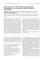
from the active site (Fig. 1). In this work
we have expressed, purified and characterized
Dm-dNK mutants carrying either N45D or N64D. We
present data that clearly points at N64D as the residue
responsible for the observed changes in the double
mutant MuD. We also present the crystal structures of
Fig. 1. Location of mutated residues. A monomer of Dm-dNK
showing the location of Asn45 and Asp64. The feedback inhibitor
dTTP is located in the active site. The P-loop and LID are labeled.
Drosophila deoxyribonucleoside kinase mutant N64D M. Welin et al.
3734 FEBS Journal 272 (2005) 3733–3742 ª 2005 FEBS
N64D in complex with its substrate dT and its inhib-
itor dTTP. Furthermore, our studies explain the cata-
lytic efficiency and sensitivity of MuD over the
wildtype Dm-dNK in terms of preference for the nucleo-
side analog AZT, and the decrease in feedback inhibi-
tion.
Results and Discussion
In vivo characterization of mutants
The Dm-dNK double mutant, N45D ⁄ N64D (MuD),
was generated by random in vitro mutagenesis [15].
When transformed into the thymidine kinase negative
E. coli strain KY895, the sensitivity of the cells
towards four nucleoside analogs with natural nucleo-
side bases but modifications at the 3¢-hydroxyl group
increased. To examine the significance of the two
amino acid exchanges for this property, we introduced
either the N45D or the N64D mutation into Dm-dNK
(lacking the 20 C-terminal residues). The resulting
mutants were first tested in two plate assays, either for
the presence of the TK activity or their ability to sensi-
tize KY895 towards AZT (Table 1).
To test the effectiveness of dT conversion, the dT
concentration in the TK selection plates was varied.
As can be concluded from Table 1, Dm-dNK and
mutant N45D could use dT more effectively than
the double mutant N45D ⁄ N64D, followed by mutant
N64D which needed the highest dT concentration to
ensure the survival of the transformed bacterial strain.
In contrast, the double mutant N45D⁄ N64D and
the mutant N64D sensitized KY895 to the same degree
to AZT (Table 1). Compared to Dm-dNK the decrease
in LD
100
for AZT was 300-fold for the double mutant
N45D ⁄ N64D and mutant N64D, but only threefold
for mutant N45D. Because in human cells AZT is
mainly a substrate for TK1 (human TK1; hTK1) we
also included this enzyme in our comparison, together
with TK from human Herpes simplex 1 virus (HSV1-
TK), which is currently the most widely used deoxy-
ribonucleoside kinase in suicide-enzyme pro-drug
therapy for cancer. As can be seen from Table 1, both
double mutant N45D ⁄ N64D and mutant N64D were
three times more efficient in killing KY895 with AZT
than hTK1 or HSV1-TK.
In vitro characterization
The relationship between velocity and substrate con-
centration was determined for the four natural deoxy-
ribonucleosides and AZT (Table 2). This confirmed the
results from Table 1 that, according to the k
cat
⁄ K
0.5
values, wildtype Dm-dNK and mutant N45D phos-
phorylate dT more efficiently than the double mutant
N45D ⁄ N64D, followed by mutant N64D. In general,
all mutants displayed a larger decrease in catalytic effi-
ciency (k
cat
⁄ K
0.5
) with the natural purine deoxyribo-
nucleosides than the pyrimidine deoxyribonucleosides,
when compared to wildtype. Mutant N64D showed
the largest decrease in catalytic efficiency, around 100–
500-fold more than mutant N45D. The decrease in
catalytic efficiency of the double mutant N45D ⁄ N64D
was between N45D and N64D suggesting that the
combined effect of the two mutations is not synergis-
tic. In fact, comparing the phosphorylation of the
natural substrates of the double mutant with the single
mutant, it seems that the mutation N45D in the
double mutant counteracts the negative effect(s) of the
N64D mutation. For phosphorylation of the thymidine
nucleoside analog AZT the picture is different; while
the double mutation N45D ⁄ N64D has increased the
efficiency for AZT, mutant N45D showed a slightly
larger decrease in efficiency than mutant N64D.
If a simultaneous presence of similar concentrations
of all four nucleoside substrates is assumed in the sur-
roundings of the wildtype and the mutant enzyme, the
difference in efficiencies between the two enzymes
should be able to be predicted using the equation,
[k
cat
⁄ K
0.5
(nucleoside analog)] ⁄ [k
cat
⁄ K
0.5
(dA) + k
cat
⁄
K
0.5
(dC) + k
cat
⁄ K
0.5
(dG) + k
cat
⁄ K
0.5
(dT) + k
cat
⁄
K
0.5
(nucleoside analog)] [22]. For the mutants N45D
and N64D and the double mutant N45D ⁄ N64D this
equation predicts an increase in catalytic efficiency for
the phosphorylation of AZT by 2.4-, 286- and 324-
fold, respectively. These values correlate quite well
with the observed changes in LD
100
for transformed
KY895 in Table 1. This suggests that the more import-
ant mutation for the observed and desired phenotype,
Table 1. Growth on TK selection plates: various plasmids were
transformed into KY895 and then the strains were examined for
growth, +, in the presence of different concentrations of thymidine
in the medium. In the last column, LD
100
values are given (in lM)
for the growth of KY895 transformed with various plasmids, on the
medium containing AZT.
dT (lgÆmL
)1
) AZT (lM)
Plasmid 0.05 1 2 10 20 50 100
pGEX-2T – – – ––––>100
pGEX-2T-Dm-dNK + ++++++ 100
pGEX-2T-double
mutant N45D ⁄ N64D
– ++++++ 0.3
pGEX-2T-mutant N45D + + + ++++ 32
pGEX-2T-mutant N64D – – + ++++ 0.3
pGEX-2T-HSV1-TK 1
pGEX-2T-hTK1 1
M. Welin et al. Drosophila deoxyribonucleoside kinase mutant N64D
FEBS Journal 272 (2005) 3733–3742 ª 2005 FEBS 3735
the death of KY895 at low AZT concentrations, is in
fact N64D.
dTTP feedback inhibition
dTTP is an efficient inhibitor of Dm-dNK with an
IC
50
value of 7 lm at 10 lm dT and 2.5 mm ATP,
whereas the double mutant N45D ⁄ N64D seems to
have lost the feedback inhibition property as reflected
by an IC
50
> 1000 lm at 2.5 mm ATP [15]. When the
two mutants, N45D and N64D were examined for
their dTTP inhibition, the feedback inhibition of
N45D is nearly unchanged (IC
50
¼ 11 lm) whereas
N64D behaved like the double mutant by having an
IC
50
> 1000 lm. The pattern of inhibition for the
N64D mutant was determined by varying thymidine
at fixed dTTP concentrations, and was found to
be predominantly competitive (K
ic
¼ 829 lm, K
iu
¼
3520 lm) in contrast to a predominantly uncompetitive
pattern observed with the Dm-dNK wildtype (K
ic
¼
16.3 lm, K
iu
¼ 4.7 lm) [15]. With ATP varied at fixed
dTTP concentrations, the kinetics was clearly compet-
itive with a K
ic
value of 1 lm. For comparison, the K
ic
value of dTTP with varied ATP for Dm-dNK wildtype
is about 200-fold lower (5.3 nm [21]). The kinetic stud-
ies, which demonstrated that mutation of residue 64
resulted in an enzyme with changed substrate specifi-
city and feedback inhibition, initiated crystallographic
studies of the mutant enzyme in complex with sub-
strate and feedback inhibitor to reveal the structural
basis for these phenomena.
Crystal structure of the N64D–dTTP complex
The Dm-dNK–dTTP complex crystallizes in a mono-
clinic form that has two dimers in the asymmetric unit.
dTTP binds as in the wildtype, as a feedback inhibitor
occupying the deoxyribonucleoside substrate site and a
part of the phosphate donor site [21]. The phosphates
of the inhibitor are tightly bound by residues of the
P-loop and LID region (residues 167–176). A Mg ion
is present in one out of the four different subunits
Table 2. Kinetic parameters of wildtype and mutant Dm-dNKs for various native nucleoside subtrates and AZT. The k
cat
values were calcula-
ted using the equation V
max
¼ k
cat
· [E] where [E] ¼ total enzyme concentration and is based on one active site ⁄ monomer. Overall, in inde-
pendent kinetic experiments, the coefficient of variation (standard deviation ⁄ mean) is less than 12% for V
max
values and less than 15% for
K
m
values.
Dm-dNK K
0.5
(lM) V
max
(U ⁄ mg) h k
cat
(s
)1
) k
cat
⁄ K
0.5
(M
)1
s
)1
)
Decrease in k
cat
⁄ K
0.5
of the mutants compared to
k
cat
⁄ K
0.5
of Dm-dNK (-fold)
dT
N45D 0.9 5.3 1 2.6 2.9 · 10
6
4.1
N64D 23.2 1.7 0.8 0.82 35344 473
N45D ⁄ N64D
a
24.2 2.5 1 1.22 50000 240
Wildtype
b
1.2 29.5 1 14.2 1.2 · 10
7
1
dC
N45D 0.9 3.4 1 1.6 1.8 · 10
6
4
N64D 118 3.4 0.8 1.6 13559 531
N45D ⁄ N64D
a
96.4 8.3 1 4.04 42000 171
Wildtype
b
2.3 34.2 1 16.5 7.2 · 10
6
1
dA
N45D 119 3.8 1 1.83 15378 6
N64D 3820 0.82 0.9 0.4 105 876
N45D ⁄ N64D
a
3166 1.7 1 0.828 260 354
Wildtype
b
225 42.7 1 20.6 92000 1
dG
N45D 412 2.7 1 1.3 3155 7.3
N64D 20350 0.24 0.5 0.12 5.9 3898
N45D ⁄ N64D
a
2004 0.156 1 0.076 38 605
Wildtype
b
665 31.3 1 15.1 23000 1
AZT
N45D 11.7 0.06 1 0.03 2564 1.7
N64D 11.1 0.074 0.8 0.037 3333 1.3
N45D ⁄ N64D
a
7.2 0.107 1 0.052 7200 0.6
Wildtype
a
8.3 0.073 1 0.036 4300 1
a
Data from [15].
b
Data from [24].
Drosophila deoxyribonucleoside kinase mutant N64D M. Welin et al.
3736 FEBS Journal 272 (2005) 3733–3742 ª 2005 FEBS
according to the difference density. The interactions
with dTTP are very similar to the interactions in the
wildtype Dm-dNK–dTTP complex, and the conforma-
tional changes of Glu52 are the same [21].
In wildtype Dm-dNK, Asn64 forms a hydrogen bond
to Glu171 as well as to the main chain amino group of
Leu66. Glu171 is part of the LID region within a loop
that also contains Glu172 that is hydrogen bonded to
the 3¢-hydroxyl group of the deoxyribose ring of dTTP.
The main chain of residues 65–66 are hydrogen bonded
to Tyr70, which forms a second hydrogen bond with
the 3¢-hydroxyl group of the substrate deoxyribose ring.
In alignments of eukaryotic deoxyribonucleoside kin-
ases, Asn64 as well as Leu66 and Glu171 are highly
conserved, even among deoxyribonucleoside kinases of
different substrate specificities.
Surprisingly, in the N64D mutant complex with
dTTP, Asp64 forms a hydrogen bond to Glu171
(Fig. 2A) which implies that one of them is protonated
in spite of a pH of 6.5 in the crystallization solution.
Because Glu171 is also stabilized by a hydrogen bond
from Arg58, it is probable that Asp64 is protonated.
Crystal structure of the N64D–dT complex
The N64D–dT structure contained dT and a sulphate
ion bound in each of the eight different subunits in
the asymmetric unit (Fig. 2B). There is well defined
density for Asp64 but very poor density for Glu171 as
well as for Glu172 that binds to the 3¢-OH in the
deoxyribose in the dTTP molecule. The LID region is
obviously very flexible (Fig. 3A) and there is no
hydrogen bond between Asp64 and Glu171 as in the
N64D–dTTP complex. In contrast to the dTTP com-
plex, Glu172 in the dT complex does not make a
hydrogen bond with the 3¢ -OH group of thymidine. In
the wildtype Dm-dNK–dT complex, Glu172 is bound
to the 3¢-OH group of the substrate while there is no
density for that interaction in the mutant structure
(Fig. 2B).
Structural basis for altered properties of the
N64D mutant
The LID region in wildtype Dm-dNK is a flexible part
of the structure that can attain slightly different posi-
tions in different complexes [16,21]. With the wildtype
enzyme, in most substrate complexes and the com-
plexes with the feedback inhibitor dTTP, the LID is
closed in over the active site. In substrate complexes,
LID arginines bind to a sulfate ion in the P-loop and
Glu172 to the 3¢-OH of the substrate. In the dTTP
complex, the phosphates are bound by the LID argi-
nines and the 3¢-OH is bound to Glu172. In these
cases, Glu171 in the LID region forms a hydrogen
bond to Asn64.
By substitution of Asn64 to Asp in the mutant
enzyme, the negative charge of Asp destabilizes the
normal interactions with Glu171. In the dT complex,
the negative charge of Asp64 repels Glu171 and the
LID region becomes more flexible and the part around
Glu171 and 172 is not visible in the electron density
maps (Fig. 3A). The absence of this part of the LID
region removes one of the hydrogen bonding inter-
AB
Fig. 2. Electron density maps. Final electron density maps for (A) the Dm-dNK N64D–dTTP complex containing the feedback inhibitor dTTP,
residues Asp64, Glu171 and Glu172, and (B) the Dm-dNK N64D–dT structure containing the same residues, the substrate dT and a sulfate
ion. The electron density maps for the protein parts (in blue) are 2Fo-Fc maps contoured at 1r. The electron density for the ligands (in green)
are Fo-Fc maps contoured at 3r before refinement. Hydrogen bonds in (A) shown as dotted lines.
M. Welin et al. Drosophila deoxyribonucleoside kinase mutant N64D
FEBS Journal 272 (2005) 3733–3742 ª 2005 FEBS 3737
actions with the 3¢-OH of the deoxyribose of the
substrate. The absence of this hydrogen bond and a
flexible LID make the substrate binding pocket larger
and provide space for the bulky 3¢-azide group. AZT
can be modeled based on the N64D–dT complex by
positioning of AZT instead of dT in its binding site
(Fig. 4).
In the complex of N64D and the feedback inhib-
itor dTTP, the LID region closes down on the inhib-
itor in the same way as in the wildtype complex in
spite of the substitution of Asn to Asp. Because of
all contacts between the phosphate groups, the LID
region is held in close interaction with dTTP. Conse-
quently, the LID region in the N64D–dTTP complex
has well-defined electron density (Fig. 3B). Glu171 is
thus forced into contact with Asp64 in spite of the
unfavorable electrostatic situation. This is overcome
by a hydrogen bonded Asp–Glu interaction that
occurs similar to the Asn–Glu interaction in the
wildtype enzyme. The energetic cost to bring the two
carboxylates of the mutant, Asp64 and Glu171,
together explains that dTTP inhibits the mutant
N64D with a considerably lower efficiency than in
the wildtype enzyme. The IC
50
value for dT phos-
phorylation is increased more than 100-fold.
The structure of the Dm-dNK N64D mutant pre-
sented above and the understanding of the feedback
regulation and substrate specificity in Dm-dNK will
now help to finalize our understanding of the struc-
ture–function relationship and also have a wide
impact on the following medical applications: the
design of novel specific pro-drug and mutant combi-
nations for gene therapy, the development of species-
specific antiviral and antibacterial nucleoside analog
based drugs, and promoting development of novel
AZT-like pro-drugs.
Fig. 3. The LID region in the two com-
plexes. Stereo view of the final electron
density for the LID region in (A) the Dm-
dNK N64D–dTTP complex and (B) the
Dm-dNK N64D–dT complex (2Fo-Fc maps
contoured at 1r).
Drosophila deoxyribonucleoside kinase mutant N64D M. Welin et al.
3738 FEBS Journal 272 (2005) 3733–3742 ª 2005 FEBS
Experimental procedures
Materials
Unlabelled nucleosides and nucleotides were from Sigma
(St Louis, MO, USA) or ICN Biochemicals (Aurora, OH).
3
H-labeled thymidine [Me-
3
H]dT (925 GBqÆmmol
)1
) and
deoxycytidine [6-
3
H]dC (740–925 GBqÆmmol
)1
) were
obtained from Amersham Corp., Piscataway, NJ, USA).
3
H-labeled deoxyadenosine [2,8-
3
H]dA (1106 GBq), deoxy-
guanosine [2,8-
3
H]dG (226 GBqÆmmol
)1
) and 3¢-azido-2¢,3¢-
dideoxythymidine [Me-
3
H]AZT (740 GBqÆmmol
)1
) were
from Moravek Biochemicals Inc. (Brea, CA, USA). When
present in the radiolabeled deoxynucleosides, ethanol was
evaporated before use.
Sequencing
Sequencing by the Sanger dideoxynucleotide method was
performed manually, using the Thermo Sequenase radio-
labeled terminator cycle sequencing kit and
33
P-labeled
ddNTPs (Amersham Corp.).
Site directed mutagenesis and expression
plasmids
Expression plasmid pGEX-2T-Dm-dNK is described in [24].
Expression plasmid pGEX-2T-MuD (pGEX-2T-double
mutant N45D ⁄ N64D) is described in [15]. The expression
vector for human TK1 (pGEX-2T-hTK1) is described else-
where [25]. The pGEX-2T-mutant N45D and pGEX-2T-
mutant N64D were constructed as follows: both mutants
were constructed by site directed mutagenesis on the plas-
mid pGEX-2T- Dm-dNK with or without truncation for
the C-terminal 20 amino acids [24]. The N45D mutation
was created with the following primers: 45D-fw (5¢-CGAG
AAGTACAAG
GACGACATTTGCCTGC-3¢) and 45D-rv
(5¢-GCAGGCAAATGTCGT
CCTTGTACTTCTCG-3¢),
where the changed nucleotide is in bold and underlined.
The N64D mutation was created with the primers 64D-fw
(5¢-CGTCAACGGGGTA
GATCTGCTGGAGC-3¢) and
64D-rv (5¢-GCTCCAGCAGAT
CTACCCCGTTGACG-3¢).
An expression plasmid for HSV1-TK was constructed as
follows: The thymidine kinase from HSV1 was amplified
using the primers HSV-for (5¢-CGCGGATCCATGGCT
TCGTACCCCGGCCATC-3¢) and HSV-rev (5¢-CCGGAA
TTCTTAGTTAGCCTCCCCCATCTCCCG-3¢) and using
the plasmid pCMV-pacTK [26] as templ ate. The PCR frag-
ment was subsequently cut by EcoRI ⁄ BamHI and ligated into
pGEX-2T vector that was also cut by EcoRI ⁄ BamHI. The
resulting plasmid was named pGEX-2T-HSV1-TK (P 632).
Test for TK activity on selection plates
The thymidine kinase deficient E. coli strain KY895 [F
–
,
tdk-1, ilv] [27], was transformed with various expression
plasmids. Overnight cultures of these transformants were
diluted 200-fold in 10% (w ⁄ v) glycerol and 2 lL drops of
the dilutions were spotted on TK selection plates [9] that
contained different dT concentrations. Only enzymes com-
plementing the TK negative E. coli strain KY895 gave rise
to colonies on this selection medium. Growth was inspected
visually after 24 h at 37 °C.
Determination of LD
100
Overnight cultures of single colonies were diluted 200-fold
in 10% (w ⁄ v) glycerol and 2 lL of these dilutions were
spotted on M9 minimal medium plates [28] supplemented
with 0.2% (w ⁄ v) glucose, 40 lgÆmL
)1
isoleucin, 40 lgÆmL
)1
valin, 100 lgÆmL
)1
ampicillin and with or without AZT.
Logarithmic dilutions of the nucleoside analog were used to
determine the lethal dose (LD
100
) of the nucleoside analog,
at which no growth of bacteria could be seen. Growth of
colonies was visually inspected after 24 h at 37 °C.
Expression and purification of recombinant
enzymes
Recombinant proteins were expressed and purified as des-
cribed previously [24].
Enzyme assay
Deoxyribonucleoside kinase activities were determined by
initial velocity measurements based on four time samples
Fig. 4. Modeling of AZT. Interactions with the substrate dT, in red,
and with AZT modeled in the substrate binding site, in blue. The
interactions with the substrate are the same in the wildtype and
the N64D mutant except for the lack of interactions between
Glu172 and 3¢-OH giving space for the azido-group of AZT. The
position of Glu172 in the wildtype structure is given in yellow.
M. Welin et al. Drosophila deoxyribonucleoside kinase mutant N64D
FEBS Journal 272 (2005) 3733–3742 ª 2005 FEBS 3739
by the DE-81 filter paper assay using tritium-labeled nucleo-
side substrates. The assay was performed as described [24].
The protein concentration was determined according to
Bradford with BSA as standard protein [29]. SDS ⁄ PAGE
was carried out according to the procedure of Laemmli [30]
and proteins were visualized by Coomassie staining.
Analysis of kinetic data
Kinetic data were evaluated by nonlinear regression ana-
lysis using the Michaelis–Menten equation v ¼ V
max
·
[S] ⁄ (K
m
+ [S]) or the Hill equation v ¼ V
max
· [S]
h
⁄
(K
0.5
h
+ [S]
h
) as described in [31]. K
m
is the Michaelis con-
stant, K
0.5
defines the value of the substrate concentration
[S] where v ¼ 0.5 V
max
and h is the Hill coefficient [32,33].
If h ¼ 1, there is no cooperativity.
The concentration of the feedback inhibitor dTTP neces-
sary for 50% inhibition (IC
50
) was determined by varying
dTTP at 10 lm dT and 2.5 mm ATP and plotting
log(v
0
) v
I
) ⁄ v
I
against log[I] where v
0
and v
I
are the velocities
without and with inhibitor, respectively. IC
50
was determined
as the intercept with the log[I] axis, where (v
0
) v
I
) ⁄ v
I
¼ 1.
The pattern of inhibition was elucidated by varying dT
at four fixed concentrations of dTTP and 2.5 mm ATP, and
analyzing the data by the Biosoft (Cambridge, UK) pro-
gram enzfitter for Windows.
Crystallization
The N64D mutant used for crystallization was truncated
for the 20 C-terminal amino acids. The C-terminal trun-
cated Dm-dNK kinases have similar enzymatic properties
as the untruncated kinases but are more stable [24]. Cry-
stals of N64D in complex with dT and dTTP were grown
by counter diffusion [34] and vapor diffusion, respectively.
The crystallization solution for the N64D mutant dT
complex was 0.15 m Mes pH 6.5, 0.3 m lithium sulphate
and 27.5% (w ⁄ v) poly(ethylene) glycol monomethyl ether
2000. The enzyme solution (20 mgÆmL
)1
including 10 mm
dT) and the crystallization solution was equally mixed in
a capillary and equilibrated for two weeks. For the N64D
complex with dTTP the crystallization solution was 0.1 m
Mes pH 6.5, 0.16 m lithium sulphate and 25% (v ⁄ v)
poly(ethylene) glycol monomethyl ether 2000. The pro-
tein solution (10 mgÆmL
)1
) including 5 mm dTTP and the
crystallization solution were mixed equally in a hanging
drop. All the crystallization trials were performed at
15 °C.
Data Collection
The N64D–dT crystals were directly flash-frozen in liquid
nitrogen. The cryoprotectant for the N64D–dTTP crystals
contained crystallization solution plus the addition of 20%
(v ⁄ v) poly(ethylene) glycol 400. The data sets for the two
complexes with dT and dTTP were collected at ID14 ⁄ EH4,
ESRF (Grenoble, France). The two data sets were indexed,
scaled and merged with mosflm [35] and scala [36]. Both
crystals belonged to the space group P2
1
and had a solvent
content of 55%. The content in the asymmetric unit for the
N64D–dT and N64D–dTTP complex corresponded to four
and two dimers, respectively.
Structure determination and refinement
The N64D–dTTP complex was solved with rigid body in
refmac5 [37] with Dm-dNK–dTTP (PDB code: 1oe0) as
a search model. The N64D–dT complex was solved with
molrep and Dm-dNK–dT as a search model (PDB code:
1ot3). The mutated residue Asn to Asp in the two com-
plexes was altered in the program o (.
uu.se/alwyn) [38]. After rigid body refinement the dT and
the dTTP complex were refined with fourfold and eight-
fold noncrystallographic averaging, respectively, in ref-
mac5, ccp4. The N64D–dT complex had a final R-value
of 27.0% and an R
free
of 28.8% while the model for
N64D–dTTP complex had an R-value of 21.3% and an
R
free
of 23.7%. The data collection and refinement statis-
tics are shown in Table 3. The coordinates have been
deposited with PDB codes: 1zmx and 1zm7.
Table 3. Data collection and refinement statistics for the N64D-dT
and N64D-dTTP complexes.
N64D-dT N64D-dTTP
Beamline ID14 ⁄ EH4, ESRF ID14 ⁄ EH4, ESRF
Wavelength (A
˚
) 0.9393 1.0
Temperature (K) 100 100
Resolution (A
˚
) 3.1 (3.27–3.10) 2.2 (2.32–2.20)
Reflections
Observed 144933 189093
Unique 39562 53387
Completeness 99.9 99.9
Rmeas(%) 12.0 (45.0) 8.3 (38,9)
I ⁄ sigmaI 12.5 (3.4) 14.5 (4.1)
Space group P2
1
P2
1
Cell Dimensions
a 69.71 67.04
b 70.34 119.27
c 224.53 68.39
beta 90.69 92.59
Content of the asymmetric unit 4 dimers 2 dimers
Refinement program Refmac5 Refmac5
R factor (%) 27.0 21.3
R
free
(%) 28.8 23.7
Root mean square deviation
Bond length (A
˚
) 0.009 0.011
Bond angles (°) 1.13 1.27
Mean B-value (A
˚
2
) 37.4 36.7
Drosophila deoxyribonucleoside kinase mutant N64D M. Welin et al.
3740 FEBS Journal 272 (2005) 3733–3742 ª 2005 FEBS
Acknowledgements
We would like to thank Marianne Lauridsen for excel-
lent technical assistance. This work was supported by
grants from the Swedish Research Council (to H.E. and
J.P.), the Swedish Cancer Foundation (to H.E.), and
Danish Research Council (to B.M P., W.K. and J.P.).
References
1 Arne
´
r ESJ & Eriksson S (1995) Mammalian deoxyribo-
nucleoside kinases. Pharmacol Ther 67, 155–186.
2 Plunkett W & Gandhi V (2001) Purine and pyrimidine
nucleoside analogs. Cancer Chemother Biol Response
Modif 19, 21–45.
3 Klopfer A, Hasenjager A, Belka C, Schulze-Osthoff K,
Dorken B & Daniel PT (2004) Adenine deoxynucleo-
tides fludarabine and cladribine induce apoptosis in a
CD95 ⁄ Fas receptor, FADD and caspase-8-independent
manner by activation of the mitochondrial cell death
pathway. Oncogene 23, 9408–9418.
4 Furman PA, Fyfe JA, St Clair MH, Weinhold K, Ride-
out JL, Freeman GA et al. (1986) Phosphorylation of
3¢-azido-3¢-deoxythymidine and selective interaction of
the 5¢-triphosphate with human immunodeficiency virus
reverse transcriptase. Proc Natl Acad Sci USA 83,
8333–8337.
5 Eriksson S, Munch-Petersen B, Johansson K & Eklund
H (2002) Structure and function of cellular deoxyribo-
nucleoside kinases. Cell Mol Life Sci 59, 1327–1346.
6 Zheng X, Johansson M & Karlsson A (2000) Retroviral
transduction of cancer cell lines with the gene encoding
Drosophila melanogaster multisubstrate deoxyribonu-
cleoside kinase. J Biol Chem 275, 39125–39129.
7 Rainov NG (2000) A phase III clinical evaluation of
herpes simplex virus type 1 thymidine kinase and
ganciclovir gene therapy as an adjuvant to surgical
resection and radiation in adults with previously
untreated glioblastoma multiforme. Hum Gene Ther 11,
2389–2401.
8 Greco O & Dachs GU (2001) Gene directed enzyme ⁄
prodrug therapy of cancer: historical appraisal and
future prospectives. J Cell Physiol 187, 22–36.
9 Black ME, Newcomb TG, Wilson HM & Loeb LA
(1996) Creation of drug-specific herpes simplex virus
type 1 thymidine kinase mutants for gene therapy. Proc
Natl Acad Sci USA 93, 3525–3529.
10 Gentry GA (1992) Viral thymidine kinases and their
relatives. Pharmac Ther 54, 319–355.
11 Chen MS & Prusoff WH (1978) Association of thymidy-
late kinase activity with pyrimidine deoxyribonucleoside
kinase induced by herpes simplex virus. J Biol Chem
253, 1325–1327.
12 Munch-Petersen B, Piskur J & Søndergaard L (1998)
The single deoxynucleoside kinase in Drosophila
melanogaster, dNK, is multifunctional and differs from
the mammalian deoxynucleoside kinases. Adv Exp Med
Biol 431, 465–469.
13 Munch-Petersen B, Piskur J & Søndergaard L (1998)
Four deoxynucleoside kinase activities from Drosophila
melanogaster are contained within a single monomeric
enzyme, a new multifunctional deoxynucleoside kinase.
J Biol Chem 273, 3926–3931.
14 Johansson M, Van Rompay AR, Degreve B, Balzarini J
& Karlsson A (1999) Cloning and characterization
of the multisubstrate deoxyribonucleoside kinase of
Drosophila melanogaster. J Biol Chem 274, 23814–
23819.
15 Knecht W, Munch-Petersen B & Piskur J (2000) Identi-
fication of residues involved in the specificity and regu-
lation of the highly efficient multisubstrate
deoxyribonucleoside kinase from Drosophila melanoga-
ster. J Mol Biol 301, 827–837.
16 Johansson K, Ramaswamy S, Ljungcrantz C, Knecht
W, Piskur J, Munch-Petersen B et al. (2001) Structural
basis for substrate specificities of cellular deoxyribonu-
cleoside kinases. Nat Struct Biol 8, 616–620.
17 Sabini E, Ort S, Monnerjahn C, Konrad M & Lavie A
(2003) Structure of human dCK suggests strategies to
improve anticancer and antiviral therapy. Nat Struct
Biol 10, 513–519.
18 Brown DG, Visse R, Sandhu G, Davies A, Rizkallah
PJ, Melitz C et al. (1995) Crystal structures of the
thymidine kinase from herpes simplex virus type-1 in
complex with deoxythymidine and ganciclovir. Nat
Struct Biol 2, 876–881.
19 Wild K, Bohner T, Folkers G & Schulz GE (1997) The
structures of thymidine kinase from herpes simplex virus
type 1 in complex with substrates and a substrate analo-
gue. Protein Sci 6, 2097–2106.
20 Welin M, Kosinska U, Mikkelsen NE, Carnrot C, Zhu
C, Wang L et al. (2004) Structures of thymidine kinase
1 of human and mycoplasmic origin. Proc Natl Acad
Sci USA 101, 17970–17975.
21 Mikkelsen NE, Johansson K, Karlsson A, Knecht W,
Andersen G, Piskur J et al. (2003) Structural basis for
feedback inhibition of the deoxyribonucleoside salvage
pathway: studies of the Drosophila deoxyribonucleoside
kinase. Biochemistry 42, 5706–5712.
22 Kokoris MS, Sabo P, Adman ET & Black ME (1999)
Enhancement of tumor ablation by a selected HSV-1
thymidine kinase mutant. Gene Ther 6, 1415–1426.
23 Sandrini M & Piskur J (2005) Deoxyribonucleoside
kinases: two enzyme families catalyze the same reaction.
Trends Biochem Sci 30, 225–228.
24 Munch-Petersen B, Knecht W, Lenz C, Søndergaard L
& Piskur J (2000) Functional expression of a multisub-
strate deoxyribonucleoside kinase from Drosophila
melanogaster and its C-terminal deletion mutants. J Biol
Chem 275, 6673–6679.
M. Welin et al. Drosophila deoxyribonucleoside kinase mutant N64D
FEBS Journal 272 (2005) 3733–3742 ª 2005 FEBS 3741
25 Berenstein D, Christensen JF, Kristensen T, Hofbauer
R & Munch-Petersen B (2000) Valine, not methionine,
is amino acid 106 in human cytosolic thymidine kinase
(TK1). Impact on oligomerization, stability, and kinetic
properties. J Biol Chem 275, 32187–32192.
26 Karreman C (1998) A new set of positive ⁄ negative
selectable markers for mammalian cells. Gene 218,
57–61.
27 Igarashi K, Hiraga S & Yura T (1967) A deoxythymi-
dine kinase deficient mutant of Escherichia coli. II.
Mapping and transduction studies with phage phi 80.
Genetics 57, 643–654.
28 Ausubel F, Brent R, Kingston RE, Moore DD, Seid-
man JG, Smith JA et al. (1995) Short Protocols in
Molecular Biology, 3rd edn. John Wiley & Sons, Inc.,
USA.
29 Bradford MM (1976) A rapid and sensitive method for
the quantitation of microgram quantities of protein util-
izing the principle of protein-dye binding. Anal Biochem
72, 248–254.
30 Laemmli UK (1970) Cleavage of structural proteins
during the assembly of the head of bacteriophage T4.
Nature 227, 680–685.
31 Knecht W, Bergjohann U, Gonski S, Kirschbaum B &
Loffler M (1996) Functional expression of a fragment of
human dihydroorotate dehydrogenase by means of the
baculovirus expression vector system, and kinetic inves-
tigation of the purified recombinant enzyme. Eur J
Biochem 240, 292–301.
32 Cornish-Bowden A (1995) Fundamentals of Enzyme
Kinetics. Portland Press Ltd, London.
33 Liebecg C (1992) IUIMB Biochemical Nomenclature and
Related Documents. Portland Press Ltd, London.
34 Ng JD, Gavira JA & Garcia-Ruiz JM (2003) Protein
crystallization by capillary counterdiffusion for applied
crystallographic structure determination. J Struct Biol
142, 218–231.
35 Leslie AGW (1992) Joint CCP4 + ESF-EAMCB News-
letter on Protein Crystallography, No. 26.
36 Collaborative Computational Project, Number 4 (1994)
The CCP4 Suite: Programs for Protein Crystallography.
Acta Crystallogr D50, 760–763.
37 Murshudov GN, Vagin AA & Dodson EJ (1997)
Refinement of macromolecular structures by the maxi-
mum-likelihood method. Acta Crystallogr D53,
240–255.
38 Jones TA, Zou JY, Cowan SW & Kjeldgaard M (1991)
Improved methods for building protein models in elec-
tron density maps and the location of errors in these
models. Acta Crystallogr 47, 110–119.
Drosophila deoxyribonucleoside kinase mutant N64D M. Welin et al.
3742 FEBS Journal 272 (2005) 3733–3742 ª 2005 FEBS