Báo cáo khoa học: The protein tyrosine phosphatase PTP-Basophil/Basophil-like Interacting proteins and molecular functions doc
Bạn đang xem bản rút gọn của tài liệu. Xem và tải ngay bản đầy đủ của tài liệu tại đây (222.01 KB, 10 trang )
REVIEW ARTICLE
The protein tyrosine phosphatase PTP-Basophil/Basophil-like
Interacting proteins and molecular functions
Kai S. Erdmann*
Department of Molecular Neurobiochemistry, Ruhr-University Bochum, Germany
The protein tyrosine phosphatase PTP-Basophil (PTP-Bas)
and its mouse homologue, PTP-Basophil-like (PTP-BL), are
high molecular mass protein phosphatases consisting of a
number of diverse protein–protein interaction modules.
Several splicing variants of these phosphatases are known to
exist thus demonstrating the complexity of these molecules.
PTP-Bas/BL serves as a central scaffolding protein facilita-
ting the assembly of a multiplicity of different proteins
mainly via five different PDZ domains. Many of these
interacting proteins are implicated in the regulation of the
actin cytoskeleton. However, some proteins demonstrate a
nuclear function of this protein tyrosine phosphatase. PTP-
Bas is involved in the regulation of cell surface expression
of the cell death receptor, Fas. Moreover, it is a negative
regulator of ephrinB phosphorylation, a receptor playing an
important role during development. The phosphorylation
status of other proteins such as RIL, IjBa and b-catenin can
also be regulated by this phosphatase. Finally, PTP-BL has
been shown to be involved in the regulation of cytokinesis,
the last step in cell division. Although the precise molecular
function of PTP-Bas/BL is still elusive, current data suggest
clearly that PTP-Bas/BL belongs to the family of PDZ
domain containing proteins involved in the regulation of
the cytoskeleton and of intracellular vesicular transport
processes.
Keywords: cytoskeleton; apoptosis; phosphatase; trafficking;
cytokinesis.
Introduction
Protein phosphorylation is one of the most prominent post-
translational modifications regulating the activity, inter-
action capability and subcellular localization of proteins.
In particular, protein tyrosine phosphorylation has been
identified as a major regulator of signal transduction in
higher eukaryotes. Protein target phosphorylation is cata-
lysed by protein tyrosine kinases and counteracted by the
activity of protein tyrosine phosphatases [1].
The family of protein tyrosine phosphatases (PTPs) is
divided into two major subtypes: (a) the receptor-like and
(b) the nonreceptor subtype. The receptor-like subtype
contains one transmembrane domain and one or two
phosphatase domains at the C-terminus, which faces the
cytosol. The nonreceptor subtype is characterized by the
lack of a transmembrane domain and is a cytosolic or
membrane-associated phosphatase [2].
The nonreceptor protein tyrosine phosphatases PTP-Bas/
BL [3,4], PTPH1/PTP-MEG [5,6] and PTPD1/PTP-RL10
[7,8] belong to a protein tyrosine phosphatase family
characterized by the presence of a Four point one/Ezrin/
Radixin/Moesin (FERM) domain (FERM-PTP family).
PTPD1 has been implicated in vesicular trafficking proces-
ses due to its interaction with a kinesin motor protein [9] and
in the regulation of the Tec tyrosine kinase family [10].
PTPH1, has been suggested to play a role in vesicular fusion
processes of the endoplasmatic reticulum [11], in cell cycle
regulation [12] and in the regulation of the tumor necrosis
factor a-convertase (TACE) [13]. This review will focus on
current knowledge of the largest member of this FERM-
PTP family, the protein tyrosine phosphatase Basophil/
Basophil-like.
The modular structure of the protein tyrosine
phosphatase-Basophil/Basophil-like
The human homologue of this protein tyrosine phosphatase
was discovered by employing a PCR-based strategy to
identify new protein tyrosine phosphatases using conserved
regions within the catalytic domain. Originally, it was
cloned from a basophil cell line, consequently, the new
protein tyrosine phosphatase was named PTP-Basophil
(PTP-Bas) [3]. Using a similar strategy, it was cloned in
parallel from a human glioma cell line and a human breast
carcinoma cell line and named PTPL1 and hPTP1e,
Correspondence to K. S. Erdmann, Department of Cell Biology,
Yale University School of Medicine, New Haven, CT, USA.
Fax: + 1 203 737 1762, Tel.: + 1 203 737 4473,
E-mail:
Abbreviations: APC, adenomatous polyposis coli protein; BP75,
bromodomain containing protein 75; CRIP2, cysteine-rich intestinal
protein 2; FERM, Four point one/Ezrin/Radixin/Moesin; GFP, green
fluorescence protein; IkBa, inhibitor of kBa; LIM, Lin-11, Isl-1,
Mec-3; PARG, PTPL1-associated RhoGAP; PIP2, 1-phosphatidyl-
inositol 4,5-biphosphate [PtdIns(4,5)P
2
]; PRK2, protein kinase
C-related kinase 2; PTP, protein tyrosine phosphatase; p75NTR,
p75 neurotrophin receptor; RIL, reversion-induced LIM protein;
siRNA, small interfering RNA; Trip6, thyroid receptor interacting
protein-6; ZRP-1, zyxin related protein-1.
*Present address: Department of Cell Biology, Yale University School
of Medicine, 06510 New Haven, CT, USA.
(Received 2 September 2003, revised 14 October 2003,
accepted 23 October 2003)
Eur. J. Biochem. 270, 4789–4798 (2003) Ó FEBS 2003 doi:10.1046/j.1432-1033.2003.03895.x
respectively [14,15]. Finally, due to its interaction with the
human cell surface receptor Fas, it was renamed as FAP-1
(Fas-associated phosphatase-1) [16]. The mouse homologue
was cloned by two independent approaches and called RIP
andalsoPTP-BL) PTP-basophil-like ) referring to its
human homologue (PTP-Bas) [4,17]. To simplify reading
and to avoid confusion within this review the human
homologue will be consequently assigned PTP-Bas and the
mouse homologue as PTP-BL.
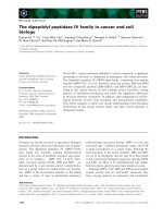
The protein tyrosine phosphatase PTP-Bas/BL is a highly
modular protein of about 2490 amino acids length (mole-
cular mass 270 kDa) (Fig. 1). The extreme N-terminus
contains a kinase noncatalytic C-lobe (KIND) domain, a
protein module identified recently that shows homology to
the regulatory C-lobe of protein kinases but that lacks
catalytic activity [18]. The function of this domain is
currently unknown but a role in mediating protein–protein
interactions has been suggested. The KIND domain is
followed by a Four-point-one/Ezrin/Radixin/Moesin
(FERM) domain. FERM domains are known to play an
important role in connecting plasma membrane receptors to
the cytoskeleton [19]. Furthermore, PTP-Bas/BL comprises
five different PSD-95/Drosophila discs large/Zonula occlu-
dens (PDZ) domains [20]. PDZ domains are protein–
protein interaction domains playing a fundamental role in
the assembly of supramolecular protein complexes [21].
Finally, the protein tyrosine phosphatase domain is located
at the extreme C-terminus of the molecule.
The complexity of the PTP-Bas/BL molecule is further
increased by alternative splicing that is observed in the
N-terminus between the FERM domain and the first PDZ
domain and within the second PDZ domain [3,4,14,16,22].
Additional minor alternative splice products have been
described for PTP-Bas predicting C-terminal truncated
versions of the PTP-Bas protein [14]. Given the presence of
at least seven protein–protein interaction domains including
the KIND, FERM and five PDZ domains, PTP-Bas/BL
functions most probably as a major scaffolding protein (see
below).
Expression pattern of PTP-BL
RNA in situ hybridization experiments show that the
expression pattern of PTP-BL is highly regulated during
development [4]. Early in development, PTP-BL is
expressed ubiquitously throughout the embryo. At later
stages, PTP-BL becomes restricted to epithelial and neur-
onal cell lineages, e.g. epithelia of the skin, oesophagus,
stomach, nasal cavity, lung, kidney, ureter, and the bladder.
Moreover, expression has been observed in the ependymal
cell layer, an epithelial cell layer surrounding the ventricles
of the brain [4]. A similar expression pattern was observed in
transgenic mice used to analyse the distribution of a
truncated version of PTP-BL lacking the portion of the
protein C-terminal to the first PDZ domain and fused to
b-galactosidase [23]. In this in vivo model, additional
expression was detected in the pigmented epithelial layer
of the eye, in the infundibulum and the anterior lobe of the
pituitary. Most interestingly, a timely regulated expression
couldbeobservedinperipheralsensoryandsympathetic
neurons.
With regard to a possible role of PTP-BL under
pathological conditions, it was demonstrated that the
human homologue PTP-Bas is up regulated in a number
of tumour cell lines [24–26]. Most strikingly, PTP-Bas is up
regulated in many ovarian tumours [27]. This is of particular
interest, as PTP-Bas has been demonstrated to mediate
resistance to Fas induced apoptosis (see below) [16]. Given
that apoptosis is one of the mechanisms to eliminate
transformed cells in a multicellular organism to avoid the
onset of cancer, a role of PTP-Bas/BL as a tumour
suppressor is conceivable.
At subcellular levels, PTP-BL shows an apical localiza-
tion in epithelial cells and is accumulated in axons and
growth cones of sympathetic and sensory neurons
[23,28,29]. Furthermore, PTP-Bas localizes to the Golgi
apparatus and to the nucleus [22,30]. Recently, it has been
shown that a specific splicing variant of PTP-BL accumu-
lates at the centrosome and at the spindle midzone during
cell division and is part of the midbody late in cytokinesis
[31] (for details see below).
Domain specific interactions of PTP-Bas/BL
Knowledge about PTP-Bas/BL has increased due to the
identification of a number of proteins interacting with one
or two of the interaction domains present in PTP-Bas/BL.
A summary of these domain specific interactions is given
below (see also Fig. 2).
The KIND-domain
Recently, the KIND domain was identified by its similarity
to the C-terminal protein kinase catalytic fold (C-lobe).
However, the absence of catalytic and activation loops
suggests that this domain is probably noncatalytic [18]. The
molecular function of this 200 amino acid new protein
module is currently unknown but given that the C-lobe
domain serves as a protein/protein interaction domain in
protein kinases a similar role for the KIND domain is
conceivable. Further experiments are needed to clarify the
molecular function of the KIND domain in PTP-Bas/BL.
The FERM-domain
FERM domains play an important role at the interface
between the plasma membrane and the cytoskeleton [19].
These domains have a length of about 300 amino acids and
can be divided into three subdomains (A–C) as revealed by
X-ray analysis. FERM domains are known to bind to a
number of cell surface receptors [19,32]. In addition, binding
Fig. 1. Modular structure of PTP-Bas/BL. PTP-Bas/BL is composed
of an N-terminal kinase noncatalytic C-lobe (KIND) domain followed
by a Four-point-one/Ezrin/Radixin/Moesin (FERM) domain. The
core of the protein comprises five different PDZ domains. The protein
tyrosine phosphatase (Phos.) domain is located at the C-terminus. The
regions of major alternative splicing are indicated by dashed lines and
red bars.
4790 K. S. Erdmann (Eur. J. Biochem. 270) Ó FEBS 2003
to 1-phosphatidylinositol 4,5-biphosphate [PtdIns(4,5)P
2
or
PIP2) has been demonstrated [33]. Most recently, it has been
reported that the FERM domain of PTP-Bas is also able to
interact with PIP2 [34]. This interaction has been suggested
to be important for membrane localization of PTP-Bas.
Moreover, the FERM domain of PTP-BL was described to
target the protein to the apical membrane in cultured
epithelial cells as well as in vivo [4,23]. Besides binding to
PIP2, colocalization as well as cosedimentation with
filamentous actin has been demonstrated suggesting an
association with the actin cytoskeleton [31]. However, there
are currently no proteins known to interact directly with the
FERM domain of PTP-Bas/BL.
PDZ domains
PDZ domains are able to interact selectively with the
C-termini of their target proteins [21,35,36]. However, there
are also examples of PDZ domain binding to internal
sequences [35,37]. Recently, it has been shown that PDZ
domains are not only protein–protein interaction domains
but can also serve as PIP (PtdInsP) binding modules [38].
PDZ domains have a length of 90 amino acids and adopt
an a/b-fold consisting of six b-sheets and two a-helices
[39,40]. Currently, only the NMR-structure of one (PDZ2)
of the five PDZ domains of PTP-Bas/BL is available [41,42].
Although the overall structure of the PDZ2 domain fits well
into the common fold of PDZ domains, it shows an unusual
foldback of the loop between b2andb3 to the backbone.
This loop has been implicated in the regulation of target
recognition of the PDZ2 domain [22,43]. Indeed the NMR-
structure of a splice variant of the PDZ2 domain (PDZ2b),
with an extended b2/b3 loop, revealed that this foldback can
even interfere with protein target binding [44].
There is an increasing number of proteins known to
interact with one or more of the PDZ domains of PTP-BL/
PTP-Bas.
PDZ1
An interaction of PDZ1 with the bromodomain-containing
protein BP75 and with the transcription regulator IjBa
was demonstrated [45,46]. Although the function of BP75
is unknown, bromodomains are thought to interact with
acetylated lysine residues that are present in histone
proteins, thus, hinting at a possible function of BP75 in
the nucleus [47]. This is in line with its nuclear localization
and the capability of BP75 to translocate the PDZ1 domain
of PTP-BL to the nucleus. Interaction of BP75 with PTP-
BL needs an intact C-terminus of BP75 although the
C-terminus alone is insufficient for binding.
Interaction of IjBa with PDZ1 is mediated via its
ankyrin repeats. IjBa is an inhibitor of the transcription
factor NFjB. A complex consisting of both proteins is
formed in the cytosol preventing NFjB from entering the
nucleus. There are two pathways involved in dissociation of
this complex. In the first pathway, IjBa is phosphorylated
on Ser32 and 36 resulting in its degradation by the ubiquitin
proteasome system. In the second pathway, IjBa is
phosphorylated on Tyr42 leading to dissociation without
degradation [48,49]. Indeed, IjBa is a substrate for PTP-Bas
phosphatase, shown by an increase in tyrosine phosphory-
lation of IjBa aftertransfectionofaPTP-Basversion
lacking phosphatase activity [46]. Furthermore, a substrate
trapping mutant of PTP-Bas was found to bind selectively
tyrosine-phosphorylated IjBa [50]. The interaction of IjBa
with PTP-Bas provides evidence for a regulation of NFjB
dependent transcription via PTP-Bas [46,51].
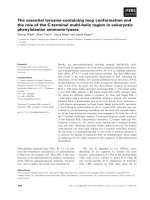
Fig. 2. PTP-Bas/BL is the central scaffolding component of a supramolecular protein complex. Domain specific interactions of PTP-Bas/BL are
indicated (for a detailed description see text). Dashed arrows indicate that an association has been determined but direct interaction was not
investigated. The interacting proteins can be divided into three groups. Known or potential regulators of the actin cytoskeleton (red), regulators of
the actin and tubulin cytoskeleton (purple) and regulators of gene transcription (green). Human Fas (hFas) does not fit in any of these groups but is
a transmembrane receptor regulating apoptosis. APC, Adenomatous polyposis coli protein; BP75, bromodomain containing protein 75; CRIP2,
cysteine-rich intestinal protein 2; PARG, PTPL1-associated RhoGAP; PIP2, 1-phosphatidylinositol 4,5-biphosphate, PRK2, protein kinase
C-related kinase 2; p75NTR, neurotrophin receptor; RIL, reversion-induced LIM (RIL); Trip6, thyroid receptor interacting protein-6; ZRP-1,
zyxin related protein-1.
Ó FEBS 2003 The protein tyrosine phosphatase PTP-Bas/BL (Eur. J. Biochem. 270) 4791
PDZ2
A number of interacting proteins were shown to interact
with the PDZ2 domain of PTP-Bas/BL. The human cell
surface protein Fas was the first protein reported to
interact with the PDZ2 domain of PTP-Bas (FAP-1) [16].
However, the corresponding mouse homologue of Fas
does not bind to PTP-BL [52] (see below). The neurotro-
phin receptor p75
NTR
has also been suggested to interact
with the PDZ2 domain of PTP-Bas [51]. Additional
proteins known to bind to the PDZ2 domain are the LIM
domain containing proteins RIL and Trip6/ZRP-1
[37,53,54]. RIL is also able to interact with PDZ4 (see
below). Little is known about the molecular functions of
RIL and Trip6/ZRP-1 but a regulatory role for the actin
cytoskeleton has been suggested for both of them.
Recently, Trip6 has been shown to accumulate at focal
adhesions and to interact with the adaptor protein
p130Cas (Crk associated substrate) implicating this mole-
cule in the regulation of cell adhesion [55]. Another
protein involved in cell adhesion regulation, the tumour
suppressor protein APC (adenomatous polyposis coli
protein), interacts selectively with the PDZ2 domain of
PTP-BL [22]. Interestingly, it has been demonstrated that
APC interacts only with one of the two splice variants of
PDZ2 (PDZ2a). The insertion of five amino acids within
the b2/b3-loop present in PDZ2b led to a complete
abolishment of the interaction with APC [22]. APC is
involved in many sporadic and inherited forms of colon
carcinoma. One important role of APC is the regulation
of the protein b-catenin. b-catenin is involved in cell
adhesion processes via the transmembrane receptor cadh-
erin and plays a role in the regulation of transcription via
the transcription factor LEF/TCF (lymphocyte enhancer
binding factor/T-cell factor) [56]. APC also interacts with
a recently identified rho exchange factor (Asef) and is
involved in the regulation of the tubulin and actin
cytoskeleton [57].
PDZ3
The only protein known to interact with the PDZ3 domain
of PTP-BL is the protein kinase C-related kinase-2 (PRK2)
[58]. PRK2 is a cytosolic serine/threonine kinase regulated
by the monomeric G-protein rho. PRK2 is implicated in the
modulation of the actin cytoskeleton and based on its
similarity to PRK1 a potential role in the regulation of
intracellular vesicular trafficking has been proposed [59–61].
Interestingly, the interaction is mediated via an unusual
PDZ domain binding motif at the C-terminus of PRK2
(DWC).
PDZ4
Proteins known to interact with the PDZ4 domain of PTP-
Bas/PTP-BL are the LIM containing proteins RIL (also
able to interact with PDZ2, see above) and CRIP2, the
rhoGAP protein PARG as well as the class B transmem-
brane ephrin receptor (ephrinB) [37,62–64]. The PDZ4
domain shows the highest variability in its target binding
motifs. RIL and CRIP2 bind via their LIM domains to the
PDZ4 domain whereas ephrinB binds via a C-terminal PDZ
domain II binding motif. However, PARG neither contains
a LIM domain nor a PDZ domain II binding consensus
sequence and the molecular mechanism of binding remains
currently unclear.
PDZ5
Currently, there are no proteins known to interact with the
PDZ5 domain although it was demonstrated that the PDZ5
domain of PTP-BL, like the PDZ2 and PDZ3 domains, is
able to interact with PIP2 [38].
Substrates of the PTP-Bas/BL protein tyrosine
phosphatase domain
Crucial for the understanding of PTP-Bas/BL function is
the identification of specific substrates of the protein
tyrosine phosphatase domain. Using recombinant gluta-
thione-S-transferase fusionproteins of the 230 amino
acids comprising phosphatase domain it has been demon-
strated that PTP-Bas/BL is a bona fide tyrosine phosphatase
[15,22]. Currently, several proteins are known to serve as
substrate for the protein tyrosine phosphatase domain of
PTP-Bas/BL. Theses proteins are RIL, IjBa and ephrinB,
which have been shown to serve as substrates in vivo using
transfected cell lines [29,37,46]. Moreover, as mentioned
above, IjBa could be coprecipitated using a substrate
trapping mutant of the PTP-Bas phosphatase domain [50].
Finally, b-catenin and c-src could be dephosphroylated
in vitro by PTP-BL phosphatase [22]. However, the func-
tional consequences of PTP-Bas/BL induced dephosphory-
lation have not been determined yet.
A potential function of PTP-Bas in Fas-
mediated apoptosis
In spite of the large number of examples of protein
interactions, a direct functional involvement of PTP-Bas/
BL in specific pathways is limited. However, a number of
reports have described a potential role of PTP-Bas in
conferring resistance to Fas-induced cell death [26,65]. Fas is
a type-I transmembrane receptor and a member of the
tumor necrosis factor-receptor/nerve growth factor recep-
tor-family [66,67]. The Fas receptor itself does not contain
catalytic activity but is able to recruit a Ôdeath signalling
complexÕ after activation by the Fas-ligand [68]. PTP-Bas
binds via its PDZ2 domain to the extreme C-terminus of
human Fas, which is known to exert a negative regulatory
effect on Fas signalling [69,70]. Indeed, it was shown that
Jurkat T leukemia cells, which do not express endogenous
PTP-Bas, were rendered more resistant to Fas-induced
apoptosis after overexpression of PTP-Bas [16]. Further
evidence for a role of PTP-Bas in regulating Fas signalling
was established by injection experiments using a tripeptide
derived from the extreme C-terminus of human Fas.
Injection of this tripeptide into a colon cancer cell line or
into thyrocytes restored sensitivity to Fas-induced cell death
[71,72]. In addition, a correlation of PTP-Bas expression
and sensitivity to Fas induced apoptosis has been observed
in a number of different tumour cell lines [16,65,73]. It was
also suggested that hepatomablastoma cells avoid apop-
tosis from coexpressing Fas and Fas-ligand by expressing
4792 K. S. Erdmann (Eur. J. Biochem. 270) Ó FEBS 2003
PTP-BAS as a negative regulator of Fas signalling [73]. An
up regulation of PTP-BAS has been observed in ovarian
cancers and ovarian cancer cell lines show a correlation of
Fas-induced cell death resistance and PTP-BAS expression
[27]. PTP-Bas has also been implicated in the escape of
HTLV1-infected T cells from Fas-mediated immune sur-
veillance and finally, down regulation of PTP-Bas has been
suggested to underlie the increased apoptotic death of
hematopoitic cells in myelodysplastic syndrome [24,74].
However, the interaction between Fas and PTP-Bas is
evolutionary not conserved, thus, mouse Fas does not
interact with PTP-BL, the mouse homologue of PTP-Bas.
Moreover, PTP-BL was not able to confer resistance or to
decrease sensitivity to human Fas induced cell death,
although PTP-BL is able to interact with human Fas via
its PDZ2 and PDZ4 domain [52]. There are also reports
unable to identify any correlation between sensitivity to
Fas-induced cell death and expression of PTP-Bas [75].
Moreover, there is evidence that PTP-Bas can even exert a
pro-apoptotic effect in human breast cancer cells, which is
associated with an early inhibition of the insulin receptor
substrate-1/PtdIns 3-kinase pathway [76].
The mechanism of how PTP-Bas is able to confer
resistance to Fas-induced apoptosis, at least in some cell
lines, is currently unclear, although regulation of tyrosine
phosphorylation of Fas has been suggested [16]. Recently
however, two reports point to a possible role of PTP-BAS in
the regulation of Fas cell surface expression.
PTP-Bas is able to regulate cell surface
expression of Fas
As mentioned above, a number of cell lines are resistant to
Fas-induced cell death. A similar observation has been
made for several pancreatic adenocarcinoma cell lines [65].
To elucidate a possible mechanism for PTP-Bas mediated
resistance to Fas-induced apoptosis, Ungefroren et al.
analysed the subcellular distribution of PTP-Bas and Fas
in Panc89 cells upon stimulation with Fas-ligand [30].
Unstimulated cells showed limited colocalization of PTP-
Bas and Fas. However, upon treatment with Fas-ligand,
colocalization of PTP-Bas and Fas was increased signifi-
cantly. Strong colocalization was then observed at the Golgi
apparatus and at peripheral vesicular structures. This
increase in colocalization to intracellular compartments
was accompanied by a strong decrease in Fas surface
expression. However, this accumulation to intracellular
stores was not observed in Capan-1 cells, a pancreatic
adenocarcinoma cell line lacking PTP-Bas expression
and being sensitive to Fas induced apoptosis. Based on
the spatial temporal relationship of PTP-Bas and Fas the
authors suggested an interfering role of PTP-Bas with the
translocation of Fas from intracellular stores to the plasma
membrane.
These results were recently confirmed and extended
analysing the expression of PTP-Bas and Fas in melanoma
cell lines [77]. Several melanoma cell lines show a correla-
tion between PTP-Bas expression and reduced cell surface
expression of a transfected Fas-GFP fusion protein. More-
over, expression of PTP-Bas in a melanoma cell line lacking
endogenous PTP-Bas (FEMX) redistributed Fas-GFP from
the cell surface to intracellular pools. Similar results were
obtained analysing the subcellular distribution of endo-
genous Fas. The inhibitory effect of PTP-Bas on cell surface
expression of Fas was dependent on the presence of the
PDZ2 and protein tyrosine phosphatase domain of PTP-
Bas. Moreover, an intact C-terminus of Fas was crucial for
the interfering role of PTP-Bas in Fas trafficking.
In summary, both reports provide strong evidence that
PTP-Bas is able to act as a negative regulator of Fas cell
surface expression giving an explanation for its inhibitory
role in Fas-induced cell death. In principle, there are two
major possibilities of how PTP-Bas could down regulate cell
surface expression of Fas (Fig. 3A). Firstly, PTP-Bas could
increase the trafficking of Fas from the cell surface to
intracellular pools, disturbing the equilibrium of endocytotic
and secretory events leading to a net decrease of Fas cell
surface expression. Secondly, the recycling of Fas as well as
the transport from intracellular stores to the cell surface
could be affected. Both possibilities are not exclusive and
given the modular complexity of PTP-Bas, an involvement
in both processes has to be considered. Taken together,
PTP-Bas joins the increasing number of PDZ-containing
proteins involved in intracellular trafficking processes
[78,79]. However, further experiments are needed to eluci-
date the precise function of PTP-Bas in receptor trafficking.
PTP-BL is a negative regulator of ephrinB
phosphorylation – the switch model
of ephrinB signalling
As described above, the ephrinB receptor interacts with the
fourth PDZ domain of PTP–Bas. This interaction has been
confirmed for mouse ephrinB and PTP-BL [29,64]. EphrinB
is a type-I transmembrane receptor with no obvious
catalytic activity in its cytoplasmic domain. However,
ephrinB is part of a dual receptor system consisting of the
ephrins and the erythropoetin-producing human hepatocel-
lular derived (eph)-receptors. Eph-receptors belong to the
large family of receptor tyrosine kinases.
The ephrin/eph-receptor system regulates an amazing
variety of developmental processes, including cell migration,
angiogenesis, segmentation and compartment boundary
formation as well as synaptogenesis and axon guidance [80–
82]. The ephrinB/eph-receptor system is able to induce a
bi-directional signalling involving src kinase family-depend-
ent tyrosine phosphorylation of conserved tyrosine residues
at the cytoplasmic domain of ephrinB. Tyrosine–phosphory-
lated ephrinB then engages the SH2/SH3 adaptor molecule,
GRB4, leading to further downstream signalling [83].
All ephrinB molecules contain a class II PDZ domain
binding motif, –YXV, at their C-terminus [64]. This motif
hasbeenshowntobecrucialfortheinteractionwiththe
fourth PDZ domain of PTP-Bas/BL (see above). PTP-BL is
able to dephosphorylate ephrinB in vitro and overexpression
of PTP-BL in HeLa cells stably transfected with ephrinB led
to a significant decrease in ephrinB tyrosine phosphoryla-
tion. This interfering effect of PTP-BL on tyrosine phos-
phorylation of ephrinB was dependent on a catalytically
active phosphatase domain [29]. After stimulation with eph-
receptor bodies, PTP-BL is recruited to ephrinB with
delayed kinetics showing a tight correlation with the kinetics
of ephrinB dephosphorylation. This led to the proposal of a
switch model of ephrinB signalling (Fig. 3C) After binding
Ó FEBS 2003 The protein tyrosine phosphatase PTP-Bas/BL (Eur. J. Biochem. 270) 4793
to the cognate eph-receptor, ephrinB becomes phosphoryl-
ated at conserved tyrosine residues leading to the recruit-
ment of SH2 domains containing proteins like GRB4. This
is followed by binding to PTP-BL and subsequent dephos-
phorylation of ephrinB, switching off the phosphotyrosine
dependent signalling and replacing it with a PDZ depending
signalling [29].
PTP-Bas/PTP-BL is involved in the regulation
of cytokinesis
Besides being involved in Fas trafficking and ephrinB
signalling, a regulatory function of PTP-BL in cytokinesis
has been suggested (Fig. 3B) [31]. Cytokinesis is the last step
of mitosis, dividing the cell into two parts after chromosome
segregation. In mammalian cells, an actin–myosin ring is
established beneath the plasma membrane accomplishing
the division of the cytoplasm by contraction. The spindle
midzone is formed in late anaphase by bundles of inter-
digitating microtubules and plays an important role in the
regulation of cytokinesis [84]. A detailed localization study
of PTP-Bas in HeLa cells revealed that its localization is
highly regulated during cell division. PTP-Bas accumulates
at the spindle midzone during anaphase and becomes part
of the midbody at the end of cytokinesis [31]. Moreover,
PTP-Bas is highly enriched at centrosomes. A domain
specific localization analysis of the mouse homologue PTP-
BL showed that PTP-BL is targeted to the midbody and to
the centrosome by a specific splice variant of the N-terminus
characterized by an insertion of 182 amino acids. Addition-
ally, it was demonstrated that the FERM domain of
PTP-BL is associated with the actin-based contractile ring
and can be cosedimented with filamentous actin (F-actin),
whereas the N-terminus can be cosedimented with micro-
tubules. Elevating the expression level of wildtype PTP-BL
or expression of PTP-BL with an inactive tyrosine phos-
phatase domain interfered with the contraction of the
contractile ring. The defects in contractile ring contraction
led to a significant increase of multinucleate cells suggesting
a regulatory role of PTP-BL in cytokinesis. Currently, the
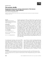
Fig. 3. Involvement of PTP-Bas/BL in different biological systems has been demonstrated. (A) PTP-Bas regulates the cell surface expression of the
human Fas receptor. There are at least two possibilities of how PTP-Bas can affect cell surface expression of Fas. PTP-Bas could regulate the
transport of Fas from the Golgi apparatus (G) to the cell surface ÔaÕ. Alternatively, PTP-Bas could increase the transport of Fas from the cell surface
to endosomal compartments (EC) ÔbÕ or from endosomal compartments to the Golgi apparatus ÔcÕ.N,nucleus;Ly,lysosome.(B)PTP-Bas/BLis
involved in the regulation of cytokinesis. During anaphase and cytokinesis PTP-Bas/BL accumulates at the spindle midzone and contractile ring.
PTP-Bas/BL associates via its FERM domain with F-actin of the contractile ring and via its N-terminus with the midzone microtubules.
Overexpression of PTP-BL leads to multinucleate cells. (C) PTP-BL is involved in the regulation of ephrinB signalling. The eph/ephrinB receptor
system evokes bi-directional signalling. After binding, eph-receptors are tyrosine phosphorylated (autophosphorylation) and ephrinB is phos-
phorylated by the src kinase family. Phosphotyrosine signalling of ephrinB is started via binding to SH2 containing proteins. PTP-BL is recruited to
ephrinB with a delayed kinetic leading to its dephosphorylation and replacing the SH2 dependent signalling by a PDZ domain dependent signalling
(switch-model of ephrinB signalling).
4794 K. S. Erdmann (Eur. J. Biochem. 270) Ó FEBS 2003
precise function of PTP-BL in cytokinesis is not known,
however, signal transduction via the small G-protein rho
plays an important role in contractile ring assembly and in
the regulation of its contraction during cytokinesis [85].
PTP-Bas/BL has been shown to interact with the rho
GTPase activating protein PARG and the rho regulated
protein kinase, PRK2, implicating PTP-BL in a rho
signaling pathway probably relevant to cytokinesis. In
additon, PTP-BL is able to interact with F-actin and with
microtubules, suggesting a role in connecting the contractile
ring with the spindle midzone microtubules thereby coordi-
nating cleavage furrow ingression with microtubule dynam-
ics. Finally, given the role of PTP-Bas in trafficking of Fas
(see above), PTP-BL could be important for targeted vesicle
transport during cleavage furrow ingression, which has been
demonstrated to be important for the final step of cyto-
kinesis [86].
Concluding remarks
PTP-Bas/BL is an exceptionally large protein tyrosine
phosphatase comprising a number of protein–protein
interaction domains. As summarized above, many proteins
have been identified as interacting with one or two of the
five PDZ domains of PTP-Bas/BL. Moreover, PTP-BL is
associated with F-actin and microtubules and binding to
PIP2 has been demonstrated. Thus, PTP-BL/Bas serves
clearly as a major scaffolding protein of a supramolecular
protein complex. However, in spite of the large number of
interacting proteins a clear cut function has not been
assigned to PTP-Bas/BL. Given the modular complexity of
PTP-Bas/BL and the number of different splicing variants it
is reasonable to assume that this protein tyrosine phospha-
tase is involved in a number of different physiological
processes. This is already reflected in the diversity of
biological systems where an involvement of PTP-BL/PTP-
Bas has already been defined (Fas trafficking, ephrinB
signaling and regulation of cytokinesis). Moreover, there is
evidence that PTP-Bas/BL is also part of a protein complex
within the nucleus. A challenge for the future will be to
elucidate whether there is a common mechanism underlying
the involvement of PTP-Bas/BL in these different systems.
Focusing on splice variant specific interactions as well as
identifying major substrates for the protein tyrosine phos-
phatase will lead to a better understanding of this fascin-
ating molecule. Moreover, mice knockout technology and
the recently developed siRNA methodology will certainly
contribute in identifying the precise molecular functions of
PTP-Bas/BL.
Acknowledgements
This work was supported by a grant from the Deutsche Forschungsg-
emeinschaft, SFB 452.
References
1. Hunter, T. (1995) Protein kinases and phosphatases: the yin and
yang of protein phosphorylation and signaling. Cell 80, 225–236.
2. Andersen, J.N., Mortensen, O.H., Peters, G.H., Drake, P.G.,
Iversen, L.F., Olsen, O.H., Jansen, P.G., Andersen, H.S., Tonks,
N.K. & Moller, N.P. (2001) Structural and evolutionary
relationships among protein tyrosine phosphatase domains. Mol.
Cell Biol. 21, 7117–7136.
3. Maekawa, K., Imagawa, N., Nagamatsu, M. & Harada, S. (1994)
Molecular cloning of a novel protein-tyrosine phosphatase con-
taining a membrane-binding domain and GLGF repeats. FEBS
Lett. 337, 200–206.
4. Hendriks, W., Schepens, J., Bachner, D., Rijss, J., Zeeuwen, P.,
Zechner,U.,Hameister,H.&Wieringa,B.(1995)Molecular
cloning of a mouse epithelial protein-tyrosine phosphatase with
similarities to submembranous proteins. J. Cell Biochem. 59,
418–430.
5. Gu, M.X., York, J.D., Warshawsky, I. & Majerus, P.W. (1991)
Identification, cloning, and expression of a cytosolic mega-
karyocyte protein-tyrosine-phosphatase with sequence homology
to cytoskeletal protein 4.1. Proc. Natl Acad. Sci. USA 88, 5867–
5871.
6. Yang, Q. & Tonks, N.K. (1991) Isolation of a cDNA clone
encoding a human protein-tyrosine phosphatase with homology
to the cytoskeletal-associated proteins band 4.1, ezrin, and talin.
Proc.NatlAcad.Sci.USA88, 5949–5953.
7. Moller, N.P., Moller, K.B., Lammers, R., Kharitonenkov, A.,
Sures, I. & Ullrich, A. (1994) Src kinase associates with a member
of a distinct subfamily of protein-tyrosine phosphatases containing
an ezrin-like domain. Proc. Natl Acad. Sci. USA 91, 7477–7481.
8. Higashitsuji, H., Arii, S., Furutani, M., Imamura, M., Kaneko,
Y., Takenawa, J., Nakayama, H. & Fujita, J. (1995) Enhanced
expression of multiple protein tyrosine phosphatases in the
regenerating mouse liver: isolation of PTP-RL10, a novel cyto-
plasmic-type phosphatase with sequence homology to cytoskeletal
protein 4.1. Oncogene 10, 407–414.
9. Dorner, C., Ciossek, T., Muller, S., Moller, P.H., Ullrich, A. &
Lammers, R. (1998) Characterization of KIF1C, a new kinesin-
like protein involved in vesicle transport from the Golgi apparatus
to the endoplasmic reticulum. J. Biol. Chem. 273, 20267–20275.
10. Jui,H.Y.,Tseng,R.J.,Wen,X.,Fang,H.I.,Huang,L.M.,Chen,
K.Y., Kung, H.J., Ann., D.K. & Shih, H.M. (2000) Protein-tyro-
sine phosphatase D1, a potential regulator and effector for Tec
family kinases. J. Biol. Chem. 275, 41124–41132.
11. Lavoie, C., Chevet, E., Roy, L., Tonks, N.K., Fazel, A., Posner,
B.I., Paiement, J. & Bergeron, J.J. (2000) Tyrosine phosphoryla-
tion of p97 regulates transitional endoplasmic reticulum assembly
in vitro. Proc. Natl Acad. Sci. USA 97, 13637–13642.
12. Zhang, S.H., Liu, J., Kobayashi, R. & Tonks, N.K. (1999) Iden-
tification of the cell cycle regulator VCP (p97/CDC48) as a sub-
strate of the band 4.1-related protein-tyrosine phosphatase
PTPH1. J. Biol. Chem. 274, 17806–17812.
13. Zheng, Y., Schlondorff, J. & Blobel, C.P. (2002) Evidence for
regulation of the tumor necrosis factor alpha-convertase (TACE)
by protein-tyrosine phosphatase PTPH1. J. Biol. Chem. 277,
42463–42470.
14. Banville, D., Ahmad, S., Stocco, R. & Shen, S.H. (1994) A novel
protein-tyrosine phosphatase with homology to both the cyto-
skeletal proteins of the band 4.1 family and junction-associated
guanylate kinases. J. Biol. Chem. 269, 22320–22327.
15. Saras, J., Claesson-Welsh, L., Heldin, C.H. & Gonez, L.J. (1994)
Cloning and characterization of PTPL1, a protein tyrosine phos-
phatase with similarities to cytoskeletal-associated proteins.
J. Biol. Chem. 269, 24082–24089.
16. Sato, T., Irie, S., Kitada, S. & Reed, J.C. (1995) FAP-1: a protein
tyrosine phosphatase that associates with Fas. Science 268,
411–415.
17. Chida,D.,Kume,T.,Mukouyama,Y.,Tabata,S.,Nomura,N.,
Thomas, M.L., Watanabe, T. & Oishi, M. (1995) Characterization
of a protein tyrosine phosphatase (RIP) expressed at a very
early stage of differentiation in both mouse erythroleukemia and
embryonal carcinoma cells. FEBS Lett. 358, 233–239.
Ó FEBS 2003 The protein tyrosine phosphatase PTP-Bas/BL (Eur. J. Biochem. 270) 4795
18. Ciccarelli, F.D., Bork, P. & Kerkhoff, E. (2003) The KIND
module: a putative signalling domain evolved from the C lobe of
the protein kinase fold. Trends Biochem. Sci. 28, 349–352.
19. Chishti, A.H., Kim, A.C., Marfatia, S.M., Lutchman, M.,
Hanspal, M., Jindal, H., Liu, S.C., Low, P.S., Rouleau, G.A.,
Mohandas, N., Chasis, J.A., Conboy, J.G., Gascard, P., Taka-
kuwa, Y., Huang, S.C., Benz, E.J. Jr, Bretscher, A., Fehon, R.G.,
Gusella, J.F., Ramesh, V., Solomon, F., Marchesi, V.T., Tsukita,
S., Tsukita, S. & Hoover, K.B. (1998) The FERM domain: a
unique module involved in the linkage of cytoplasmic proteins to
the membrane. Trends Biochem. Sci. 23, 281–282.
20. Kennedy, M.B. (1995) Origin of PDZ (DHR, GLGF) domains.
Trends Biochem. Sci. 20, 350.
21. Sheng, M. & Sala, C. (2001) PDZ domains and the organization of
supramolecular complexes. Annu. Rev. Neurosci. 24, 1–29.
22. Erdmann, K.S., Kuhlmann, J., Lessmann, V., Herrmann, L.,
Eulenburg, V., Muller, O. & Heumann, R. (2000) The Adeno-
matous Polyposis Coli-protein (APC) interacts with the protein
tyrosine phosphatase PTP-BL via an alternatively spliced PDZ
domain. Oncogene 19, 3894–3901.
23. Thomas, T., Voss, A.K. & Gruss, P. (1998) Distribution of a
murine protein tyrosine phosphatase BL-beta-galactosidase fusion
protein suggests a role in neurite outgrowth. Dev. Dyn. 212,
250–257.
24. Arai, M., Kannagi, M., Matsuoka, M., Sato, T., Yamamoto, N. &
Fujii, M. (1998) Expression of FAP-1 (Fas-associated phospha-
tase) and resistance to Fas-mediated apoptosis in T cell lines
derived from human T cell leukemia virus type 1-associated
myelopathy/tropical spastic paraparesis patients. AIDS Res. Hum.
Retroviruses 14, 261–267.
25. Lee, S.H., Shin, M.S., Lee, H.S., Bae, J.H., Lee, H.K., Kim, H.S.,
Kim,S.Y.,Jang,J.J.,Joo,M.,Kang,Y.K.,Park,W.S.,Park,J.Y.,
Oh,R.R.,Han,S.Y.,Lee,J.H.,Kim,S.H.,Lee,J.Y.&Yoo,N.J.
(2001) Expression of Fas and Fas-related molecules in human
hepatocellular carcinoma. Hum. Pathol. 32, 250–256.
26. Li, Y., Kanki, H., Hachiya, T., Ohyama, T., Irie, S., Tang, G.,
Mukai, J. & Sato, T. (2000) Negative regulation of Fas-mediated
apoptosis by FAP-1 in human cancer cells. Int. J. Cancer 87,
473–479.
27. Meinhold-Heerlein, I., Stenner-Liewen, F., Liewen, H., Kitada, S.,
Krajewska, M., Krajewski, S., Zapata, J.M., Monks, A., Scudiero,
D.A., Bauknecht, T. & Reed, J.C. (2001) Expression and potential
role of Fas-associated phosphatase-1 in ovarian cancer. Am.
J. Pathol. 158, 1335–1344.
28. Cuppen, E., Wijers, M., Schepens, J., Fransen, J., Wieringa, B. &
Hendriks, W. (1999) A FERM domain governs apical confine-
ment of PTP-BL in epithelial cells. J. Cell Sci. 112, 3299–3308.
29. Palmer, A., Zimmer, M., Erdmann, K.S., Eulenburg, V., Porthin,
A.,Heumann,R.,Deutsch,U.&Klein,R.(2002)EphrinB
phosphorylation and reverse signaling: regulation by Src kinases
and PTP-BL phosphatase. Mol. Cell 9, 725–737.
30. Ungefroren, H., Kruse, M.L., Trauzold, A., Roeschmann, S.,
Roeder,C.,Arlt,A.,Henne-Bruns,D.&Kalthoff,H.(2001)
FAP-1 in pancreatic cancer cells: functional and mechanistic stu-
dies on its inhibitory role in CD95-mediated apoptosis. J. Cell Sci.
114, 2735–2746.
31. Herrmann, L., Dittmar, T. & Erdmann, K.S. (2003) The Protein
Tyrosine Phosphatase PTP-BL Associates with the Midbody and
Is Involved in the Regulation of Cytokinesis. Mol. Biol. Cell 14,
230–240.
32. Pearson, M.A., Reczek, D., Bretscher, A. & Karplus, P.A. (2000)
Structure of the ERM protein moesin reveals the FERM domain
fold masked by an extended actin binding tail domain. Cell 101,
259–270.
33. Hamada, K., Shimizu, T., Matsui, T., Tsukita, S. & Hakoshima,
T. (2000) Structural basis of the membrane-targeting and
unmasking mechanisms of the radixin FERM domain. EMBO J.
19, 4449–4462.
34. Bompard, G., Martin, M., Roy, C., Vignon, F. & Freiss, G. (2003)
Membrane targeting of protein tyrosine phosphatase PTPL1
through its FERM domain via binding to phosphatidylinositol
4,5-biphosphate. J. Cell Sci. 116, 2519–2530.
35. Hillier, B.J., Christopherson, K.S., Prehoda, K.E., Bredt, D.S. &
Lim, W.A. (1999) Unexpected modes of PDZ domain scaffolding
revealed by structure of nNOS-syntrophin complex. Science 284,
812–815.
36. Kornau, H.C., Schenker, L.T., Kennedy, M.B. & Seeburg, P.H.
(1995) Domain interaction between NMDA receptor subunits and
the postsynaptic density protein PSD-95. Science 269, 1737–1740.
37. Cuppen, E., Gerrits, H., Pepers, B., Wieringa, B. & Hendriks, W.
(1998)PDZmotifsinPTP-BLandRILbindtointernalprotein
segments in the LIM domain protein RIL. Mol. Biol. Cell 9,
671–683.
38. Zimmermann, P., Meerschaert, K., Reekmans, G., Leenaerts, I.,
Small, J.V., Vandekerckhove, J., David, G. & Gettemans, J. (2002)
PIP (2)-PDZ domain binding controls the association of syntenin
with the plasma membrane. Mol. Cell 9, 1215–1225.
39. Morais Cabral, J.H., Petosa, C., Sutcliffe, M.J., Raza, S.,
Byron, O., Poy, F., Marfatia, S.M., Chishti, A.H. & Lidding-
ton, R.C. (1996) Crystal structure of a PDZ domain. Nature 382,
649–652.
40.Doyle,D.A.,Lee,A.,Lewis,J.,Kim,E.,Sheng,M.&
MacKinnon, R. (1996) Crystal structures of a complexed and
peptide-free membrane protein-binding domain: molecular basis
of peptide recognition by PDZ. Cell 85, 1067–1076.
41. Kozlov, G., Gehring, K. & Ekiel, I. (2000) Solution structure of
the PDZ2 domain from human phosphatase hPTP1E and its
interactions with C-terminal peptides from the Fas receptor.
Biochemistry 39, 2572–2580.
42. Walma, T., Spronk, C.A., Tessari, M., Aelen, J., Schepens, J.,
Hendriks, W. & Vuister, G.W. (2002) Structure, dynamics and
binding characteristics of the second PDZ domain of PTP-BL.
J. Mol. Biol. 316, 1101–1110.
43. Kozlov, G., Banville, D., Gehring, K. & Ekiel, I. (2002) Solution
structure of the PDZ2 domain from cytosolic human phosphatase
hPTP1E complexed with a peptide reveals contribution of the
beta2-beta3 loop to PDZ domain–ligand interactions. J. Mol. Biol.
320, 813–820.
44. Kachel,N.,Erdmann,K.S.,Kremer,W.,Wolff,P.,Gronwald,
W., Heumann, R. & Kalbitzer, H.R. (2003) Structure determin-
ation and ligand interactions of the PDZ2b domain of PTP-Bas
(hPTP1E): Splicing-induced modulation of ligand specificity.
J. Mol. Biol. 334, 143–155.
45. Cuppen, E., van Ham, M., Pepers, B., Wieringa, B. & Hendriks,
W. (1999) Identification and molecular characterization of BP75,
a novel bromodomain-containing protein. FEBS Lett. 459,
291–298.
46. Maekawa,K.,Imagawa,N.,Naito,A.,Harada,S.,Yoshie,O.&
Takagi, S. (1999) Association of protein-tyrosine phosphatase
PTP-BAS with the transcription-factor-inhibitory protein Ikappa-
Balpha through interaction between the PDZ1 domain and
ankyrin repeats. Biochem. J. 337, 179–184.
47. Horn, P.J. & Peterson, C.L. (2001) The bromodomain: a
regulator of ATP-dependent chromatin remodeling? Front Biosci.
6, D1019–D1023.
48. Imbert, V., Rupec, R.A., Livolsi, A., Pahl, H.L., Traenckner, E.B.,
Mueller-Dieckmann, C., Farahifar, D., Rossi, B., Auberger, P.,
Baeuerle, P.A. & Peyron, J.F. (1996) Tyrosine phosphorylation of
I kappa B-alpha activates NF-kappa B without proteolytic
degradation of I kappa B-alpha. Cell 86, 787–798.
49. Baeuerle, P.A. & Baltimore, D. (1996) NF-kappa B: ten years
after. Cell 87, 13–20.
4796 K. S. Erdmann (Eur. J. Biochem. 270) Ó FEBS 2003
50. Nakai, Y., Irie, S. & Sato, T.A. (2000) Identification of Ikappa-
Balpha as a substrate of Fas-associated phosphatase-1. Eur.
J. Biochem. 267, 7170–7175.
51. Irie, S., Hachiya, T., Rabizadeh, S., Maruyama, W., Mukai, J., Li,
Y., Reed, J.C., Bredesen, D.E. & Sato, T.A. (1999) Functional
interaction of Fas-associated phosphatase-1 (FAP-1) with p75
(NTR) and their effect on NF-kappaB activation. FEBS Lett. 460,
191–198.
52. Cuppen, E., Nagata, S., Wieringa, B. & Hendriks, W. (1997) No
evidence for involvement of mouse protein-tyrosine phosphatase-
BAS-like Fas-associated phosphatase-1 in Fas-mediated apopto-
sis. J. Biol. Chem. 272, 30215–30220.
53. Cuppen, E., van Ham, M., Wansink, D.G., de Leeuw, A., Wier-
inga, B. & Hendriks, W. (2000) The zyxin-related protein TRIP6
interacts with PDZ motifs in the adaptor protein RIL and the
protein tyrosine phosphatase PTP-BL. Eur. J. Cell Biol. 79,
283–293.
54. Murthy, K.K., Clark, K., Fortin, Y., Shen, S.H. & Banville, D.
(1999) ZRP-1, a zyxin-related protein, interacts with the second
PDZ domain of the cytosolic protein tyrosine phosphatase
hPTP1E. J. Biol. Chem. 274, 20679–20687.
55. Yi, J., Kloeker, S., Jensen, C.C., Bockholt, S., Honda, H., Hirai,
H. & Beckerle, M.C. (2002) Members of the Zyxin family of LIM
proteins interact with members of the p130Cas family of signal
transducers. J. Biol. Chem. 277, 9580–9589.
56. Behrens,J.,vonKries,J.P.,Kuhl,M.,Bruhn,L.,Wedlich,D.,
Grosschedl, R. & Birchmeier, W. (1996) Functional interaction
of beta-catenin with the transcription factor LEF-1. Nature 382,
638–642.
57. Kawasaki, Y., Sato, R. & Akiyama, T. (2003) Mutated APC and
Asef are involved in the migration of colorectal tumour cells. Nat.
Cell Biol. 5, 211–215.
58. Gross, C., Heumann, R. & Erdmann, K.S. (2001) The protein
kinase C-related kinase PRK2 interacts with the protein tyrosine
phosphatase PTP-BL via a novel PDZ domain binding motif.
FEBS Lett. 496, 101–104.
59. Vincent, S. & Settleman, J. (1997) The PRK2 kinase is a potential
effector target of both Rho and Rac GTPases and regulates actin
cytoskeletal organization. Mol. Cell Biol. 17, 2247–2256.
60. Mellor, H., Flynn, P., Nobes, C.D., Hall, A. & Parker, P.J. (1998)
PRK1 is targeted to endosomes by the small GTPase, RhoB.
J. Biol. Chem. 273, 4811–4814.
61. Gampel, A., Parker, P.J. & Mellor, H. (1999) Regulation of epi-
dermal growth factor receptor traffic by the small GTPase rhoB.
Curr. Biol. 9, 955–958.
62. vanHam,M.,Croes,H.,Schepens,J.,Fransen,J.,Wieringa,B.&
Hendriks, W. (2003) Cloning and characterization of mCRIP2, a
mouse LIM-only protein that interacts with PDZ domain IV of
PTP-BL. Genes Cells 8, 631–644.
63. Saras, J., Franzen, P., Aspenstrom, P., Hellman, U., Gonez, L.J. &
Heldin, C.H. (1997) A novel GTPase-activating protein for Rho
interacts with a PDZ domain of the protein-tyrosine phosphatase
PTPL1. J. Biol. Chem. 272, 24333–24338.
64. Lin, D., Gish, G.D., Songyang, Z. & Pawson, T. (1999) The
carboxyl terminus of B class ephrins constitutes a PDZ domain
binding motif. J. Biol. Chem. 274, 3726–3733.
65. Ungefroren, H., Voss, M., Jansen, M., Roeder, C., Henne-Bruns,
D., Kremer, B. & Kalthoff, H. (1998) Human pancreatic adeno-
carcinomas express Fas and Fas ligand yet are resistant to Fas-
mediated apoptosis. Cancer Res. 58, 1741–1749.
66. Itoh, N., Yonehara, S., Ishii, A., Yonehara, M., Mizushima, S.,
Sameshima, M., Hase, A., Seto, Y. & Nagata, S. (1991) The
polypeptide encoded by the cDNA for human cell surface antigen
Fas can mediate apoptosis. Cell 66, 233–243.
67. Oehm, A., Behrmann, I., Falk, W., Pawlita, M., Maier, G., Klas,
C.,Li-Weber,M.,Richards,S.,Dhein,J.&Trauth,B.C.(1992)
Purification and molecular cloning of the APO-1 cell surface
antigen, a member of the tumor necrosis factor/nerve growth
factor receptor superfamily. Sequence identity with the Fas anti-
gen. J. Biol. Chem. 267, 10709–10715.
68. Peter, M.E., Krammer, P.H. (2003) The CD95 (APO-1/Fas) DISC
and beyond. Cell Death. Differ. 10, 26–35.
69. Itoh, N. & Nagata, S. (1993) A novel protein domain required for
apoptosis. Mutational analysis of human Fas antigen. J. Biol.
Chem. 268, 10932–10937.
70.Saras,J.,Engstrom,U.,Gonez,L.J.&Heldin,C.H.(1997)
Characterization of the interactions between PDZ domains of the
protein-tyrosine phosphatase PTPL1 and the carboxyl-terminal
tail of Fas. J. Biol. Chem. 272, 20979–20981.
71. Yanagisawa, J., Takahashi, M., Kanki, H., Yano-Yanagisawa,
H.,Tazunoki,T.,Sawa,E.,Nishitoba,T.,Kamishohara,M.,
Kobayashi, E., Kataoka, S. & Sato, T. (1997) The molecular
interaction of Fas and FAP-1. A tripeptide blocker of human Fas
interaction with FAP-1 promotes Fas-induced apoptosis. J. Biol.
Chem. 272, 8539–8545.
72. Myc,A.,Arscott,P.L.,Bretz,J.D.,Thompson,N.W.&Baker,
J.R. Jr (1999) Characterization of FAP-1 expression and function
in thyroid follicular cells. Endocrinology 140, 5431–5434.
73. Lee, S.H., Shin, M.S., Lee, J.Y., Park, W.S., Kim, S.Y., Jang, J.J.,
Dong, S.M., Na, E.Y., Kim, C.S., Kim, S.H. & Yoo, N.J. (1999)
In vivo expression of soluble Fas and FAP-1: possible mechanisms
of Fas resistance in human hepatoblastomas. J. Pathol. 188,
207–212.
74. Mundle, S.D., Mativi, B.Y., Bagai, K., Feldman, G., Cheema, P.,
Gautam, U., Reza, S., Cartlidge, J.D., Venugopal, P., Shetty, V.,
Gregory,S.A.,Robin,E.,Rifkin,S.,Shah,R.&Raza,A.(1999)
Spontaneous down-regulation of Fas-associated phosphatase-1
may contribute to excessive apoptosis in myelodysplastic mar-
rows. Int. J. Hematol. 70, 83–90.
75. Komada, Y., Inaba, H., Zhou, Y.W., Zhang, X.L., Tanaka, S.,
Azuma, E. & Sakurai, M. (1997) mRNA expression of Fas
receptor (CD95)-associated proteins (Fas-associated phosphatase-
1/FAP-1, Fas-associating protein with death domain/FADD, and
receptor-interacting protein/RIP) in human leukaemia/lymphoma
cell lines. Br. J. Haematol. 99, 325–330.
76. Bompard, G., Puech, C., Prebois, C., Vignon, F. & Freiss, G.
(2002) Protein-tyrosine phosphatase PTPL1/FAP-1 triggers
apoptosis in human breast cancer cells. J. Biol. Chem. 277, 47861–
47869.
77. Ivanov, V.N., Lopez, B.P., Maulit, G., Sato, T.A., Sassoon, D. &
Ronai, Z. (2003) FAP)1 association with Fas (Apo-1) inhibits Fas
expression on the cell surface. Mol. Cell Biol. 23, 3623–3635.
78. Cao, T.T., Deacon, H.W., Reczek, D., Bretscher, A. & von Zas-
trow, M. (1999) A kinase-regulated PDZ–domain interaction
controls endocytic sorting of the beta2-adrenergic receptor. Nature
401, 286–290.
79. Swiatecka-Urban, A., Duhaime, M., Coutermarsh, B., Karlson,
K.H., Collawn, J., Milewski, M., Cutting, G.R., Guggino,
W.B., Langford, G. & Stanton, B.A. (2002) PDZ domain
interaction controls the endocytic recycling of the cystic fibrosis
transmembrane conductance regulator. J. Biol. Chem. 277, 40099–
40105.
80. Grunwald, I.C., Korte, M., Wolfer, D., Wilkinson, G.A., Uns-
icker, K., Lipp, H.P., Bonhoeffer, T. & Klein, R. (2001) Kinase-
independent requirement of EphB2 receptors in hippocampal
synaptic plasticity. Neuron 32, 1027–1040.
81. Wilkinson, D.G. (2001) Multiple roles of EPH receptors and
ephrins in neural development. Nat. Rev. Neurosci. 2, 155–164.
82. Kullander, K. & Klein, R. (2002) Mechanisms and functions of
Eph and ephrin signalling. Nat. Rev. Mol. Cell Biol. 3, 475–486.
83. Cowan, C.A. & Henkemeyer, M. (2001) The SH2/SH3 adaptor
Grb4 transduces B-ephrin reverse signals. Nature 413, 174–179.
Ó FEBS 2003 The protein tyrosine phosphatase PTP-Bas/BL (Eur. J. Biochem. 270) 4797
84. Glotzer, M. (2001) Animal cell cytokinesis. Annu. Rev. Cell Dev.
Biol. 17, 351–386.
85. Prokopenko, S.N., Saint, R. & Bellen, H.J. (2000) Untying the
Gordian knot of cytokinesis. Role of small G proteins and their
regulators. J. Cell Biol. 148, 843–848.
86. Skop, A.R., Bergmann, D., Mohler, W.A. & White, J.G. (2001)
Completion of cytokinesis in C. elegans requires a brefeldin
A-sensitive membrane accumulation at the cleavage furrow apex.
Curr. Biol. 11, 735–746.
4798 K. S. Erdmann (Eur. J. Biochem. 270) Ó FEBS 2003