Báo cáo khoa học: Tracking interactions that stabilize the dimer structure of starch phosphorylase from Corynebacterium callunae Roles of Arg234 and Arg242 revealed by sequence analysis and site-directed mutagenesis doc
Bạn đang xem bản rút gọn của tài liệu. Xem và tải ngay bản đầy đủ của tài liệu tại đây (438.01 KB, 11 trang )
Eur. J. Biochem. 270, 2126–2136 (2003) Ó FEBS 2003
doi:10.1046/j.1432-1033.2003.03562.x
Tracking interactions that stabilize the dimer structure of starch
phosphorylase from Corynebacterium callunae
Roles of Arg234 and Arg242 revealed by sequence analysis and site-directed
mutagenesis
Richard Griessler1,2,3, Alexandra Schwarz1,3, Jan Mucha2 and Bernd Nidetzky1,3
1
Institute of Food Technology and 2Centre of Applied Genetics, University of Agricultural Sciences, Vienna, Austria;
Institute of Biotechnology, Graz University of Technology, Austria
3
Glycogen phosphorylases (GPs) constitute a family of
widely spread catabolic a1,4-glucosyltransferases that are
active as dimers of two identical, pyridoxal 5¢-phosphatecontaining subunits. In GP from Corynebacterium callunae,
physiological concentrations of phosphate are required
to inhibit dissociation of protomers and cause a 100-fold
increase in kinetic stability of the functional quarternary
structure. To examine interactions involved in this large
stabilization, we have cloned and sequenced the coding gene
and have expressed fully active C. callunae GP in Escherichia
coli. By comparing multiple sequence alignment to structurefunction assignments for regulated and nonregulated GPs
that are stable in the absence of phosphate, we have scrutinized the primary structure of C. callunae enzyme for
sequence changes possibly related to phosphate-dependent
dimer stability. Location of Arg234, Arg236, and Arg242
within the predicted subunit-to-subunit contact region made
these residues primary candidates for site-directed muta-
genesis. Individual Arg fi Ala mutants were purified and
characterized using time-dependent denaturation assays in
urea and at 45 °C. R234A and R242A are enzymatically
active dimers and in the absence of added phosphate, they
display a sixfold and fourfold greater kinetic stability of
quarternary interactions than the wild-type, respectively.
The stabilization by 10 mM of phosphate was, however, up
to 20-fold greater in the wild-type than in the two mutants.
The replacement of Arg236 by Ala was functionally silent
under all conditions tested. Arg234 and Arg242 thus partially destabilize the C. callunae GP dimer structure, and
phosphate binding causes a change of their tertiary or
quartenary contacts, likely by an allosteric mechanism,
which contributes to a reduced protomer dissociation rate.
Glycogen phosphorylases (GPs) catalyse degradation of
glycogen and structurally related reserve polysaccharides
in the cytosol to provide energy via the branch point
metabolite a-D-glucose-1-phosphate. All known GPs are
functional homodimers composed of % 90-kDa subunits
and require pyridoxal 5¢-phosphate (PLP) cofactor for
activity [1–7]. Although a very low basal activity may be
present in the holoenzyme protomer, quarternary interactions clearly determine physiological levels of phosphorylase activity and are a prerequisite for the regulatory
properties of eukaryotic GPs [8–10]. Forces that stabilize
the dimer structure of GP are therefore essential to
optimal enzyme function under physiological boundary
conditions. GPs are a/b proteins that display a twodomain fold in which the N-terminal domain and the
C-terminal domain are separated by a catalytic site cleft.
The structural elements that comprise the subunit–subunit
interface are located in the N-terminal domain. The dimer
contact regions of regulated and nonregulated GPs share
structural similiarity overall, but differ on the molecular
level [3–7].
Starch phosphorylase (StP) from the soil bacterium
Corynebacterium callunae is a member of the GP family
[11]. Its activity is not under control of common allosteric
effectors of mammalian GPs such as AMP or D-glucose6-phosphate. The enzyme differs from structurally wellcharacterized GPs [3–7] because it requires physiological
concentrations of phosphate (% 5 mM) for stability of the
functional oligomeric structure [12]. Binding of phosphate
to a protein site different from the site where the substrate
phosphate binds causes apparent tightening of quarternary
interactions present in StP and leads to a 100-fold increase
in kinetic stability of the active dimer [12]. The very low
in-vitro lifetime of StP activity in the absence of phosphate
(% 30 min [12]) suggests that this stabilizing effect might be
Correspondence to B. Nidetzky, Institute of Biotechnology, Graz
University of Technology, Petersgasse 12/I, A-8010 Graz, Austria.
Fax: + 43 316 873 8434, Tel.: + 43 316 873 8400;
E-mail:
Abbreviations: GP, glycogen phosphorylase; rmGP, rabbit muscle GP;
MalP, maltodextrin phosphorylase; EcMalP, MalP from Escherichia
coli; PLP, pyridoxal 5¢-phosphate; StP, starch phosphorylase.
Enzymes: a-glucan (glycogen, starch, maltodextrin) phosphorylase
(1,4-a-D-glucan:phosphate a-D-glucosyltransferase) (EC 2.4.1.1).
Note: The genomic sequence of C. callunae that comprises the entire
structural gene of starch phosphorylase is available under the
GenBank accession number: AY102616.
(Received 28 November 2002, accepted 7 March 2003)
Keywords: interface; oxyanion; phosphate; stabilization;
subunit dissociation.
Ó FEBS 2003
Dimer stability of bacterial starch phosphorylase (Eur. J. Biochem. 270) 2127
important in the physiology of C. callunae. In light of the
fact that interactions apparently critical to a stable and
active protein conformation converge at the dimer interface
of GP, we considered oxyanion-dependent stability of the
StP dimer to be a significant problem and hence worth
examining. We thus turned our attention to the primary
structure of StP and report here the cloning, sequencing and
heterologous expression in Escherichia coli of the gene
encoding this enzyme. In an effort to identify sequence
changes relevant to GP dimer stability, we compared
multiple sequence alignment with secondary structure
assignments and dimer contacts in structurally characterized
GPs [3–7,13–15]. Through this process, the main element of
the dimer interface in GPs, the so-called TOWER helix, was
allocated to the linear sequence of StP. In rabbit muscle GP
(rmGP) and likewise other GPs [3–7,13–15], this helix forms
intimate contacts with its counterpart helix on the opposite
subunit. Three arginine residues are located within the
predicted TOWER helix of StP. Because arginine are
common components of interfaces of oligomeric proteins
and frequently show interaction with oxyanion ligands such
as phosphate or sulfate, the ÔTOWER argininesÕ of StP were
selected as candidates to be replaced by alanine-scanning
site-directed mutagenesis. We have assessed the role of each
arginine for oxyanion-dependent stability of the StP dimer.
Experimental procedures
(GT)CT(AGT)GC-3¢; and reverse (p2), 5¢-GC(CT)TC
(AGT)GGCA(AGT)(AC)AC(AGC)GT(AG)TGGTT(AGT)
GT-3¢. Polymerase chain reactions (50 lL) were carried out
with a Hybaid thermocycler (Thermo Life Sciences) and
used 200 ng of chromosomal template DNA, 40 pmol sense
and antisense primers, 150 lmol dNTPs in PCR buffer
(Promega). PCRs consisted of 30 cycles of 30 s denaturation
at 95 °C, followed by 20 s primer annealing at 55 °C and
40 s elongation at 72 °C. The final extension step was
carried out at 72 °C for 7 min. The resulting PCR product
was gel-purified and placed into a pUC 18 vector (Life
Technologies) using SmaI cleavage and blunt-end cloning.
This recombinant vector was transferred by electroporation
into competent cells of E. coli DH5a (Stratagene), and after
plasmid purification the insert was subjected to dideoxy
sequencing with an ABI Prism 310 Genetic Analyzer
(Applied Biosystems) using Universal Primer (Amersham
Pharmacia Biotech). The PCR product was used as specific
probe for Southern blot hybridization experiments. Fragments of interest were cloned into pBluescript II SK(+/–)
(Life Technologies) via BamHI and HindIII restriction sites
and used to generate a partial genomic library of C. callunae
DNA. Colony hybridization with the PCR probe was used
to screen this library. Positive clones were sequenced in both
senses of the DNA at the VBC Genomics Sequencing
Service Facility of the University of Vienna using the Ôprimer
walkingÕ method.
Materials
Construction of expression plasmids
Natural StP from C. callunae DSM 20145 was produced
and purified by reported procedures [11]. Materials and
assays for measuring enzyme activities in the directions of
a-glucan degradation and synthesis have been described
elsewhere [11,12]. Restriction endonucleases, T4DNA ligase
and Taq DNA polymerase were obtained from Promega.
Pfu DNA polymerase, alkaline phosphatase, RNase, and
positively charged nylon membranes were from Roche. The
expression vectors pQE 30 and pQE 70, the gel extraction
kit Qiaex II, and the plasmid purification kit were from
Qiagen.
The following primer set was designed to amplify the entire
open reading frame of the StP gene by using PCR:
Preparation of an oligonucleotide probe
for the StP gene
Chromosomal DNA from C. callunae DSM 20145 was
prepared by incubating approximately 200 mg of wet cell
mass suspended in 10 mM Tris/HCl buffer, pH 7.6, containing 1 mM EDTA and 15 mg lysozyme (Sigma) for 3 h
at 37 °C. To this mixture were added 3 mL of a solution of
0.4 M NaCl, 0.7% (w/v) SDS and 1 mgỈmL)1 proteinase K
(Sigma) in 10 mM Tris/HCl buffer, pH 8.2. After incubation
at 50 °C for 5 h, protein was precipitated with 1 mL of 6 M
NaCl and removed by centrifugation at 13 000 g. The DNA
in the supernatant was precipitated with ethanol and
purified by standard protocols [16].
From a comparison of GP sequences in the GenBank
database, two well-conserved peptides, GNGGLGRL (residues 131–138 in rmGP) and TNHTLMPEAL (residues
374–383 in rmGP), were chosen and reverse translated into
a pair of degenerated PCR primers: forward (p1), 5¢-GG
(ACT)AA(CT)GG(GCT)GGT(CT)T(AG)GG(ACT)CG
pNter (5¢-CGCGCATGCAGCCCTGAAAAACAGCC-3¢)
derived from the authentic N-terminus of StP [11], and
pCter (5-ACGC-GTCGACCTACTTTTTAACAGCAG
GAGTTG-3¢)
where SphI and SalI restriction sites are underlined.
The 50-lL reaction mixture contained 200 ng of C. callunae DNA, 50 pmol of pNter and pCter, 0.1 mM dNTPs,
Pfu polymerase buffer, 5 units of Pfu DNA polymerase and
was subjected to 35 cycles of 1 min denaturation at 95 °C,
1 min annealing at 52 °C and 3 min elongation at 72 °C.
The resulting PCR product was blunt-end subcloned into
a SmaI-digested pUC 19 vector, yielding pUC 19-StP.
pUC 19-StP was then digested with SphI and SalI and
cloned in-frame into the SphI/SalI site of the pQE 30 vector
to produce a fusion protein bearing an N-terminal metal
affinity tag (RGSHHHHHHGSA). Competent cells of
E. coli XL1 Blue (Stratagene) were transformed with the
pQE 30 vector containing the DNA insert (pQE 30-StP). In
order to obtain nontagged recombinant StP, the StP gene
was cloned in the SphI site of the expression vector pQE 70.
The C-terminal His-tag provided by the plasmid was deleted
by inserting a stop codon in the C-terminal primer. The
following primers were synthesized to amplify the open
reading frame,
pNter (5¢-CGCGCATGCCTGAAAAACAGCCACTCC-3¢),
pCter (5¢-ACGGCATGCTTAAACAGCAGGAGTTGG-3¢),
where restriction sites are underlined. The resulting recombinant StP lacked Ser1.
Ó FEBS 2003
2128 R. Griessler et al. (Eur. J. Biochem. 270)
Site-directed mutagenesis
The single point mutations were introduced by the
PCR-based overlap extension method [17]. The following
mutagenic oligonucleotide primers were used where the
mismatched bases are underlined: 5¢-ATCGAAGCC
GAGCGCGTTTCC-3¢ (R234A); 5¢-GAACGCGAGGCC
GTTTCC-3¢ (R236A); and 5¢-GATATCTGCGCCGT
GCTC-3¢ (R242A).
A 1400-bp fragment of the StP gene, obtained by
digestion of pQE 30-StP with SphI and Eco91I, was used
as a template. The flanking primers were pNter and pEco91I
(5¢-CCAGATCGGTTACCCAATCATCGGAACCG-3¢).
PCR conditions were as described above except for the
annealing temperature which was 50 °C. Plasmid mini-prep
DNA was subjected to dideoxy sequencing to verify that the
desired mutation had been introduced and that no misincorporation of nucleotides had occurred as a result of the
DNA polymerase. Each mutagenized fragment was then
cloned into the residual pQE 30-StP vector.
Expression of the StP gene in Escherichia coli
Cells of E. coli XL1 Blue harbouring pQE 30-StP (or pQE
70-StP) were grown in media that contained tryptone
(10 gỈL)1), yeast extract (5 gỈL)1), and NaCl (10 gỈL)1) and
ampicillin (100 lgỈmL)1). After the optical density at
600 nm had reached a value of approximately 1, the initial
temperature of 37 °C was reduced to 25 °C, and gene
expression was induced with 0.5 mM of isopropyl thio-b-Dgalactoside for 12 h. Cells were harvested by centrifugation
(2000 g for 15 min) and diluted approximately twofold with
50 mM potassium phosphate buffer, pH 7.0. The suspension was passed three times through a 1-inch French
pressure cell (Aminco), and cell debris was removed by
centrifugation (10 000 g for 30 min). The resulting supernatant was used further. Expression of the mutagenized StP
genes, placed into the pQE 30 expression vector, was
performed in exactly the same way as just described for the
wild-type.
Purification and characterization of recombinant StP
and mutants thereof
Recombinant wild-type StP was purified by a reported
protocol [18]. The following procedure was used to purify
His-tagged StP and mutants thereof. The E. coli cell extract
(100 mg protein) was applied to a 10-mL copper-loaded
chelating Sepharose fast flow resin column (Amersham
Pharmacia Biotech; 16 mm diameter) equilibrated with a
50 mM triethanolamine buffer, pH 7.0, containing 20 mM of
sodium sulfate. Bound protein was eluted with a linear
gradient from 0 to 250 mM imidazole in the same buffer
(pH 7.0). Fractions containing phosphorylase activity were
pooled and brought to 65% saturation in ammonium
sulfate. The protein pellet obtained after centrifugation
(10 000 g for 30 min) was dissolved in a small volume of
300 mM potassium phosphate buffer, pH 7.0, and incubated at 60 °C for 40 min. Note that heat treatment inactivates any remaining endogenous E. coli maltodextrin
phosphorylase [19]. After centrifugation and concentration using 30-kDa Microsep tubes (Pall Filtron), further
purification was carried out by size exclusion chromatography on Superose 12 Prep Grade (Amersham Pharmacia
Biotech; 16 mm diameter, 140 mL) equilibrated with
50 mM phosphate buffer, pH 7.0, containing 0.2 M NaCl.
The methods used for the characterization of the activity
and the stability of the recombinant enzymes were those
described in detail elsewhere for natural StP [11,12,18].
Unless mentioned otherwise, a continuous coupled enzyme
assay at 30 °C was used to measure phosphorylase activity
using as the substrate 30 gỈL)1 of maltodextrin (dextrin
equivalent 19.4; Agrana [11]) dissolved in a 50 mM
potassium phosphate buffer, pH 7.0, containing 5 lM of
glucose 1,6-bisphosphate, 2.5 mM of NAD+, rabbit muscle
phosphoglucomutase (8 units; Boehringer), and glucose
6-phosphate dehydrogenase (3 units; Sigma). One unit of
activity (1 U) refers to 1 lmol NADH produced under the
given conditions. Binding of phosphate and sulfate to StP
or mutants thereof was determined by using a previously
reported procedure in which inhibition of quenching of
cofactor fluorescence by iodide was measured [12]. CD
spectroscopic measurements were carried out as described
recently [12] using protein solutions (0.1 mgỈmL)1 ± 10%)
in a 20 mM Mops buffer, pH 7.0. CD data are expressed in
terms of molar ellipticity.
Results
Cloning and sequencing of the StP gene
Using the PCR primers p1 and p2, a 710-bp fragment was
amplified from chromosomal C. callunae DNA, blunt-end
cloned into pUC 18, and sequenced. The sequence similarity search clearly indicated that this fragment was a part of a
putative phosphorylase gene. The [a-P32dCTP]-labeled
PCR fragment was used as a probe for Southern blot
hybridization to C. callunae genomic DNA that had been
exhaustively digested with different endonucleases. A strong
hybridization to a BamHI fragment of approximately
2.9 kb and a HindIII fragment of approximately 4.2 kb
was found (Fig. 1, panel A). These fragments were cloned
into pBluescript II SK(+/–), and positive clones were
identified by colony hybridization. The sequence of the
HindIII fragment comprised the entire StP gene except for
158 nucleotides corresponding to the N-terminal part of
C. callunae StP (Fig. 1). The BamHI fragment included this
part of the open reading frame, as shown in Fig. 1 (panel B).
Further gene sequencing revealed the absence of another
open reading frame % 1000 bp upstream of the start codon
and % 2000 bp downstream of the stop codon of the StP
gene. The entire open reading frame for StP consisted of
2388 bp encoding a protein of 796 amino acids. The
calculated molecular mass of the StP subunit is 90 603 Da,
in good agreement with the value of 88 000 obtained from
protein characterization [11].
Identification of dimer contact regions in StP
from the structural alignment of StP with other GPs
An alignment of the amino acid sequences of StP, rmGP
and maltodextrin phosphorylase from Escherichia coli
(EcMalP) is shown in Fig. 2. rmGP and EcMalP were
chosen for structure-based sequence comparison because
Ó FEBS 2003
Dimer stability of bacterial starch phosphorylase (Eur. J. Biochem. 270) 2129
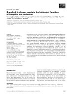
Fig. 1. Southern blot analysis for C. callunae genomic DNA (A) and
results of DNA library screening (B) using a 0.71 kb PCR probe for the
StP gene. Lanes 1–4 of the autoradiogram in panel A show the
hybridization patterns of the 32P-labeled PCR probe with C. callunae
DNA (50 lg) digested for up to 3 days with different endonucleases
(10–20 U) as indicated. DNA fragments were separated on a 0.8%
agarose gel and after transfer to a nylon membrane (Roche) allowed to
hybridize with the PCR probe overnight at 60 °C. Arrows on the right
and left of the blot indicate the sizes (in kb) of the main hybridizing
DNA fragments of which the BamHI and HindIII fragments were
cloned to give a partial genomic DNA library. After screening using
colony hybridization with the 710-bp PCR probe, positive clones were
selected and the inserts sequenced. The bottom of the figure (B) indicates the positions of BamHI and HindIII fragments, relative to the
entire StP structural gene.
they represent prototypes of regulated and nonregulated
GPs, respectively, and both have well-established structurefunction relationships. StP is 41% identical to rmGP and
42% identical to EcMalP. Figure 2 maps structural and
functional elements of rmGP [4] and EcMalP [3] onto the
linear sequence of StP and thus measures the extent of
conservation of the respective ÔsitesÕ in StP. As in other GPs,
the catalytic site and the PLP binding site of StP are virtually
identical to the corresponding sites in rmGP and EcMalP.
By contrast, the regulatory sites of rmGP are almost
completely lost in StP. Interestingly, there is only small
sequence identity between StP and EcMalP in the segments
of the sequence that correspond to regulatory sites of rmGP.
Hudson et al. [14] have classified dimer contacts in rmGP
into three relatively independent networks of interacting groups. The first two networks, often dubbed the cap¢a2-b7 interface, are mediated by residues in the ultimate
N-terminal part of the rmGP protomers and are associated
with control by allosteric effectors and covalent phosphorylation, as shown in Fig. 2. These dimer contact pairs of
rmGP are conserved to a very low degree in EcMalP and
likewise StP. The third network constitutes the major dimer
contact region in GP and involves the so-called TOWER
(a7) helices (residues 266–277 in rmGP) and the subsequent
gate loops of adjacent subunits. While specific interactions
between the subunits at the TOWER interface vary
considerably among different GPs [3–7], the position of
the a7 helix in the primary structures of rmGP, human liver
GP, yeast GP, and EcMalP is very well conserved.
Therefore, the TOWER-GATE region could be easily
assigned to the sequence of StP, as shown by underlining in
Fig. 2 (StP residues 231–242). Considering the involvement
of the TOWER interface of rmGP in signal transmission
from regulatory sites into the active centre [4–6,15], it was
interesting that residues in StP corresponding to the a7 helix
of rmGP exhibited a higher degree of similarity to the
mammalian enzyme than to EcMalP. Furthermore, the
occurrence of three arginine residues within the TOWER
helix of StP at positions 234, 236 and 242 was interesting
(Fig. 2). Arginines are common components of protein
interfaces and occur frequently at oxyanion-binding protein
sites [20,21]. Residues Arg234, Arg236, and Arg242 were
thus targets for site-directed mutagenesis, and their PCRbased replacement by alanine was chosen to eliminate all
electrostatic interactions at the respective position. It is
worth noting that Arg234 and Arg242 are positionally
conserved in all mammalian a-glucan phosphorylases while
the same positions show considerable variation in the
related enzymes from bacteria, fungi and plants.
Expression of the wild-type and mutagenized StP genes
in E. coli, and purification and characterization
of recombinant enzymes
Following induction with isopropyl thio-b-D-galactoside
using the conditions described in Experimental procedures,
a specific phosphorylase activity of approximately
10 mg)1 (± 15% SD) was measured in cell extracts of
E. coli XL1 Blue cells transformed with either pQE 30-StP
or pQE 70-StP. Comparison of this figure with the known
specific activity of 30 mg)1 for pure natural StP [11]
shows that recombinant StP corresponded to % 30% of the
total soluble E. coli protein. Recombinant wild-type and
His-tagged StP, and likewise StP mutants were purified to
apparent homogeneity and all were recovered in approximately 25 ± 5% yield. Like native StP, His-tagged StP and
all StP mutants contained 0.8–1.0 mol of PLP per mol of
90-kDa protomer, as expected if incorporation of cofactor
during folding of the recombinant proteins had taken place
correctly. Circular dichroism (Fig. 3) and Fourier-transform infrared spectra (not shown) of the purified recombinant proteins were recorded in an effort to identify
alterations in structure, relative to the natural wild-type
enzyme [12,18], as a result of recombinant protein production and mutagenesis. There were no traceable differences
between CD spectra of natural and recombinant StP (not
shown). The CD spectrum of His-tagged StP and CD
spectra of R234A and R242A mutants were not superimposable (Fig. 3), but the overall picture is one of close
structural similarity among the wild-type and the two
mutants. Therefore, site-specific replacements of Arg234
and Arg242 did not cause gross changes in the composition
of secondary structural element in the two mutants, relative
Ó FEBS 2003
2130 R. Griessler et al. (Eur. J. Biochem. 270)
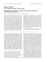
Fig. 2. Comparison of the StP amino acid
sequence with the sequences of EcMalP and
rmGP. The alignment was performed with the
MEGALIGN program using CLUSTALW with
standard settings. Amino acids conserved are
shaded in black. Catalytic and regulatory sites
of rmGP [14] are marked above the sequence,
as follows: a, AMP-binding site; c, caffeine/
purine inhibitor site; g, active site residues; p,
residues involved in covalent phosphorylation;
s, glycogen storage sites; v, pyridoxal phosphate binding site. Residues contributing to
the dimer interface of rmGP are indicated
using the letter, d. The primary structure of
StP is 41% and 42% identical to the sequences
of rmGP and EcMalP, respectively, indicating
overall conservation of the structural fold
[3,4]. The positions of the TOWER helices in
EcMalP and rmGP are underlined by a thick
line, and the mutations (to be reported later)
are indicated by arrows. Also, note that the
natural enzyme isolated from C. callunae [11]
lacks Ser1.
to the wild-type. The specific activities of recombinant StP,
His-tagged StP, and the R234A and R236A mutants were
identical within the experimental error of ± 10% to the
specific activity of StP isolated from C. callunae [11]. In the
standard assay of phosphorylase activity (Experimental
section), the R242A mutant displayed only 10% of wildtype activity. However, under conditions of saturation in
a-glucan substrate (30 gỈL)1 of maltodextrin) and phosphate (500 mM), the R242A mutant had a specific activity
approximately 40% that of the wild-type. (A discontinuous
assay was used here because the high phosphate concentration interferes with coupled enzyme measurements [11]).
The result reveals that maximum reaction rate and substrate
affinity are both decreased in the R242A mutant, compared
to the wild-type. Although this implies that the replacement
Arg242 by alanine is not without effect on steps involved in
enzymic catalysis, we point out that in wild-type StP, loss of
active site integrity and subunit dissociation occur as,
clearly, kinetically uncoupled events at an elevated temperature [12]. Therefore, the analysis of steady-state kinetic
data for the R242A mutant and the wild-type must not be
interpreted to weaken the comparative evaluation of
stabilities of the same enzymes, which follows later.
The N-terminal His-tag causes formation
of an active StP tetramer
Preparations of His-tagged StP that were > 98% pure by
the criterion of a single protein band in SDS/PAGE (not
shown) eluted from a Superose 12 size exclusion column in
Ó FEBS 2003
Dimer stability of bacterial starch phosphorylase (Eur. J. Biochem. 270) 2131
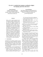
Fig. 3. Comparison of CD spectra of wild-type StP (solid line), R234A
mutant (dotted line), and R242A mutant (dashed line). Spectra were
recorded in a 20 mM Mops buffer, pH 7.0, not containing oxyanion.
Note that the value of protein concentration (0.1 mgỈmL)1) contained
10% error and may be partly responsible for observed differences in
molar ellipticity at 222 nm.
mutants gave elution profiles that were superimposable to
that of the wild-type. The StP tetramer was of interest
because mammalian GP is known to form tetramers at high
protein concentrations. These tetramers are inactive but
dissociate into active dimers when glycogen is present [4]. To
determine whether dissociation of the StP tetramer could be
induced in the presence of substrate, we subjected the
purified tetramer fraction to size exclusion chromatography
(SEC) under conditions where the elution buffer contained a
saturating concentration of maltohexaose (20 mM) or a-Dglucose-1-phosphate (20 mM). The tetramer was completely
stable for the time of the experiment (% 2 h) when one of
the above ligands was present. In marked contrast to
observations made with His-tagged StP, the recombinant
StP lacking the metal affinity fusion eluted as a single
protein peak from the Superose 12 column. Its estimated
molecular mass was 180 kDa. Automated Edman degradation of this recombinant StP yielded the sequence, ProGlu-Lys-Gln, for the N-terminal tetrapeptide of the
recombinant wild-type, which is in accordance with the
authentic N-terminal sequence of native StP [11]. Therefore,
the N-terminal metal affinity peptide appears to be responsible for the observed tetramer : dimer ratio of % 0.2 in Histagged StP, and likewise the Arg fi Ala mutants thereof.
The results suggest that if the occurrence of tetrameric and
dimeric forms of His-tagged StP truly represents an altered
oligomerization equilibrium, relative to wild-type StP, and is
not an artifact of the protein folding process in the E. coli
cytosol, the conversion of the tetramer into its constituent
dimers must take place at a slow rate. The data suggest that
the use of amino-terminal affinity tags may not be ideal for
studies of GP structure. However, we emphasize that
dimer : tetramer heterogeneity of His-tagged wild-type StP
was not changed in the His-tagged mutants and did thus not
affect the conclusions of this work.
Determination of dissociation constants of binary
enzyme–oxyanion complexes
Fig. 4. Elution profile of purified recombinant His-tagged StP upon
analytical gel filtration on a Superose 12 column. Approximately 100 lg
of protein in 100 lL triethanolamine buffer (50 mM, pH 7.0) were
applied to the column (20 mL; 1.6 cm diameter) equilibrated with
50 mM potassium phosphate buffer, pH 7.0, containing 0.2 M NaCl.
Elution was carried out with the same phosphate buer at a ow rate
ă
of 0.25 mLặmin)1 using an Aktaexplorer 100 system (Amersham
Pharmacia Biotech) and detection at 280 nm. Calibration of the sizing
column was performed using appropriate protein standards of known
molecular mass. Apparent molecular masses of 180 kDa and 360 kDa
were determined for the eluting protein fractions in this figure.
two fractions of well-defined apparent molecular masses, as
shown in Fig. 4: a major 180-kDa fraction corresponding to
the dimer and containing approximately 85% of the total
protein, and another fraction that accounted for the
remainder protein and displayed a molecular mass of
360 kDa, as expected for a StP tetramer. The minor protein
fraction had the same specific enzyme activity as the dimeric
wild-type. A monomer fraction was not observed. The StP
Fluorescence titration assays [12] were carried out with Histagged StP and the R234A and R242A mutants and yielded
dissociation constants for enzyme–sulfate (KdSO4) and
enzyme–phosphate (KdPi) complexes. These Kd values are
summarized in Table 1. The Kd values for the His-tagged
wild-type enzyme agree closely with the corresponding
values measured recently for native StP (KdSO4 ¼ 4;
KdPi ¼ 16) [12]. The data also reveal that the replacement
of the guanidinium side chain of arginine by a methyl side
chain of alanine in the R234A and R242A mutants caused
only a small effect on the binding of sulfate. An approximately 2.5-fold increase in KdSO4 was observed for the
R234A mutant, compared to the wild-type. The KdSO4 value
for the R242A mutant was very similar to that of the wildtype. These observations are not consistent with a scenario
in which the original side chains of Arg234 and Arg242
participate in binding the sulfate dianion. If these side chains
provided direct interactions with sulfate, a much larger
increase in KdSO4 would be expected for the mutants in
comparison to wild-type. We did not observe any significant
inhibition of quenching of PLP fluorescence in the R234A
and R242A mutants in the presence of phosphate at levels
of 10 mM and 20 mM, relative to a control that did not
Ó FEBS 2003
2132 R. Griessler et al. (Eur. J. Biochem. 270)
Table 1. Comparison of stabilities of recombinant wild-type StP and two enzyme variants in urea and thermal denaturation experiments at pH 7.0.
The experiments were carried out in 50 mM triethanolamine buffer, pH 7.0, and used 200 lgỈmL)1 of protein in each assay. Other conditions and
procedures were as reported previously [11,12,18]. n.a., not applicable because no significant change in iodide quenching of cofactor fluorescence
occurred in the presence of phosphate up to a concentration of 20 mM.
Cm at 30 °C (M)/t1/2 at 45 °C (min)
Enzyme
KdSO4 (mM)/KdPi (mM)
No oxyanion added
+ sulfatea
Wild-type
4.5 ± 0.5/18 ± 4
1.17 ± 0.03/3.2 ± 0.1
2.95 ± 0.10/stableb
R234A
R242A
9 ± 3/n.a.
3.8 ± 0.4/n.a.
2.60 ± 0.06/20 ± 1
2.00 ± 0.02/12 ± 0.5
4.45 ± 0.05/stableb
2.93 ± 0.02/stableb
+ phosphatea
5.2
(3.45
3.55
2.27
±
±
±
±
0.2c/stableb,c
0.03d/stableb,d)
0.02d/stableb,d
0.02d/43 ± 5d
a
Potassium phosphate and ammonium sulfate were used. Unless indicated, the oxyanion concentrations matched the respective Kd values. It
was shown in separate control experiments that the cation, K+ or NH4+, had no influence on stabilities of wild-type StP and mutants
thereof. b Being stable means that no significant inactivation occurred during a 0.5-h long incubation at 45 °C. c,d Data obtained in the
presence of c 20 mM and d 10 mM phosphate.
contain the oxyanion. This could result if the site-directed
replacement of Arg234 and Arg242 strongly weakened
binding of phosphate or if it altered the conformational
change in response to phosphate binding. Considering that
values of KdSO4 are not very sensitive to the mutations, the
latter interpretation would seem to be more likely, but the
relatively high KdPi value for the wild-type prevents any firm
conclusion on the mutants.
Stability of recombinant wild-type StP and TOWER helix
mutants thereof as revealed in urea and thermal
denaturation experiments
To compare the stabilities of StP and Arg fi Ala mutants
thereof, we carried out urea denaturation assays in which
protein concentration and incubation time were constant
parameters, and [urea] was varied in steps of 0.25 M
between 0.0 and 6.0 M. The chosen assay monitors enzyme
inactivation that is completely irreversible and thus
provides a measure of the kinetic stability of the respective
enzyme under the conditions used. For each protein, the
dependence of percentage of remaining enzyme activity on
[urea] was analyzed under conditions in which either no
oxyanion was present, or phosphate or sulfate was added
in a concentration corresponding approximately to the
dissociation constant (Kd) of the respective binary enzyme–
phosphate or enzyme–sulfate complex at 30 °C (Table 1).
Saturation in oxyanion was not attempted to avoid
possible interferences of stability measurements by a
lyotropic anion effect in the presence of high concentrations of phosphate or sulfate. Using nonlinear least squares
regression analysis with the SIGMAPLOT 2000 programme
(SPSS Inc.), data were fitted to Eqn (1), which describes a
sigmoidal decrease of enzyme activity (EA) with increasing
concentration of denaturant,
EA (urea) ẳ a=ẵ1 ỵ expbẵurea cÞ
ð1Þ
where a, b, and c are parameters (which are not derived
from any formal mechanism of denaturation of StP). The
apparent denaturation midpoint (Cm) is calculated by using
Eqn (1) and the respective parameter estimates, and
corresponds to the urea concentration where half the
original enzyme activity has been lost.
Cm values for wild-type StP and two Arg fi Ala mutants
thereof are summarized in Table 1. Results for the R236A
mutant are not shown in the Table because the stabilities of
this mutant and the wild-type were identical within limits of
experimental error (DCm % ± 0.15 M) under all conditions
examined. The Cm values in Table 1 reveal large stabilizing
effects of the Arg fi Ala replacements at positions 234 and
242 for conditions in which no oxyanion was present. Note
that values of the parameter b, which is a measure of the
slope of the decrease in EA as [urea] increases, showed little
variation in dependence of the enzyme or the reaction
conditions and were in the range )0.43 to )0.49. The extra
stability brought about by the mutations is reflected by
significant shifts of the Cm values for the mutants, relative to
that for the wild-type, to higher urea concentrations by
% 1 M or greater. The stabilization of wild-type StP by a
half-saturating concentration of phosphate can be expressed
quantitatively by a dramatic up-shift in Cm value by 4.0 M,
compared to the control reaction lacking phosphate. The
observed increase in Cm value effected by a sulfate level
matching KdSO4 was 1.2 M, suggesting that under the
conditions used, the StP–sulfate complex displays a much
smaller kinetic stability than the StP–phosphate complex.
The DCm-values for the wild-type serve as a frame of
reference for analyzing the stabilities of the mutants. Taking
into account the large stabilization of wild-type StP by
bound phosphate, it was unfortunate that KdPi values were
not accessible for the R234A and R242A mutants and so
defined conditions with regard to saturation in oxyanion
were possible only for sulfate.
Irrespective of the added oxyanion, observed DCm-values
for the R242A mutant were smaller than corresponding
values for the wild-type (Table 1). Considering that sulfate
binding takes place with almost identical affinities in wildtype StP and the R242A mutant and assuming that this
reflects similar sulfate binding modes in both proteins, the
results show that the binding event as such is not sufficient
for sulfate to induce a large stabilization, which in turn is
mirrored in the value of DCm. It is important to recognize
therefore that denaturation midpoints in the presence of
half-saturating levels of sulfate were identical within the
experimental error for the wild-type and the R242A
mutant. The simplest explanation of this finding is that
Ó FEBS 2003
Dimer stability of bacterial starch phosphorylase (Eur. J. Biochem. 270) 2133
the enzyme-sulfate complexes of wild-type and R242A
mutant share similar kinetic stabilities; and that the
Arg fi Ala replacement at position 242 offsets the stabilizing effect of sulfate binding in the wild-type to the extent
that this mutation stabilizes the enzyme when no sulfate is
present (Fig. 5B). Interestingly therefore bound sulfate
stabilized the R234A mutant and the wild-type equally.
Hence, although Arg234 is clearly destabilizing in unligated
StP, site-directed mutagenesis of the side chain of Arg234
into the methyl side chain of alanine did not diminish the
stabilizing effect of sulfate binding in comparison to wildtype, as it was observed for the R242A mutant. This result is
interesting because it leads to a different interpretation of
the role of Arg234 and Arg242 for oxyanion-dependent
stability of StP. The stabilization brought about by the
presence of 10 mM of phosphate was substantially smaller
for the R234A mutant (DCm %1 M) than the wild-type
(DCm %2.3 M). Even in the absence of a KdPi value for
R234A (and likewise R242A), the comparison at a fixed
phosphate level is relevant. It shows that site-specific
replacement in each mutant either decreases the affinity
for phosphate, relative to the wild-type, or lowers the kinetic
stability of the mutant-phosphate complex, relative to the
same wild-type complex. Figure 5 illustrates this point by
comparing the dependence of DCm-values on the concentrations of phosphate and sulfate for wild-type StP and the
R242A mutant. The results show a marked preference
for stabilization by sulfate over stabilization by phosphate
in the mutant, which is clearly different to what
was observed for the wild-type. Note that the separation of the parallel lines in panel B of Fig. 5 corresponds to
the difference in Cm-values for the R242A mutant and the
wild-type under conditions in which no sulfate was added.
The data in Fig. 5 can be used to roughly estimate the
apparent half-saturation constants (app K) for the stabilization of the wild-type (app KSO4 %17 mM; app
KPi %28 mM) and the R242A mutant (app KSO4 %12 mM;
app KPi % 130 mM).
Thermal stabilities of wild-type and Arg fi Ala mutants
were determined at 45 °C and are shown in Table 1. In the
absence of added oxyanion, the R242A and R234A mutants
were 3.8- and 6.2-fold more stable than the wild-type,
respectively. No significant inactivation of StP and the two
enzyme variants was seen over an incubation time of 30 min
in the presence of sulfate concentration matching KdSO4. In
the presence of 10 mM phosphate, wild-type and the R234A
mutant were stable while the R242A mutant displayed
significant loss of activity.
Discussion
Fig. 5. Stabilization of wild-type StP and the R242A mutant by phosphate (A) and sulfate (B) against urea denaturation. Results show DCmvalues, which report the difference between Cm at the shown oxyanion
concentration and the Cm measured in buffer lacking oxyanion. The
data are presented as a double reciprocal plot to emphasize the saturatable dependence of DCm on [oxyanion]. However, extrapolation to
infinite [oxyanion] must be made with caution (hence, the broken lines)
because of the additional lyotropic anion effect. Also note that in panel
A, lines do not have identical intercept values. Experiments were carried out at 30 °C in 50 mM triethanolamine buffer, pH 7.0, using
conditions reported in the text.
The goal of the present paper was to advance the
relationships between structure and oxyanion-dependent
stability of StP from Corynebacterium callunae. Cloning,
sequencing, and heterologous expression of the gene
encoding StP were essential requirements for the utilization
of site-directed mutagenesis to examine the functional roles
of potentially important amino acid residues that were
identifiable through analysis of the StP primary structure.
The results have revealed clearly that Arg234 and Arg242 of
the TOWER interface region of StP partially destabilize the
dimer structure of the unligated enzyme so that loss of these
residues in the Arg fi Ala mutants leads to significantly
higher kinetic stability. Phosphate binding appears to cause
a change in interactions of these arginines, most probably by
an allosteric mechanism as discussed below, contributing to
the observed stabilization. An unexpected finding was that
replacements of Arg242 and Arg234 induced a large
apparent preference for sulfate over phosphate with regard
to the stabilizing effect.
2134 R. Griessler et al. (Eur. J. Biochem. 270)
Relationships between StP structure and oxyaniondependent kinetic stability
Previous studies have shown that subunit dissociation
occurs as an early step during denaturation of StP at
elevated temperatures (30 °C) [12] or in urea (R. Griessler,
& B. Nidetzky, unpublished observations). Under conditions of dilute protein and in the absence of free PLP, loss of
oligomer structure is accompanied by immediate release of
cofactor from the StP subunit. Therefore, it is not detectably
reversible on the time scale of the assay for phosphorylase
activity (% 1–2 min) [12]. Measurement of irreversible
inactivation of StP can thus serve as a useful reporter of
the protomer dissociation event. It would seem likely
therefore that observed changes in Cm and t1/2-values for
irreversible inactivation in urea and at elevated temperatures, brought about by site-specific amino acid replacements in the dimer contact region of StP and likewise,
oxyanion bound at the enzyme oxyanion site, result from
altered kinetic barriers for subunit dissociation, relative to
unliganded wild-type StP. We stress, however, that based on
the available data, it is not possible to rule out completely a
contribution of thermodynamic effects to the measured
kinetic stabilities.
Arginine residues are known for their prevalence in both
intra- and inter-chain interfaces [22,23] where the charged
guanidinium group is often involved in formation of strong
intermolecular hydrogen bonds. Such non-covalent interactions have been hypothesized to stabilize multidomain
and oligomeric proteins by strengthening either the network
of interfacial contacts or the tertiary bonds that prevail in
the segment of the interface. Site directed mutagenesis has
been used, in a few instances though, to verify the role of
arginines as stabilizing elements of dimer contact regions
[24,25]. Therefore, irrespective of the exact orientation of
Arg234, Arg236, and Arg242 at the TOWER interface
region of StP, it was unexpected that two out of three
enzyme variants harboring the Arg fi Ala substitution
exhibited a considerably greater kinetic stability in thermal
and urea denaturation studies than the wild-type. Hydrogen-bonding or other electrostatic interactions involving the
ÔTOWER argininesÕ are obviously not optimized for kinetic
stability. In this scenario, oxyanions could have a stabilizing
effect if their binding was capable of either decreasing
nonfavorable contacts between protomers or increasing the
favorable ones. This could occur by various mechanisms,
but likely an allosteric one in which oxyanion binding
affects the tertiary and/or quarternary interactions involving
Arg234 and Arg242 thus leading to a greater stability.
For the interpretation of the kinetic stabilities of the
R234A and R242A mutants, it is most useful to first
consider the effects of phosphate and sulfate on conformation and stability of wild-type StP. It was shown here that
under conditions of half-saturation in oxyanion, phosphate
stabilizes the wild-type much more efficiently against
denaturation by urea than sulfate, the difference in DCmvalue (which is the increase in Cm compared to the
unliganded enzyme when oxyanion is present) being as
large as 2.2 M. A greater stability of the enzyme-phosphate
complex than the enzyme–sulfate complex correlates well
with a greater compactness of the former complex,
as revealed recently by comparing iodide quenching of
Ó FEBS 2003
cofactor fluorescence in StP saturated with phosphate and
sulfate [12]. The results for wild-type StP imply that a
conformational change in protein structure accompanies the
oxyanion-binding event and is required for kinetic stability.
The extent of the structural rearrangement is larger for a
phosphate than a sulfate ligand, suggesting that more
binding energy from the StP–oxyanion interaction can be
translated into a stabilized protein conformation when
phosphate is bound.
The comparison of Kd values for enzyme-sulfate complexes of wild-type and the two Arg fi Ala mutants reveals
that a direct participation of the side chains of Arg234 or
Arg242 in binding of sulfate is not likely. However, both
arginines, clearly, take part in the just described oxyaniondependent conformational relay of wild-type StP, and
analysis of R234A and R242A mutants serves to emphasize
the differential effect of bound phosphate and sulfate in the
wild-type. In both mutants, however, mainly R242A,
phosphate has lost much of the stabilizing potential
originally present in the wild-type. Expressed as the ratio
of DCm (M) and [phosphate] (M), the phosphate-specific
stabilization is % 30 for the wild-type, but only % 10 and
% 1 for the R234A and R242A mutant, respectively. The
situation is different for sulfate, which stabilizes wild-type
StP and the R234A mutant to approximately the same
extent. In the R242A mutant, the stabilizing effect of the
Arg fi Ala replacement in the unligated protein is offset by
the smaller stabilization when sulfate is bound, compared to
wild-type StP. In conclusion, these data can be summarized
to yield the following hypothetical model of the stabilization
of StP by oxyanions. Phosphate binding at an allosteric site,
perhaps within the subunit, leads to propagation of a
conformational change into the dimer contact region of the
protein. Arg242 is a key residue implicated in this structural
rearrangement and may even have an active role in relaying
the phosphate-dependent and to a lesser extent though, the
sulfate-dependent conformational switches. Arg234 appears
to be part of the relay when phosphate is bound, but not
when sulfate is bound.
Comparison of StP with rmGP and other a-glucan
phosphorylases
Structure-function studies of rmGP are highly relevant for
the interpretation of results for StP. First of all, a
dissociative mechanism of thermal denaturation of rmGP,
similar to that proposed for StP, has been reported recently
[26]. A major difference between rmGP and StP, however,
pertains to the moderate effect that oxyanions have on
rmGP stability [26]. Secondly, Fletterick and coworkers
have mutated TOWER helix residues of rmGP, among
them Arg277, which is the rmGP counterpart of Arg242,
into alanine and characterized the variant enzymes structurally and with respect to allosteric activation by AMP [27].
Their conclusion from a detailed comparison of intersubunit
contacts in X-ray structures of wild-type rmGP and R277A
was that the Arg fi Ala replacement would destabilize
significantly the quaternary interactions originally present in
the muscle enzyme. Keeping in mind the limitations of using
irreversible inactivation as a measure of global protein
stability, our results then suggest that Arg242 in StP must
participate in interactions clearly different from those of the
Ó FEBS 2003
Dimer stability of bacterial starch phosphorylase (Eur. J. Biochem. 270) 2135
corresponding residue in rmGP. Interestingly, mutating
Arg242 and Arg277 had similar effects on the catalytic
competence of StP and rmGP, respectively, resulting in each
case, in a significant decrease in specific activity, compared
to the wild-type level. The side chains of Arg269 and Arg277
make direct hydrogen bonds across the dimer interface of
activated rmGP with side chains of Asn250¢ and Asn270¢,
respectively, on the adjacent subunit. Each of the two
asparagines of rmGP is replaced positionally by a glutamate
in StP. Considerations of charge and packing arrangements
suggest that if Arg234 and Arg242 were truly involved in
inter-subunit interactions analogous to those seen in rmGP
structures, bonding across the interface of StP should be
stronger, compared to rmGP, which is unlikely in light of
the experimental evidence for StP. Another interesting
difference between StP and rmGP revealed by structurebased sequence comparison pertains to contacts of Arg277
(and likewise Arg242) within the subunit. In rmGP, this
arginine forms a charged hydrogen bond with the carboxylate group of Glu162 [4,27]. In StP, Glu162 is positionally
replaced by an arginine (Arg142), and this is a likely reason
for different atomic environments of Arg242 in StP and
Arg277 in rmGP.
Multiple alignment of the first one-hundred a-glucan
phosphorylase sequences identified through screening of the
nonredundant data bases with the StP primary structure
using the BLAST 2 program (not shown) revealed an
interesting conservation pattern for positions equivalent
to Arg142 and Arg242 of StP. In all but two cases, namely
a-glucan phosphorylases from Corynebacterium glutamicum
(Q8NQW4) and Fusobacterium nucleatum (Q8RF61), the
pair of arginine residues found in StP is not observed. A pair
of amino acids with oppositely charged side chains,
glutamate (or aspartate) at position 142 and arginine (or
lysine) at position 242, occurs most frequently in the aligned
sequences. Several other pairwise combinations of amino
acids are possible, but bulk and charge at a certain position
appear not to be conserved across all organisms and cell
types. To give two examples for structurally characterized
enzymes, EcMalP has a glutamine-lysine pair whereas
Saccharomyces cerevisiae glycogen phosphorylase has a
glutamate-alanine pair. However, an interesting generalization is that enzymes from (hyper)thermophilic bacteria and
archaea contain a conserved pair of lysine (position 142)
and glutamic acid (position 242). It would be interesting
therefore to examine if positional charge reversal for
extremophilic structures, compared to most other a-glucan
phosphorylase sequences including the mammalian ones, is
related to increased stability [28–30]. Furthermore, our
comparisons show that if an arginine residue occurs at
position 142 in a-glucan phosphorylases, position 242 is
generally taken by an alanine, serine, or threonine. These
residues whose side chains are uncharged and sterically less
demanding than the side chain of arginine may be primed
to avoid unfavorable (destabilizing) interactions with or
relayed to counterpart Arg142. This interpretation of the
sequence changes among aligned a-glucan phosphorylases
is in excellent agreement with the observed kinetic stability
of R242A mutant of StP, compared to wild-type enzyme. It
also provides a rational for an allosteric mechanism of
stabilization of StP by oxyanion binding. In light of the fact
that Arg242 is conserved in all mammalian GPs and
considering that the R242A mutant shows only 40% of
wild-type activity, it seems probable that Arg242 has been
selected in StP for so far unknown reasons of enzyme
function.
Acknowledgements
Financial support from the Austrian Science Funds (P-15118-MOB to
B.N.) is gratefully acknowledged.
References
1. Palm, D., Klein, H.W., Schinzel, R., Buehner, M. & Helmreich,
E.J. (1990) The role of pyridoxal 5¢-phosphate in glycogen phosphorylase catalysis. Biochemistry 29, 1099–1107.
2. Schinzel, R. & Nidetzky, B. (1999) Bacterial a-glucan phosphorylases. FEMS Microbiol. Lett. 171, 73–79.
3. Watson, K.A., Schinzel, R., Palm, D. & Johnson, L.N. (1997) The
crystal structure of Escherichia coli maltodextrin phosphorylase
provides an explanation for the activity without control in this
basic archetype of a phosphorylase. EMBO J. 16, 1–14.
4. Johnson, L.N. (1992) Glycogen phosphorylase: control by phosphorylation and allosteric effectors. FASEB J. 6, 2274–2282.
5. Rath, V.L. & Fletterick, R.J. (1994) Parallel evolution in two
homologues of phosphorylase. Nat. Struct. Biol. 1, 681–690.
6. Newgard, C.B., Hwang, P.K. & Fletterick, R.J. (1989) The family
of glycogen phosphorylases: structure and function. Crit. Rev.
Biochem. Mol. Biol. 24, 69–99.
7. Buchbinder, J.L., Rath, V.L. & Fletterick, R.J. (2001) Structural
relationships among regulated and unregulated phosphorylases.
Annu. Rev. Biophys. Biomol. Struct. 30, 191–209.
8. Tu, J.-I. & Graves, D.J. (1973) Association-dissociation properties
of sodium borohydride-reduced phosphorylase b. J. Biol. Chem.
248, 4617–4622.
9. Feldmann, K., Zeisel, H. & Helmreich, E. (1972) Interactions
between native and chemically modified subunits of matrix-bound
glycogen phosphorylase. Proc. Natl Acad. Sci. USA 69, 2278–
2282.
10. Tagaya, M., Shimomura, S., Nakano, K. & Fukui, T. (1982)
A monomeric intermediate in the reconstitution of potato
apophosphorylase with pyridoxal 5¢-phosphate. J. Biochem. 91,
589–597.
11. Weinhausel, A., Griessler, R., Krebs, A., Zipper, P., Haltrich, D.,
ă
Kulbe, K.D. & Nidetzky, B. (1997) a-1,4-D-glucan phosphorylase
of gram-positive Corynebacterium callunae: isolation, biochemical
properties and molecular shape of the enzyme from solution X-ray
scattering. Biochem. J. 326, 773–783.
12. Griessler, R., D’Auria, S., Tanfani, F. & Nidetzky, B. (2000)
Thermal denaturation pathway of starch phosphorylase from
Corynebacterium callunae: oxyanion binding provides the glue that
efficiently stabilizes the dimer structure of the protein. Protein Sci.
9, 1149–1161.
13. Watson, K.A., McCleverty, C., Geremia, S., Cottaz, S., Driguez,
H. & Johnson, L.N. (1999) Phosphorylase recognition and phosphorolysis of its oligosaccharide substrate: answers to a long
outstanding question. EMBO J. 18, 4619–4632.
14. Hudson, J.W., Golding, G.B. & Crerar, M.M. (1993) Evolution of
allosteric control in glycogen phosphorylase. J. Mol. Biol. 234,
700–721.
15. Lin, K., Hwang, P.K. & Fletterick, R.J. (1997) Distinct phosphorylation signals converge at the catalytic center in glycogen
phosphorylases. Structure 5, 1511–1523.
16. Sambrook, J., Fritsch, E.F. & Maniatis, T. (1989) Molecular
Cloning: A Laboratory Manual, 2nd edn. Cold Spring Harbor
Laboratory Press, Cold Spring Harbor, NY, USA.
Ó FEBS 2003
2136 R. Griessler et al. (Eur. J. Biochem. 270)
17. Higuchi, R., Krummel, B. & Saiki, R.K. (1988) A general method
of in vitro preparation and specific mutagenesis of DNA fragments: study of protein and DNA interactions. Nucleic Acids Res.
16, 7351–7367.
18. Griessler, R., Pickl, M., D’Auria, S., Tanfani, F. & Nidetzky, B.
(2001) Oxyanion-mediated protein stabilization: differential roles
of phosphate for preventing inactivation of bacterial a-glucan
phosphorylases. Biocat. Biotrans. 19, 379–398.
19. Griessler, R., D’Auria, S., Schinzel, R., Tanfani, F. & Nidetzky, B.
(2000) Mechanism of thermal denaturation of maltodextrin
phosphorylase from Escherichia coli. Biochem. J. 346, 225–263.
20. Chakrabarti, P. (1993) Anion binding sites in protein structures.
J. Mol. Biol. 234, 463–482.
21. Copley, R.R. & Barton, G.J. (1994) A structural analysis of
phosphate and sulphate binding sites in proteins. Estimation of
propensities for binding and conservation of phosphate binding
sites. J. Mol. Biol. 242, 321–329.
22. Nandi, C.L., Singh, J. & Thornton, J.M. (1993) Atomic
environments of arginine side chains in proteins. Protein Eng. 6,
247–259.
23. Jones, S., Marin, A. & Thornton, J.M. (2000) Protein domain
interfaces: characterization and comparison with oligomeric protein interfaces. Protein Eng. 13, 77–82.
24. Mrabet, N.T., Van den Broeck, A., Van den Brande, I., Stanssens,
P., Laroche, Y., Lambeir, A.M., Matthijssens, G., Jenkins, J.,
Chiadmi, M. & van Tilbeurgh, H. (1992) Arginine residues as
stabilizing elements in proteins. Biochemistry 31, 2239–2253.
25. Prasanna, V., Gopal, B., Murthy, M.R.N., Santi, D.V. &
Balaram, P. (1999) Effect of amino acid substitutions at the
subunit interface on the stability and aggregation properties of a
26.
27.
28.
29.
30.
dimeric protein: role of Arg 178 and Arg 218 at the dimer interface
of thymidylate synthase. Proteins 34, 356–368.
Kurganov, B.I., Kornilaev, B.A., Chebotareva, N.A., Malikov,
V.P., Orlov, V.N., Lyubarev, A.E. & Livanova, N.B. (2000) Dissociative mechanism of thermal denaturation of rabbit skeletal
muscle glycogen phosphorylase b. Biochemistry 39, 13144–13152.
Buchbinder, J.L., Guinovart, J.J. & Fletterick, R.J. (1995)
Mutations in paired a-helices at the subunit interface of glycogen
phosphorylase alter homotropic and heterotropic cooperativity.
Biochemistry 34, 6423–6432.
Takata, H., Takaha, T., Okada, S., Takagi, M. & Imanaka, T.
(1998) Purification and characterization of a-glucan phosphorylase from Bacillus stearothermophilus. J. Ferment. Bioeng. 85, 156–
161.
Xavier, K.B., Peist, R., Kossmann, M., Boos, W. & Santos, H.
(1999) Maltose metabolism in the hyperthermophilic archaeon
Thermococcus litoralis: purification and characterization of key
enzymes. J. Bacteriol. 181, 3358–3367.
Bibel, M., Brettl, C., Gosslar, U., Kriegshaeuser, G. & Liebl, W.
(1998) Isolation and analysis of genes for amylolytic enzymes
of hyperthermophilic bacterium Thermotoga maritima. FEMS
Microbiol. Lett. 158, 9–15.
Supplementary Material
The following material is available from http://www.
blackwellpublishing.com/products/journals/suppmat/EJB/
EJB3562/EJB3562sm.htm