Báo cáo khoa học: Co-operation of domain-binding and calcium-binding sites in the activation of gelsolin pptx
Bạn đang xem bản rút gọn của tài liệu. Xem và tải ngay bản đầy đủ của tài liệu tại đây (343.48 KB, 8 trang )
Co-operation of domain-binding and calcium-binding sites
in the activation of gelsolin
Emeline Lagarrigue
1
, Sutherland K. Maciver
2
, Abdellatif Fattoum
3
, Yves Benyamin
1
and Claude Roustan
1
1
UMR 5539 (CNRS) Laboratoire de motilite
´
cellulaire (Ecole Pratique des Hautes Etudes), Universite
´
de Montpellier 2, France;
2
Genes and Development Group, Department of Biomedical Sciences, University of Edinburgh, Scotland, UK;
3
Centre de Recherches de Biochimie Macromole
´
culaire, UPR 1086 (CNRS), Montpellier, France
Gelsolin is an abundant calcium dependent actin filament
severing and capping protein. In the absence of calcium the
molecule is compact but in the presence of calcium, as its six
similar domains alter their relative position, a generally more
open configuration is adopted to reveal the three actin
binding sites. It is generally held that a Ôhelical-latchÕ at the
C-terminus of gelsolin’s domain 6 (G6), binds domain 2 (G2)
to keep gelsolin in the calcium-free compact state, and that
the crutial calcium binding site(s) reside in the C-terminal
half of gelsolin perhaps involving the C-terminal helix itself
has to be bound to release this latch. Here we provide
evidence for a calcium dependent conformational change
within G2 (K
d
¼15 l
M
). We also report a calcium
dependent binding site for the C-terminus (G4–6) within
G2 and delimit this further to a specific region formed by
residues 203–225 and 159–193. It is known that the acti-
vation of gelsolin involves multiple calcium binding
events (around 6) the first of which (in G6) may release
the latch. We propose that the calcium-dependent con-
formational change in G2 may be a subsequent step that
is necessary for the dissociation of G2 from G4–6, and
that this movement occurs in sympathy with calcium
induced conformational changes within G6 by the physi-
cal coupling of the two calcium binding sites within G2
and G6. Additional calcium binding in other domains then
result in the complete opening and activation of the
gelsolin molecule.
Keywords: actin; gelsolin; cytoskeleton; severing; calcium
activation.
Gelsolin is a calcium-activated actin filament severing and
capping protein found in many tissues and in the plasma of
vertebrates (for a review, see [1]). It belongs to a wider group
of actin-binding proteins that share a number of repeated
domains; six in the case of gelsolin itself, adseverin and
villin, and three in capG, fragmin and severin (for a review,
see [2]). The binding of calcium to gelsolin and to actin
bound gelsolin is complex. Free gelsolin binds at least six
calcium ions. These sites, coordinated solely by gelsolin have
been termed type II [3]. The affinity of type II sites varies
greatly. High affinity calcium sites (K
d
1 l
M
) have been
identified [4–6] and two of these have been localized within
G4–6 [7]. A body of evidence suggests that calcium binding
by G4–6 affords calcium-sensitivity to the whole gelsolin
molecule [8,9]. Sites have been identified by biochemical
means within G4-5 (K
d
2 l
M
) and G5–6 (K
d
0.2 l
M
)
[9], and crystallographic studies (S. Kolappan, J. Gooch,
A. Weeds & P. McLaughlin, Wellcome Centre for Cell
Biology, University of Edinburgh, UK, personal commu-
nication, [3]) have shown that calcium ions are bound by
both G5 and G6 (sites IIG5 and IIG6 [3]. Low affinity
calcium-binding sites (K
d
1m
M
) have also been detected
[10,11]. A site has been inferred to lie within G2-3 by
proteolysis susceptibility and molecular radius changes [12],
and this site has been narrowed further to G2 (IIG2) and
tentatively suggested to have a dissociation constant of
32 l
M
[11]. Additionally, a calcium ion (IG1) is ÔtrappedÕ
between actin and G1 [13,14], and by actin and G4 (IG4)
[3,15], these calcium sites, coordinated by both gelsolin and
actin have been termed type I sites [3].
In the absence of calcium, gelsolin cannot bind actin as its
three [5,16] identified actin binding sites residing in G1, G2
and G4 [7,16,17] are not accessible. In the presence of
calcium, gelsolin becomes activated by the unfolding of the
whole molecule so that the F-actin binding region in G2 is
exposed allowing the molecule to make the initial contact
with the actin filament. Whereas gelsolin is opened by
0.1–1 l
M
calcium [12,18,19], the ternary actin: gelsolin
complex is only stable at calcium concentrations exceeding
Correspondence to C. Roustan, UMR 5539(CNRS) UM2 CC107,
Place E. Bataillon 34095 Montpellier Cedex 5, France.
Fax: + 33 0467144927,
E-mail:
Abbreviations: G1-6, The six repeated domains of gelsolin; FITC,
fluorescein 5-isothiocyanate; 1,5-I-AEDANS, N-iodoacetyl-N¢-(sulfo-
1-naphthyl)-ethylenediamine; BACNHS, biotinamidocaproate
N-hydroxyl-succinimide ester; G-actin, monomeric actin;
F-actin, filamentous actin.
Note: we have adopted the labeling system introduced by Choe et al.
[Choe, H., Burtnick, L.D., Mejillano, M., Yin, H.L., Robinson, R.C.
& Choe, S. (2002) J. Mol. Evol. 324, 691–702.] for the various Calcium-
binding sites so that IG1 is the type I binding site within G2 and IIG6 is
the type II binding site within G6. Type I binding sites are coordinated
by gelsolin and actin whereas type II sites are coordinated solely by
gelsolin residues.
Note: webpages are available at />umr5539/, and
/>(Received 20 December 2002, revised 10 March 2003,
accepted 26 March 2003)
Eur. J. Biochem. 270, 2236–2243 (2003) Ó FEBS 2003 doi:10.1046/j.1432-1033.2003.03591.x
30 l
M
[19], and the most rapid severing rate of actin
filaments occurs at 300 l
M
[20]. Accumulating evidence
[3,12,18,21] suggests the following mechanism for the
activation of gelsolin by calcium. Low calcium concentra-
tions are proposed [18,21,22] to ÔunlatchÕ the connection
between G2 and G6, but higher concentrations are required
to break salt bridges between other domains until the
gelsolin is fully ÔopenÕ, then additional calcium ions are
requiredtobindactintoG1andtoG4forseveringand
capping. The details of the latch helix structure in the
presence of calcium are not clear as there is no density for
this region in the available crystallographic solutions [3,15],
so we cannot tell if calcium ions are bound directly to the
latch helix to inhibit its binding to G2. However, this seems
unlikely as is has been shown that gelsolin without the
C-terminal helix alters neither the number nor the affinity
of gelsolin’s calcium binding sites [18].
Here, we confirm that G2 shows calcium-dependent
conformational changes and that peptides derived from it
show calcium-dependent binding to G4–6. Therefore the
calcium-dependence shown by G2 may have a role in the
multistep mechanism of calcium-activation of gelsolin prior
to binding actin.
Methods
Proteins and peptides
Human gelsolin domain 2 (G2) and domains 4–6 (G4–6)
were produced in Escherichia coli BL21(pLysS) and
BL21(de3), respectively, using the pMW172 vector [23],
following induction of expression with isopropyl thio b-
D
-
galactoside. G2 (residues 151–266 of human serum gelsolin)
was purified from the soluble fraction of the bacteria [24],
and G4–6 (residues 407–755) [25] was purified from
inclusion bodies [26].
Gelsolin G4–6 domain was selectively cleaved by trypsin.
The domain (225 lgÆmL
)1
) was incubated in 0.1
M
Tris HCl
pH 7.5 supplemented with either 1 m
M
CaCl
2
or 1 m
M
EGTA in the presence of trypsin (9 lg/mL). The proteolysis
was stopped by addition of an antiprotease mixture
(Complete) (1 : 25 mass/vol.) purchased from Roche SA
(Mannheim, Germany). To identify the cleavage products,
the digest was labeled at cysteine residues by treatment with
1,5-I-AEDANS (10 molar excess) [27] for 4 h at room
temperature, in the presence of 1% SDS. The labeling
reaction was stopped by addition of 0.1
M
b-mercapto-
ethanol. The digest was then analyzed by SDS/PAGE.
Antibodies directed towards gelsolin G4–6 domain were
elicited in rabbits according to [28]. Anti-IgG antibodies
labeled with alkaline phosphatase were purchased from
Sigma (Dorset, UK).
Synthetic peptides derived from gelsolin sequences 159–
193 and 203–225 [17] were prepared on solid phase support
using a 9050 Milligen PepSynthesizer (Millipore) according
to the Fmoc/tBu system. The crude peptides were
de-protected and thoroughly purified by preparative
reverse-phase HPLC. The purified peptides were shown to
be homogenous by analytical HPLC. Electrospray mass
spectra, carried out in the positive ion mode using a Trio
2000 VG Biotech Mass spectrometer (Altrincham), were in
line with the expected structures.
Gelsolin G2 and G4–6 domains were labeled by FITC as
described elsewhere [29]. Biotinylation of G2 domain by
BACNHS was performed as reported previously [30]. Excess
reagent was eliminated by chromatography on a PD10
column (Pharmacia) in 0.1
M
NaHCO
3
buffer pH 8.6.
Immunological techniques
The ELISA technique [31], described previously in detail
[32] was used to monitor interaction of ligands with
synthetic peptides or G4–6 domain. G4–6 domain
(1 lgÆmL
)1
) or synthetic peptides (1 lgÆmL
)1
)in50m
M
NaHCO
3
/Na
2
CO
3
, pH 9.5, were immobilized on plastic
microtiter wells. The plate was then saturated with 0.5%
gelatin/3% gelatin hydrolysate in 140 m
M
NaCl, 0.05%
tween 20, 10 m
M
Phosphate buffer, pH 7.4. Experiments
with coated fragments were performed in 0.1
M
KCl, 20 m
M
Tris pH 7.2. Binding was monitored at 405 nm using
alkaline phosphatase-labeled anti-IgG antibodies (dilution
1/1000) or alkaline phosphatase labeled streptavidin (dilu-
tion 1/1000). Control assays were carried out in wells
saturated with gelatin and gelatin hydrolysate used alone.
Each assay was conducted in triplicate and the mean value
plotted after subtraction of nonspecific absorption. The
binding parameters (apparent dissociation constant K
d
and
the maximal binding A
max
) were determined by nonlinear
fitting A ¼ A
max
· [L]/(K
d
+[L])(relation1)whereA is
the absorbance at 405 nm and [L] the ligand concentration,
by using the
CURVE FIT
software developed by Kevin Raner
Software, Mt Waverley, Victoria, Australia. Additional
details on the different experimental conditions are given in
the figure legends.
Fluorescence measurements
Fluorescence experiments were conducted with a LS 50
Perkin–Elmer luminescence spectrometer. Spectra for FITC
labeled proteins were obtained in 0.1
M
KCl 20 m
M
Tris/
HCl buffer pH 7.4, with the excitation wavelength set at
470 nm. Fluorescence changes were deduced from the area
of the emission spectra of fluorescein isothiocyanate (FITC)
between 505 and 525 nm. The parameters K
d
(apparent
dissociation constant) and A
max
(maximum effect) were
calculated by nonlinear fitting of the experimental data
points as for ELISA (relation 1), or by using the following
equation (relation 2), DF ¼ 1/2 A
max
· [E]
)1
{([E] +
[L] + K
d
) ) (([E] + [L] + K
d
)
2
) 4[E] · [L])
0.5
}where
[E] is the concentration of the fluorescent protein. The
maximum fluorescence change (A
max
) at infinite substrate
concentration expressed as percentage variation from initial
fluorescence: F1 – F°/F°
*
100 was calculated by the
relation F 1 – F°/F° ¼ A
max
/F° where F° and F1 are
fluorescence intensities for zero and infinite ligand concen-
trations, respectively.
Analytical methods
Protein concentrations were determined by UV absorbency
using a Varian MS 100 spectrophotometer. Gelsolin
domain concentrations were determined spectrophotomet-
rically using values of A
280
(1 cm
)1
) ¼ 15.5 l
M
for G4–6,
and 79 l
M
for G2. These extinction coefficients were
Ó FEBS 2003 Calcium activation of gelsolin (Eur. J. Biochem. 270) 2237
calculated by tryptophan, tyrosine, and cysteine content
[33]. Electrophoresis was carried out on 15% (w/v)
polyacrylamide slab gels (SDS/PAGE 15%) according to
Laemmli [34] and stained with Coomassie blue or observed
by UV for fluorescent gels before staining. The 14–97 kDa
molecular weight marker kits were from Biorad. Calcium
concentrations in the calcium-EGTA buffer system were
experimentally measured by fluorometry using indo 1 as
indicator [35].
Results
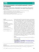
Cleavage of G4–6 domain by trypsin
A recent detailed study [19] reported that the exposure of
various tryptic cleavage sites in gelsolin is actin, and
calcium, dependent. In fact, several investigations using
namely circular dichroism and light scattering [9,12,15,18]
showed the occurrence of major conformational changes
in the regulatory C-terminal half of gelsolin upon calcium
binding. In an initial experiment, we tested the susceptibility
of the C-terminal domain to proteolysis. As shown in
Fig. 1A, tryptic digestion give rise to two fragments, one of
30 kDa and the other of 16 kDa. In order to identify these
fragments, we have labeled the unique cysteine residue
(Cys645) located in domain G6 with 1,5-I-AEDANS. As
this residue is buried in the native molecule [15,22], the
labeling was carried out after proteolytic digestion and SDS
unfolding (see Methods). Figure 1A shows that only the
16 kDa band is fluorescent. This result demonstrates that
the cleavage occurs in the loop between G5 and G6 domain
and is in accord with the unpublished results reported
previously [19].
As depicted in Fig. 1B, the tryptic cleavage was faster in
EGTA than in calcium, suggesting that the orientation
between the two domains is different and the junction more
accessible in EGTA.
Calcium induced change in the G4–6 and G2 domains
Conformation changes induced by calcium binding were
monitored by two approaches. First, intrinsic tryptophan
fluorescence of G4–6 domain was measured in the presence
of increasing calcium concentrations (between 1 n
M
and
1m
M
). An increase in fluorescence intensity was observed at
submicromolar calcium concentrations (Fig. 2). In a second
experiment, conformational changes were detected from the
extrinsic fluorescence measurements of FITC-labeled G4–6
domain. A biphasic relationship was observed (Fig. 2)
producing two transitions in fluorescence intensity, one at
1.5 l
M
, and another around 0.1 l
M
. These transitions
reflecting conformational changes correlate well with the two
binding sites (IIG5 and IIG6, K
d
¼ 2and0.2l
M
, respect-
ively) detected and measured by equilibrium dialysis [7].
Intrinsic fluorescence of G2 domain was also measured as a
function of calcium concentration (Fig. 2 insert). A simple
reduction of fluorescence was observed with half maximum
change occurring at a calcium concentration of 15 l
M
.
Interaction of G2 segment with G4–6 domain
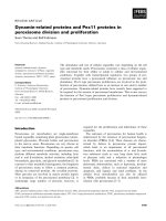
In the crystallographic model of gelsolin in the EGTA form
[22], G2 is tightly interacting with G6 segment (Fig. 3). Two
regions in G2 domain appear included in this interface. The
Fig. 2. Effect of calcium on the G4–6 tryptophan and FITC labeled
G4–6 fluorescence emission. Aliquots of calcium (0.1 m
M
solution)
were added successively to unlabeled G4–6 or labeled G4–6 in 0.1
M
KCl, 20 m
M
Tris buffer pH 7.4 in the presence of 0.1 m
M
EGTA.
Changes in fluorescence intensities corresponding to tryptophan
emission (s) or FITC emission (d) are plotted vs. free calcium con-
centration expressed as pCa ¼ log(1/[Ca
2+
]). Calcium concentrations
were determined experimentally (see Material and methods). Inset:
effect of calcium on the G2 tryptophan fluorescence emission. Fluo-
rescence changes were plotted vs. free calcium concentrations.
Fig. 1. Effect of calcium on susceptibility of gelsolin G4–6 domain to
tryptic digestion. (A) Identification of the two fragments (30 kDa and
16 kDa) produced by proteolysis in the presence of 1 m
M
EGTA
followed by 1,5-I-AEDANS labeling as described in Material and
methods. Molecular weight markers (lane T). G4–6 digest chemically
modified by IEADANS and revealed with Coomassie blue (lane 1) or
upon UV lamp (lane 2). (B) Digestion of G4–6 domain by trypsin
(trypsin/G4–6 ratio: 1/25 w/w) for 10 min molecular weight markers
(lane T). G4–6 domain (46 kDa) before proteolysis (lane 1). G4–6
digest in the presence of 0.1 m
M
calcium (lane 2) and in the presence of
0.1 m
M
EGTA (lane 3). Molecular weight marker are phosphory-
lase B (97.4 kDa), bovine serum albumin (66.2 kDa), ovalbumin
(45 kDa), carbonic anhydrase (31 kDa), soybean trypsin inhibitor
(21.5 kDa) and lysozyme (14.4 kDa).
2238 E. Lagarrigue et al. (Eur. J. Biochem. 270) Ó FEBS 2003
sequence 203–225, including one of the actin-binding sites, is
in interaction with the C-terminal a-helix of G6 domain.
The sequence 159–193, including the second actin interface,
appears also in interaction with G6 domain. Therefore, we
tested the interaction of G4–6 domain with G2 domain by
two independent methods.
In ELISA experiments, G4–6 fragment was immobilized
on the plastic microtiter plate and the binding of biotinyl-
ated G2 domain was revealed by using alkaline phosphatase
labeled streptavidin. Figure 4 shows that binding occurs in
the presence of EGTA as well in the presence of calcium
although a better affinity is observed in the former case.
These data were confirmed by studies in solution using
fluorescence measurements. G2 or G4–6 domains were
labeled by FITC and increasing concentrations of G4–6 or
G2 were added, respectively. Figure 5A shows a decrease in
the fluorescence intensity of FITC labeled G2 domain in the
presence of EGTA. Analysis of these data shows that the
fluorescence intensity decrease, extrapolated to infinite
concentration of G4–6, is low (4%). Therefore, only a
rough estimation of the apparent K
d
(about 0.5 l
M
)canbe
obtained. This result shows that G4–6 fragment interaction
induces a conformational change in G2 domain in the
presence of EGTA. Calcium was without effect (Fig. 5A),
but it is known that calcium did promote important changes
[11,36], which would explain the present results. Conse-
quently, further analyses, using FITC labeled G4–6, were
carried out. Labeled G4–6 was incubated with increasing
concentrations of G2 (between 0 and 7.6 l
M
)andthe
changes in fluorescence were monitored. Saturation curves
were observed in the presence of EGTA or calcium and
apparent K
d
s of 0.8 and 3.5 l
M
, respectively, were deter-
mined (Fig. 5B and Table 1). These values and those
obtained above from ELISA show a better interaction of
G2 in the presence of EGTA than calcium. The differences
in the absolute values observed between the two methods
are likely to be due to the heterogeneous phases used in
ELISA. More interestingly, a significant difference in the
maximum fluorescence enhancement determined in EGTA
and calcium (about 8% and 30%, respectively) possibly
reflects changes in domain conformation and in interfaces
produced under these two conditions. In order to deduce a
more precise relationship between G2 interaction and G4–6
calcium-induced conformation, the maximum fluorescence
enhancement extrapolated for infinite G2 concentrations
was determined at various calcium concentrations. As
shown in Fig. 6, a change in the fluorescence from EGTA to
calcium states was observed in the 0.1 l
M
range. This
important result suggests that the transition would be linked
to the high affinity calcium site in G4–6 [7], that we now
know to be IIG6 [3] (see Discussion). In addition, apparent
K
d
values were also estimated from the same fluorescence
experiments performed in the presence of different calcium
concentrations. As shown in Fig. 6 insert, an apparent K
d
transition occurs around 15 l
M
, a value which is observed
for the binding of calcium to G2 domain.
Footprint of G2 on gelsolin G4–6 domain
Two sites within the G2 domain appear involved in the
G2- G4–6 interface in the presence of EGTA (Fig. 3). The
interaction of these two sites (residues 159–193 and 203–
225) was first tested by ELISA. The interaction of G4–6
with the plate-coated peptides was revealed using specific
antibodies to G4–6. The results summarized in Table 1
Fig. 3. X-ray crystallographic structure of gelsolin in the absence of
calcium (Burtnick et al. [22]) showing the interface between the G6
C-terminal domain and G2. The G6 domain is coloured green and its
C-terminal alpha helix is coloured blue. The G2 domain is coloured
yellow and its sequences in contact with G6, sequences 159–193 and
203–225 are coloured red and purple, respectively.
Fig. 4. Binding of gelsolin domain G2 with G4–6 monitored by ELISA.
Coated G4–6 was reacted with the biotinylated G2 domain in the
presence of 1 m
M
EGTA (d) or in the presence of 1 m
M
CaCl
2
(s)at
the concentrations indicated. Binding was monitored at 405 nm using
alkaline phosphatase labeled streptavidin. Percentage binding was
plotted vs. G2 concentrations.
Ó FEBS 2003 Calcium activation of gelsolin (Eur. J. Biochem. 270) 2239
show that the two peptides interacted with G4–6 in the
presence of EGTA. In contrast, only the 153–193 peptide
interacted in the presence of calcium (Table 1). These results
were confirmed in solution. For this aim, FITC labeled
G4–6 was mixed with each peptide supplemented with
EGTA or calcium. In the presence of EGTA, changes in the
fluorescence intensity (Fig. 7A,B) were obtained with the
two fragments. A maximum fluorescence quenching of 4%
and fluorescence enhancement of 4% were calculated for
159–193 and 203–225 peptides, respectively. When calcium
is present in the medium, binding of peptide 153–193 to
labeled G4–6 induces an important decrease in fluorescence
intensity (15%) (Fig. 7). The last result demonstrates that
the binding of the latter peptide causes a somewhat different
conformational change in G4–6 domain. In contrast no
effect is observed for the 203–225 fragment, accordingly to
ELISA experiments.
Discussion
Since its discovery as an actin-depolymerizing factor, gelsolin
has now been implicated in a number of important pathways
such as apoptosis, oncogenic transformation, signal trans-
duction and amyloidosis (reviewed [2]). All of these
pathways are likely to involve calcium activation, the process
by which various binding sites become (especially actin)
available for interaction. In this paper we confirm that
calcium causes a large conformational change in the
C-terminal half of gelsolin (G4–6) that can be monitored
in a number of different techniques. We also show that G2
undergoes a conformational change upon binding calcium
by a site that has a slightly higher affinity than was previously
assumed. This study also provides some details on the
interface between these two gelsolin domains (G2 with
G4–6) whose dissociation is pivotal in the activation of
gelsolin.
Calcium-dependent conformational changes in G4–6
That the C-terminal half of gelsolin (G4–6) binds calcium,
leading to the activation of the whole gelsolin molecule
Fig. 5. Binding of gelsolin G2 domain with G4–6 monitored by fluor-
escence measurements. (A) Interaction of FITC labeled G2 domain
(0.2 l
M
) with G4–6 was carried out in 0.1
M
KCl 20 m
M
Tris buffer
pH 7.4. Change in fluorescence emission spectra of FITC was recorded
at various G4–6 concentrations (0–2.1 l
M
) in the presence of 1 m
M
EGTA (d)or1m
M
CaCl
2
(s). (B) Binding of G2 domain with FITC
labeled G4–6 (0.3 l
M
) determined by fluorescence. The experiment
was carried out in 0.1
M
KCl, 20 m
M
Tris buffer pH 7.4 supplemented
with 1 m
M
EGTA (d)or1m
M
CaCl
2
(s).
Fig. 6. Effect of calcium on the fluorescence of the FITC labeled G4–6/
G2 complex. Maximum fluorescence enhancement (% initial fluores-
cence) extrapolated to infinite G2 concentration is plotted vs. free
calcium concentration (pCa). Inset, apparent K
d
s for G2. Interactions
with FITC labeled G4–6 are plotted vs. pCa.
Table 1. Binding of G2 and derived peptide to G4–6.
EGTA K
d
(l
M
) Calcium K
d
(l
M
)
Fluorescence ELISA Fluorescence ELISA
G2 0.5–0.8 0.3 3 1
159–193 1 1 2–3 2
203–225 3 3 None None
2240 E. Lagarrigue et al. (Eur. J. Biochem. 270) Ó FEBS 2003
through large structural changes is well established [8,9]. We
have shown that a calcium sensitive proteolysis between G5
and G6 occurs in agreement with the structural data [3,15]
that shows a long random coil connecting G5 to G6.
Previous data from biochemical studies on G4–6 have
detect two calcium binding site one within G4-5
(K
d
¼ 2 l
M
), the other within G5–6 (K
d
¼ 0.2 l
M
)[7].
New data (Koloppan et al. 2003) collected with actin-free
G4–6 identify calcium bound to G5 and G6 only, a finding
in agreement with Choe et al. [3] who hypothesized that a
third site, IIG4, became coordinated solely by G4 residues
only when actin was bound (were this to be the case then
IIG4 is a site with properties between a site I and a site II
type). Taken together it seems that two sites are bound by
G4–6 in the absence of actin, one of these, IIG5 has a
moderate affinity (K
d
¼ 2 l
M
) in agreement of this study
and previous work [7], the other IIG6 has a higher affinity.
The slight difference observed between the present study
(K
d
¼ 0.1 l
M
) and the previous study [7] who measured a
K
d
of 0.2 l
M
, may be attributable to conformational
differences in the site in the context of G4–6 compared to
G5–6 or a degree of cocooperativity between calcium site
(see below).
A calcium-dependent conformational change in G2
A number of studies have concluded that calcium induces
conformational changes within G2 [11,36]. A calcium
binding site has been detected in G1–3 [12], and others
[3,11] have located this site within G2 itself. However, this
previous [11] study estimated a low affinity (K
d
¼32 l
M
)
whereas we have estimated a slightly higher affinity
(K
d
¼15 l
M
). We have also found a calcium-dependent
conformational change in G2 this may explain why the
reactivity of the thiol groups within G2 are calcium-sensitive
[37,38].
The G2 G4–6 interface
As expected we have found that G2 binds to G4–6 with high
affinity (K
d
¼0.5–0.8 l
M
)inEGTA,weakerbinding
was evident in the presence of 1 m
M
calcium (Fig. 5). In
addition a change in the interface occurs during calcium
binding to IIG6 site (Fig. 6). The interaction between
gelsolin C-terminal helix (residues 744–755) and the helix of
G2 is well known. We have confirmed this interaction
biochemically and found a calcium dependent interaction
between G4–6 and the peptide 203–225 derived from G2.
The calcium sensitivity of this interaction is probably due to
IIG6 as the coordinating residues that comprise the IIG2
site are not all present within the peptide. In addition to this
expected interaction, we have also detected binding of G4–6
to a second G2-derived peptide 159–193. This interaction
is only marginally calcium sensitive and of higher affinity
(Table 1). The EGTA structure [22] reveals that this peptide
makes salt bridge contacts (R168–D669 and R169–D670)
and that hydrophobic interactions also occur such as those
between V170-V657, with G6. In summary, G2 binds to
G4–6 through two distinct interfaces. Binding site 1 involves
G2 203–225 and G6 744–755 and binding site 2 involves
G2 region around R168-R69 and G6 D669-D670.
‘Unlatching’ and dissociation of the G2 and G6
connection
If the last 23 amino-acids are removed from gelsolin
mutants, the requirement for calcium for actin-binding
is lessened but not abolished [39], similarly it has been
determined that adseverin which naturally lacks the
C-terminal helix has a similar calcium requirement than
the helix minus gelsolin mutant [21]. Together with recent
observations on the structure of gelsolin [3,36], our new
data on binding site 2 is compatible with the following
Fig. 7. Effect of calcium on the interaction of two sequences involved in the G2–G4–6 interface monitored by fluorescence. Binding of FITC labeled
G4–6 (0.3 l
M
) with two synthetic peptides derived from G2 domain: (A) sequence 203–225 and (B), sequence 159–193. The experiments were
performed in 0.1
M
KCl, 20 m
M
Tris buffer pH 7.4 supplemented with 1 m
M
EGTA (d)or1m
M
CaCl
2
(s).
Ó FEBS 2003 Calcium activation of gelsolin (Eur. J. Biochem. 270) 2241
explanation. It is likely that G2 remains held to G4–6
through binding site 2 in the absence of site 1.
Occupancy of IIG2 has been proposed to disrupt site 1
[36], by disruption of hydrophobic and salt bridges between
G2 helix a1 and the C-terminal helix of G6. It is proposed
that binding of IIG6 disrupts the salt bridges between
D669 and R168, and between D670 and R169. Cooper-
ativity between IIG2 and IIG6 has been suggested to occur
as a result of the breaking of site 1 and site 2, as breakage
of either connection frees up ligands to coordinate calciums
in either type II site [3]. Calcium binding to whole gelsolin
has been found to be cooperative [40] it is possible that this
is due to coordination of calcium sites as is proposed for
IIG2 and IIG6, in addition to the general opening of the
molecule. Cooperation of the sites would also explain why
the dissociation of G2 from G4–6 occurs at 100 n
M
calcium (Fig. 6), whereas we have measured a K
d
value
of 15 l
M
, and others estimate 32 l
M
[11] for calcium
binding to IIG2. Occupancy of IIG6 may indirectly alter
IIG2 so that it too becomes a high affinity site. We
measure the IIG6 K
d
to be 100 n
M
(Fig. 2) in agreement
with this model.
Acknowledgements
This research was supported by grants from AFM.
References
1. Sun, H.Q., Yamamoto, M., Mejillano, M. & Yin, H.L. (1999)
Gelsolin, a multifunctional actin regulatory protein. J. Biol. Chem.
274, 33179–33182.
2. Kwiatkowski, D.J. (1999) Functions of geloslin: motility, signa-
ling, apoptosis, cancer. Curr. Op. Cell Biol. 11, 103–108.
3. Choe, H., Burtnick, L.D., Mejillano, M., Yin, H.L., Robinson,
R.C. & Choe, S. (2002) The calcium activation of gelsolin: Insights
from the 3A structure of the G4–G6/actin complex. J. Mol. Evol.
324, 691–702.
4. Yin, H.L. & Stossel, T.P. (1980) Purification and structural
properties of gelsolin, a calcium-activated regulator protein of
macrophages. J. Biol. Chem. 255, 9490–9493.
5. Bryan, J. (1988) Gelsolin has three actin-binding sites. J. Cell Biol.
106, 1553–1562.
6. Weeds,A.G.,Gooch,J.,Pope,B.P.&Harris,H.E.(1986)Pre-
paration and characterization of pig plasma and platelet gelsolins.
Eur. J. Biochem. 161, 69–76.
7. Pope, B., Maciver, S. & Weeds, A.G. (1995) Localization of the
calcium-sensitive actin monomer binding site in gelsolin to seg-
ment 4 and identification of calcium-binding sites. Biochemistry
34, 1583–1588.
8. Patkowski, A., Seils, J., Hinssen, H. & Dorfmuller, T. (1990) Size,
shape parameters and calcium-induced conformational change of
the gelsolin molecule. A dynamic light scattering study. Biopoly-
mers 30, 427–435.
9. Hellweg, T., Hinssen, H. & Eimer, W. (1993) The Ca
2+
-induced
conformational change of gelsolin is located in the carboxyl-
terminal half of the molecule. Biophys. J. 65, 799–805.
10. Doi, Y., Kim, F. & Kido, S. (1990) Weak binding of divalent
cations to plasma gelsolin. Biochemistry 29, 1392–1397.
11. Zapun, A., Grammatyka, S., Deral, G. & Vernet, T. (2000) Cal-
cium-dependent conformational stability of modules 1 and 2 of
human gelsolin. Biochem. J. 350, 873–881.
12. Pope, B.J., Gooch, J.T. & Weeds, A.G. (1997) Probing the effects
of calcium on gelsolin. Biochemistry 36, 15848–15855.
13. McLaughlin, P.J., Gooch, J.T., Mannherz, H.G. & Weeds, A.G.
(1993) Structure of gelsolin segment-1-actin complex and the
mechanism of filament severing. Nature 364, 685–692.
14. Weeds, A.G., Gooch, J., McLaughlin, P., Pope, B., Bengtsdotter,
M. & Karlsson, R. (1995) Identification of the trapped calcium in
the gelsolin segment 1-actin complex: implications for the role
of calcium in the control of gelsolin activity. FEBS Lett. 360,
227–230.
15. Robinson, R.C., Mejillano, M., Le, V.P., Burtnick, L.D., Yin,
H.L. & Choe, S. (1999) Domain movement in gelsolin: a calcium-
activated switch. Science 286, 1939–1942.
16. Feinberg, J., Capony, J.P., Benyamin, Y. & Roustan, C. (1993)
Definition of the EGTA–independent interface involved in the
serum gelsolin actin complex. Biochem. J. 293, 813–817.
17. Renoult, C., Blondin, L., Fattoum, A., Ternent, D., Maciver,
S.K., Raynaud, F., Benyamin, Y. & Roustan, C. (2001) Binding of
gelsolin domain 2 to actin. An actin interface distinct from that of
gelsolin domain 1 and from ADF/cofilin. Eur. J. Biochem. 268,
6165–6175.
18. Lin, K M., Mejillano, M. & Yin, H.L. (2000) Ca
2+
regulation of
gelsolin by its C-terminal tail. J. Biol. Chem. 275, 27746–27752.
19. Khaitlina, S. & Hinssen, H. (2002) Ca-dependent binding of actin
to gelsolin. FEBS Lett. 521, 14–28.
20. Kinosian, H.J., Newman, J., Lincoln, B., Selden, L.A., Gershman,
L.C. & Estes, J.E. (1998) Ca
2+
regulation of gelsolin activity:
binding and severing of F-actin. Biophys. J. 75, 3101–3109.
21. Lueck, A., Brown, D. & Kwiatkowski, D.J. (1998) The actin-
binding proteins adseverin and gelsolin are both hightly expressed
but differentially localized in kidney and intestine. J. Cell Sci. 111,
3633–3643.
22. Burtnick, L.D., Koepf, E.K., Grimes, J., Jones, E.Y., Stuart, D.I.,
McLaughlin, P.J. & Robinson, R.C. (1997) The crystal structure
of plasma gelsolin: implications for actin severing, capping and
nucleation. Cell 90, 661–670.
23. Way, M., Pope, B., Gooch, J., Hawkins, M. & Weeds, A.G. (1990)
Identification of a region of segment 1 of gelsolin critical for actin
binding. EMBO J. 9, 4103–4109.
24. Way, M., Pope, B. & Weeds, A.G. (1992) Evidence for functional
homology in the F-actin binding domains of gelsolin and alpha-
actinin: implications for the requirements of severing and capping.
J. Cell Biol. 119, 835–842.
25. Way, M., Gooch, J., Pope, B. & Weeds, A.G. (1989) Expression
of human plasma gelsolin in E. coli anddissectionofactin
binding sites by segmental deletion mutagenesis. J. Cell Biol. 109,
593–605.
26. Pope, B., Way, M. & Weeds, A.G. (1991) Two of the three actin-
binding domains of gelsolin bind to the same subdomain of actin.
FEBS Lett. 280, 70–74.
27. Takashi, R. (1979) Fluorescence energy transfer between sub-
fragment-1 and actin points in the rigor complex of actosubfrag-
ment-1. Biochemistry 18, 5164–5169.
28. Me
´
jean, C., Hue
´
, H.K., Pons, F., Roustan, C. & Benyamin, Y.
(1988) Cation binding sites on actin: a structural relationship
betweenantigenicepitopesandcationexchange.Biochem. Bio-
phys. Res. Comm. 152, 368–375.
29. Miki, M. & dos Remedios, C.G. (1988) Fluorescence quenching
studies of fluorescein attached to Lys-61 or Cys-374 in actin:
effects of polymerization, myosin subfragment-1 binding, and
tropomyosin-troponin binding. J. Biochem. (Tokyo). 104, 232–
235.
30. Papa, I., Astier, C., Kwiatek, O., Lebart, M C., Raynaud, F.,
Benyamin, Y. & Roustan, C. (1999) Use of a chaotropic anion
iodide in the purification of Z-line proteins: isolation of CapZ.
from fish white muscle. Prot. Expr. Purif. 17,1–7.
31. Engvall, E. (1980) Enzyme immunoassay ELISA and EMIT.
Methods Enzymol. 70, 419–439.
2242 E. Lagarrigue et al. (Eur. J. Biochem. 270) Ó FEBS 2003
32. Me
´
jean,C.,Lebart,M.C.,Poyer,M.,Roustan,C.&Benyamin,Y.
(1992) Localization and identification of actin structures involved
in the filamin-actin interactio. Eur. J. Biochem. 209, 555–562.
33. Gill, S.C. & von Hippel, P.H. (1989) Calculation of protein
extinction coefficients from amino acid sequence data. Anal. Bio-
chem. 182, 319–326.
34. Laemmli, U.K. (1970) Cleavage of structural proteins during the
assembly of the head of bacteriophage T4. Nature 227, 680–685.
35. Grynkeiwicz, G., Poenie, M. & Tsien, R.Y. (1985) A new gen-
eration of calcium indicators with greatly improved fluorescence
properties. J. Biol. Chem. 260, 3440–3450.
36. Kazmirski, S.L., Isaacson, R.L., An, C., Buckle, A., Johnson,
C.M., Daggett, V. & Fersht, A.R. (2002) Loss of a metal-binding
site in gelsolin leads to familial amyloidosis-Finnish type. Nature
Struct. Biol. 9, 112–116.
37. Kilhoffer, M C. & Ge
´
rard, D. (1985) Fluorescence study of bre-
vin, the Mr 92,000 actin-capping and -fragmenting protein isolated
form serum. Effect of Ca
2+
on protein conformation. Biochem-
istry 24, 5653–5660.
38. Rouayrenc, J P., Fattoum, A., Me
´
gean, C. & Kassab, R. (1986)
Characterization of the Ca
2+
-induced conformational change in
gelsolin and identification of interaction regions between actin and
gelsolin. Biochemistry 25, 3859–3867.
39. Kwiatkowski, D.J., Janmey, P.A. & Yin, H.L. (1989) Identifica-
tion of critical functional and regulatory domains in gelsolin.
J. Cell Biol. 108, 1717–1726.
40. Gremm, D. & Wegner, A. (1999) Co-operative binding of Ca
2+
ions to the regulatory binding sites of gelsolin. Eur. J. Biochem.
262, 330–334.
Ó FEBS 2003 Calcium activation of gelsolin (Eur. J. Biochem. 270) 2243