Báo cáo khoa học: Expression, purification and characterization of the second Kunitz-type protease inhibitor domain of the human WFIKKN protein pot
Bạn đang xem bản rút gọn của tài liệu. Xem và tải ngay bản đầy đủ của tài liệu tại đây (283.6 KB, 7 trang )
Expression, purification and characterization of the second
Kunitz-type protease inhibitor domain of the human WFIKKN protein
Alinda Nagy, Ma
´
ria Trexler and La
´
szlo
´
Patthy
Institute of Enzymology, Biological Research Center, Hungarian Academy of Sciences, Budapest, Hungary
Recently we have described a novel secreted protein (the
WFIKKN protein) that consists of multiple types of prote-
ase inhibitory modules, including two tandem Kunitz-type
protease inhibitor-domains. On the basis of its homologies
we have suggested that the WFIKKN protein is a multi-
valent protease inhibitor that may control the action of
different proteases. In the present work we have expressed
the second Kunitz-type protease inhibitor domain of the
human protein WFIKKN in Escherichia coli, purified it
by affinity chromatography on trypsin-Sepharose and its
structure was characterized by CD spectroscopy. The
recombinant protein was found to inhibit trypsin
(K
i
¼ 9.6 n
M
), but chymotrypsin, elastase, plasmin, pan-
creatic kallikrein, lung tryptase, plasma kallikrein, thrombin,
urokinase or tissue plasminogen activator were not inhibited
by the recombinant protein even at 1 l
M
concentration. In
view of the marked trypsin-specificity of the inhibitor it is
suggested that its physiological target may be trypsin.
Keywords: Kunitz-domain; multidomain protease inhibitor;
serine proteinases; trypsin.
Recently we have identified two closely related human
proteins (WFIKKN and WFIKKNRP) each of which
contain a WAP-domain, a Follistatin/Kazal domain, an
Immunoglobulin-domain, two Kunitz-domains and an
NTR-domain [2,3]. The tissue expression pattern of the
two proteins, however, is markedly different suggesting
that they have distinct biological roles. Whereas the
WFIKKNRP gene is expressed primarily in ovary, testis
and brain, the most significant expression of the WFIKKN
gene is observed in adult pancreas, liver and thymus.
In view of the presence of WAP-, Kazal-, Kunitz- and
NTR-modules (which are frequently involved in inhibition
of proteases) in a single multidomain protein we have
suggested that these proteins function as multivalent
protease inhibitors.
In order to test this hypothesis, in the present work we
have expressed the second Kunitz-type protease inhibitor
domain of the human protein WFIKKN in Escherichia coli.
Our structural studies on the recombinant protein have
shown that the protein adopts a structure typical of the
Kunitz-domain family. The recombinant protein was found
to show remarkable specificity for trypsin in contrast to
its lack of activity for elastase, chymotrypsin and various
proteases with trypsin-like specificity.
Experimental procedures
Restriction enzymes, PCR primers, vectors, bacterial
strains
Restriction enzymes were purchased from Promega (Madi-
son, WI, USA) and New England Biolabs (Beverly, MA,
USA). The M13 sequencing reagents used for dideoxy
sequencing of cloned DNA fragments were from Promega.
PCR primers were obtained from Integrated DNA Tech-
nologies (Coralville, IA, USA). Plasmid pMed23 was
from P. Venetianer (Biological Research Center, Szeged,
Hungary). E. coli strain JM109 was used to propagate
and amplify expression plasmids. The pMed23 expression
plasmid contains an ampicillin resistance gene for the
selection of the positive clones [4].
Proteases and protease substrates
Bovine trypsin (Sigma-Aldrich, St. Louis, MO, USA),
bovine elastase (Serva, Heidelberg, Germany), bovine
pancreatic alpha-chymotrypsin (Worthington, Lakewood,
NJ, USA), bovine thrombin, human plasmin, human lung
tryptase, human high molecular mass urokinase, human
tissue plasminogen activator, human plasma kallikrein and
porcine pancreatic kallikrein (Calbiochem, Affiliate of
Merck, Darmstadt) were commercial preparations.
The synthetic substrates N-succinyl-Ala-Ala-Pro-
Phe-pNA and N-a-benzoyl-
L
-Arg-pNA (L-BAPNA) were
purchased from Sigma,
D
-Val-Leu-Lys-pNA and
D
-Pro-Phe-Arg-pNA were from Serva. Glu-Gly-Arg-pNA,
D
-Ile-Pro-Arg-pNA, Bz-Phe-Val-Arg-pNA,
D
-Val-Leu-
Arg-pNA and succinyl-Ala-Ala-Ala-pNA were obtained
Correspondence to L. Patthy, Institute of Enzymology, Biological
Research, Center, Hungarian Academy of Sciences, Budapest,
Karolina u´ t 29, H-1113, Hungary.
Fax: + 361 4665 465, Tel.: + 361 2093 537,
E-mail:
Abbreviations: BPTI, bovine pancreatic trypsin inhibitor; NPGB,
p-nitrophenyl-p-guanidinobenzoate; pNA, p-nitroanilide; TFPI, tissue
factor pathway inhibitor; WAP, whey acidic protein.
Definition: The nomenclature for the substrate amino acid residues
Pn-P4-P3-P2-P1-P¢1-P¢2-P¢3-P’n., where -P1-P¢1- denotes the
hydrolyzed bond, and Sn-S4-S3-S2-S1-S¢1-S¢2-S¢3-S¢4denote
the corresponding enzyme binding sites is described fully in [1].
(Received 16 December 2002, revised 5 March 2003,
accepted 26 March 2003)
Eur. J. Biochem. 270, 2101–2107 (2003) Ó FEBS 2003 doi:10.1046/j.1432-1033.2003.03593.x
from Bachem (Bubendorf, Switzerland). p-Nitrophenyl-
p-guanidinobenzoate was a product of Fluka (Buch,
Switzerland).
Cloning and expression of the second Kunitz-type
protease inhibitor module of human WFIKKN protein
On the basis of the known sequence of the human
WFIKKN mRNA (GenBank accession number AF422194)
we have designed PCR primers for the amplification of the
cDNA segment encoding its second Kunitz-domain. The
DNA segment coding for the second Kunitz-module of
human WFIKKN protein (residues Asp357–Pro412) was
amplified with the 5¢-GAG TCG ACC GAC GCC TGC
GTG CTG CCT GC-3¢ sense, and 5¢-GCA AGC TTA
CGG CAC GGG GCA GGC ATC CTC-3¢ antisense
primers from a plasmid containing the cDNA coding for
WFIKKN protein. The amplified DNA was digested with
HindIII and SalI restriction endonucleases and ligated into
M13mp18 Rf digested with the same enzymes. The sequence
of the cloned DNA was verified by dideoxy sequencing.
The DNA fragment encoding the second Kunitz-module
of WFIKKN was excised from M13mp18 by HincII/
HindIII digestion and ligated into pMed23 expression
vector cut with PvuII/HindIII. E. coli JM109 cells were
transformed with the ligation mixture and plated on LB
medium (1% tryptone, 0.5% yeast extract, 1% NaCl)
containing 100 lgÆmL
)1
ampicillin.
E. coli JM109 cells carrying the expression vector were
grown, and expression of b-galactosidase fusion proteins
was induced with 100 l
M
isopropyl thio-b-
D
-galactoside.
The fusion products were isolated from inclusion bodies by
dissolving them in 60 mL of 0.1
M
Tris/HCl, 8
M
urea,
10 m
M
EDTA, 0.1
M
dithiothreitol (Sigma-Aldrich),
pH 8.0. The solution was incubated at 25 °Cfor60min
with constant stirring. Insoluble cellular debris were
removed by centrifugation and the solubilized proteins
were chromatographed on a Sephacryl S-300 column
equilibrated with 100 m
M
Tris/HCl, 8
M
urea, 10 m
M
EDTA, 0.1% 2-mercaptoethanol. The fractions containing
the fusion proteins were identified by SDS/PAGE and
pooled. The isolated recombinant proteins were refolded by
dialysis against 100 m
M
Tris and 10 m
M
EDTA pH 8.0
buffer, for 24 h, then against 0.1
M
ammonium bicarbonate
pH 8.0 buffer.
The b-galactosidase moiety of the recombinant fusion
protein was removed by limited elastase digestion.
The recombinant protein (1 mgÆmL
)1
) was dissolved in 0.1
M
ammonium bicarbonate buffer and incubated with
10 lgÆmL
)1
elastase (Serva) at 25 °C for 60 min. The
reaction was arrested with 2 m
M
phenylmethanesulfonyl
fluoride (Serva) and the protein was lyophilized. The digested
recombinant protein was separated from the b-galactosidase
fragment on Sephadex G-50 column, equilibrated with 0.1
M
ammonium bicarbonate, pH 8.0. Fractions containing the
Kunitz-module were pooled, and lyophilized.
The protein was further purified by trypsin-Sepharose
affinity chromatography according to described procedures
[5,6]. The protein was dissolved in 50 m
M
Tris-HCl pH 7.5
and applied on a 5-mL trypsin-Sepharose column. The
column was washed with four volumes of 50 m
M
Tris-HCl
pH 7.5 and the bound protein was eluted with 100 m
M
glycine/HCl buffer, pH 2.0. The pH of the eluted fraction
was adjusted to 8.0, the protein was desalted on a G-25
Sephadex column equilibrated with 0.1
M
ammonium
bicarbonate pH 8.0 buffer, and lyophilized.
Sequence analysis of the purified protein with a
PE-Applied Biosystems Ltd Procise protein sequencing
system showed that the elastase cleavage occurred at the
boundary of the b-galactosidase region of the b-gal fusion
protein. The amino acid sequence of the resulting puri-
fied protein was RTDACVLPAVQGPCRGWEPRWAYS
PLLQQCHPFVYGGCEGNGNNFHSRESCEDACPVP,
where the residues corresponding to the second Kunitz
domain of human WFIKKN are in bold. The N-terminal
residues RT are part of the vector construct.
Protein analyses
The composition of protein samples was analysed by tricine/
SDS/PAGE using 16% slab gels under both reducing and
nonreducing conditions [7]. The gels were stained with
Coomassie brilliant Blue G-250. The concentration of the
recombinant Kunitz-module was determined using the
extinction coefficient 14300
M
)1
Æcm
)1
. The extinction coef-
ficient was determined by using the online protein analysis
tool,
PROTPARAM
( />html).
Circular dichroism spectroscopy
CD spectra were measured over the range of 190–250 nm by
using a JASCO J-720 spectropolarimeter thermostatted with
a Neslab RT-111 water bath. The measurements were
carried out in 1 mm pathlength cells and protein solutions of
approximately 0.1 mgÆmL
)1
in 10 m
M
Tris/HCl, pH 8.0
buffer. All spectra were measured at 25 °C with a 8-s time
constant and a scan rate of 10 nmÆmin
)1
. The spectral slit
width was 1.0 nm. All measurements represent the computer
average of three scans. Secondary structure of the recom-
binant protein was estimated from the CD spectra with the
CDPRO
software ( />~
sreeram/
CDPro/index.html [8–10]). Thermal unfolding of the protein
was monitored at 203 nm at a heating rate of 60 °CÆh
)1
.
Effect of the recombinant protein on the activity
of proteases
The activity of the proteases on synthetic peptide-pNA
substrates was monitored spectrophotometrically using a
Carry 300 Scan spectrophotometer. Hydrolysis of peptide-
pNA conjugates was monitored at 410 nm and the initial
rates of the reaction were determined.
In the case of bovine trypsin, stock solutions were
prepared in 1 m
M
HCl, 20 m
M
CaCl
2
, the active site
concentration of trypsin was determined by titration with
NPGB according to a described procedure [11]. Stock
solutions of the Kunitz-module were prepared in 25 m
M
Tris, 5 m
M
CaCl
2
pH 7.5 buffer.
The kinetic parameters of trypsin-catalysed hydrolysis
of Bz-Phe-Val-Arg-pNA were determined by incubating
trypsin (30 n
M
final concentration) in 25 m
M
Tris, 5 m
M
CaCl
2
,pH7.5for5minat37°C, after which Bz-Phe-Val-
Arg-pNA (100–400 l
M
final concentration) was added and
2102 A. Nagy et al. (Eur. J. Biochem. 270) Ó FEBS 2003
the enzymatic formation of pNA was monitored at 410 nm,
employing a De of 8800
M
)1
Æcm
)1
.
The value of the equilibrium constant for the inhibition of
trypsin by the Kunitz-module was determined by measuring
its inhibitory effect on the enzymatic hydrolysis of Bz-Phe-
Val-Arg-pNA substrate at 37 °C. Aliquots of 250 lL assay
mixtures containing 30 n
M
enzyme and 15, 30, 60 and
150 n
M
inhibitor were incubated for 5 min at 37 °Cin
25 m
M
Tris, 5 m
M
CaCl
2
, pH 7.5 buffer. Bz-Phe-Val-Arg-
pNA (100–400 l
M
final concentration) was then added and
the activity was recorded. All experiments were run three
times. The enzymatic hydrolysis of the substrate was always
corrected for spontaneous hydrolysis.
The dissociation constant of the trypsin–inhibitor com-
plex, K
i
was determined from the replot of the apparent K
m
values vs. the inhibitor concentration at which they were
obtained.
In the case of chymotrypsin, plasmin, thrombin, tissue
plasminogen activator and plasma kallikrein, the proteases
were preincubated for 30 min at 37 °Cin50m
M
Tris,
100 m
M
NaCl, 2 m
M
CaCl
2
, 0.01% Triton X-100 pH 7.5
buffer in the presence of increasing inhibitor concentrations
(up to 1 l
M
final concentration of the inhibitor). The
reactions were initiated by adding the appropriate sub-
strate specific for the enzyme. The reaction mixtures
contained the following initial enzyme and substrate con-
centrations: alpha-chymotrypsin was measured at an
enzyme concentration of 50 n
M
and 80 l
M
N-succinyl-
Ala-Ala-Pro-Phe-pNA substrate concentration; human
plasmin at 10 n
M
enzyme and 300 l
MD
-Val-Leu-Lys-
pNA substrate concentration; bovine thrombin at 100 n
M
enzyme and 200 l
M
Bz-Phe-Val-Arg-pNA substrate con-
centration; human plasma kallikrein at 3 n
M
enzyme and
650 l
MD
-Pro-Phe-Arg-pNA substrate concentration, and
the inhibition of human tissue plasminogen activator was
measured at 44 n
M
enzyme and 100 l
MD
-Ile-Pro-Arg-pNA
concentration.
In the case of elastase, pancreatic kallikrein, lung tryptase,
urokinase activity was monitored following preincubation
of the protease with inhibitor (up to 1 l
M
final concentra-
tion of the inhibitor) for 30 min at 37 °C in the appropriate
buffer (see below). Reactions were initiated with substrate
to achieve the following initial component concentrations:
bovine elastase in 100 m
M
Tris, 0.05% Triton X-100,
pH 8.0 with [E
0
] ¼ 38 n
M
and 600 l
M
succinyl-Ala-Ala-
Ala-pNA; porcine pancreatic kallikrein in 50 m
M
Tris,
100 m
M
NaCl, 2 m
M
CaCl
2
, 0.01% Triton X-100 pH 8.4
with [E
0
] ¼ 16 UÆmL
)1
and 200 l
MD
-Val-Leu-Arg-pNA;
human lung tryptase in 50 m
M
Tris, 120 m
M
NaCl,
44 lgÆmL
)1
heparin pH 7.5 with [E
0
] ¼ 22 n
M
and
100 l
M
N-a-Benzoyl-
L
-Arg-pNA; human urokinase in
50 m
M
Tris, 10 m
M
EDTA, 50 m
M
NaCl, 0.5% Triton
X-100 pH 8.0 with [E
0
] ¼ 30 n
M
and 300 l
M
Glu-Gly-
Arg-pNA.
Sequence analyses
The amino acid sequences of human WFIKKN protein
(AAL18839), human WFIKKNRP protein (AAL77058),
bovine pancreatic trypsin inhibitor (bpt1_bovin, P00974),
human bikunin (ambp_human, P02760), human Alzhei-
mer’s disease amyloid a4 protein precursor (a4_human,
P05067) and human type 1 and type 2 hepatocyte growth
factor activator inhibitors (spt1_human, O43278; spt2_
human, O43291) were taken from NCBI’s protein sequence
databases.
By searching genomic databases of Fugu rubripes (http://
bahama.jgi-psf.org/fugu/bin/fugu_search; i.
nlm.nih.gov/PMGifs/Genomes/fugu.html; http://fugu.
hgmp.mrc.ac.uk/blast/blast.html) with the human WFIKKN
and WFIKKNRP sequences as query sequences we
have identifed three pufferfish genes/proteins with the
same domain organization as human WFIKKN and
WFIKKNRP. An ortholog of the human WFIKKN
protein (on Scaffold 218), two genes closely related to the
human WFIKKNRP protein (WFIKKNRP1 on Scaffold
1054, WFIKKNRP2 on scaffolds 19035 and 2327) were
identified in the genome of F. rubripes. Using human
WFIKKN and WFIKKNRP sequences as query sequences
we have identified the C-terminal part (containing only the
C-terminal Kunitz- and NTR-domains) of a WFIKKNRP
related protein of the Cephalochordate Branchiostoma
belcheri in NCBI’s EST database (AU234635).
Multiple alignments of the amino acid sequences of
Kunitz-domains were constructed using
CLUSTAL W
[12].
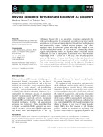
Fig. 1. Far UV circular dichroism spectra of the second Kunitz–type
protease inhibitor module of human WFIKKN. The solid line indicates
the spectrum of the recombinant protein, the dotted line indicates the
CDPro-predicted spectrum of a protein consisting of 0.051 regular
b-strand, 0.062 distorted b-strand, 0.110 regular a-helix, 0.183 distor-
ted a-helix, 0.284 turn and 0.309 unordered structure. Spectra were
recorded in 10 m
M
Tris/HCl, pH 8.0 at 25 °Cusing0.1mgÆmL
)1
of
protein.
Ó FEBS 2003 The second Kunitz-type domain of WFIKKN protein (Eur. J. Biochem. 270) 2103
Results and discussion
Structural characterization of the recombinant
Kunitz-module of human WFIKKN protein
The circular dichroism spectra of the second Kunitz-module
of WFIKKN protein (hereafter referred to as WFIKKN-
KU2) are very similar to those of other members of the
Kunitz-domain family [13,14] inasmuch as it is also
characterized by a deep trough at 203 nm and a shoulder
at 215 nm (cf. Figure 1). Analysis of the spectra with the
CDPRO
software predicted 5.1% regular b-strand, 6.2%
distorted b-strand 11.0% regular a-helix, 18.3% distorted
a-helix, 28.4% turn and 30.9% unordered structure.
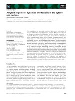
Fig. 2. Temperature dependence of the CD spectra of the second Kunitz–type protease inhibitor module of human WFIKKN protein. (A) Changes in
the CD of the protein were monitored at 203 nm in 10 m
M
Tris/HCl buffer, pH 8.0, during the course of heating from 40 °Cto90 °Cataheating
rate of 60 °CÆh
)1
. (B) Melting temperature was determined by derivative processing of changes in CD (cf. part A) using the
J
-700
STANDARD
ANALYSIS
program for
WINDOWS
, v1.30.00.7 (JASCO).
Fig. 3. Alignment of the sequences of the second Kunitz-modules of the human and fugu WFIKKN proteins (wfikkn_hu_2; wfikkn_fugu_2) with the
second Kunitz-domains of the human and the two fugu WFIKKNRP proteins (wfikknrp_hu_2; wfikknrp1_fugu_2; wfikknrp2_fugu_2), wfikkn_hu_2),
the Kunitz domain of WFIKKNRP of the amphioxus Branchiostoma belcheri (WFIKKN_BRABE), and the Kunitz domains of bovine pancreatic
trypsin inhibitor (bpt1_bovin), human bikunin (ambp_human_1, ambp_human_2), human Alzheimer’s disease amyloid a4 protein precursor (a4_human)
and human type 1 and type 2 hepatocyte growth factor activator inhibitors (spt1_human_1, spt1_human_2, spt2_human_1, spt2_human_2). In the
bottom line (+) signs mark the P5, P4, P3, P2, P1, P¢1, P¢2, P¢3, P¢4 positions, while residues of the secondary sites are indicated by dots. In the top
line, asterisks highlight the P1 and P¢2 sites. Residues conserved in at least 50% of the aligned sequences are shown by white letters on a black
background. Conserved residues are grouped as follows: F,Y,W; I,L,V,M; R,K; D,N; E,Q; T,S.
2104 A. Nagy et al. (Eur. J. Biochem. 270) Ó FEBS 2003
The presence of both b-strands and a-helices in
WFIKKN-KU2 is consistent with the fact that all homo-
logues of WFIKKN-KU2 are known to contain b-strands
and a-helices in equivalent positions [14–19]. In view of the
fact that the structure of the Kunitz inhibitor of the sea
anemone Stichodactyla helianthus is nearly identical with
that of the bovine pancreatic trypsin inhibitor despite a mere
35% of sequence similarity between the two proteins [16] we
can assume that the structure of WFIKKN-KU2 (41%
identical with the sequence of BPTI) also has a typical
Kunitz-fold.
The thermal unfolding of the recombinant WFIKKN-
KU2 protein has been characterized by monitoring changes
of CD spectra. As shown in Fig. 2, changes in the CD
spectra at 203 nm reflect a single, sharp transition with a T
m
value of 61 °C, indicating that the protein collapses in a
highly cooperative fashion. It should be noted that the
thermal stability of WFIKKN-KU2 is somewhat lower
than that of the closely related bovine pancreatic trypsin
inhibitor or the chymotrypsin inhibitor of Bungarus fasci-
atus which have been shown to retain most of their native
structure at 80 °C [14].
Functional characterization of the second
Kunitz-domain of the WFIKKN protein
In view of the fact that an arginine residue is present in the
P1 position of WFIKKN-KU2 (Fig. 3), it was not unex-
pected that the recombinant WFIKKN-KU2 protein did
not inhibit the proteolytic action of chymotrypsin or
elastase even when tested at 100 l
M
final concentration.
(This observation has permitted the use of elastase to
remove the b-galactosidase portion from the refolded fusion
protein; see Experimental procedures).
Next, we studied the effect of the WFIKKN-KU2
protein on trypsin and a panel of other serine proteases
with specificity for Arg-X or Lys-X peptide bonds. These
studies have shown that WFIKKN-KU2 is an efficient
inhibitor of trypsin, the dissociation constant for its complex
with trypsin (K
i
) was 9.6 n
M
(Fig. 4).
WFIKKN-KU2 was found to display a striking speci-
ficity for trypsin. When the inhibitor was employed at 1 l
M
final concentration, complete inhibition of trypsin was
achieved, but no detectable inhibition was observed in
the case of plasmin, lung tryptase, plasma kallikrein, throm-
bin, urokinase, tissue plasminogen activator, pancreatic
kallikrein, chymotrypsin or elastase. Such a marked trypsin-
specificity is somewhat unusual among Kunitz-domains.
For example, the Kunitz domains of BPTI, amyloid
precursor protein, amyloid precursor protein homolog
display broader specificity, inasmuch as at 1 l
M
concentra-
tion they inhibit chymotrypsin, glandular kallikrein, plas-
min as well as trypsin [5].
We suggest that the explanation for such a marked
trypsin specificity of WFIKKN-KU2 lies in the presence of
a Trp-residue at the P¢2 site of the inhibitor. In the case of
Kunitz-domains it is now well established that the primary
sites interacting with the target proteases (and determining
their protease-specificity) are found in a short segment
containing the second conserved cysteine, a secondary site
contacting the target proteases includes residues adjacent to
the fourth conserved cysteine ([18] cf. Fig. 3). Among all the
contact sites, the P1 and the P¢2 site play the most critical
roles in determining the target specificity of a Kunitz
inhibitor [18]. The P1 site interacts with the S1 binding
pocket (residues 189–195, 214–220 of target proteases), the
P¢2 site interacts with the S¢2 pocket (residues 151, 192–193
of the target proteases).
As shown in Fig. 3., the putative functional sites deter-
mining the target-specificity of the WFIKKN-KU2 domain
are quite similar to the corresponding segments of other
Kunitz-domains, with one major exception: a Trp residue is
found in the P¢2 position. A survey of the sequences of
Kunitz domains deposited in public databases has revealed
that the WFIKKN-KU2 domain and its pufferfish ortholog
are unique in that they are the only ones which have a bulky
Trp residue at this position.
A key determinant of the hydrophobic S¢2 binding
pocket of trypsins is the side-chain of Tyr151 [18]. The
importance of this residue is underlined by the fact that in
the case of the second Kunitz-domain of TFPI its complex
with trypsin is stabilized by favorable stacking interaction
of Tyr17 (the P¢2 residue of the inhibitor) with the Tyr151
side-chain of trypsin [17]. It seems probable that the
aromatic Trp residue at the P¢2 position of WFIKKN-
KU2 also makes favorable contacts with the Tyr151 of
Fig. 4. Lineweaver–Burk plots of the activity of trypsin (30 n
M
)recor-
ded at different concentrations of the second Kunitz-type protease
inhibitor domain of WFIKKN (0, 15, 30, 60 or 150 n
M
). Hydrolysis of
Bz-Phe-Val-Arg-pNA was monitored at 37 °Cin25m
M
Tris, 5 m
M
CaCl
2
, pH 7.5 buffer. The inhibition constant was calculated by
replotting the apparent K
m
values (inset).
Ó FEBS 2003 The second Kunitz-type domain of WFIKKN protein (Eur. J. Biochem. 270) 2105
trypsin. It is noteworthy in this respect that the majority of
the proteases tested in the present study have nonaromatic
residues in positions equivalent to Tyr151 of trypsin (Thr
in bovine chymotrypsin, Leu in bovine elastase, Ile in
human plasma kallikrein, Gly in human plasmin, Gln in
human thrombin, Pro in human lung tryptase), raising the
possibility that the inability of WFIKKN-KU2 to inhibit
these proteases is partly due to the lack of such a favorable
interaction of the P¢2 Trp with the target enzymes. The fact
that the Trp residue at the P¢2 position of WFIKKN-KU2
is conserved from pufferfish to human (cf. Fig. 3) is
consistent with the notion that this residue has a major
functional importance.
In view of the marked trypsin-specificity of WFIKKN-
KU2 it seems plausible to assume that its physiological
function is to inhibit trypsin. It should be pointed out,
however, that the affinity of WFIKKN-KU2 toward
pancreatic trypsin (K
i
¼ 9.6 · 10
)9
M
) is somewhat weaker
than that observed for many other Kunitz inhibitors for
their specific target proteases. For example, the Kunitz-
domains of placental bikunin (hepatocyte growth factor
activator inhibitor type 2) inhibit their target proteases
(plasmin, plasma kallikrein) with K
i
values in the
10
)9
)10
)10
M
range [20], the second Kunitz-domain of
tissue factor pathway inhibitor inhibits factor Xa with a K
i
value of 1.5 · 10
)10
M
[17].
The relatively high K
i
value of isolated WFIKKN-KU2
domain towards pancreatic trypsin raises the possibility that
its primary physiological target may be a trypsin-like
protease distinct from pancreatic trypsin. Nevertheless, it
is likely that the trypsin inhibitory activity of WFIKKN-
KU2 has physiological relevance. First, the affinity of the
second Kunitz-domain for trypsin may be higher in the case
of the intact WFIKKN protein than that of the isolated
WFIKKN-KU2 domain. Second, for an inhibitor to be
physiologically efficient only its local concentration has to
be higher than its K
i
value. Our observation that the
WFIKKN gene is expressed primarily in the pancreas [2,3]
suggests that the local concentration of the WFIKKN
protein in this organ may reach levels high enough to
control pancreatic trypsin activity.
The biological role of the WFIKKN protein is not limited
to the pancreas. We have shown previously that in addition
to pancreas, the protein is also expressed in liver, lung and
kidney [2,3]. The fact that human trypsins 1, 2 and 3 are also
expressed in liver, lung and kidney [21] is consistent with the
notion that the WFIKKN protein may also serve as a
trypsin inhibitor in these tissues.
Acknowledgements
This work was supported by grants NKFP (National Research &
Development Program of Hungary) 1/044/2001 and AKP (Research
Fund of the Hungarian Academy of Sciences) 2000-8 3.3. The authors
wish to thank Andra
´
s Patthy (Agricultural Biotechnology Center,
Go
¨
do
¨
ll
}
oo, Hungary) for sequence analyses of recombinant proteins.
References
1. Schechter, I. & Berger, A. (1967) On the size of the active site in
proteases. I. Papain. Biochem. Biophys. Res. Commun. 27, 157–
162.
2. Trexler, M., Ba
´
nyai, L. & Patthy, L. (2001) A human protein
containing multiple types of protease-inhibitory modules. Proc.
Natl Acad. Sci., USA 98, 3705–3709.
3. Trexler, M., Ba
´
nyai, L. & Patthy, L. (2002) Distinct
expression pattern of two related human proteins containing
multiple types of protease-inhibitory modules. Biol. Chem.
383, 223–228.
4. Lukachovich, T., Boros, I. & Venetianer, P. (1987) New regulatory
features of the promoters of an Escherichia coli rRNA gene.
J. Bacteriol. 169, 272–277.
5. Petersen, L.C., Bjorn, S.E., Norris, F., Norris, K., Sprecher, C.
& Foster, D.C. (1994) Expression, purification and
characterization of a Kunitz-type protease inhibitor domain
from human amyloid precursor protein homolog. FEBS Lett. 338,
53–57.
6. Tanaka, A.S., Andreotti, R., Gomes, A., Torquato, R.J.,
Sampaio, M.U. & Sampaio, C.A. (1999) A double headed serine
proteinase inhibitor – human plasma kallikrein and elastase
inhibitor – from Boophilus microplus larvae. Immunopharma-
cology. 45, 171–177.
7. Schagger, H. & von Jagow, G. (1987) Tricine-sodium dodecyl
sulfate-polyacrylamide gel electrophoresis for the separation of
proteins in the range from 1 to 100 kDa. Anal. Biochem. 166,
368–379.
8. Sreerama, N., Venyaminov, S.Y. & Woody, R.W. (1999) Esti-
mation of the number of a-helical and b-strand segments in pro-
teins using CD spectroscopy. Protein Sci. 8, 370–380.
9. Sreerama, N. & Woody, R.W. (2000) Estimation of protein sec-
ondary structure from circular dichroism spectra: comparison of
CONTIN, SELCON, CDSSTR methods with expanded reference
set. Anal Biochem. 287, 252–260.
10. Sreerama, N., Venyaminov, S.Y. & Woody, R.W. (2001) Analysis
of protein circular dichroism spectra based on the tertiary struc-
ture classification. Anal Biochem. 299, 271–274.
11. Chase, T. & Shaw, E. (1967) p-Nitrophenyl-p-guanidinobenzoate
HCl: a new active site titrant for trypsin. Biochem. Biophys. Res.
Commun. 29, 508–514.
12. Thompson, J.D., Higgins, D.G. & Gibson, T. (1994) CLUS-
TALW: Improving the sensitivity of progressive multiple sequence
alignment through sequence weighting, position-specific gap
penalties and weight matrix choice. Nucleic Acids Res. 12, 4673–
4680.
13. Fioretti, E., Iacopino, G., Angeletti, M., Barra, D., Bossa, F. &
Ascoli, F. (1985) Primary structure and antiproteolytic activity of
a Kunitz-type inhibitor from bovine spleen. J. Biol. Chem. 260,
11451–11455.
14. Chen, C., Hsu, C.H., Su, N.Y., Lin, Y.C., Chiou, S.H. & Wu, S.H.
(2001) Solution structure of a Kunitz-type chymotrypsin inhibitor
isolated from the elapid snake Bungarus fasciatus. J. Biol. Chem.
276, 45079–45087.
15. Wlodawer, A., Nachman, J., Gilliland, G.L., Gallagher, W. &
Woodward, C. (1987) Structure of form III crystals of bovine
pancreatic trypsin inhibitor. J. Mol. Biol. 198, 469–480.
16. Antuch, W., Berndt, K.D., Chavez, M.A., Delfin, J. & Wuthrich,
K. (1993) The NMR solution structure of a Kunitz-type protei-
nase inhibitor from the sea anemone Stichodactyla helianthus. Eur.
J. Biochem. 212, 675–684.
17. Burgering, M.J., Orbons, L.P., van der Doelen, A., Mulders, J.,
Theunissen,H.J.,Grootenhuis,P.D.,Bode,W.,Huber,R.&
Stubbs, M.T. (1997) The second Kunitz domain of human tissue
factor pathway inhibitor: cloning, structure determination and
interaction with factor Xa. J. Mol. Biol. 269, 395–407.
18. Scheidig, A.J., Hynes, T.R., Pelletier, L.A., Wells, J.A. &
Kossiakoff, A.A. (1997) Crystal structures of bovine chymo-
trypsin and trypsin complexed to the inhibitor domain of
Alzheimer’s amyloid beta-protein precursor (APPI) and basic
2106 A. Nagy et al. (Eur. J. Biochem. 270) Ó FEBS 2003
pancreatic trypsin inhibitor (BPTI): engineering of inhibitors with
altered specificities. Protein Sci. 6, 1806–1824.
19. Xu, Y., Carr, P.D., Guss, J.M. & Ollis, D.L. (1998) The crystal
structure of bikunin from the inter-alpha-inhibitor complex: a
serine protease inhibitor with two Kunitz domains. J. Mol. Biol.
276, 955–966.
20. Delaria, K.A., Muller, D.K., Marlor, C.W., Brown, J.E., Das,
R.C., Roczniak, S.O. & Tamburini, P.P. (1997) Characterization
of placental bikunin, a novel human serine protease inhibitor.
J. Biol. Chem. 272, 12209–12214.
21. Koshikawa, N., Hasegawa, S., Nagashima, Y., Mitsuhashi, K.,
Tsubota, Y., Miyata, S., Miyagi, Y., Yasumitsu, H. & Miyazaki,
K. (1998) Expression of trypsin by epithelial cells of various tis-
sues, leukocytes, and neurons in human and mouse. Am. J. Pathol.
153, 937–944.
Ó FEBS 2003 The second Kunitz-type domain of WFIKKN protein (Eur. J. Biochem. 270) 2107