Báo cáo khoa học: Genomic structure, expression and characterization of a STAT5 homologue from pufferfish (Tetraodon fluviatilis) ppt
Bạn đang xem bản rút gọn của tài liệu. Xem và tải ngay bản đầy đủ của tài liệu tại đây (604.07 KB, 14 trang )
Genomic structure, expression and characterization of a STAT5
homologue from pufferfish (
Tetraodon fluviatilis
)
Shu-Chiun Sung
1,2
, Ting-Jia Fan
1
, Chih-Ming Chou
3
, Jiann-Horng Leu
1
, Ya-Li Hsu
4
, Shui-Tsung Chen
1
,
Yueh-Chun Hsieh
1
and Chang-Jen Huang
1
1
Graduate Institute of Life Science, National Defense Medical Center, Taipei, Taiwan;
2
Institute of Biological Chemistry,
and
4
Zoology, Academia Sinica, Taipei, Taiwan;
3
Department of Biochemistry, Taipei Medical University, Taipei, Taiwan
The STAT5 (signal transducer and activator of transcription
5) gene was isolated and characterized from a round-spotted
pufferfish genomic library. This gene is composed of 19
exons spanning 11 kb. The full-length cDNA of Tetraodon
fluviatilis STAT5 (TfSTAT5) contains 2461 bp and encodes
a protein of 785 amino acid residues. From the amino acid
sequence comparison, TfSTAT5 is most similar to mouse
STAT5a and STAT5b with an overall identity of 76% and
78%, respectively, and has < 35% identity with other
mammalian STATs. The exon/intron junctions of the
TfSTAT5 gene were almost identical to those of mouse
STAT5a and STAT5b genes, indicating that these genes are
highly conserved at the levels of amino acid sequence and
genomic structure. To understand better the biochemical
properties of TfSTAT5, a chimeric STAT5 was generated by
fusion of the kinase-catalytic domain of carp Janus kinase 1
(JAK1) to the C-terminal end of TfSTAT5. The fusion
protein was expressed and tyrosine-phosphorylated by its
kinase domain. The fusion protein exhibits specific DNA-
binding and transactivation potential toward an artificial
fish promoter as well as authentic mammalian promoters
such as the b-casein promoter and cytokine inducible SH2
containing protein (CIS) promoter when expressed in both
fish and mammalian cells. However, TfSTAT5 could not
induce the transcription of b-casein promoter via rat pro-
lactin and Nb2 prolactin receptor. To our knowledge, this is
the first report describing detailed biochemical characteri-
zation of a STAT protein from fish.
Keywords: DNA binding; pufferfish; signal transduction;
STAT5; transactivation.
Signal transducers and activators of transcription (STATs)
are a family of latent cytoplasmic transcription factors that
are activated in response to various extracellular polypep-
tide ligands such as cytokines, growth factors and hormones
[1–3]. Extensive studies in mammalian systems have estab-
lished a paradigm for the activation mechanism of STAT
molecules. Mammalian STATs can be activated by either
Janus kinase (JAK)-dependent or JAK-independent path-
ways. In the JAK–STAT pathway, binding of cognate
ligand to the receptor triggers a series of sequential events
including the association of the receptor with JAKs, tyrosine
phosphorylation of the receptor by activated JAKs, and
generation of the docking site for SH2-containing STAT
proteins. The recruitment of STATs to the receptor leads to
their tyrosine phosphorylation by the JAK kinases (JAK-
dependent). STATs can also be activated by receptor
tyrosine kinases, such as insulin receptor [4], epidermal
growth factor receptor [5], and mutant fibroblast growth
factor receptor [6], as well as by v-Src [7] and v-Abl [8]. The
phosphorylated STATs form homo- or hetero-dimers and
then translocate to the nucleus where they bind to specific
DNA sequences and transactivate the downstream target
gene.
The activation of STAT is an evolutionally conserved
mechanism by which signals can be transduced from
membrane to the nucleus rapidly. In mammals, there are
seven distinct STAT proteins [9,10], while only one related
molecule has been found in Drosophila [11,12] and in the
malaria vector Anopheles gambiae [13]. Among mammalian
STATs, STAT1 is critical for interferon function as well as
innate immunity [14,15], while STAT3 is required for
interleukin (IL)-6 signalling in haematopoietic cells as well
as antiapoptosis [16–18]. In addition, STAT5a and STAT5b
have been shown to play important roles in growth,
lactation and haematopoiesis [19–21]. In contrast with the
broad range of biological effects derived from STAT1,
STAT3, and STAT5, STATs 2, 4, and 6 have relatively
restricted functions, centred on immune response regula-
tion. STAT2 is activated only by a/b-interferon, STAT4 by
IL-12andSTAT6byIL-4andIL-13[10,22,23].
STAT5a was originally identified as a mammary gland
factor that can be activated by prolactin treatment and bind
Correspondence to C J. Huang, Institute of Biological Chemistry,
Academia Sinica, 128, Sec 2, Academia Road., Taipei, Taiwan 115.
Fax: + 886 2 2788 9759, Tel.: + 886 2 2785 5696,
E-mail:
Abbreviations: STAT, signal transducer and activator of transcription;
JAK, Janus kinase; CIS, cytokine-inducible SH2-containing
protein; HA, haemagglutinin; EMSA, electrophoretic mobility
shift assay; IL, interleukin; CAT, chloramphenicol acetyltransferase;
Luc, luciferase; PRL, prolactin; ST, STAT.
Note: the nucleotide sequences reported in this paper have been
submitted to GenBank and assigned accession numbers AF307108
and AF394166.
(Received 7 April 2002, revised 20 September 2002,
accepted 20 November 2002)
Eur. J. Biochem. 270, 239–252 (2003) Ó FEBS 2003 doi:10.1046/j.1432-1033.2003.03380.x
to the promoter of the b-casein gene to regulate milk
production [24]. STAT5b is closely related to STAT5a: the
proteins not only have 95% amino acid identity but are
also functionally redundant in some ways [25]. Moreover,
STAT5 is activated by a variety of cytokines such as IL-2,
IL-3, IL-5, IL-7, IL-15, granulocyte/macrophage colony-
stimulating factor, erythropoietin, growth hormone, and
thrombopoietin [26]. Many cytokines activating STAT5
transcriptional activity play important roles in the estab-
lishment of myeloid and lymphoid lineages. To ascertain the
functional roles of the STAT5 proteins, knockout mice with
deletion of STAT5a and/or STAT5b [27–30] were gener-
ated. The most evident phenotypes of gene knockout mice
are with prolactin and growth hormone function and T-cell
proliferation. In contrast, haematopoietic lineages were only
slightly affected.
Studies of the JAK–STAT pathway have been mostly
in mammals with only few being performed in lower
vertebrates. Recently, two STAT homologues, STAT1
andSTAT3,wereisolatedfromzebrafish(Danio rerio)
[31]. We have previously identified four distinct genes
encoding JAK1, JAK2, JAK3 and Tyk2 from the
pufferfish (Tetraodon fluviatilis) [32,33]. In this study, we
report the isolation and characterization of the STAT5
gene from T. fluviatilis (TfSTAT5), the third member of
the STAT family from fish, to establish the evolutionary
relationship of STAT5 between mammals and teleosts.
T. fluviatilis posseses an extremely compact genome and
can be maintained in aquaria. Compared with Fugu [34],
T. fluviatilis is easily obtained making it an alternative
puffer fish model for comparative genome study [35]. A
complete JAK–STAT pathway has not been established
in fish. STAT5 is the logical choice of a STAT gene to
complete the JAK–STAT pathway, because prolactin and
the prolactin receptor have both been cloned in fish
[36,37]. Therefore, in this work, we also constructed a
chimeric T. fluviatilis STAT5 to characterize DNA-bind-
ing specificity and transactivation property of TfSTAT5.
Moreover, STAT5 appears to be a good target gene,
because in mammals it has been shown to regulate diverse
aspects of growth and health [26]. As T. fluviatilis is a
commercial aquarium species, any information about its
growth or health could be valuable.
Materials and methods
Isolation of the
STAT5
gene from a
T. fluviatilis
genomic library
Genomic DNA was prepared from the liver of the round-
spotted pufferfish (T. fluviatilis) with DNAzol reagent (Life
Technologies). Using lambda FIXII as a cloning vector
(Stratagene), the genomic library was constructed and
amplified as reported previously [32]. Two degenerate prim-
ers, ST5F (5¢-ACNTT(T/C)TGGCA(A/G)TGGTT (T/C)
GA-3¢) and ST5R (5¢-AANCGNA(G/A)NA(G/A) (A/G)
AANGTNCC-3¢) were generated based on the sequence of
two conserved regions, TFWQWFD and GTFLLRF in
most mammalian STAT proteins. With these primers and
pufferfish genomic DNA as template, PCR was performed
to isolate all possible STAT5 homologues from pufferfish
genomic DNA. One DNA fragment of 322 bp containing
partial genomic sequences of the pufferfish STAT5 (data
not shown) was used as a probe to screen the genomic
library mentioned above. The probe was labelled using a
digeoxygenin DNA Labelling Kit (Boehringer Mannheim).
Hybridization, washing, and the following chemilumines-
cence detection were performed according to manufac-
turer’s manual. From 1 · 10
7
plaques, three positive phage
clones were isolated and the genomic DNA fragments were
subcloned.
To investigate whether there is another STAT5 gene in
the T. fluviattilis genome, degenerate primers were designed
from two stretches of cDNA sequences,
677
RPKDEVF and
698
YVKPQIKQ (see Fig. 2), in which two different genomic
fragments were obtained for STAT5a and STAT5b genes,
respectively, in mouse (Mus musculus) genome. PCR
reaction was performed at low stringency (annealing at
42 °C) with T. fluviattilis genomic DNA as template. The
PCR products were cloned into pGEM-T easy vector and
sequenced.
Subcloning, DNA sequence analysis and phylogenetic
analysis
Parental clones and restriction fragment subclones were
sequenced using PRISM Ready Reaction Dye Deoxy
Termination Cycle sequencing Kit (Applied Biosystems)
on an Applied Biosystems 310 automated DNA
sequencer. Sequence assembly and alignment were per-
formed using the Genetics Computer Group software
program. The exon/intron boundaries were determined
by alignment of the encoded protein sequences with
those of mammalian STAT2 [38], STAT3 [39], and
STAT5 [40] genes. Phylogenetic analysis, based on the
sequences of the DNA-binding domain, linker region and
SH2 domain, were performed using
CLUSTRAL X
program
[41,42].
RACE
The 5¢ and the 3¢ end of TfSTAT5 mRNA were obtained
by the RACE technique using the Marathon cDNA
amplification kit (Clontech) according to the supplier’s
instructions.
The 5¢ RACE was performed with a 27-mer sense primer
(AP1) specific for the adaptor and a STAT5-specific primer
ST5R11 (5¢-CTGGTATTTCTGGGACATGGTCTCC-3¢)
whereas the second-round PCR was carried out with a
nested 23-mer sense primer (AP2) and a nested gene-specific
antisense primer ST5R12 (5¢-GACGAGCCTCTGCTC
GGTGTAAAGG-3¢). Similarly, 3¢ RACE was performed
with two rounds of PCR, first with AP1 primer and ST5F5
(5¢-AGGCTCAGGACATGCTGATGTCC-3¢)andthen
with AP2 primer and ST5F6 (5¢-TTCAGTGACTCTG
AGATCGGAGG-3¢). According to the sequences of the
clones obtained from 5¢ and 3¢ RACE, primers ST5F17
(5¢-ACTCGAGGTGTTGAAGATGGCAGTGTGG-3¢)
containing a XhoI site and ST5R17 (5¢-CCTCTAGAGGT
TAAAGGTCAGGACTGCTGG-3¢) containing a XbaI
site were synthesized and used to amplify full-length
TfSTAT5 cDNA using first-strand cDNA prepared from
pufferfish gill tissues as template. The PCR products were
cloned into pGEM-T vector (Promega) and sequenced.
240 S C. Sung et al. (Eur. J. Biochem. 270) Ó FEBS 2003
RNA isolation and RT/PCR
Total RNA was isolated from gill, heart, intestine, liver,
kidney, and testis of T. fluviatilis using the RNAzol reagent
(Tel.Test, Inc.) following the manufacturer’s instructions.
First-strand cDNA from different tissue was amplified from
2–5 lg total RNA using 10 pmole oliogT primer in a 25 lL
reaction containing 30 U RNasin (Promega), 1 m
M
dNTP,
10 m
M
dithiothreitol, and 300 U Superscript II (Life
Technologies). Incubation was performed at 42 °Cfor
1h,and2lL of the resulting reaction containing the single-
stranded cDNA template were used for subsequent PCR
amplification. PCR was performed in a 50-lLreaction
mixture containing 200 ng b-actin primers (ActF, 5¢-CCT
CCGGTCGTACCACTGGTAT-3¢ and ActR, 5¢-CAAC
GGAAGGTCTCATTGCCGATCGTG-3¢)orTfSTAT5
primers (ST5F34, 5¢-GACAGTGGATGGCTATGTGAA
ACCA-3¢ and STR15, 5¢-CCGCGCAAATCTAACTACG
ACAGTCC-3¢, corresponding to sequences in exon 17 and
the 3¢ untranslated region respectively), 1.5 m
M
MgCl
2
,
0.2 m
M
dNTP and 0.5 U ExTaq (Takara Shuzo Co.). The
conditions for amplification were 96 °C for 2 min; 35 cycles
of 96 °Cfor1min,50°Cfor30s,and72°C for 30 s; and
final extension at 72 °C for 5 min. A negative control was
performed in the absence of RNA. The products were
resolved on a 1.5% agarose gel.
Plasmid construction
The expression vector, pTf-STAT5-HA, was constructed
by inserting the full-length TfSTAT5 cDNA between the
XhoIandXbaI sites of pHA-YUN, which is derived from
pcDNA3 and has a haemagglutinin (HA) tag located
between the KpnI site and multiple cloning sites. The
pHA-YUN plasmid was kindly provided by H. J. Kung
(University of California at Davis Cancer Center, Sacra-
mento, CA, USA). The JH1 domain of carp JAK1 kinase
[43] was generated by PCR with primers containing
BamH1 and XhoI restriction sites on both ends, and
cloned into the corresponding sites in pHA-YUN to
generate pHA-JH1. The full-length TfSTAT5 cDNA was
reamplified by PCR with primers containing SmaIand
Kpn I restriction sites on both ends, and cloned into the
HindIII filled-in and KpnI sites in pHA-JH1 to generate
pTf-STAT5-HA-JH1. The MmSTAT5 cDNA cloned into
expression vector pECE was a kind gift from H. J. Kung
[44]. The cDNA of MmSTAT5 was recloned into
pcDNA3 with XbaIandEcoRI. The rat Nb2 PRL-R
cDNA,kindlyprovidedbyL.Y.Yu-Lee[45],was
released with EcoRI from pECE vector and inserted into
pcDNA3 expression vector.
The reporter plasmids pb-casein-CAT containing rat
b-casein gene promoter ()344 to )1) linked to the
chloramphenicol acetyltransferase (CAT) gene and pCIS-
luc containing mouse cytokine inducible SH2 containing
protein (CIS) gene promoter ()646 to )1) linked to the fire
fly luciferase (Luc) reporter gene were kindly provided by
W. Doppler (Institut fur Medizinische Chemie und Bioche-
mie, Innsbruck, Austria) and A. Yoshimura (Institute of
Life Science, Kurume University, Kurume, Japan), respect-
ively. The other reporter construct p(ST5)
2
-TK-CAT was
generated by cloning two copies of the ST5-binding motif
into the HindIII site of pTK-CAT, which carries a minimal
thymidine kinase (TK)2 gene promoter ()364 to +122)
isolated from round-spotted pufferfish [33]. The resulting
clone was confirmed by DNA sequencing.
In vitro
transcription and translation
The TNT coupled transcription/translation system pur-
chased from Promega was used to analyse the in vitro
synthesized TfSTAT5-HA and TfSTAT5-HA-JH1 accord-
ing to the manufacturer’s instructions. Briefly, 0.2–2 lg
plasmid DNA is added to the master mixture and incubated
for 90 min at 30 °C. The synthesized proteins are then
analysed by SDS/PAGE and visualized by autoradio-
graphy. For subsequent bandshift analysis and Western
blotting, [
35
S]methionine was replaced by cold methionine
(final concentration 1 m
M
).
Cell cultures
Monkey kidney fibroblast COS-1 cells were grown in
Dulbecco’s modified Eagle’s medium supplemented with
10% fetal bovine serum, penicillin G (50 UÆmL
)1
), strepto-
mycin (50 lgÆmL
)1
), and
L
-glutamine (2 m
M
) in a humidified
atmosphere of 5% CO
2
at 37 °C. Carp fin epitheloid CF cells
[46] were grown in Leibovitz’s L-15 medium supplemented
with 10% foetal bovine serum, penicillin G (50 UÆmL
)1
),
streptomycin (50 lgÆmL
)1
), and independent of CO
2
at 28 °C.
Preparation of whole cell lysates and nuclear extracts
All of the plasmids used for transfections were purified by
Qiagen plasmid purification kit (Qiagen Inc., Hilden,
Germany). Approximately 50% confluent cells were tran-
siently transfected for 5 h at 37 °C for COS-1 cells and
28 °C for CF cells, respectively, with 4 lg pTf-STAT5-HA,
pTf-STAT5-HA-JH1, or pHA-YUN DNA per 100 mm
dish using LIPOFECTAMIN/PLUS kit (Life Technol-
ogies) according to the manufacturer’s instructions. Whole
cell lysates were prepared at 48 h after transfection. In
general, cells were washed twice with NaCl/P
i
and lysed in
the lysis buffer (50 m
M
Tris/HCl pH 7.5, 150 m
M
NaCl,
1% Triton X-100, 1 m
M
EDTA) containing 0.2 m
M
Na
2
VO
4
,and2m
M
phenylmethylsulfonyl fluoride [47].
Extracts were centrifuged at 4 °Cfor10minat13000g
and the resulting supernatants were used for subsequent
immunoprecipitations. Nuclear extracts were prepared
according to the procedures described previously [48].
Briefly, cells were washed twice with NaCl/P
i
and solubilized
with buffer A (20 m
M
Hepes, 1 m
M
KCl, 1 m
M
EDTA,
1m
M
dithiothreitol, 1 m
M
phenylmethanesulfonyl fluoride,
0.1 m
M
Na
2
VO
4
, 0.2% NP-40, 10% glycerol, 1 lgÆmL
)1
for
aprotinin, pepstatin, and leupeptin, pH 7.9) on ice for
30 min. Cell lysates were centrifuged at 4 °Cfor2minat
13 000 g andthepelletswereextractedwithbufferB
(20 m
M
Hepes, 350 m
M
NaCl, 10 m
M
KCl, 1 m
M
EDTA,
1m
M
DTT, 1 m
M
phenylmethanesulfonyl fluoride, 0.1 m
M
Na
2
VO
4
, 0.2% NP-40, 10% glycerol, 1 lgÆmL
)1
for apro-
tinin, pepstatin, and leupepetin, pH 7.9). The extracts were
centrifuged at 4 °C for 5 min at 13 000 g, and supernatants
were quickly frozen and stored at )70 °C for subsequent use
of electrophoretic mobility shift assay (EMSA).
Ó FEBS 2003 Expression and characterization of pufferfish STAT5 (Eur. J. Biochem. 270) 241
Antibodies, immunoprecipitation and Western blotting
Monoclonal antibodies against HA tag and phospho-
tyrosine (PY-99) were from Santa Cruz Biotechnology.
Polyclonal antisera specifically recognizing TfSTAT5 was
generated from rabbit using a His-tagged TfSTAT5 fusion
protein as antigen. The TfSTAT5 fusion protein was
generated by amplification of the DNA fragment encoding
the C-terminal end (amino acid residues 546–728) of
TfSTAT5 by PCR followed by cloning of the PCR
product into the His-Tag expression vector pQE30
(Qiagen). The His-tagged TfSTAT5 protein was expressed
in E. coli and purified using Ni–NTA agarose column
(Qiagen) under denaturing condition (1.5% sarcosine in
NaCl/P
i
) as described previously [43]. The purified protein
was used to immunize New Zealand White rabbits by the
intrasplenic immunization method [49]. The polyclonal
antibodies were harvested according to the previously
described method. For immunoprecipitation, anti-
TfSTAT5 antibody (2 lg) was added to whole cell lysates
and incubated overnight at 4 °C. The immune complexes
were precipitated by a further incubation with protein A/
G-Sepharose (Santa Cruz Biotechnology), the immuno-
precipitates were then washed twice with lysis buffer and
eluted by boiling for 5 min in sample buffer. The
immunoprecipitates or in vitro transcription/translation
products were separated by SDS/PAGE (10% polyacryl-
amide), then transferred to a nitrocellulose membrane and
blocked with 4% skim milk in NaCl/P
i
. The membane
was then incubated with monoclonal antibodies PY-99 or
anti-HA monoclonal antibodies (Santa Cruz Biotechno-
logy), and visualized by the enhanced chemiluminescent
detection system (NEN Life Science Products) according
to the manufacturer’s instructions.
EMSA
EMSA was as described previously [50]. Briefly, reactions
were performed by the addition of nuclear extracts or
products of in vitro transcription and translation in the
presence of
32
P-labelled double-stranded oligonucleotides
probes (10 000 c.p.m.Ælg
)1
) to the binding buffer [10 m
M
Tris/HCl pH 7.5, 50 m
M
NaCl, 1 m
M
EDTA, 1 m
M
dithiothreitol, 5% glycerol, 2 lg poly (dI/dC)]. Excess of
unlabelled or mutant oligonucleotides were added as
competitors as indicated. After incubation at room tem-
perature for 30 min, the reaction mixture was loaded on a
5% polyacrylamide gel in 0.5 · Tris/borate/EDTA and run
at 200 V for 2 h at room temperature. The oligonuclotides
probes used in this study were commercially available,
corresponding to the DNA-binding sites for mammalian
STATs (Santa Cruz). The sequences of the upper strand of
the normal and the corresponding mutant oligonucleotides
are listed in Table 3. The probes were prepared by annealing
the upper and lower strands of oligonucleotides, one of
which was end-labelled with [c-
32
P]ATP by using T4
polynucleotide kinase (Boehringer Mannheim).
Transactivation assay
For transactivation assay, transfections were performed in
six-well plates. One lg reporter plasmids pb-casein-CAT,
pCIS-luc or p(ST5)
2
-TK-CAT, was cotransfected with
100 ng of expression plasmid pTf-STAT5-HA or pTf-
STAT5-HA-JH1 using LIPOFECTAMIN/PLUS kit (Life
Technologies). The pSV-b-galactosidase vector (Promega)
carrying the SV40 promoter linked to the b-galactosidase
gene and a Renilla luciferase construct containing the TK
promoter linked to the Renilla luciferase were used to
normalize transfection efficiency in CAT assay and luci-
ferase assay, respectively. For CAT assay, cells were
harvested at 48 h after transfection, CAT and b-galactosi-
dase activities from the cell extracts were measured accord-
ing to the published procedures [51]. The acetylated
products of the CAT assay were separated by TLC,
developed with chloroform/methanol (95 : 5, v/v) and
visualized by autoradiography. The data was quantitated
by a PhosphoImager (Bio-Imaging Analyser BAS 2000,
Fuji, Japan). Similarly, cells were harvested and assayed for
luciferase activity using a dual luciferase assay kit FireLite
(Packard; Groninge, BK, Netherland) according to the
manufacturer’s instructions. Final luciferase activity was
obtained after normalization with Renilla luciferase activity.
To investigate the transactivation potential of TfSTAT5
on b-casein-CAT construct via Nb2 PRL-R, 500 ng Nb2
PRL-R expression construct, 500 ng TfSTAT5 or
MmSTAT5 expression construct, and 500 ng b-casein-
CAT reporter were cotransfected into COS-1 cells. At
24 h after transfection, cells were stimulated or not
1 lgÆmL
)1
rat prolactin for a further 24 h. Cell extracts
were prepared by using a reported lysis buffer (Promega)
and CAT activity was analysed as described above.
Results
Isolation and genomic organization of the pufferfish
STAT5
gene
The STAT genes from pufferfish were isolated by PCR
amplification using degenerate primers containing sequences
corresponding to two stretches of amino acid residues
TFWQWFD and GTFLLRF that are conserved among
most STAT proteins [52]. Using T. fluviatilis genomic DNA
as template, three PCR products were obtained (data not
shown). One of these products, 322 bp in length, encodes a
portion of the protein that is highly homologous to
mammalian STAT5a and STAT5b. This DNA fragment
was used as probe to screen a genomic phage library of the
round-spotted puffer fish [32,33]. Three positive phage
clones, S1, S2 and S3, with similar restriction map, were
isolated. The S1 clone was chosen for further characteriza-
tion and sequence determination. Fig. 1 summarizes the
restriction map of the S1 clone.
A total of 11 kb of the T. fluviatilis STAT5 (TfSTAT5)
gene was completely sequenced by conventional subcloning
strategy in combination with automated sequencing. The
complete sequence was deposited in GenBank with an
accession number AF307108. To fully characterize the
STAT5 transcript, 5¢ and 3¢ RACE were performed to
obtain untranslated regions of STAT5 message (data not
shown). TfSTAT5 is composed of 19 exons and 18 introns.
All exon/intron boundaries identified conformed to the GT/
AG splice donor/acceptor rule [53]. The sizes of the introns
varied considerably, ranging from 79 bp (intron 6) to
242 S C. Sung et al. (Eur. J. Biochem. 270) Ó FEBS 2003
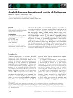
1381 bp (intron 1) with an average size of 427 bp (Table 1).
The first exon contains the 5¢-untranslated region and the
second exon contains the putative translation initiation site
(Fig. 1).
To investigate further whether there are two related
STAT5 genes in the pufferfish genome, we used a pair of
degenerate primers, which were derived from two highly
conserved regions (677RPKDEVF and 698YVKPQIKQ),
to amplify genomic fragments in which the intron sizes are
different in mammalian STAT5a and STAT5b. However,
we only obtained one PCR product whose sequences were
identical to those of TfSTAT5 as mentioned above (data not
shown).
Sequence comparison and phylogenic analysis
In addition, to resolve the genomic structure of TfSTAT5,
the full-length cDNA was synthesized by PCR amplifica-
tion using first-strand cDNA prepared from T. fluviatilis
gill and specific primers designed from the results of 5¢-
and 3¢-RACE. After amino acid sequence comparison,
TfSTAT5 showed 76% and 78% identity to mouse
STAT5a and STAT5b (MmSTAT5a and 5b) and less
than 34% to other vertebrate STATs (Table 2). In
addition, the 10 C-terminal amino acid sequence of
TfSTAT5 is more similar to that of MmSTAT5b than
to that of MmSTAT5a. The comparison also reveals that
all the major functional domains identified in mammalian
STAT proteins are also found in TfSTAT5, including an
N-terminal protein interaction domain, coiled-coil domain,
DNA-binding domain, SH2 domain, and C-terminal
transactivation domain. Furthermore, a tyrosine (Y698)
was also identified in the C-terminal transactivation
domain that is to be phosphorylated by JAK kinases
during activation. As shown in Fig. 2, the N-terminal
protein interaction domain, the coiled-coil domain, and
C-terminal transactivation domain are encoded by exons
2–5, 5–8, and 17–19, respectively, while the SH2 domain is
encoded by exons 15–16 and the DNA-binding domain by
exons 9–12. These results suggest that TfSTAT5 and
MmSTAT5 proteins displayed conserved features consis-
tent with their conservation in genomic structure.
Table 1. Exon-intron organization of theTf STAT5gene.
Exon
no.
Exon size
(bp)
3¢ end of
the exon
5¢ end of
the intron
Intron size
(bp)
3¢ end of
the intron
5¢ end of
the next exon
Amino acid
interrupted
1 >24 GAA GGC AAG AG tatgtgtggc 1381 tcccaactag GGT GTT GAA
2 138 GGG CAG CTG TG gtgagtcgcc 270 acgtatgtag G GAT GCA ATT Trp 43 (2)
3 157 AGT CAG CTT AAG gtgagtcttg 1042 tctctttaag AGC ACG TAT Lys 95 (3)
4 90 GAG GCC ACC AAT gtgagtagga 614 tatttaacag TCT AGT TCT Asn125 (3)
5 175 CGT ATC CAG G gtgagtctgt 226 ccccacacag CT CAG CTG TCC Ala184 (1)
6 131 AAA TAC CGA CTG gtaaacccaa 79 atgataccag GAC CTG GCA Leu227 (3)
7 152 CTG CAG TCA TG gtgagttgtc 231 tgcataacag G TGT GAG AAG Trp278 (2)
8 156 CTG GTT ACC AG gtatctgcct 932 ttttctacag C ACC TTT ATT Ser331 (2)
9 80 AAC ACA AGG AA gtaagttcaa 535 tctgccacag T GAA AGC AGT Ans391 (2)
10 88 TTC AGG AAC ATG gtgagtgcct 306 atccttgtag TCC TTG AAG Met420 (3)
11 123 TTT CAA GTG AAG gtaagagagc 172 ctctgcacag ACG TTA TCA Lys461 (3)
12 93 TTT GCA GAG CCG gtgagcacgt 158 ctgtccacag GGT CGG GTG Pro492 (3)
13 207 CAG TTT AAC AGG gtcagcacca 568 ctgtttacag GAG AGT CTT Arg561 (3)
14 95 TGG AAC GAC GG gtaagggaac 172 ttttttttag A GCC ATA CTG Gly593 (2)
15 143 AAC AAA GCA G gtatattcag 434 atgtttccag GT GAG AGA ATG Gly640 (1)
16 156 CCG CCC CTT T gtaagcaacc 139 tcacctaaag CC AAA GCA GTG Ser693 (1)
17 52 GTC GTG CCA GA gtaagtgaca 368 ccgtgcacag G TTT ACT ACA Glu710 (2)
18 108 TAC CCG CCT AT gtaagccact 654 tcctgtccag G AGC GAC TCC Met746 (2)
19 354 ATCCTGGACGCAGACGGAGACTTCG
ACCTGGA CGACACCATGGACGTGGC
CAGG (the end of the pufferfish STAT5 gene)
Fig. 1. Genomic organization of the T. fluviatilis STAT5 gene. Exons are indicated by boxes numbered from 1 to 19. The coding regions are shown
as filled boxes whereas the 5¢-and3¢-untranslated regions are shown as open boxes. Introns and the 5¢-and3¢-flanking regions are indicated by solid
lines. The restriction map was shown above the genomic structure. Restriction endonuclease sites are B, BamHI;E,EcoRI;H,Hind III; K, Kpn I;
S, Sal I; Xb, Xba I; Xh, Xho I.
Ó FEBS 2003 Expression and characterization of pufferfish STAT5 (Eur. J. Biochem. 270) 243
To investigate the evolutionary relationship between
Tf STAT5 and other STAT family members, the amino
acid sequences of highly conserved regions including the
DNA-binding domain, linker domain, and SH2 domain
of STAT proteins were used to perform phylogenic
analyses. As illustrated in Fig. 3, Tf STAT5, MmSTAT5a,
MmSTAT5b, MmSTAT6 and Drosophila STAT
(DmSTAT) are closely related to each other, constituting
a proposed ancient class of the STAT family. In contrast,
mouse STAT1, STAT2 analyses suggest that two mam-
malian STAT5 genes diverged from a STAT5 locus in
teleosts. It has been proposed that in mammals two
closely related STAT5 genes resulted from a recently gene
duplication [54].
Tf
STAT5
mRNA expression
The expression pattern of TfSTAT5 mRNA in tissues such
as brain, gill, intestine, liver, kidney and testis from adult
T. fluviatilis was analysed by RT/PCR. As a negative
control, a PCR reaction without RNA was performed. As
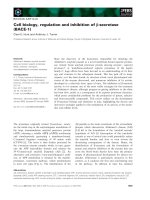
shown in Fig. 4, a 321-bp DNA fragment could be
amplified from all tissues examined. Compared with the
expression level of b-actin mRNA, TfSTAT5 was expressed
at similar levels in all tissues examined. These results
suggested that STAT5 was expressed ubiquitously in
T. fluviatilis, consistent with the expression pattern of its
mouse homologoue, MmSTAT5a and MmSTAT5b [25].
In vitro
translated
Tf
STAT5-HA-JH1 fusion protein
can be tyrosine-phosphorylated and bind
to mammalian STAT5 responsive elements
The fact that Tf STAT5 protein displayed high similarity
in amino acid sequences to Mm STAT5 prompted us to
compare the biochemical properties of Tf STAT5 and
Mm STAT5. We adopted the method used by Berchtold
et al. [55] to generate constitutively active Tf STAT5
variant and to characterize its biochemical properties in
acellfreesystem.Thestrategyweusedwastogeneratea
fusion protein Tf STAT5-HA-JH1 containing Tf STAT5
and the JH1 domain of the carp JAK1 [43]. The full-
length Tf STAT5 cDNA was inserted into a eukaryotic
expression vector pcDNA3 with HA-tag (Invitrogen) to
generate pTf-STAT5-HA, followed by fusing the JH1
domain (286 amino acids, position 869–1154) of carp
JAK1 to the C terminus of Tf STAT5-HA to generate
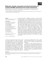
Tf STAT5-HA-JH1. In vitro transcription and translation
products of Tf STAT5-HA and Tf STAT5-HA-JH1 can
be recognized by mAb against HA (Fig. 5A, lanes 1 and
2). After incubation with mAb PY-99 against phospho-
tyrosine, only Tf STAT5-HA-JH1 was reactive to anti-
body PY-99 (Fig. 5A, lane 4), demonstrating that the JH1
domain of carp JAK1 could elicit tyrosine phosphoryla-
tion activity to autophosphorylate or transphosphorylate
Tf STAT5-HA-JH1 in vitro.
To further investigate DNA-binding characteristics of
Tf STAT5 in vitro, several commercially available mamma-
lian DNA-binding motifs for STATs (Santa Cruz, Table 3)
were used to perform EMSAs. From these results,
Tf STAT5-HA-JH1 displayed specific and strong binding
to the ST5 probe (Fig. 5B, lanes 2, 3, and 4) and Int16 probe
(Fig. 5B, lanes 6, 7, and 8) which has been found in intron 16
of the T. fluviatilis JAK1 gene [32], but did not bind to other
probes ST1, ST3, ST4, ST6, the c-interferon-activated site,
and the sis-inducible element (data not shown). Unlabelled
or mutant oligonucleotides in a 50-fold excess over radio-
active probe were included in the binding reaction to
distinguish the binding specificity. The core sequence of
ST5 is TTCTAGGAA whereas the core sequence of Int16 is
TTCTTGGAA. These sequences resemble the consensus
sequence described for human STAT5 (TTCTNA/GGAA)
[56].
Table 2. Pairwise amino acid sequence comparisons of Tf STAT5 and other known STATs. All protein sequences were aligned pairwise using the
Geneworks nucleic acid and protein sequence analysis software 2.5 from Intelligenetics, Inc. The numbers represent percent amino acid identity.
Accession numbers for all sequences are: mouse STAT1 (P42225), STAT2 (AAD38329), STAT3 (P42227), STAT4 (P42228), STAT5a (P42230),
STAT5b (P42232), and STAT6 (P52663).
Mm STAT5b Mm STAT5a Mm STAT6 Mm STAT1 Mm STAT3 MmSTAT4 Mm STAT2
Tf STAT5 78 76 31 23 25 25 18
Mm STAT5b 91 30 24 25 26 18
Mm STAT5a 30 24 25 25 18
Mm STAT6 19 19 20 20
Mm STAT1 50 50 31
Mm STAT3 45 29
Mm STAT4 29
Fig. 2. Comparison of the exon/intron organization for TfSTAT5,
MmSTAT5a,andMmSTAT5b genes. The encoded amino acid
sequences, represented by single-letter codes, were aligned using the
PILEUP
program (Genetic Computer Group). Gaps are introduced to
optimize alignment and shown as dashes. Spliced sites are indicated by
down-pointing arrowheads. Consensus amino acids (Con) indicate
identity in all three sequences. It is obvious that the spliced sites are
almost identical. The boundaries of N-terminal protein interaction
domains, coiled-coil domains, DNA-binding domains, linker regions
and SH2 domains are indicated by arrowed brackets. The tyrosine
residue phosphorylated upon activation is indicated by black star. Two
pairs of degenerate primers used to probe STAT homologue and
explore the second STAT5 gene from T. fluviatilis genomic DNA are
indicated by black arrows. Accession numbers for the three sequences
are TfSTAT5 (AF307108), MmSTAT5a (AF049104) and
MmSTAT5b (AC021632).
244 S C. Sung et al. (Eur. J. Biochem. 270) Ó FEBS 2003
Ó FEBS 2003 Expression and characterization of pufferfish STAT5 (Eur. J. Biochem. 270) 245
Expression and DNA-binding ability of
Tf
STAT5-HA-JH1
fusion protein in COS-1 cell
The expression plasmid pTf-STAT5-HA-JH1 or pTf-
STAT5-HA was transiently transfected into COS-1 cells
to investigate the expression of Tf STAT5-HA-JH1 fusion
protein in mammalian cells. Whole cell lysates were
prepared and immunoprecipitated with polyclonal antibod-
ies against Tf STAT5, followed by Western blotting with
antibodies against HA or PY-99. As shown in Fig. 6A, the
fusion proteins of TfSTAT5-HA and TfSTAT5-HA-JH1
were expressed in COS-1 cells (Fig. 6A, lanes 2 and 3), but
only TfSTAT5-HA-JH1 could be tyrosine-phosphorylated
(Fig. 6A, lane 6). This result indicated that TfSTAT5-HA-
JH1 could be constituively active and underwent tyrosine-
phosphorylation in COS-1 cells.
An EMSA was performed with nuclear extract prepared
from COS-1 cells transiently transfected with the expression
plasmid pTf-STAT5-HA-JH1 to examine the DNA-binding
specificity of the fusion protein TfSTAT5-HA-JH1 in vivo.
AsshowninFig.6B,TfSTAT5-HA-JH1 could specifically
bind to ST5 and Int16 motifs demonstrating that constitu-
tively tyrosine-phosphorylated TfSTAT5-HA-JH1 could
elicit DNA-binding activity in vivo.
Transactivation potential of
Tf
STAT5-HA-JH1 fusion
protein in mammalian COS-1 cells and carp CF cells
To test the transactivation potential of the fusion protein
TfSTAT5-HA-JH1, the expression vector pTf-STAT5-HA
or pTf-STAT5-HA-JH1 was cotransfected into carp CF
cells with a reporter plasmid, p(ST5)
2
-TK-CAT, containing
two copies of synthetic ST5-binding motifs upstream of
T. fluviatilis TK2 minimal promoter [33]. When the reporter
plasmid p(ST5)
2
-TK-CAT was cotransfected with pTf-
STAT5-HA-JH1 expression vector, a three- to fourfold
increase of CAT activity was observed compared to that of
cotransfection with pTf-STAT5-HA expression vector
(Fig. 7A). These data indicated that the chimeric
TfSTAT5-HA-JH1 protein not only had specific DNA-
binding activity, but also had transactivation ability toward
an artificial reporter plasmid in fish cells.
Furthermore, two reporter plasmids containing the mam-
malian b-casein gene promoter or CIS gene promoter were
used to evaluate transactivation properties of fusion protein
TfSTAT5-HA-JH1. The b-casein gene is the target gene of
the PRL-STAT5 signalling pathway [19] and CIS can be
induced by the activation of STAT5 [57]. These two genes
were originally identified as immediate early genes responsive
to cytokine stimulation, which contain STAT5 responsive
elements in their promoter regions. Now, they are widely
used for evaluation of STAT5 activation in mammals. The
b-casein-CAT reporter construct, containing the promoter
region ()344 to )1) ofthe b-casein gene and the CAT reporter
gene, when cotransfected with pTf-STAT5-HA-JH1, exhib-
ited 20-fold and 14-fold increase in CAT activity compared
with the cotransfection with pTf-STAT5-HA in COS-1 cells
Fig. 3. Phylogenic tree of Tf STAT5 and other STAT family members.
The amino acid sequences of DNA-binding and SH2 domains of
Tf STAT5 were aligned with those of nine known STAT proteins. The
phylogenic tree was constructed by using the
NEIGHBOR
-
JOINING
pro-
gram together with bootstrap analysis using 1000 replicates provided
by
CLUSTRAL X
. Branch lengths are proportional to sequence diver-
gence. Branch labels record the stability of the branches over 1000
bootstrap replicates. GenBank accession numbers of the sequences
used are follows: Caenorhabditis elegans (CeSTAT, Z70754); Dro-
sophila melanogaster (DmSTAT, Q24151); mouse STAT1
(MmSTAT1, P42225); MmSTAT2 (AAD38329); MmSTAT3
(P42227); MmSTAT4 (P42228); MmSTAT5a (P42230); MmSTAT5b
(P42232); and MmSTAT6 (P52633).
Fig. 4. Tissue distribution of TfSTAT5 transcripts by RT/PCR. Total
RNA (2–5 lg) from tissues of T. fluviatili were subjected to RT/PCR
analysis. The resulting PCR products were electrophoresed on 1.2%
agarose gel containing ethidium bromide. A negative control was run
simultaneously. Top, A DNA fragment of 321 bp was amplified from
different tissues using TfSTAT5-specific primers. Bottom, The inten-
sity of the 342-bp DNA fragment using b-actin specific primers
amplified from T. fluviatili tissues was used to evaluate the relative
amount of cDNA used in each PCR.
246 S C. Sung et al. (Eur. J. Biochem. 270) Ó FEBS 2003
and carp CF cells, respectively (Fig. 7B). Similarly, the CIS-
Luc reporter construct, containing the promoter region
()646 to )1) of the CIS1 gene and Luc reporter gene, was
cotransfected with pTf-STAT5-HA-JH1 or pTf-STAT5-HA
in COS-1 cells and CF cells. As shown in Fig. 7C, when the
CIS-Luc reporter was cotransfected with pTf-STAT5-HA-
JH1, the luciferase activity was increased fourfold and
fivefold, in COS-1 cells and CF cells, respectively, compared
with that of cotransfection with pTf-STAT5-HA. The
transactivation potential of constitutively activated STAT5
isolated from T. fluviatilis towards a CIS-Luc reporter gene
in COS-1cells suggested that TfSTAT5 might be functionally
equivalent to mammalian STAT5. Moreover, constitutively
activated TfSTAT5 can drive the mammalian promoter in
fish cells, suggesting that the signalling pathway from STAT5
toward the target gene CIS1 may be conserved between
teleosts and mammals.
Tf
STAT5 was not able to activate b-casein promoter
via rat Nb2-PRL-R
The in vitro translation product of fusion protein TfSTAT5-
JH1 exhibited the same DNA-binding specificity as mam-
malian STAT5, and when expressed in COS cells,
TfSTAT5-JH1 can activate b-casein and the CIS promoter.
However, the full functionality of TfSTAT5 with respect to
its mammalian orthologues remains unclear. To address this
Fig. 5. Expression, tyrosine phosphorylation and DNA binding of TfSTAT5-HA-JH1 fusion protein in vitro. In vitro transcription and translation
products of TfSTAT5-HA (lane 1) and TfSTAT5-HA-JH1 (lane 2) were detected by Western blot analysis (A) using anti-HA mAb (lanes 1 and 2),
or anti-phosphotyrosine mAb, PY99 (lanes 3 and 4). In (B), the same product of TfSTAT5-HA-JH1 fusion protein was incubated with labelled
probes ST5 (lanes 2–4), and Int16 (lanes 6–8), followed by EMSA. In vitro transcription and translation product of pu-STAT5-HA was used as
controls (lanes 1, and 5). Binding is completely abolished by the addition of 50-fold molar excess of cold competitor oligonucleotide (lanes 3 and 7),
but remains unchanged when mutant oligonucleotide is added at a 50-fold molar excess (lanes 4 and 8).
Table 3. Oligonucleotide sequences used to determine DNA binding
specificity of Tf STAT5-HA-JH1. STAT binding motifs are underlined
while mutant sequences are indicated by bold.
Probe Sequences
GAS AAGTACTTTCAG
TTTCATATTACTCTA
mut. GAS AAGTACTTTCAGTGGTCTATTACTCTA
SIE GTGCAT
TTCCCGTAAATCTTGTCTACA
mut. SIE GTGCATCCACCGTAAATCTTGTCTACA
STAT1 CATGTTATGCATA
TTCCTGTAAGTG
mut. STAT1 CATGTTATGCATATTGGAGTAAGTG
STAT3 GATCC
TTCTGGGAATTCCTAGATC
mut. STAT3 GATCCTTCTGGGCCGTCCTAGATC
STAT4 GAGCCTGAT
TTCCCCGAAATGATGAGC
mut. STAT4 GAGCCTGATTTCTTTGAAATGATGAGC
STAT5 AGAT
TTCTAGGAATTCAATCC
mut. STAT5 AGATTTAGTTTAATTCAATCC
STAT6 CCGCTGTTGCTCAATCGAC
TTCCCAA
GAACA
mut. STAT6 CCGCTGTTGCTCAATCGACTAGCCAA
GAACA
Int16 GCCGTGTAGT
TTCTTGGAAATTTCTGG
mut. Int16 GCCGTGTAGTTTAGATTAAATTTCTGG
Ó FEBS 2003 Expression and characterization of pufferfish STAT5 (Eur. J. Biochem. 270) 247
question, TfSTAT5 or MmSTAT5 was cotransfected with
Nb2 PRL-R and b-casein reporter into COS-1 cells. The
expression of TfSTAT5 or MmSTAT5 was confirmed by
Western blotting (data not shown). Nb2 PRL-R was
originally isolated from a pre-T rat lymphoma cell line
Nb2 [58]. Compared to long-form prolactin receptor, Nb2
Fig. 7. The fusion protein TfSTAT5-HA-JH1 activates gene expression in COS-1 cells and carp CF cells. Carp CF cells were cotransfected with 1 lg
of reporter construct (ST5)
2
-Tyk-CAT and 0.1 lg of expression plasmid encoding TfSTAT5-HA or TfSTAT5-HA-JH1 as indicated (A).
Transfection with TK2-CAT reporter was used as the negative control. COS-1 cells and carp CF cells were cotransfected with 0.1 lg expression
plasmid encoding TfSTAT5-HA or TfSTAT5-HA-JH1 and 1 lg reporter construct b-casein–CAT (B) or CIS-Luc (C) as indicated. Cell lysates
were prepared at 48 h after transfection to determine either CAT activities (A and B) or luciferase activities (C). Transfection efficiency was
normalized with the results of a simultaneous b-galactosidase assay. The data obtained were means of three independent experiments, with standard
deviations.
Fig. 6. Expression, tyrosine phosphorylation and DNA binding of TfSTAT5-HA-JH1 fusion protein in vivo. (A) COS-1 cells were transiently
transfected with plasmid pHA-YUN (lanes 1 and 4) as a control, pTf-STAT5-HA (lanes 2 and 5) or pTf-STAT5-HA-JH1 (lanes 3 and 6). Cell
lysates were immunoprecipated with an anti-TfSTAT5 antibody, then separated by SDS/PAGE (10% acrylamide) and subjected to Western blot
analysis by using anti-HA (lanes 1–3) or antiphosphotyrosine mAb, PY99 (lanes 4–6). (B) COS-1 cells were transiently transfected with expression
plasmid encoding pTf-STAT5-HA-JH1. Nuclear extracts were prepared at 48 h after transfection and incubated with labelled probes ST5 (lanes 1–
4), and Int16 (lanes 5–8). Nuclear extracts of COS-1 cells transiently transfected with p-Tf-STAT5-HA served as controls (lanes 1, and 5). A 50-fold
excess of unlabelled or mutant oligonucleotides of ST5, and Int16 probes were included in binding reactions as indicated.
248 S C. Sung et al. (Eur. J. Biochem. 270) Ó FEBS 2003
PRL-Rcontaineda198-aminoaciddeletioninthecytop-
lasmic domain. However, this form of PRL-R was able to
transactivate b-casein CAT reporter to a similar level to the
long-form of PRL-R [59]. As shown in Fig. 8, when Nb2
PRL-R was cotransfected with MmSTAT5, CAT activity
was increased by fourfold in response to mammalian
prolactin stimulation (Fig. 8, lane 3 and 4). In contrast,
when TfSTAT5 was cotransfected with Nb2 PRL-R, no
significant increase in CAT activity was detected (Fig. 8.
lane 5 and lane 6). This result indicated that TfSTAT5 was
not able to transmit the prolactin signal via the mammalian
Nb2 prolactin receptor.
Discussion
In this study, we have determined the genomic structure of
T. fluviatilis STAT5 gene. The fusion protein TfSTAT5-
JH1 was expressed and its biochemical properties including
DNA-binding specificity and transactivation potential was
characterized in mammalian and fish cells.
Earlier studies first determined the complete genomic
structures of human STAT2 and partial STAT1 gene. The
human STAT2 gene contains 24 exons with genomic
structure extremely similar to the STAT1 gene [38].
Recently, the genomic structures of mouse STAT3,
STAT5a, STAT5b and zebrafish STAT3 have also been
identified and characterized [40]. The MmSTAT5b and
MmSTAT5a genes have 19 and 20 exons, respectively, while
MmSTAT3 gene has 24 exons. From the comparison of
intron/exon junctions, it is proposed that these STAT genes
evolved from a common primordial gene, followed by
specific insertion of intron sequences into this primordial
gene resulting in the STAT5 and STAT3 genes as two
different lineages [40]. The positions of introns in the
TfSTAT5 gene (Table 1, and Fig. 2) were almost identical
to those of MmSTAT5a and MmSTAT5b, indicating that
both MmSTAT5 and TfSTAT5 genes are highly conserved
in their genomic structure. This data is consistent with the
result obtained from phylogenic analysis (Fig. 3). All
mammalian STAT genes are clustered in tandem at three
different chromosomes, and this feature is proposed to be
the result of successive gene duplication events, and finally, a
very recent duplication that led to the formation of tandem
STAT5a and STAT5b genes that encoded two proteins with
91% identity [54]. From genomic library screening and two
types of PCR as described in Results, there appears to be
only one STAT5geneintheT. fluviatilis genome, which is
possibly the first direct evidence that the tandem duplication
of the STAT5 locus seen in mammalian genomes is a
derived trait of the tetrapod lineage.
Recently, a variety of constitutively activated STATs has
been reported. Changes of two amino acid residues, H298R
and S710F, resulted in a constitutively active STAT5 [60].
One mutation is located upstream of the DNA-binding
domain (H298R) while the other is in the C-terminal
activation domain (S710F). In the case of STAT6, two
amino acid residue changes in the SH2 domain (V547A and
V548A) generated a STAT6 mutant that is activated
without IL-4 stimulation [61]. Moreover, a fusion protein
consisting of STAT5 and the kinase domain of JAK2 was
also reported to act as a constitutively active form of STAT5
independent of cytokines and their cognate receptors [55]. In
this study, we first chose the JH1 domain of carp JAK1 [43]
to be fused to the C-terminal end of TfSTAT5. The fusion
protein showed tyrosine kinase activity and was phosphor-
ylated on tyrosine (Figs 5A and 6A), and exhibited specific
DNA-binding activity to the mammalian STAT5-binding
element (Figs 5B and 6B). This result indicates that the
biochemical properties of TfSTAT5 is conserved with
mammalian STAT5, consistent with the highly similar
feature in genomic organization and amino acid sequences
of STAT5 between mammals and teleosts. We further
constructed another pTf-STAT5-HA-JH1 consisting of the
JH1 domain derived from pufferfish JAK2 [33] to investi-
gate the effect of different JH1 domain on the transactiva-
tion potential of the fusion protein. This chimeric protein
had the same DNA-binding activity towards the STAT5-
binding motif and the same transactivation activity as the
fusion protein with the JH1 domain from carp JAK1 (data
not shown).
Several components involved in the JAK-STAT pathway
have been identified in fish. For example, STAT1 from
zebrafish with an overall identitiy of 63.9% to the mouse
homologue can substitute for mammalian STAT1 to
support the survival of the STAT1-deficient U3A human
cell line [31]. Moreover, zebrafish STAT3 is highly conserved
with mouse STAT3 (86.5% identity), and its gene is
expressed in the central nervous system in a similar manner
to that in the mammalian system. In addition to these two
STATs, JAK2a from zebrafish with an overall identity of
65% to mouse JAK2 is demonstrated to be involved in
erythropoiesis consistent with those observed in mouse [62].
Taking the finding of these studies together with the
observation that TfSTAT5 identified in this work has
identical genomic organization and very high sequence
homology (79% identity) to MmSTAT5 led us to postulate
that TfSTAT5 may be functionally equivalent to mamma-
lian STAT5. Indeed, there is no apparent difference in the
biochemical properties of TfSTAT5 and mammalian
Fig. 8. Effects of MmSTAT5 and Tf STAT5 on the trancription of the
b-casein promoter via Nb2 PRL-R. COS cells were cotransfected with
the Nb2 PRL-R expression clone, the b-casein–CAT construct and the
STAT construct as indicated. At 24 h after transfection, cells were
stimulated with or without 1 lgÆmL
)1
rat prolactin for another 24 h.
Cell extracts were prepared and subjected to CAT assay. Each trans-
fectionalsoincludedtheb-galactosidase gene linked to SV40 promoter
as an internal control to monitor the transfection efficiency. Three
independent experiments were carried out with similar results.
Ó FEBS 2003 Expression and characterization of pufferfish STAT5 (Eur. J. Biochem. 270) 249
STAT5 with respect to DNA binding (Figs 5 and 6).
However, in this study TfSTAT5 failed to activate the
b-casein promoter via rat Nb2 PRL-R (Fig. 8). This might
be because TfSTAT5 could not be recruited to the receptor
complexes or could not be phosphorylated by JAK2 kinase
constitutively associated with mammalian PRL-R. Amino
acid comparison revealed that the overall identity between
fish PRL-R and mammalian PRL-R is only 37% [37]. The
sequence identity between the cytoplasmic domain of fish
PRL-R and mammalian PRL-R is even lower (27%) [37].
That the cytoplasmic domain of mammalian PRL-R might
not provide an optimal docking site is likely to be the reason
that fish STAT5 failed to be activated by mammalian PRL.
So far, no complete JAK-STAT pathway has been
established in fish. The only known example of ligand-
receptor involved in a K-STAT pathway in fish is prolactin
and its receptor. The first fish prolactin receptor was cloned
and characterized from tilapia [37], and more recently,
other fish prolactin receptors have been cloned from
goldfish [63] and sea bream [64]. In mammals, prolactin is
known to play important roles in milk production and
other physiological functions, but its primary action in fish
is responsible for osmo-regulation during freshwater
adaptation [65]. In this study, STAT5 could not be
activated by mammalian PRL. However, two forms of
tilapia prolactin tiPRL188 and tiPRL177 were demonstra-
ted that could mediate expression of a reporter gene fused
with lactogenic hormone responsive element in HEK293
cell transiently transfected with ti PRLR [66]. This result
suggested that the effect of prolactins on osmo-regulation
might be dependent on the JAK-STAT pathway. Further
study is necessary to clarify whether fish STAT5 could be
activated by fish prolactin via the fish prolactin receptor
and result in the induction of responsive genes. Several
lines of evidence indicate that the a1 subunit of Na
+
/K+-
ATPase in gill is a possible target that is responsive to
prolactin stimulation for osmo-regulation [67]. However,
information about the promoter region and transcriptional
regulation of this gene is not available in fish.
Acknowledgements
We thank W. Doppler and A. Yoshimura for kindly providing reporter
plasmids, b-casein-CAT and CIS-Luc, respectively. We also thank
L. Y. Yu-Lee for providing the Nb2 PRL-R expression clone. We are
grateful to S P. Hwang, G D. Chang, J Y. Chen, and W C. Lin for
critically reading the manuscript and helpful discussions. This research
was supported by grants from the National Science Council (NSC-89-
2311-B-001-195), Taiwan, Republic of China.
References
1. Schindler, C. & Darnell, J.E. Jr (1995) Transcriptional responses
to polypeptide ligands: the JAK-STAT pathway. Annu. Rev.
Biochem. 64, 621–625.
2. Ihle, J.N. & Kerr, I.M. (1995) Jaks and Stats in signaling by the
cytokine receptor superfamily. Trends Genet. 11, 69–74.
3. Wilks, A.F. & Harpur, A.G. (1994) Cytokine signal transduction
and the JAK family of protein tyrosine kinases. Bioessays 16,
313–320.
4. Chen, J., Sadowski, H.B., Kohanski, R.A. & Wang, L.H. (1997)
Stat5 is a physiological substrate of the insulin receptor. Proc. Natl
Acad. Sci. USA 94, 2295–2300.
5. Sartor, C.I. & Dziubinski, M.L., Yu, C.L., Jove, R. & Ethier, S.P.
(1997) Role of epidermal growth factor receptor and STAT-3
activation in autonomous proliferation of SUM-102PT human
breast cancer cells. Cancer Res. 57, 978–987.
6. Su, W.C., Kitagawa, M., Xue, N., Xie, B., Garofalo, S., Cho, J.,
Deng,C.,Horton,W.A.&Fu,X.Y.(1997)ActivationofStat1by
mutant fibroblast growth-factor receptor in thanatophoric dys-
plasia type II dwarfism. Nature 386, 288–292.
7. Yu, C.L., Meyer, D.J., Campbell, G.S., Larner, A.C., Carter-Su,
C., Schwartz, J. & Jove, R. (1995) Enhanced DNA-binding
activity of a Stat3-related protein in cells transformed by the Src
oncoprotein. Science 269, 81–83.
8. Danial, N.N., Pernis, A. & Rothman, P.B. (1995) Jak-STAT sig-
naling induced by the v-abl oncogene. Science 269, 1875–1877.
9. Darnell, J.E. Jr, Kerr, I.M. & Stark, G.R. (1994) Jak-STAT
pathways and transcriptional activation in response to IFNs and
other extracellular signaling proteins. Science 264, 1415–1421.
10. Darnell, J.E. Jr (1997) STATs and gene regulation. Science 277,
1630–1635.
11. Hou, X.S., Melnick, M.B. & Perrimon, N. (1996) Marelle acts
downstream of the Drosophila HOP/JAK kinase and encodes a
protein similar to the mammalian STATs. Cell 84, 411–419.
12. Yan, R., Small, S., Desplan, C., Dearolf, C.R. & Darnell, J.E. Jr
(1996) Identification of a STAT gene that functions in Drosophila
development. Cell 84, 421–430.
13. Barillas-Mury, C., Seeley, D. & Kafatos, F.C. (1999) Anopheles
gambiae Ag-STAT, a new insect member of the STAT family
is activated in response to bacterial infection. EMBO J. 18, 959–
967.
14. Durbin, J.E., Hackenmiller, R., Simon, M.C. & Levy, D.E. (1996)
Targeted disruption of the mouse Stat1 gene results in compro-
mised innate immunity to viral disease. Cell 84, 443–450.
15.Meraz,M.A.,White,J.M.,Sheehan,K.C.,Bach,E.A.,Rodig,
S.J., Dighe, A.S., Kaplan, D.H., Riley, J.K., Greenlund, A.C.,
Campbell,D.,Carver-Moore,K.,DuBois,R.N.,Clark,R.,
Aguet, M. & Schreiber, R.D. (1996) Targeted disruption of the
Stat1 gene in mice reveals unexpected physiologic specificity in the
JAK-STAT signaling pathway. Cell 84, 431–442.
16. Fukada, T., Hibi, M., Yamanaka, Y., Takahashi-Tezuka, M.,
Fujitani, Y., Yamaguchi, T., Nakajima, K. & Hirano, T. (1996)
Two signals are necessary for cell proliferation induced by a
cytokine receptor gp130: involvement of STAT3 in anti-apoptosis.
Immunity 5, 449–460.
17. Takeda, K., Noguchi, K., Shi, W., Tanaka, T., Matsumoto, M.,
Yoshida, N., Kishimoto, T. & Akira, S. (1997) Targeted disrup-
tion of the mouse Stat3 gene leads to early embryonic lethality.
Proc.NatlAcad.Sci.USA94, 3801–3804.
18. Catlett-Falcone, R., Landowski, T.H., Oshiro, M.M., Turkson, J.,
Levitzki, A., Savino, R., Ciliberto, G., Moscinski, L., Fernandez-
Luna, J.L., Nunez, G., Dalton, W.S. & Jove, R. (1999)
Constitutive activation of Stat3 signaling confers resistance to
apoptosis in human U266 myeloma cells. Immunity 10, 105–115.
19. Gouilleux, F., Wakao, H., Mundt, M. & Groner, B. (1994) Pro-
lactin induces phosphorylation of Tyr694 of Stat5 (MGF), a
prerequisite for DNA binding and induction of transcription.
EMBO J. 13, 4361–4369.
20. Akira, S. (1999) Functional roles of STAT family proteins: lessons
from knockout mice. Stem Cells 17, 138–146.
21. Frank, D.A. (1999) STAT signaling in the pathogenesis and
treatment of cancer. Mol. Med. 5, 432–456.
22. Ihle, J.N. (1996) STATs: signal transducers and activators of
transcription. Cell 84, 331–334.
23. Leonard, W.J. & O’Shea, J.J. (1998) Jaks and STATs: biological
implications. Annu. Rev. Immunol. 16, 293–322.
24. Wakao, H., Gouilleux, F. & Groner, B. (1994) Mammary gland
factor (MGF) is a novel member of the cytokine regulated
250 S C. Sung et al. (Eur. J. Biochem. 270) Ó FEBS 2003
transcription factor gene family and confers the prolactin
response. EMBO J. 13, 2182–2191.
25. Liu, X., Robinson, G.W., Gouilleux, F., Groner, B. & Hennigh-
ausen, L. (1995) Cloning and expression of Stat5 and an additional
homologue (Stat5b) involved in prolactin signal transduction in
mouse mammary tissue. Proc. Natl Acad. Sci. USA 92, 8831–8835.
26. Takeda, K. & Akira, S. (2000) STAT family of transcription
factors in cytokine-mediated biological responses. Cytokine
Growth Factor Rev. 11, 199–207.
27. Liu, X., Robinson, G.W., Wagner, K.U., Garrett, L., Wynshaw-
Boris, A. & Hennighausen, L. (1997) Stat5a is mandatory for adult
mammary gland development and lactogenesis. Genes Dev. 11,
179–186.
28. Udy, G.B., Towers, R.P., Snell, R.G., Wilkins, R.J., Park, S.H.,
Ram, P.A., Waxman, D.J. & Davey, H.W. (1997) Requirement of
STAT5b for sexual dimorphism of body growth rates and liver
gene expression. Proc. Natl Acad. Sci. USA 94, 7239–7244.
29. Teglund, S., McKay, C., Schuetz, E., van Deursen, J.M., Strav-
opodis,D.,Wang,D.,Brown,M.,Bodner,S.,Grosveld,G.&
Ihle, J.N. (1998) Stat5a and Stat5b proteins have essential and
nonessential, or redundant, roles in cytokine responses. Cell 93,
841–850.
30. Socolovsky, M., Fallon, A.E., Wang, S., Brugnara, C. & Lodish,
H.F. (1999) Fetal anemia and apoptosis of red cell progenitors in
Stat5a
–/–
5b
–/–
mice: a direct role for Stat5 in Bcl-X (L) induction.
Cell 98, 181–191.
31. Oates, A.C., Wollberg, P., Pratt, S.J., Paw, B.H., Johnson, S.L.,
Ho, R.K., Postlethwait, J.H., Zon, L.I. & Wilks, A.F. (1999)
Zebrafish stat3 is expressed in restricted tissues during embryo-
genesis and stat1 rescues cytokine signaling in a STAT1-deficient
human cell line. Dev. Dyn. 215, 352–370.
32. Leu, J.H., Chang, M.S., Yao, C.W., Chou, C.K., Chen, S.T. &
Huang, C.J. (1998) Genomic organization and characterization of
the promoter region of the round-spotted pufferfish (Tetraodon
fluviatilis) JAK1 kinase gene. Biochim. Biophys. Acta 1395, 50–56.
33. Leu, J.H., Yan, S.J., Lee, T.F., Chou, C.M., Chen, S.T., Hwang,
P.P., Chou, C.K. & Huang, C.J. (2000) Complete genomic orga-
nization and promoter analysis of the round-Spotted pufferfish
JAK1, JAK2, JAK3, and TYK2 genes. DNA Cell Biol. 19, 431–
446.
34. Brenner, S., Elgar, G., Sandford, R., Macrae, A., Venkatesh, B. &
Aparicio, S. (1993) Characterization of the pufferfish (Fugu)gen-
ome as a compact model vertebrate genome. Nature 366, 567–571.
35. Crnogorac-Jurcevic, T., Brown, J.R., Lehrach, H. & Schalkwyk,
L.C. (1997) Tetraodon fluviatilis, a new puffer fish model for
genome studies. Genomics 41, 177–181.
36. Rentier-Delrue, F., Swennen, D., Prunet, P., Lion, M. & Martial,
J.A. (1989) Tilapia prolactin: molecular cloning of two cDNAs
and expression in Escherichia coli. DNA 8, 261–270.
37. Sandra, O., Sohm, F., de Luze, A., Prunet, P., Edery, M. & Kelly,
P.A. (1995) Expression cloning of a cDNA encoding a fish pro-
lactin receptor. Proc. Natl Acad. Sci. USA 92, 6037–6041.
38. Yan, R., Qureshi, S., Zhong, Z., Wen, Z. & Darnell, J.E. Jr (1995)
The genomic structure of the STAT genes: multiple exons in
coincident sites in Stat1 and Stat2. Nucl Acids Res. 23, 459–463.
39. Shi, W., Inoue, M., Minami, M., Takeda, K., Matsumoto, M.,
Matsuda, Y., Kishimoto, T. & Akira, S. (1996) The genomic
structure and chromosomal localization of the mouse STAT3
gene. Int. Immunol. 8, 1205–1211.
40. Miyoshi, K., Cui, Y., Riedlinger, G., Lehoczky, J., Zon, L., Oka,
T., Dewar, K. & Hennighausen, L. (2001) Structure of the mouse
stat 3/5 locus: evolution from drosophila to zebrafish to mouse.
Genomics 71, 150–155.
41. Higgins, D.G. & Sharp, P.M. (1988) CLUSTAL: a package for
performing multiple sequence alignment on a microcomputer.
Gene 73, 237–244.
42. Higgins, D.G., Thompson, J.D. & Gibson, T.J. (1996) Using
CLUSTAL for multiple sequence alignments. Methods Enzymol.
266, 383–402.
43. Chang, M.S., Chang, G.D., Leu, J.H., Huang, F.L., Chou, C.K.,
Huang, C.J. & Lo, T.B. (1996) Cloning, characterization and
genomic structure of carp JAK1 kinase gene. DNA Cell Biol. 15,
827–844.
44. Lechner, J., Welte, T., Tomasi, J.K., Bruno, P., Cairns, C.,
Gustafsson, J. & Doppler, W. (1997) Promoter-dependent synergy
between glucocorticoid receptor and Stat5 in the activation of
beta-casein gene transcription. J. Biol. Chem. 272, 20954–20960.
45. Luo, G. & -Lee, L.Y. (1997) Transcriptional inhibition by Stat5.
Differential activities at growth- related versus differentiation-
specific promoters. J. Biol. Chem. 272, 26841–26849.
46. Chen, S.N. & Kou, G.H. (1986) Establishment, characterization
and application of 14 cell lines from warm-water fish. In
Invertebrate and Fish Tissue Culture (Kurstak E. & Kuroda Y.,
eds), pp. 218–227. Japan Societies Press, Springer-Verlag, Tokyo.
47. Ariyoshi, K., Nosaka, T., Yamada, K., Onishi, M., Oka, Y.,
Miyajima, A. & Kitamura, T. (2000) Constitutive activation of
STAT5 by a point mutation in the SH2 domain. J. Biol. Chem.
275, 24407–24413.
48. Hong, T.S., Chen, S.T., Tang, T.K., Wang, S.C. & Chang, T.H.
(1989) The production of polyclonal and monoclonal antibodies in
mice using novel immunization methods. J. Immunol. Methods
120, 151–157.
49. Harlow, E. & Lane, D. (1988) Antibodies - A Laboratory Manual,
pp. 283–316. Cold Spring Harbor Laboratory Press, Cold Spring
Harbor, New York.
50. Schmitt-Ney,M.,Doppler,W.,Ball,R.K.&Groner,B.(1991)
Beta-casein gene promoter activity is regulated by the hormone-
mediated relief of transcriptional repression and a mammary-
gland-specific nuclear factor. Mol. Cell Biol. 11, 3745–3755.
51. Herbomel, P., Bourachot, B. & Yaniv, M. (1984) Two distinct
enhancers with different cell specificities coexit in the regulatory
region of polyoma. Cell 39, 653–662.
52. Horvath, C.M. (2000) STAT proteins and transcriptional
responses to extracellular signals. Trends Biochem. Sci. 25, 496–
502.
53. Breathnach, R., Benoist, C., O’hare, K., Gannon, F. & Chambon,
P. (1978) Ovalbumin gene: evidence for a leader sequence in
mRNA and DNA sequences at the exon-intron boundaries. Proc.
NatlAcad.Sci.USA75, 4853–4857.
54. Copeland, N.G., Gilbert, D.J., Schindler, C., Zhong, Z., Wen, Z.,
Darnell, J.E. Jr, Mui, A.L., Miyajima, A., Quelle, F.W., Ihle, J.N.
& Jenkins, N.A. (1995) Distribution of the mammalian Stat gene
family in mouse chromosomes. Genomics 29, 225–228.
55. Berchtold, S., Moriggl, R., Gouilleux, F., Silvennoinen, O., Bei-
senherz,C.,Pfitzner,E.,Wissler,M.,Stocklin,E.&Groner,B.
(1997) Cytokine receptor-independent, constitutively active vari-
ants of STAT5. J. Biol. Chem. 272, 30237–30243.
56. Ehret, G.B., Reichenbach, P., Schindler, U., Horvath, C.M., Fritz,
S., Nabholz, M. & Bucher, P. (2001) DNA binding specificity of
different STAT proteins. Comparison of in vitro specificity with
natural target sites. J. Biol. Chem. 276, 6675–6688.
57. Matsumoto, A., Masuhara, M., Mitsui, K., Yokouchi, M., Oht-
subo, M., Misawa, H., Miyajima, A. & Yoshimura, A. (1997) CIS,
a cytokine inducible SH2 protein, is a target of the JAK-STAT5
pathway and modulates STAT5 activation. Blood 89, 3148–3154.
58. Ali, S., Pellegrini, I. & Kelly, P.A. (1991) A prolactin-dependent
immune cell line (Nb2) expresses a mutant form of prolactin
receptor. J. Biol. Chem. 266, 20110–20117.
59. Ali, S., Edery, M., Pellegrini, I., Lesueur, L., Paly, J., Djiane, J. &
Kelly, P.A. (1992) The Nb2 form of prolactin receptor is able to
activate a milk protein gene promoter. Mol. Endocrinol. 6, 1242–
1248.
Ó FEBS 2003 Expression and characterization of pufferfish STAT5 (Eur. J. Biochem. 270) 251
60. Onishi, M., Nosaka, T., Misawa, K., Mui, A.L., Gorman, D.,
McMahon, M., Miyajima, A. & Kitamura, T. (1998) Identifica-
tion and characterization of a constitutively active STAT5 mutant
that promotes cell proliferation. Mol. Cell Biol. 18, 3871–3879.
61. Daniel, C., Salvekar, A. & Schindler, U. (2000) A gain-of-function
mutation in STAT6. J. Biol. Chem. 275, 14255–14259.
62. Oates,A.C.,Brownlie,A.,Pratt,S.J.,Irvine,D.V.,Liao,E.C.,Paw,
B.H., Dorian, K.J., Johnson, S.L., Postlethwait, J.H., Zon, L.I. &
Wilks, A.F. (1999) Gene duplication of zebrafish JAK2 homologs
is accompanied by divergent embryonic expression patterns: only
jak2a is expressed during erythropoiesis. Blood 94, 2622–2636.
63. Tse, D.L., Chow, B.K., Chan, C.B., Lee, L.T. & Cheng, C.H.
(2000) Molecular cloning and expression studies of a prolactin
receptor in goldfish (Carassius auratus). Life Sci. 66, 593–605.
64. Santos, C.R., Brinca, L., Ingleton, P.M. & Power, D.M. (1999)
Cloning, expression, and tissue localisation of prolactin in adult
sea bream (Sparus aurata). Gen. Comp. Endocrinol. 114, 57–66.
65. Manzon, L.A. (2002) The role of prolactin in fish osmoregulation:
areview.Gen. Comp. Endocrinol. 125, 291–310.
66. Sohm, F., Pezet, A., Sandra, O., Prunet, P., de Luze, A. &
Edery, M. (1998) Activation of gene transcription by tilapia
prolactin variants tiPRL188 and tiPRL177. FEBS Lett. 438, 119–
123.
67.Seidelin,M.&Madsen,S.S.(1999)Endocrinecontrolof
Na
+
,K
+
-ATPase and chloride cell development in brown
trout (Salmo trutta): interaction of insulin-like growth
factor-I with prolactin and growth hormone. J. Endocrinol. 162,
127–135.
252 S C. Sung et al. (Eur. J. Biochem. 270) Ó FEBS 2003