Báo cáo khoa học: Modeled ligand-protein complexes elucidate the origin of substrate specificity and provide insight into catalytic mechanisms of phenylalanine hydroxylase and tyrosine hydroxylase pptx
Bạn đang xem bản rút gọn của tài liệu. Xem và tải ngay bản đầy đủ của tài liệu tại đây (317.63 KB, 11 trang )
Modeled ligand-protein complexes elucidate the origin of substrate
specificity and provide insight into catalytic mechanisms
of phenylalanine hydroxylase and tyrosine hydroxylase
Astrid Maaß
1
, Joachim Scholz
2,3
and Andreas Moser
2
1
Fraunhofer-Institute for Algorithms and Scientific Computing (SCAI), Schloss Birlinghoven, Sankt Augustin, Germany;
2
Neurochemistry Research Group, Department of Neurology, Medical University of Lu
¨
beck, Lu
¨
beck, Germany;
3
Neural Plasticity Research Group, Department of Anesthesia and Critical Care, Massachusetts General Hospital
and Harvard Medical School, Charlestown, Massachusetts, USA
NMR spectroscopy and X-ray crystallography have provi-
ded important insight into structural features of phenyl-
alanine hydroxylase (PAH) and tyrosine hydroxylase (TH).
Nevertheless, significant problems such as the substrate
specificity of PAH and the different susceptibility of TH
to feedback inhibition by
L
-3,4-dihydroxyphenylalanine
(
L
-DOPA) compared with dopamine (DA) remain unre-
solved. Based on the crystal structures 5pah for PAH and
2toh for TH (Protein Data Bank), we have used molecular
docking to model the binding of 6(R)-
L
-erythro-5,6,7,8-
tetrahydrobiopterin (BH
4
) and the substrates phenylalanine
and tyrosine to the catalytic domains of PAH and TH.
The amino acid substrates were placed in positions common
to both enzymes. The productive position of tyrosine
in THÆBH
4
was stabilized by a hydrogen bond with BH
4
.
Despite favorable energy scores, tyrosine in a position trans
to PAH residue His290 or TH residue His336 interferes with
the access of the essential cofactor dioxygen to the catalytic
center, thereby blocking the enzymatic reaction. DA and
L
-DOPA were directly coordinated to the active site iron via
the hydroxyl residues of their catechol groups. Two alter-
native conformations, rotated 180° around an imaginary
iron–catecholamine axis, were found for DA and
L
-DOPA
in PAH and for DA in TH. Electrostatic forces play a key
role in hindering the bidentate binding of the immediate
reaction product
L
-DOPA to TH, thereby saving the enzyme
from direct feedback inhibition.
Keywords: phenylalanine hydroxylase; tyrosine hydroxylase;
substrate specificity; catecholamines; feedback inhibition.
Phenylalanine hydroxylase (PAH, EC 1.14.16.1), tyrosine
hydroxylase (TH, EC 1.14.16.2), and tryptophan hydroxy-
lase (TPH, EC 1.14.16.4) constitute a family of closely
related aromatic amino acid hydroxylases sharing structural
as well as functional features [1,2]. The three enzymes are
each composed of an N-terminal regulatory domain and a
C-terminal region containing a highly conserved catalytic
core domain and a tetramerization domain [3]. Studies using
partial proteolysis or heterologous expression of truncated
enzymes have shown that the C-terminal amino acids 165–
479 of rat TH [4] and the C-terminal residues 142–410 of rat
PAH [5] retain the catalytic activity of these enzymes.
Sequence comparison reveals that the catalytic domains of
TH and PAH possess 65% sequence identity and 80%
homology [3] (Fig. 1). In a catalytic mechanism shared by
PAH and TH, an aromatic amino acid is hydroxylated
within the highly conserved active site containing a single,
iron(II) atom. Dioxygen and 6(R)-
L
-erythro-5,6,7,8-tetra-
hydrobiopterin (BH
4
) are essential cosubstrates of the
reaction. A coupled hydroxylation of the amino acid and
the pterin takes place after all three substrates (BH
4
,
dioxygen, amino acid) have bound to the active site [6,7].
TH and PAH are subject to feedback inhibition by
L
-3,4-dihydroxyphenylalanine (
L
-DOPA), dopamine (DA),
noradrenaline and adrenaline. These end products of
catecholamine synthesis are competitive inhibitors vs. BH
4
and lead to oxidation of the catalytic iron [8,9].
Analyses of truncated forms of rat TH, rat and human
PAH by means of X-ray crystallography have provided
insight into the three-dimensional structure of the catalytic
domains of the two enzymes [10–13]. X-ray crystallography
of 7,8-dihydrobiopterin (7,8-BH
2
) bound to truncated rat
TH and human PAH has identified amino acid residues
critical for the positioning of this oxidized cosubstrate in the
second coordination sphere of the catalytic iron [13,14]. The
impact of the structural identity of the pterin cosubstrate on
TH activity has been shown in a kinetic study using
synthetic pterin analogs [15]. Spectroscopic investigations of
Correspondence to A. Maaß, Fraunhofer Institute for Algorithms
and Scientific Computing (SCAI), Schloss Birlinghoven,
53754 Sankt Augustin, Germany.
Fax: + 49 2241 142656, Tel.: + 49 2241 142481,
E-mail:
Abbreviations:BH
4
,6(R)-
L
-erythro-5,6,7,8,-tetrahydrobiopterin;
7,8-BH
2
, 7,8-dihydrobiopterin; DA, dopamine;
L
-DOPA,
L
-3,4-dihydroxyphenylalanine; PAH, phenylalanine hydroxylase;
TH, tyrosine hydroxylase.
Enzymes:aromatic
L
-amino acid decarboxylase (EC 4.1.1.28);
phenylalanine hydroxylase (phenylalanine-4-hydroxylase;
EC 1.14.16.1); tryptophan hydroxylase (tryptophan-5-monooxygenase;
EC 1.14.16.4); tyrosine hydroxylase (tyrosine-3-monooxygenase;
EC 1.14.16.2).
(Received 9 October 2002, revised 29 November 2002,
accepted 16 December 2002)
Eur. J. Biochem. 270, 1065–1075 (2003) Ó FEBS 2003 doi:10.1046/j.1432-1033.2003.03429.x
TH and crystallographic data obtained from binary com-
plexes of catecholamines and truncated human PAH have
demonstrated that the two hydroxyl groups of the catechol
moiety bind to the catalytic iron [16–18].
Despite the similarities between TH and PAH regarding
thestructureoftheactivesiteandthecatalyticmecha-
nism, there is one striking difference: TH accepts also
phenylalanine as substrate, with K
m
increased by a factor
of six and V
max
decreased by a factor of four compared
with tyrosine [19]. In contrast, PAH is not able to further
hydroxylate its product tyrosine. Mutation studies have
revealed the significance of single amino acids or larger
portions within the catalytic domain for substrate affinity
and substrate specificity of PAH and TH [19–21].
However, it still remains unresolved as to which actual
features of the structural environment defining the active
site underlie the substrate specificity of PAH and TH.
Rapid conversion of active cosubstrates and substrates
into their products makes it difficult to produce complexes
that are sufficiently stable to be crystallized or undergo
NMR spectroscopy. Recently, molecular modeling based
on crystal structures [22] or NMR spectroscopy [23] of
PAH with bound substrate analogs has been employed to
elucidate ligand binding to the active site of this enzyme.
Multiple sequence alignment and knowledge of the crystal
coordinates of PAH and TH has been used to model the
full length structure of TPH [24].
We have modeled the catalytic sites of PAH and TH and
introduced BH
4
and the amino acid substrates phenyl-
alanine or tyrosine by molecular docking in order to
investigate structural properties responsible for the differ-
ence in the substrate specificity of the enzymes. In a separate
set of docking experiments, we modeled the inhibition of
PAH and TH by catecholamine end products to explain the
reduced susceptibility of TH to feedback inhibition by
L
-DOPA compared with DA.
Experimental procedures
Ligand–protein complexes were generated based on the
crystal structures 5pah for PAH and 2toh for TH (Protein
Data Bank) [13,17]. The software
FLEXX
, version 1.7.6 was
used for ligand docking [25]. The complexes were optimized
by force-field energy minimization using
CHARMM
,version
23.2 [26].
CHARMM
and
CAMLAB
, version 1.0 were applied to
calculate the total energy in aqueous solution [27].
Construction of ligand–protein complexes
The active sites in the docking runs included all atoms
within a radius of 8.0 A
˚
around the reference ligands
7,8-BH
2
or DA in the crystal structures 2toh or 5pah,
respectively. Iron(II) was parametrized for
FLEXX
as a
divalent cation. Assuming that dioxygen replaced one of the
iron-bound crystal water molecules [13,17], one of these
water molecules served as a placeholder within the iron
coordination sphere. TH residue 300, specified as meta-
tyrosine, was reverted to phenylalanine, as this residue has
been hydroxylated artificially during crystallization [28].
The positions of crystal water molecules within the coordi-
nation sphere and of hydrogen atoms added to the protein
structure were optimized by 100 steps of conjugate gradient
energy minimization with the dielectric constant e ¼ 2r and
convergence ensured throughout.
Ligand structures were divided into fragments and
reconstructed stepwise within the active site using
FLEXX
.
As alternative placements of the ligand fragments are
possible, a set of conformations resulted, which were ranked
based on their energy score [29]. Placements close to the true
conformation are supposed to have low energy and will
occupy the top ranks.
Energy minimization
Each complex was subjected to 600 steps of conjugate
gradient energy minimization (e ¼ 2r). Ligands and iron-
bound water molecules were allowed to move freely,
whereas the protein and the iron atom were fixed. Atomic
partial charges of the ligands were calculated using the
Charge-Templates method (Quanta, MSI). A cut-off value
of 15 A
˚
was applied in the computation of coulombic
interactions.
Energy calculation
The energy-minimized structures were re-ranked according
to their total energy in aqueous solution. The total energy
was modeled as the sum of the
CHARMM
force field energy in
a homogeneous dielectric medium (e ¼ 4) plus the solvation
energy calculated by
CAMLAB
. The nonpolar contribution to
the solvation energy was assumed to be proportional to the
solvent-accessible surface of the complex with a surface
tension constant of 84 JÆA
˚
)2
. This value is derived from the
distribution coefficients for alkanes in polar and nonpolar
solvents [27]. The polar portion was estimated by solving the
Poisson–Boltzmann equation [30,31] twice using a fast
multigrid finite difference solver [32]. First, the electrostatic
energy was calculated for a heterogeneous system with the
dielectric constants e
internal
¼ 4 inside the molecular surface
of the complex, and e
external
¼ e
H2O
¼ 78.5 outside. The
molecular surface was defined by the van der Waals radii of
atoms composing the complex. Secondly, the electrostatic
energy was computed assuming a homogeneous dielectric
system, with the dielectric constants e
internal
¼ e
external
¼ 4.
The electrostatic contribution to the solvation energy was
obtained by the difference between the two electrostatic
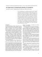
Fig. 1. Alignment of amino acid residues composing the catalytic
domains of PAH and TH. Residues identical in both enzymes are
highlighted. Dark gray bars indicate amino acid residues that possess
at least one atom within a distance of 14 A
˚
from the catalytic iron and,
based on this criterion, were included in energy calculations.
1066 A. Maaß et al. (Eur. J. Biochem. 270) Ó FEBS 2003
energies. The total electric charge was +6 for 5pah and )16
for 2toh, as 2toh comprises the catalytic core domain and
the tetramerization domain. BH
4
is uncharged. The aroma-
tic amino acids phenylalanine and tyrosine were implemen-
ted in their zwitterionic state with a protonated amine group
and a carboxylate moiety, resulting in a total charge of zero.
DA possesses an electric charge of +1.
We restricted the region of the complex for which the
total energy was calculated to those amino acid residues and
molecules that had at least one atom within a radius of 14 A
˚
around the iron atom. This sphere included about 50% of
the catalytic domain and comprised the entire binding
pocket containing the ligand (Fig. 1).
Analysis of results
For each ligand-protein pair, this procedure led to a set of
200–300 diverse structure predictions and relative total
energies. Considering that the relevant parts of the con-
formational space were probed and that the relative total
energy is a reasonable approximation of the free energy, the
structure with the lowest total energy should be closest to
the true structure of the complex in solution. However,
conformations with low energy values were discarded if the
predicted ligand position extended into the regulatory
domain of PAH [33]. Assuming that all reactive ligand
groups are placed in close proximity to the active site iron
immediately before the enzymatic reaction, only placements
within a defined ligand-iron distance were considered
relevant. According to the X-ray crystallographic structures
of TH and PAH with bound 7,8-BH
2
(2toh, 1dmw), the
carbonyl oxygen of 7,8-BH
2
is the atom closest to the active
site iron with a distance of 3.6 or 3.8 A
˚
, respectively.
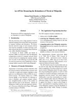
Therefore, conformations of BH
4
with a maximal distance
d
BH
4
-Fe
of 4.5 A
˚
were included in further analyses (Fig. 2).
As an oxygen atom may be placed between the ring of the
amino acid substrates and the active site iron, the maximal
distance d
Tyr-Fe
or d
Phe-Fe
between the center of the ring and
the iron atom was defined as 6.5 A
˚
(Fig. 2). As previous
spectroscopic and X-ray crystallographic studies have
suggested a tight bidentate coordination of catecholamines
towards the iron atom, 5.0 A
˚
was set as the maximal
distance d
L
-DOPA-Fe
or d
DA-Fe
between an imaginary line
connecting the oxygen atoms of the two catechol hydroxyl
groups and the iron (Fig. 2). This distance criterion would
allow bidentate, monodentate and other binding modes to
be included in further analysis.
Results
Pterin binding to the catalytic domains of PAH and TH
Docking of the native cosubstrate BH
4
into the crystal
structure of the PAH catalytic domain yielded a total of
286 conformations. Out of these, 44 conformations were
considered relevant as the distance between the pterin
carbonyl oxygen and the PAH catalytic iron atom (d
BH
4
-Fe
)
waslessthan4.5A
˚
(Fig. 3). The energetically most
favorable conformer corresponded with the position of
7,8-BH
2
bound to PAH at the first coordination sphere of
the iron atom as previously determined by NMR spec-
troscopy (rmsd 2.4 A
˚
) [23] and X-ray crystallography
(rmsd 2.1 A
˚
) [14]. The pterin backbone was close to the
aromatic ring of PAH residue Phe254, with a relative tilt of
about 20°. The guadinium moiety was anchored by an
H-bond between N1 and the amine group of Leu249. The
distance between N4 and the side chain of Glu286 was
4.6 A
˚
. This allows a putative water molecule to be placed
in between, which stabilizes the complex by additional
H-bonds (Fig. 3).
Docking BH
4
into the crystal structure of TH produced
300 conformations; in 46 out of these, the distance d
BH
4
-Fe
was shorter than 4.5 A
˚
(Fig. 3). The energetically most
favorable placement was very similar to the conformation of
BH
4
in PAH (rmsd 2.3 A
˚
). The pterin ring was in close
contact to Phe300 with the two ring planes tilted by 32°.The
carbonyl oxygen of BH
4
coordinated directly to the iron and
the guanidinium moiety was fixed by an H-bond between
the proton of N2 and the backbone oxygen of Gln310
(Fig. 3). This conformation of BH
4
coincided with the
conformation of BH
4
in TH previously computed by Alma
˚
s
et al.using
DOCK
4.0 (rmsd 2.2 ± 0.2 A
˚
) [15]. However, it
differed from the conformation of 7,8-BH
2
in the binary
complex identified by X-ray crystallography [13]. In the
latter study, the position of 7,8-BH
2
was rotated by 180° and
characterized by a p-stacking interaction of the planar
pterin moiety with TH residue Phe300, tilted by 10°.
We manually docked BH
4
into TH to further investigate
this alternative pterin position. The energetically most
favorable manual placement was in good agreement with
the rotated position of 7,8-BH
2
cocrystallized in TH (rmsd
1.5 A
˚
). The pterin backbone was close to the aromatic ring
of Phe300, tilted by 45°, and the carbonyl oxygen was again
coordinated towards the iron atom. The 2-hydroxyl group
of BH
4
formed an H-bond to the carboxylate group of
Glu332. The distance d
BH
4
-Fe
was 2.27 A
˚
, thus similar to
2.30 A
˚
in the conformer obtained by automated docking
(Fig. 3). The lowest total energy in the group of manually
docked conformations was 11.4 kcalÆmol
)1
compared with
1.5 kcalÆmol
)1
for the most favorable conformer in auto-
mated docking (Fig. 3). The difference mainly resulted from
Fig. 2. Ligands docked into the crystal structure of PAH and TH.
Placements suggested by
FLEXX
were considered relevant and included
in further analyses if the indicated distances between the ligands and
the iron atom at the active site of the enzymes were within defined
limits.
Ó FEBS 2003 Modeled ligand–protein complexes of PAH and TH (Eur. J. Biochem. 270) 1067
presumably artificial straining of the deeply buried BH
4
-
sidechain, caused by the minimization conditions applied.
Hence both rotamers should be treated as equivalent. The
equivalence of the two conformations was underscored by
the fact that the orientation of the pterin cosubstrate did not
affect the subsequent placement of amino acid substrates in
the ternary complexes with TH.
The position of amino acid substrates in the complexes
with PAHÆBH
4
and THÆBH
4
After docking the native substrate phenylalanine into
PAHÆBH
4
, 73 candidate positions with a distance d
Phe-Fe
between the center of the aromatic ring and the TH iron not
exceeding 6.5 A
˚
were included in further analysis. In the
conformation with the lowest total energy (13.5 kcalÆmol
)1
),
the distance between the phenylalanine ring center and the
iron atom was 4.96 A
˚
. The carboxylate moiety of phenyl-
alanine was anchored by H-bonds to PAH residue Arg270.
Another H-bond was formed between the carboxylate
group of phenylalanine and the amine group of Thr278. The
ammonium group of phenylalanine formed an H-bond to
the carbonyl oxygen of Thr278 (Fig. 4). Table 1 summar-
izes relevant energy components for the interactions of
phenylalanine in the complex with PAHÆBH
4
. This phenyl-
alanine position provided by
FLEXX
and the calculated
hydrogen bonds to surrounding PAH residues are in
agreement with X-ray crystallographic data of the phenyl-
alanine analog 3-(2-thienyl)-
L
-alanine bound to PAH [22].
In another, energetically equivalent conformation the
phenyl ring occupied the same position but the ammonium
group now formed an H-bond with Ser349, while the salt
bridge between the carboxylate group and Arg270 is
maintained (not shown). This latter position matches the
conformation of phenylalanine in the complex with
PAHÆ7,8-BH
2
that was previously calculated after restraints
from NMR spectroscopy [23] (rmsd 2.09 A
˚
and 1.29 A
˚
,
respectively).
Docking of tyrosine into PAHÆBH
4
produced a set of 179
conformations. Out of these, 55 conformations fulfilled the
distance criterion of d
Tyr-Fe
being smaller than 6.5 A
˚
.Only
eight conformations displayed the expected coordination of
the tyrosine aromatic ring towards the iron atom. However,
the hydroxyl group of the aromatic ring was placed trans to
His290. In contrast to the anchoring of the native substrate
phenylalanine, the ammonium group of tyrosine formed an
H-bond with the carboxyl moiety of Pro279 (Table 1). As
shown in Fig. 4, this position of tyrosine in PAH differed
significantly from that of phenylalanine.
FLEXX
provided 318 possible conformations of tyrosine in
THÆBH
4
. A set of 150 conformations was regarded as
relevant. The position with the lowest total energy corres-
ponded to the conformation of tyrosine in PAH (rmsd
1.12 A
˚
). In this position, the hydroxyl moiety of tyrosine
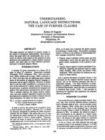
Fig. 3. Placement of BH
4
in the catalytic site of PAH and TH. Distance-energy diagrams obtained after docking BH
4
into the active sites of PAH (A)
and TH (B). Total energies of candidate positions provided by automated
FLEXX
calculations are given as s, total energies of conformations obtained
by manual docking are shown as ·. (C) Superposition of BH
4
placements in the catalytic sites of PAH and TH. Enzyme (PAH/TH) residues mentioned
in the text are displayed based on Protein Data Bank files 5pah and 2toh by using the program
RASMOL
(Sayle, R., Glaxo Wellcome Research and
Development, Stevenage, Hertfordshire, UK). Protein structures are depicted as sticks with carbon atoms colored purple, nitrogen atoms blue and
oxygen atoms red. The iron at the center of the active side is colored orange. Atoms coordinating directly to the iron atom are shown as balls. The top-
scoring conformation obtained by
FLEXX
for BH
4
in PAH is shown in red, for BH
4
in TH according to the results of the automated docking in blue,
and for BH
4
in TH according to the manual docking in the X-ray crystallographic mode in green. Hydrogens are omitted for clarity.
1068 A. Maaß et al. (Eur. J. Biochem. 270) Ó FEBS 2003
was again oriented towards the catalytic iron atom of TH,
with a distance of 2.16 A
˚
between the hydroxyl oxygen and
the iron. In analogy to the position of tyrosine in PAHÆBH
4
,
the hydroxyl group was placed trans to TH residue His336.
The ammonium group of tyrosine formed an H-bond to
Asp425 (Table 2). As pointed out below, this position is
likely to hinder the access of the essential reaction cofactor
dioxygen to the catalytic center. In contrast to the placement
of tyrosine in PAHÆBH
4
however,
FLEXX
provided a second
conformation for tyrosine, which was almost identical to the
position predicted for the native substrate phenylalanine in
the complex with PAHÆBH
4
. This conformation was
characterized by a salt bridge between the carboxylate
group and the guanidinium moiety of Arg316 (Table 2).
The ammonium group of tyrosine was surrounded by the
backbone oxygens of TH residues Ser324 and Pro325
(Fig. 4). The orientation of the pterin cosubstrate in the
complex with TH did not have an effect on the position of
tyrosine. However, the orientation of BH
4
in the second-
ranking conformation of tyrosine in THÆBH
4
corresponded
to that of 7,8-BH
2
cocrystallized with TH [13] and in this
orientation, N3 of BH
4
exhibited a stabilizing H-bond to the
tyrosine hydroxyl group.
The best placement of phenylalanine in THÆBH
4
coincided with the second-ranking position of tyrosine,
and it also corresponded to the position obtained by
phenylalanine in PAHÆBH
4
(rmsd 1.65 A
˚
). Forty-eight out
of 319 calculated conformations were considered relevant
here. In the energetically most favorable position (total
energy 1.3 kcalÆmol
)1
), the center of the aromatic ring was
placed at a distance of 5.86 A
˚
from the active-site iron,
compared with 4.95 A
˚
for tyrosine in THÆBH
4
(Fig. 4).
The increase in the distance is caused by an additional
iron-bound water molecule required for the docking of
phenylalanine.
Table 1. Intermolecular interaction energy contributions for the relevant amino acid placements in PAHÆBH
4
. Energy values for van der Waals
interactions (E
vdW
), coulombic (electrostatic) interactions (E
Coulomb
) and H-bonds of the ligand to neighboring protein residues are given in
kcalÆmol
)1
.
PAH residue
Phenylalanine in PAHÆBH
4
Tyrosine in PAHÆBH
4
E
vdW
E
Coulomb
H-bonds PAH residue E
vdW
E
Coulomb
H-bonds
Arg270 – 1.12 – 6.04 – 0.90 Leu248 – 0.75 – 0.21
Met276 – 0.74 1.16 Pro279 – 1.38 – 2.13 – 2.53
His277 – 1.45 – 1.10 Glu280 – 1.54 – 0.28
Thr278 – 2.65 – 4.41 – 3.00 Pro281 – 3.20 – 0.16
Pro279 – 0.50 0.23 His385 – 1.13 0.48
Glu280 – 1.31 – 1.36 Trp326 – 1.48 0.07
Pro281 – 1.73 – 0.09 Glu330 – 1.92 2.20
Asp282 – 0.37 0.57 Val379 – 1.28 – 0.39
His285 – 3.16 – 0.08
Glu330 – 1.08 – 1.80 Iron atom – 17.32 – 15.86
Phe331 – 0.85 0.09 BH
4
– 2.78 – 1.62 – 2.69
Gly346 – 1.50 0.09
Ser349 – 1.85 1.08
Ser350 – 1.41 0.10
Val379 – 0.12 – 0.38
Iron atom – 0.04 2.90
Fig. 4. Amino acid substrates docked into complexes of PAH or TH
with bound BH
4
. Superimposed are the top-scoring confomations of
phenylalanine in PAH (light green) and tyrosine in PAH (red). The
tyrosine conformation in TH with the lowest total energy is shown in
darker green; the second-ranking tyrosine conformation (blue) cor-
responds closely to the position of phenylalanine in PAH. The place-
ment of phenylalanine in TH is given in light green. BH
4
in the
corresponding complexes is shown in the same color as the amino acid
substrate.
Ó FEBS 2003 Modeled ligand–protein complexes of PAH and TH (Eur. J. Biochem. 270) 1069
Differences in the binding of
L
-DOPA and DA
to the active site iron of PAH and TH
In agreement with previous results from X-ray crystallo-
graphy [17], a common mode of bidentate binding was
found when the catecholamine end products
L
-DOPA and
DA were docked into PAH. The two hydroxyl groups of
the catechol moiety formed a tight chelate complex with
the iron atom at the center of the active site.
FLEXX
yielded
189 conformations of
L
-DOPA and 213 conformations of
DA in 5pah; 32 candidate positions for
L
-DOPA and 55
positions for DA were within a distance of 5.0 A
˚
.Nine
independent predictions for
L
-DOPA converged to the same
local minimum, which was characterized by an H-bond
between the amine group of
L
-DOPA and the backbone
oxygen of PAH residue Leu249, and a second H-bond
between the
L
-DOPA carboxyl moiety and the Leu249
nitrogen (Fig. 5). The next favorable conformation was
rotated by 180° around an imaginary axis passing between
the two hydroxyl groups of the catechol moiety (Fig. 5).
Thedifferenceof5.5kcalÆmol
)1
in the total energy of the
two orientations presumably represents an overestimate
resulting from energy minimization in the absence of solvent
molecules. For DA, the second favorable conformation was
also rotated by 180°. Here, the difference between the two
rotamers was 0.3 kcalÆmol
)1
, thus negligible. Apparently, in
PAH two equivalent positions exist for
L
-DOPA and DA,
with the plane of the catechol ring rotated 180°.
The docking of DA into TH yielded 216 relevant
conformations. DA bound directly to the active site iron
(Fig. 5). The amine end freely stuck out of the active site
crevice, analogous to the placement of DA in PAH (rmsd
Table 2. Intermolecular interaction energies for the placement of tyrosine or phenylalanine in the complex of THÆBH
4
. Energy contributions from
van der Waals interactions (E
vdW
), coulombic (electrostatic) interactions (E
Coulomb
) and H-bonds between ligands and neighboring protein residues
are shown. Values are expressed in kcalÆmol
)1
.
Tyrosine in THÆBH
4
(unproductive position)
Tyrosine in THÆBH
4
(productive position)
Phenylalanine
in THÆBH
4
TH residue E
vdW
E
Coulomb
H-bonds TH residue E
vdW
E
Coulomb
H-bonds TH residue E
vdW
E
Coulomb
H-bonds
Leu294 – 2.57 – 0.23 Arg316 – 1.06 – 4.78 Arg316 – 0.34 – 10.02 – 0.68
Pro325 – 1.52 – 0.16 Met322 – 0.36 0.68 Met322 – 0.14 0.22
Glu326 – 1.53 – 0.54 His323 – 2.19 – 1.85 His323 – 0.57 – 0.71
Pro327 – 2.91 – 0.03 Ser324 – 2.51 – 4.81 – 4.90 Ser324 – 1.57 – 3.88 – 3.31
His331 – 0.72 0.17 Pro325 – 1.88 – 1.44 Pro325 – 1.01 – 1.40
Trp372 – 1.53 0.04 Glu326 – 1.31 – 0.74 Glu326 – 1.52 – 0.08
Glu376 – 1.27 1.85 Pro327 – 1.85 – 0.24 Pro327 – 2.45 – 0.48
Asp425 – 0.40 – 13.12 – 1.03 Asp328 – 0.37 1.33 Asp328 – 0.49 3.68
His331 – 2.87 0.44 His331 – 2.97 – 0.75
Iron atom – 15.62 – 12.89 Glu376 – 1.62 0.39 Glu376 – 1.64 – 0.57
BH
4
– 2.95 1.00 Phe377 – 1.10 0.12 Phe377 – 1.11 0.10
Gly392 – 1.27 0.14 Gly392 – 1.16 0.16
Ser395 – 1.32 0.73 Ser395 – 2.38 1.00
Ser396 – 1.33 )1.04 Ser396 – 0.86 – 0.37
Asp425 – 0.19 )3.90 Asp425 – 0.33 – 3.17
Iron atom – 2.49 )4.26 Iron atom – 0.05 – 0.20
Fig. 5. Binding of catecholamine end products at the catalytic site. (A) High-scoring conformations of the catecholamines
L
-DOPA (blue, darker
green) and DA (red, light green) in the catalytic site of PAH. (B) High-scoring positions of
L
-
DOPA
(red) and DA (blue, green) docked into the
catalytic site of TH.
1070 A. Maaß et al. (Eur. J. Biochem. 270) Ó FEBS 2003
2.15 A
˚
). Like in PAH, two conformations of DA in TH,
rotated 180° around their iron-catecholamine axis,
possessed similar energy levels (2.3 kcalÆmol
)1
). Due to the
different rotamer of the iron-fixing Glu376 in TH, the
oxygen atom trans to His331 was slightly pushed aside
(0.68 A
˚
).
In contrast to the position of
L
-DOPAorDAinPAH
and DA in TH, only one predicted placement of
L
-DOPA in
TH was compatible with a bidentate binding mode (Fig. 5).
However, the total energy of this conformation was
8.5 kcalÆmol
)1
. This is about 6.0 kcalÆmol
)1
higher than
the total energy of the most likely, monodentate conforma-
tion among the 33 placements of
L
-DOPA with a distance
between the catechol moiety and the iron atom of less than
5.0 A
˚
. Bidentate binding of
L
-DOPA to the active site iron
was prevented by electrostatic forces: the
L
-DOPA carb-
oxylate group was repelled by the negatively charged TH
residue Asp425. In PAH, the residue corresponding to TH
Asp425 is a neutral Val379 so that the charged carboxylate
group of
L
-DOPA does not interfere with the tight,
bidentate binding of the catechol moiety to the active site
iron.
In the catalytic domains of both amino acid hydroxylases,
the positions of
L
-DOPA and DA overlapped with the
binding site of BH
4
. This was true for both orientations of
the pterin cosubstrate in TH. Consequently, based on the
results from our molecular docking experiments, it can be
predicted that the two catecholamines compete with BH
4
for binding to the active site. Once
L
-DOPA or DA have
formed a chelate complex with the catalytic iron, enzyme
function must be significantly impaired due to the restricted
access of the essential cosubstrate BH
4
.
Discussion
The aim of the present study was to model critical steps
during substrate binding, catalysis and feedback inhibition
of PAH and TH by molecular docking. The computational
approach allowed first, the creation of binary complexes of
the natural pterin cosubstrate BH
4
and the catalytic domain
of the enzymes, followed by the docking of amino acid
substrates. We thereby mimicked the highly ordered
sequence of substrate binding in TH [6] that was recently
also proposed for PAH [22].
Molecular docking of BH
4
into the catalytic domain of
PAH resulted in a conformation corresponding to the
position and orientation of 7,8-BH
2
in PAH determined
by NMR spectroscopy [23] and X-ray crystallography
[14]. The distance between the C4a-Atom of BH
4
and the
iron was 5.97 A
˚
, thus closer to the value of 6.06 A
˚
in the
crystal structure [14] than to the distance of 4.3 A
˚
measured by NMR spectroscopy [23]. Exactly the same
distance was found for BH
4
in the recent crystallographic
study of Andersen et al. [22]. Three H-bonds fixed the
cosubstrate to the protein. The N3-bound proton was at
3.71 A
˚
from the carboxylate-group of Glu286, so that
additional water-mediated H-bonds might anchor the
guanidinium moiety in the binding pocket [22]. The BH
4
sidechain made hydrophobic contacts to Ala322 and
Tyr325, two PAH residues that were recently associated
with mutations in hyperphenylalaninemia or phenol-
ketonuria, respectively [34,35].
ThebestplacementofBH
4
in TH in our experiments
differed from the reported X-ray crystallographic structure
of 7,8-BH
2
bound to TH [13] by a 180° rotation of BH
4
around its C4a–C8a bond. The natural pterin cosubstrate
was anchored by two H-bonds and several hydrophobic
interactions, which included p-stacking with Phe300. The
distance between the metal atom of TH and the pterin
carbon C4a that is hydroxylated during the enzymatic
reaction, was 4.2 A
˚
,wellbelow5.6A
˚
as measured in the
X-ray crystallography study [13]. On the contrary, the
conformer calculated by
FLEXX
was similar to the orienta-
tion of cosubstrate analogs including 7,8-BH
2
bound to a
recombinant, cobalt(II)-substituted human TH isoform 1,
examined by proton NMR spectroscopy [36]. In this study,
the distance between C4a of the pterin analog and the iron
atom has been measured as 3.7 A
˚
. Our coordinates of the
pterin position are also in agreement with a recent study
using the program
DOCK
4.0 [37] to model the conformation
of BH
4
and a series of pterin analogs placed into the crystal
structure of the TH catalytic domain [15]. On the other
hand, equivalent energy scores were obtained when BH
4
was manually docked into TH according to the orientation
determined by X-ray crystallography [13]. Consequently,
our results support the conception that in fact two
alternative orientations of the pterin cosubstrate exist within
the binding site of TH [13–15]. The two conformers
apparently possess a similar total energy but seem to be
differentially favored depending on the experimental con-
ditions.
Almost identical positions were obtained for phenylala-
nine docked into PAHÆBH
4
or THÆBH
4
and for the second-
ranking conformation of tyrosine docked into THÆBH
4
,
which probably represents the productive position. In this
position, the carboxylate moiety of the amino acids was
anchored by Arg270 in PAH or Arg316 in TH, respectively.
The pterin orientation in TH did not have a major effect on
the position of the amino acid substrate. However, a
stabilizing hydrogen bond was formed between the tyrosine
hydroxyl moiety at position C4 and BH
4
in the complex with
TH when the pterin cosubstrate was oriented following the
crystallographic coordinates of 7,8-BH
2
in TH [13]. Site-
directed mutagenesis of recombinant rat TH has previously
demonstrated the critical significance of the salt bridge
formed between the carboxylate group of the amino acid
substrate and the guanidinium moiety of Arg316. A
replacement of Arg316 with lysine was associated with an
increase of K
Tyr
by a factor of at least 400 compared with the
wild type [20]. As Arg316 forms another buried salt bridge to
Asp328, replacing Asp328 with serine might render the
guanidinium group of Arg316 more mobile. As a result,
binding of tyrosine to the catalytic site would become less
stable, explaining the increase of K
Tyr
byafactorof60inthe
mutant enzyme [20]. This substrate position calculated by
FLEXX
is in agreement with a recent proton NMR spectro-
scopic study of a complex consisting of dimeric human PAH
(residues Gly103 to Gln428), 7,8-BH
2
and phenylalanine
[23]. As stated in the latter study, the distance between the
hydroxylation site of phenylalanine, the C4 carbon atom,
and the catalytic iron is 4.34 A
˚
[23]. This is in good fit with
4.62 A
˚
determined by
FLEX
X for the common position of
phenylalanine in the complex with PAH or TH. The distance
appears optimal for accepting an iron bound oxygen.
Ó FEBS 2003 Modeled ligand–protein complexes of PAH and TH (Eur. J. Biochem. 270) 1071
The energetically most favorable placement of tyrosine in
PAHÆBH
4
and surprisingly, also in THÆBH
4
differed sub-
stantially from this common position of the amino acid
substrates. Here, the hydroxyl group of tyrosine was
coordinated towards the active-site iron as expected, but
tyrosine formed a hydrogen bond between its ammonium
group and Pro279 in PAH or Asp425 in TH. Our data
suggest that in TH, electrostatic attraction of the tyrosine
ammonium moiety by the negatively charged Asp425
(distance 3.4 A
˚
) and the formation of an H-bond counteract
the repulsion of the tyrosine carboxylate moiety by the same
protein residue (distance 5.3 A
˚
). Anchored in this position,
tyrosine interferes with the hydroxylation of BH
4
at carbon
C4a, which is required for catalysis. The orientation of BH
4
in PAH and in TH implies that dioxygen must be bound
trans to His290 or His336, respectively, in order to obtain
access to the pterin C4a. The only alternative position of
dioxygen would be trans to Glu330 in PAH or Glu376 in
TH. However, the distance between dioxygen in this
position and the pterin C4a would be approximately 5 A
˚
,
hence incompatible with hydroxylation. In addition, the
coordination of the aromatic ring of tyrosine gives it an
unfavorable position for accepting an electrophile. Daubner
et al. [21] have demonstrated by combined mutations of
PAH that the replacement with aspartate of residue Val379,
which corresponds to TH residue Asp425, provides only
very low rates of
L
-DOPA formation compared with TH.
On the other hand, according to the experimentally
established sequence of substrate binding in TH [6], the
binding of dioxygen prior to the amino acid will promote
the placement of tyrosine in the second-ranking, but
productive position.
While the unproductive orientation of tyrosine to the
catalytic domain of PAH sufficiently explains the specificity
of this enzyme for its native substrate phenylalanine,
additional factors outside the catalytic domain may be
relevant, too. It has been shown that the substrate specificity
of PAH and TH is enhanced though not determined by
mechanisms involving the N-terminal regulatory domain
[19]. Our models were restricted to the catalytical domains
of PAH and TH so that regulatory changes in the
N-terminal regions were not investigated. It is conceivable
that phenylalanine exerts an allosteric effect on PAH after
binding to the regulatory domain [33]. The assumption of a
fixed active-site crevice structure precludes the observation
of conformational alterations upon phenylalanine binding
as recently described by Andersen et al. [22]. In this study,
the placement of tyrosine in PAH was modeled after
crystallographic coordinates of the phenylalanine analog
3-(2-thienyl)-
L
-alanine bound to BH
4
ÆPAH. Preserving in
their model both the position of the main chain and the
orientation of the ring structure, the authors concluded that
tyrosine is not accepted by PAH as amino acid substrate
because its hydroxyl oxygen is sterically hindered by the side
chain of Trp326 [22]. The sterical interference of tyrosine in
this primary substrate position will certainly be important
for its release from the active site as product after
phenylalanine hydroxylation. According to the present
results, however, the preconditions of this model appear
too rigid if one considers tyrosine as an independent ligand
or possible alternative substrate of PAH. In this case,
the most probable position of tyrosine in PAH, defined by
the total energy, differs essentially from the position of the
native substrate phenylalanine. Whether binding of tyrosine
in this position triggers a large change in the protein
structure as shown after binding of 3-(2-thienyl)-
L
-alanine
[22] needs to be investigated.
Feedback inhibition by catecholamine end products is a
major regulatory factor both for PAH and TH. Molecular
docking of
L
-DOPA or DA into the crystal structure of
either amino acid hydroxylase provided a plausible and
energetically favorable conformation of the complex with
direct binding of the catecholamine inhibitors to the active-
site iron. Bidentate binding of the TH iron by the two
hydroxyl groups of the DA catechol moiety has been
suggested based on studies using resonance Raman spectro-
scopy [38], or a combination of electron paramagnetic
resonance (EPR), extended X-ray absorption fine structure
(EXAFS) and Mo
¨
ssbauer spectroscopy [16]. This binding
mode was later demonstrated by X-ray crystallography in a
binary complex of truncated PAH with bound catechol-
amines
L
-DOPA, DA, noradrenaline and adrenaline [17].
The position of the amine group of the catecholamine
inhibitors is less well defined. According to our results, the
amine groups of
L
-DOPA and DA freely stick out of the
active-site pocket in both TH and PAH. Two distinct
conformers of
L
-DOPA and DA in the complex with PAH
were assigned comparably favorable energy scores. The
conformers differed by 180° rotation around an imaginary
iron-catecholamine axis that runs between the two hydroxyl
groups of the catechol moiety. In agreement with a previous
X-ray crystallographic investigation of binary PAHÆcate-
cholamine complexes [17], neither of the two conformers
was preferred.
We found the same ambiguous binding pattern for DA in
TH, but not for
L
-DOPA. Instead, the predicted position of
L
-DOPA in the bidentate binding mode was assigned a high
energy. This is attributable to the electrostatic repulsion of
its negatively charged carboxylate group by TH residue
Asp425, whereas in PAH, the corresponding residue
represents neutral Val379. A monodentate, therefore less
tight and also less stable binding of
L
-DOPA to the catalytic
iron was predicted by F
LEX
X. Consequently, direct inhibi-
tion of TH by its native product
L
-DOPA would be
impeded compared with DA. In vitro experiments have
shown that concentrations of
L
-DOPA between 10- and
15-fold higher than DA are necessary to inhibit by 50%
recombinant human TH isoforms 1 and 2 [39]. Our findings
suggest that in TH, electrostatic forces play a key role in
hindering an immediate interference of the reaction product
with the catalytic center. The negative charge of the
corresponding TH residue Asp425 is a limiting factor for
the access of
L
-DOPA to the catalytic iron, thereby reducing
product inhibition.
As the binding sites of catecholamines and BH
4
overlap
in both amino acid hydroxylases, they compete with each
other for obtaining access to the catalytic site. Moreover,
bidentate binding of catecholamines to the catalytic iron(II)
causes its oxidation to the ferric form. Stoichiometric
amounts of DA rapidly induce iron oxidation and enzyme
inactivation [40]. Due to the stability of the generated
complex, catecholamines turn into almost irreversible
inhibitors [3,8]. Considering that catecholamines are redu-
cing agents, oxidation of the catalytic iron provoked by the
1072 A. Maaß et al. (Eur. J. Biochem. 270) Ó FEBS 2003
binding of these inhibitors must surprise. We hypothesize
that after binding to the iron atom, catecholamines activate
dioxygen in analogy to the activation of dioxygen by BH
4
during catalysis. According to our hypothesis, a highly
reactive iron-oxo intermediate would form together with the
generation of DA quinone and a hydroxyl anion (Fig. 6).
As the remaining iron-bound oxygen atom is now neigh-
boring solvent water molecules instead of an electron-rich
nucleophile such as the aromatic ring in tyrosine or
phenylalanine, the intermediate is likely to decompose into
oxidized iron and a highly reactive hydroxyl radical (Fig. 6).
The generation of reactive oxygen species during in vitro
tyrosine hydroxylation has been reported previously,
although it was attributed to partial uncoupling of BH
4
oxidation during catalysis [41]. In our model, the generation
of reactive oxygen species depends on the stoichiometric
equilibrium of the aromatic amino acid hydroxylase, the
cosubstrate BH
4
and catecholamine end products. A
clinically important shift in this equilibrium may occur in
Parkinson’s disease, as patients are systemically treated with
L
-DOPA, which is intracerebrally decarboxylized to DA.
Post mortem investigations of parkinsonian brains and
animal studies have shown that degenerating dopaminergic
neurons in the substantia nigra are specifically vulnerable to
reactive oxygen species due to a reduction in their antioxi-
dative defense systems such as glutathion [42]. As an
unwanted side-effect, high doses of
L
-DOPA may add to the
oxidative stress by tipping the balance towards TH inhibi-
tion and iron oxidation.
Acknowledgment
The authors wish to thank Marcus Gastreich, Jannis Apostolakis,
Joachim Selbig and Volker Schu
¨
nemann for constructive discussions
and helpful comments on the manuscript. This research was supported
in part by the Faculty of Medicine of the Medical University of Lu
¨
beck
(MUL J031).
References
1. Kappock, T.J. & Caradonna, J.P. (1996) Pterin-dependent amino
acid hydroxylases. Chem. Rev. 96, 2659–2756.
2. Fitzpatrick, P.F. (1999) Tetrahydropterin-dependent amino acid
hydroxylases. Ann. Rev. Biochem. 68, 355–381.
3. Flatmark, T. & Stevens, R.C. (1999) Structural insight into the
aromatic amino acid hydroxylases and their disease-related
mutant forms. Chem. Rev. 99, 2137–2160.
4. Walker, S.J., Liu, X., Roskoski, R. & Vrana, K.E. (1994) Catalytic
core of rat tyrosine hydroxylase: terminal deletion analysis of
bacterially expressed enzyme. Biochim. Biophys. Acta 1206, 113–
119.
5. Dickson, P.W., Jennings, I.G. & Cotton, R.G.H. (1994) Deli-
neation of the catalytic core of phenylalanine hydroxylase and
identification of glutamate 286 as a critical residue for pterin
function. J. Biol. Chem. 269, 20369–20375.
Fig. 6. Hydroxylation of tyrosine and a possible sequence of events related to the feedback inhibition of TH by the representative catecholamine DA.
The substrates of TH bind in a highly ordered sequence, starting with (1) BH
4
followed by (2) dioxygen and (3) the amino acid substrate tyrosine. (4)
During the catalytic step, one oxygen atom reacts with BH
4
to produce 4a-OH- BH
4
. The second oxygen atom forms an iron-oxo-intermediate
before the actual hydroxylation of the tyrosine aromatic ring takes place. (5) After its release from the catalytic site, (6)
L
-DOPA is decarboxylated
to DA by the aromatic
L
-amino acid decarboxylase (EC 4.1.1.28). (7) DA can bind to the TH catalytic iron via the hydroxyl groups of its catechol
moiety in a bidentate fashion. (8) DA activates dioxygen in analogy to the catalytic activation of dioxygen by BH
4
during catalysis. (9) As a result, a
highly reactive iron-oxo intermediate is generated. (10) In the following reaction step, DA quinone and a hydroxyl anion are formed. In the presence
of solvent water and due to the lack of an electron-rich nucleophile, the intermediate decomposes into oxidized iron and a highly reactive hydroxyl
radical. (11) BH
4
is required to reduce the ferric iron and re-establish TH activity.
Ó FEBS 2003 Modeled ligand–protein complexes of PAH and TH (Eur. J. Biochem. 270) 1073
6. Fitzpatrick, P.F. (1991) Steady-state kinetic mechanism of rat
tyrosine hydroxylase. Biochemistry 30, 3658–3662.
7. Fitzpatrick, P.F. (1991) Studies of the rate-limiting step in the
tyrosine hydroxylase reaction: alternate substrates, solvent isotope
effects, and transition-state analogues. Biochemistry 30, 6386–
6391.
8. Kumer, S.C. & Vrana, K.E. (1996) Intricate regulation of tyrosine
hydroxylase activity and gene expression. J. Neurochem. 67, 443–
462.
9. Ramsey, A.J. & Fitzpatrick, P.F. (2000) Effects of phosphoryl-
ation on binding of catecholamines to tyrosine hydroxylase: spe-
cificity and thermodynamics. Biochemistry 39, 773–778.
10. Erlandsen, H., Fusetti, F., Martinez, A., Hough, E., Flatmark, T.
& Stevens, R.C. (1997) Crystal structure of the catalytic domain of
human phenylalanine hydroxylase reveals the structural basis for
phenylketonuria. Nat. Struct. Biol. 4, 995–1000.
11. Goodwill, K.E., Sabatier, C., Marks, C., Raag, R., Fitzpatrick,
P.F. & Stevens, R. (1997) Crystal structure of tyrosine hydroxylase
at 2.3 A
˚
and its implications for inherited neurodegenerative dis-
eases. Nat. Struct. Biol. 4, 578–585.
12. Fusetti, F., Erlandsen, H., Flatmark, T. & Stevens, R.C. (1998)
Structure of tetrameric human phenylalanine hydroxylase and
its implications for phenylketonuria. J. Biol. Chem. 237, 16962–
16967.
13. Goodwill, K.E., Sabatier, C. & Stevens, R.C. (1998) Crystal
structure of tyrosine hydroxylase with bound cofactor analogue
andironat2.3A
˚
resolution: self-hydroxylation of Phe300 and the
pterin-binding site. Biochemistry 37, 13437–13445.
14. Erlandsen, H., Flatmark, T. & Stevens, R.C. (2000) Crystal
structure and site-specific mutagenesis of pterin-bound human
phenylalanine hydroxylase. Biochemistry 39, 2208–2217.
15. Alma
˚
s, B., Toska, K., Teigen, K., Groehn, V., Pfleiderer, W.,
Martinez, A., Flatmark, T. & Haavik, J. (2000) A kinetic and
conformational study on the interaction of tetrahydropteridines
with tyrosine hydroxylase. Biochemistry 39, 13676–13686.
16. Meyer-Klaucke,W.,Winkler,H.,Trautwein,A.X.,Nolting,H.F.
& Haavik, J. (1996) Mo
¨
ssbauer, electron-paramagnetic-resonance
and X-ray-absorption fine-structure studies of the iron environ-
ment in recombinant human tyrosine hydroxylase. Eur. J. Bio-
chem. 241, 432–439.
17. Erlandsen, H., Flatmark, T., Stevens, R.C. & Hough, E. (1998)
Crystallographic analysis of the human phenylalanine hydroxylase
catalytic domain with bound catechol inhibitors at 2.0 A
˚
resolu-
tion. Biochemistry 37, 15638–15646.
18. Schu
¨
nemann, V., Meier, C., Meyer-Klaucke, W., Winkler, H.,
Trautwein, A.X., Knappskog, P.M., Toska, K. & Haavik, J.
(1999) Iron coordination geometry in full-length, truncated, and
dehydrated forms of human tyrosine hydroxylase studied by
Mo
¨
ssbauer and X-ray absorption spectroscopy. J. Biol. Inorg.
Chem. 4, 223–231.
19. Daubner, S.C., Hillas, P.J. & Fitzpatrick, P.F. (1997) Characteri-
zation of chimeric pterin-dependent hydroxylases: contributions
of the regulatory domains of tyrosine and phenylalanine hydro-
xylase to substrate specificity. Biochemistry 36, 11574–11582.
20. Daubner, S.C. & Fitzpatrick, P. (1999) Site-directed mutants of
charged residues in the active site of tyrosine hydroxylase.
Biochemistry 38, 4448–4454.
21. Daubner, S.C., Melendez, J. & Fitzpatrick, P. (2000) Reversing the
substrate specificities of phenylalanine and tyrosine hydroxylase:
aspartate 425 of tyrosine hydroxylase is essential for 1-DOPA
formation. Biochemistry 39, 9652–9661.
22. Andersen, O.A., Flatmark, T. & Hough, E. (2002) Crystal struc-
ture of the ternary complex of the catalytic domain of human
phenylalanine hydroxylase with tetrahydrobiopterin and 3-(2-
thienyl)-
L
-alanine, and its implications for the mechanism of
catalysis and substrate activation. J. Mol. Biol. 320, 1095–1108.
23. Teigen, K., Frøystein, N. & Martinez, A. (1999) The structural
basis of the recognition of phenylalanine and pterin cofactors by
phenylalanine hydroxylase: implications for the catalytic
mechanism. J. Mol. Biol. 294, 807–823.
24. Jiang, G.C T., Yohrling, G.J., Schmitt, I.V.J.D. & Vrana, K.E.
(2000) Identification of substrate orienting and phosphorylation
sites within tryptophan hydroxylase using homology-based
molecular modeling. J. Mol. Biol. 302, 1005–1017.
25. Rarey, M., Kramer, B., Lengauer, T. & Klebe, G. (1996) A fast
flexible docking method using an incremental construction algo-
rithm. J. Mol. Biol. 261, 470–489.
26. Brooks, B.R., Bruccoleri, R.E., Olafson, B.D., States, D.J.,
Swaminathan, S. & Karplus, M. (1983) CHARMm: a program
for macromolecular energy, minimization and dynamics calcula-
tion. J. Comp. Chem. 4, 187–217.
27. Hoffmann, D., Kramer, B., Washio, T., Steinmetzer, T., Rarey,
M. & Lengauer, T. (1999) Two-stage method for protein-ligand
docking. J. Med. Chem. 42, 4422–4433.
28. Ellis, H.R., Daubner, S.C., McCulloch, R.I. & Fitzpatrick, P.F.
(1999) Phenylalanine residues in the active site of tyrosine
hydroxylase: mutagenesis of Phe300 and Phe309 to alanine and
metal ion-catalyzed hydroxylation of Phe300. Biochemistry 38,
10909–10914.
29. Bohm, H J. (1994) The development of a simple empirical scoring
function to estimate the binding constant for a protein-ligand
complex of known three-dimensional structure. J. Comput. Aided
Mol. Des. 8, 243–256.
30. Warwicker, J. & Watson, H.C. (1982) Calculation of the electric
potential in the active site cleft due to alpha-helix dipoles. J. Mol.
Biol. 157, 671–679.
31. Klapper, I., Hagstrom, R., Fine, R. & Honig, B. (1986) Focusing
of electric fields in the active site of Cu-Zn superoxide dismutase:
effects of ionic strength and amino-acid modification. Proteins 1,
47–59.
32. Hoffmann,D.,Washio,T.,Gessler,K.&Jacob,J.(1998)Tackling
concrete problems in molecular biophysics using Monte Carlo and
related methods: glycosylation, folding, solvation. In: Proceedings
of the Workshop on: Monte Carlo Approach to Biopolymers and
Protein Folding (Grassberger, P., Barkema, G. & Nadler, W., eds),
pp. 153–170. World Scientific Publishing, River Edge, Singapore.
33. Kobe, B., Jennings, I.G., House, C.M., Michell, B.J., Goodwill,
K.E., Santarsiero, B.D., Stevens, R.C., Cotton, R.G.H. & Kemp,
B.C. (1999) Structural basis of autoregulation of phenylalanine
hydroxylase. Nat. Struct. Biol. 6, 442–448.
34. Svensson, E., Eisensmith, R.C., Dworniczak, B., van Dobeln, U.,
Hagenfeld, L., Horst, J. & Woo, S.L. (1992) Two missense
mutations causing mild hyperphenylalaninemia associated with
DNA haplotype 12. Hum. Mutat. 1, 129–137.
35. Nowacki, P., Byck, S., Prevost, L. & Scriver, C.R. (1998) PAH
Mutation Analysis Consortium Database: 1997. Prototype for
relational locus-specific mutation databases. Nucleic Acids Res. 26,
220–225.
36. Martinez, A., Vageli, O., Pfleiderer, W. & Flatmark, T. (1998)
Proton NMR studies on the conformation of the pterin cofactor
bound at the active site of recombinant human tyrosine hydro-
xylase. Pteridines 9, 44–52.
37. Meng, E.C., Schichet, B.K. & Kuntz, I.D. (1992) Automated
docking with grid-based energy evaluation. J. Comput. Chem. 13,
505–524.
38. Michaud-Soret, I., Andersson, K.K., Que, L. Jr & Haavik, J.
(1995) Resonance Raman studies of catecholate and phenolate
complexes of recombinant human tyrosine hydroxylase.
Biochemistry 34, 5504–5510.
39. Alma
˚
s, B., Le Bourdelles, B., Flatmark, T., Mallet, J. & Haavik, J.
(1992) Regulation of recombinant human tyrosine hydroxylase
isozymes by catecholamine binding and phosphorylation.
1074 A. Maaß et al. (Eur. J. Biochem. 270) Ó FEBS 2003
Structure/activity studies and mechanistic implications. Eur.
J. Biochem. 209, 249–255.
40. Haavik, J., Martinez, A., Olafsdottir, S., Mallet, J. & Flatmark, T.
(1992) The incorporation of divalent metal ions into recombinant
human tyrosine hydroxylase apoenzymes studied by intrinsic
fluorescence and
1
H-NMR spectroscopy. Eur. J. Biochem. 210,
23–31.
41. Haavik, J., Alma
˚
s, B. & Flatmark, T. (1997) Generation of
reactive oxygen species by tyrosine hydroxylase: a possible
contribution to the degeneration of dopaminergic neurons?
J. Neurochem. 68, 328–332.
42. Blum, D., Torch, S., Lambeng, N., Nissou, M., Benabid, A.L.,
Sadoul, R. & Verna, J.M. (2001) Molecular pathways involved in
the neurotoxicity of 6-OHDA, dopamine and MPTP: contri-
bution to the apoptotic theory in Parkinson’s disease. Progr.
Neurobiol. 65, 135–172.
Ó FEBS 2003 Modeled ligand–protein complexes of PAH and TH (Eur. J. Biochem. 270) 1075