Báo cáo khoa học: Mapping of chorismate mutase and prephenate dehydrogenase domains in the Escherichia coli T-protein doc
Bạn đang xem bản rút gọn của tài liệu. Xem và tải ngay bản đầy đủ của tài liệu tại đây (178.64 KB, 7 trang )
Mapping of chorismate mutase and prephenate dehydrogenase
domains in the
Escherichia coli
T-protein
Shuqing Chen
1,
*, Sarah Vincent
2
, David B. Wilson
1
and Bruce Ganem
2
1
Department of Molecular Biology and Genetics and
2
Department of Chemistry and Chemical Biology, Cornell University, NY, USA
The Escherichia coli bifunctional T-protein transforms
chorismic acid to p-hydroxyphenylpyruvic acid in the
L
-tyrosine biosynthetic pathway. The 373 amino acid
T-protein is a homodimer that exhibits chorismate mutase
(CM) and prephenate dehydrogenase (PDH) activities, both
of which are feedback-inhibited by tyrosine. Fifteen genes
coding for the T-protein and various fragments thereof were
constructed and successfully expressed in order to charac-
terize the CM, PDH and regulatory domains. Residues 1–88
constituted a functional CM domain, which was also
dimeric. Both the PDH and the feedback-inhibition activities
were localized in residues 94–373, but could not be separated
into discrete domains. The activities of cloned CM and PDH
domains were comparatively low, suggesting some cooper-
ative interactions in the native state. Activity data further
indicate that the PDH domain, in which NAD, prephenate
and tyrosine binding sites were present, was more unstable
than the CM domain.
Keywords: chorismate mutase; E. coli T-protein; prephenate
dehydrogenase.
The final step in the biosynthesis of tyrosine in Escherichia
coli and other enteric bacteria is the transamination of
p-hydroxyphenylpyruvate, which is produced in two
sequential chemical reactions from chorismic acid in
nature’s shikimic acid metabolic pathway [1,2]. In the first
reaction, chorismate undergoes a Claisen rearrangement to
form prephenate, which is catalyzed by chorismate mutase
(CM; EC 5.4.99.5). In the second reaction, prephenate
undergoes NAD
+
-mediated oxidative decarboxylation to
p-hydroxyphenylpyruvate, which is catalyzed by prephenate
dehydrogenase (PDH; EC 1.3.1.12). In E. coli, both the
CM and PDH activities are located in a single, bifunctional
protein known as the T-protein, which is encoded by the
tyrA gene. Tyrosine (Tyr) is an end product inhibitor of
both CM and PDH, and induces aggregation of the
T-protein [3]. An analogous bifunctional protein in E. coli,
known as the P-protein, contains CM and prephenate
dehydratase (PDT), and catalyzes the transformation of
chorismate into phenylpyruvate in the biosynthetic pathway
to phenylalanine.
Domain mapping studies on the P-protein (386 amino
acids, homodimer, molecular mass 43 kDa) have estab-
lished that the CM, PDT, and regulatory activities reside
in discrete, separable domains that can be subcloned and
expressed [4–7]. The structure of the P-protein CM
domain (residues 1–109), which has been solved by X-ray
crystallography, reveals the key structural motif respon-
sible for noncovalent dimer formation in the wild-type
protein. However, biochemical studies aimed at mapping
the various functional domains in the T-protein suggest a
more complex spatial relationship of the catalytic
sites. Primary sequence alignments between the T- and
P-proteins indicate that CM in the T-protein is also
located at the N-terminus, although the sequences share
only approximately 25% similarity. Mutagenesis studies
on the T-protein and kinetic studies using substrate
analogs suggested that the CM and PDH reactions
occurred at overlapping [8] or perhaps closely proximal
[9] active sites. Strong evidence for two separate CM and
PDH active sites comes from pH rate profile analyses [10]
and from various substrate and product-based inhibitors
that affect the two catalytic activities with differing
degrees of selectivity [11]. At one extreme, a widely
studied oxabicyclic mutase inhibitor has been shown to
inhibit CM activity in the T-protein without affecting
PDH activity [9]. More recently, a tricyclic diacid was
reported to inhibit PDH activity in the T-protein without
affecting CM activity [12].
The main objectives of this study were to investigate
the various domain substructures, interactions, and allos-
teric effects in the E. coli T-protein by genetically
engineering and expressing fragments of tyrA.Using
these techniques, we hoped to determine whether the CM
and PDH activities could be separated into discrete,
properly folded entities displaying good catalytic activity.
We also hoped to ascertain whether a separate regulatory
domain existed within the T-protein that was responsible
for Tyr-induced end-product inhibition and T-protein
aggregation. Finally, we hoped to gain an understanding
Correspondence to B. Ganem, Department of Chemistry
and Chemical Biology, Baker Laboratory, Cornell University,
Ithaca, NY 14853-1301 USA.
Fax: + 1 607 255 6318, Tel.: + 1 607 255 7360,
E-mail:
Abbreviations: CM, chorismate mutase; PDT, prephenate dehydra-
tase; PDH, prephenate dehydrogenase; WT, wild-type.
Enzymes: chorismate mutase (EC 5.4.99.5); prephenate
dehydrogenase (EC 1.3.1.12).
*Present address: College of Pharmaceutical Science,
Zhejiang University, Hangzhou 310031, P.R. China.
(Received 25 October 2002, revised 11 December 2002,
accepted 19 December 2002)
Eur. J. Biochem. 270, 757–763 (2003) Ó FEBS 2003 doi:10.1046/j.1432-1033.2003.03438.x
of the detailed molecular interactions involved in
T-protein dimerization.
Experimental procedures
Materials
Unless indicated otherwise, all chemicals and biochemicals
were purchased from Sigma, and enzymes were purchased
from New England Biolabs.
Strain
E. coli BL21 Gold (DE3) competent cells (Stratagene) were
used as the host for cloning, plasmid preparation and
protein expression.
Recombinant DNA method
The tyrA gene, which codes for the T-protein, was
subcloned from plasmid pKB45, a derivative of pMB9
that contains a 6-kb segment of E. coli chromosomal
DNA [13]. Several primers (Table 1) were used to
amplify specific fragments from pKB45. NdeIandXhoI
sites were introduced into the primers at the N- and
C-terminal coding sites, respectively, of the target
fragments. A His tag was attached to the C-terminus
of the wild-type (WT) T-protein as a means of
simplifying the previously reported isolation [14] and
purification [15] procedures. C-terminal His-tags were
also attached to each fragment to facilitate subsequent
purification. In order to promote the fidelity of PCR,
GC-rich PCR kits were employed in amplification.
DNA sequencing (Cornell BioResource Center) was
carried out on every new plasmid to confirm that no
mutations had been introduced by PCR. Novagen
pET26b+ was used as the vector for all cloning. It
has a kanamycin-resistant gene to facilitate screening for
transformants.
Expression
All strains harboring plasmids were grown in LB (Luria–
Bertani) medium or on LB plates containing kanamycin
(60 lgÆmL
)1
). All strains were grown in LB containing
kanamycin (60 lgÆmL
)1
)at37°C for seed cultures and in
LB without antibiotics inoculated 1 : 50 for large-scale
enzyme production.
Isolation and purification of the T-protein
and cloned fragments thereof
After induction with 1 m
M
isopropyl b-
D
-thiogalactoside at
D
660
¼ 0.8 and growth at 30 °C for 2.5 h, cells were
collected by centrifugation at 10 000 g at 4 °C for 25 min.
Cell pellets were resuspended in cold binding buffer (5 m
M
imidazole, 0.5
M
NaCl, 20 m
M
Tris/HCl, pH 7.9), and the
cells were ruptured at 2000 p.s.i. using a French press.
Purification of the intact, His-tagged T-protein and of
its cloned fragments was performed on His-tag resin
(Novagen) following the manufacturer’s protocol. Peptide
1–88, without a His-tag, was obtained by mutating residue
89 to create a stop codon. The expressed peptide was
purified by Q-Sepharose and Ultragel ACA54 column
chromatography.
Proteolytic digestion
The purified T-protein was partly digested with papain by
varying the time and quantity of papain. T-protein
(20 lg) was dissolved in 100 lLof0.1
M
NH
4
Ac,
0.004
M
EDTA, 0.01
M
cysteine (pH 6.8) and 0.4 lLof
0.1 mgÆmL
)1
(1 : 50 ratio) or 2 lLof0.01mgÆmL
)1
(1 : 1000 ratio) of papain were added. The reaction was
incubated at 37 °Cand10lL samples were removed into
tubes containing SDS gel loading buffer and put into a
boiling water bath for 3 min at 0, 15, 30, 45, 60, 90, and
120 min intervals. All samples were then run on SDS/
PAGE gels.
Enzyme assays
Chorismate mutase and prephenate dehydrogenase activity
assays were performed according to Davidson et al.[14]
with 1 m
M
chorismate or 0.2 m
M
prephenate and 2 m
M
NAD, respectively. One unit of enzyme was defined as the
amount of enzyme required to produce 1 lmol of product
per minute at 37 °C. Specific activity was expressed as units
per mg of protein.
Kinetic studies
Enzyme assays of the T-protein and derived fragments in
the presence of Tyr were run at effector concentrations from
0to0.3m
M
, with substrate concentrations ranging from 0
to 1 m
M
or 2 m
M
based on the K
m
value to be measured.
Controls were run for every assay. Values for the maximal
Table 1. Primers used to clone T-protein peptides.
Primer Sequence
T01 5¢-GGT AGA CTC GAG TCA GTG GTG GTG GTG GTG GTG CTG GCG ATT GTC ATT CGC CTG ACG C-3¢
T02 5¢-GCT TAA GAG GTT TCA TAT GGT TGC TGA ATT G-3¢
PDH96 5¢-GGA TTT AAA ACA CAT ATG CCG TCA CTG CGT CCG GTG-3¢
PDH93 5¢-CGA CAA AGG ACA TAT GCA ACT TTG TCC GTC ACT GCG-3¢
PDH101 5¢-CCG TCA CTG CAT ATG GTG GTT ATC GTC GGC G-3¢
PDH93-336 5¢-CCA GTG CTC CAC CTC GAG TCA GTG GTG GTG GTG GTG GTG CTT ATC GCC CTG CTC CAG CAA-3¢
PDH93-316 5¢-CAA CTC AAT CGC CTC GAG TCA GTG GTG GTG GTG GTG GTG GAT TAA CGC CAG ATT ACG CTC TG-3¢
PDH93-296 5¢-GCT CTG ACG ACA TAA TCT CGA GTC AGT GGT GGT GGT GGT GGT GAG CCA ACA GTC GCC CGA CC-3¢
PDH93-276 5¢-CAT CGC CAG CTC AAG CTC GAG TCA GTG GTG GTG GTG GTG GTG AAG TTG CTC AAG CTG AAC AT-3¢
CM1-94 5¢-GCC ACC GCC GAC CTC GAG TCA GTG GTG GTG GTG GTG GTG AAG TGT TTT AAA TCC TTT GTC-3¢
CM1-108 5¢-CGA GAG GGT CAG CTC GAG TCA GTG GTG GTG GTG GTG GTG ACC GCC ACC GCC GAC GAT-3¢
758 S. Chen et al.(Eur. J. Biochem. 270) Ó FEBS 2003
velocity (V
max
) and the Michaelis constant (K
m
)were
determined using standard rate equations in conjunction
with the curve fitting options in the
KALEIDAGRAPH
program
(Abelbeck Software).
N-terminal analysis
Samples of the proteolytic bands were prepared for
N-terminal sequencing by electroblotting from the SDS
gels after electrophoresis. An Immobilen-P membrane was
prewet in methanol, and electrotransfer was performed
following the manufacturer’s procedure (50 V, 1 h).
Membranes were stained with 0.1% Commassie bright
blue for 10 min and destained in 90% methanol, 7%
acetic acid to a clear background. The band was cut out
and N-terminal sequencing was performed on a PE/
Applied Biosystems Procise 492 by the Cornell Bio-
Resource Center.
Molecular mass estimation
Molecular masses were determined by SDS gel electro-
phoresis under denatured conditions and gel exclusion
HPLC for determination of native molecular masses.
Standard molecular mass markers (Invitrogen BenchMark
Prestained Protein Ladder) were run on 12% or 17% SDS/
PAGE gels. A 600E Waters HPLC was used with a
Pak Glass 300SW 8 · 300 mm column and 50 m
M
Tris/
HCl, pH 8.0, 50 m
M
NaCl buffer at a flow rate of
0.75 mLÆmin
)1
. A Bio-Rad gel filtration standard was used
to prepare a standard curve.
Chemical cross-linking
The C-terminal His-tagged T-protein was chemically cross-
linked by a modified procedure as follows: 0.05 mg of
T-protein was dissolved in 20 mL of 50 m
M
KH
2
PO
4
/
K
2
HPO
4
buffer (pH 6.0), and 50% glutaraldehyde
(0.83 mL) was added to give a final concentration of 2%.
The reaction was run at room temperature for 22 h, then
0.5 mL of freshly prepared 2
M
NaBH
4
/0.1
M
NaOH was
added to quench the reaction. After standing at room
temperature for 20 min, 20 lL of 10% sodium deoxycho-
late in 0.1
M
NaOH was added followed by 0.5 mL of 100%
trichloroacetic acid (w/v) and the mixture was incubated
until the deoxycholate and protein precipitated. The sam-
ples were centrifuged at 20 000 g for 20 min and the pellets
were immediately dissolved in SDS/PAGE loading buffer
containing dithiothreitol, boiled for 3 min and analyzed by
electrophoresis on SDS/PAGE, using 17% acrylamide
gels for proteins having molecular mass < 20 kDa and
12% acrylamide gels for proteins having molecular
mass > 20 kDa.
Results
Expression
Expression levels for all fragments lacking the native
N-terminal sequence (plasmids PSQC2,3,4,5,6,7,8,13)
were low. Good levels of expression were observed with all
other fragments. By working at lower temperature (30 °C),
the formation of inclusion bodies was suppressed, and
expressed fragments were isolated from the soluble fractions.
Activity of wild-type T-protein
In assays of the WT T-protein, the specific activity for CM
was 130 unitsÆmg protein
)1
,andthatforPDHwas
98 unitsÆmg protein
)1
(Table 3). Both values were in good
agreement with those determined by Davidson et al.[13].
However, prolonged storage of purified His-tagged
T-protein at )80 °C, whether in storage buffer (0.1
M
sodium citrate : 10% glycerol : 1 m
M
dithiothreitol, pH 7.5)
or in assay buffer (0.1
M
Mes, 0.051
M
N-ethylmorpholine,
0.01
M
diethanolamine, 1 m
M
EDTA, 1 m
M
dithiothreitol,
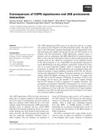
10% glycerol, pH 7.5) resulted in the loss of virtually all
PDH activity (Fig. 1). Activity losses were somewhat
smaller when protein was stored in the assay buffer. Because
of the instability of the T-protein, all assays were performed
on fresh enzyme. Controls indicated negligible loss of
activity on the day that assays were conducted. The specific
activity values reported in Table 3 were relative to freshly
prepared enzyme (100% activity), and represented the
highest values determined from the initial assays.
Proteolysis studies
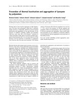
When papain was used to digest the T-protein under
limiting conditions (papain : T-protein ¼ 1 : 1000), a con-
sistent pattern of fragments was detected having molecular
mass values centered around 30 kDa and 10 kDa (Fig. 2).
The N-terminal sequence of the 30 kDa fragment was
determined to be TLCPSLRPVVIV, which corresponded to
residues 93–104 of the T-protein. Essentially identical results
were obtained when the T-protein was digested in the
presence of Tyr (300 l
M
), but without NAD
+
.
Digestions carried out in the presence of higher con-
centrations of papain (papain : T-protein ¼ 1:50) for
limited periods of time revealed that the 30 kDa fragment
Fig. 1. CM and PDH activity lost during storage.
Ó FEBS 2003 E. coli T-protein catalytic and regulatory domains (Eur. J. Biochem. 270) 759
disappeared almost completely within 30 min, while the
10 kDa fragment was still detectable after 60 min (Fig. 3).
Activity of cloned T-protein fragments
Guided by the proteolysis results and using appropriately
selected primer pairs, 14 new plasmids (Table 2) were
constructed and used to express T-protein fragments
corresponding to various regions of the T-protein
sequence. The expressed proteins were designated with
abbreviations indicating their T-protein origin and inclu-
sive residues.
The specific activities of both CM and PDH were
determined for all engineered T-protein fragments
(Table 3). The data indicate that all peptides containing
the N-terminal 88 residues of the T-protein (entries 9–15)
exhibited CM activity. However, the specific activity of all
CM-active T-protein fragments was low. Even the largest
such fragment, T/1–336, exhibited only approximately 5%
of the native T-protein’s activity. The Michaelis constant,
K
m
for CM activity in T/1–88 and T/1–336 were
1.7 ± 0.1 m
M
and 2.4 ± 0.5 m
M
, respectively. By com-
parison, K
m
for the T-protein was 0.23 m
M
. None of the
fragments exhibiting CM activity displayed PDH activity.
T-protein fragments T/93–373 and T/96–373 (Table 3,
entries 2 and 3) retain 25–50% of the PDH activity of the
T-protein, but are devoid of CM activity. Fragment
T/101–373 lacked PDH activity suggesting that residues
97–100 of the T-protein were essential for it (Table 3).
Several additional T-protein fragments were studied
(Table 3, entries 4–8) to refine the site of PDH activity.
Fragments T/101–373, T/93–277, T/93–297, T/93–316, and
T/93–336 displayed neither CM activity nor PDH activity.
Expression levels of the truncated proteins in Table 3
entries 2–8 were significantly lower than for proteins in
entries 9–15, which retained the native N-terminus.
Feedback inhibition by Tyr
In the absence of NAD
+
, the CM activity of fragments
T/1–88, T/1–94 and T/1–108 was unaffected by Tyr at
concentrations up to 300 l
M
. The CM activity of fragment
Table 2. Primer pairs used in constructing plasmids for cloning
T-protein fragments.
Plasmid Primer
T-protein
fragment
PSQC1 T02, T01 T/1–373
(T-protein)
PSQC2 PDH93, T01 T/93–373
PSQC3 PDH96, T01 T/96–373
PSQC4 PDH101, T01 T/101–373
PSQC5 PDH93, PDH93-276 T/93–277
PSQC6 PDH93, PDH93-296 T/93–297
PSQC7 PDH93, PDH93-316 T/93–316
PSQC8 PDH93, PDH93-336 T/93–336
pSQC24 T/1–88
pSQC9 T02, CM1-94 T/1–94
pSQC10 T02, CM1-108 T/1–108
pSQC11 T02, PDH93-276 T/1–276
pSQC12 T02, PDH93-296 T/1–296
pSQC13 T02, PDH93-316 T/1–316
pSQC14 T02, PDH93-336 T/1–336
Fig. 2. Proteolytic digestion of the T-protein by papain at a ratio of
1 : 1000 (w/w). Lane 1, molecular mass standards; lane 2, 0 min; lane
3, 15 min; lane 4, 30 min; lane 5, 45 min; lane 6, 60 min; lane 7,
90 min; lane 8, 120 min.
Fig. 3. The proteolytic digestion of T-protein by papain at a ratio of
papain/T-protein ¼ 1:50(w/w).Lane 1, 0 min (enzyme added; some
digestion observed); lane 2, 30 min; lane 3, 60 min; lane 4, molecular
mass ladder.
Table 3. CM and PDH activities of cloned segments of the T-protein.
Enzyme activity
(UÆmg
)1
)
Entry Plasmids Protein fragment CM PDH
1 PSQC1 T/1–373 (T-protein) 130 98
2 PSQC2 T/93–373 0 25.2
3 PSQC3 T/96–373 0 55.0
4 PSQC4 T/101–373 0 0
5 PSQC5 T/93–277 0 0
6 PSQC6 T/93–297 0 0
7 PSQC7 T/93–316 0 0
8 PSQC8 T/93–336 0 0
9 pSQC24 T/1–88 1.8 0
10 pSQC9 T/1–94 11.4 0
11 pSQC10 T/1–108 8.1 0
12 pSQC11 T/1–276 10.1 0
13 pSQC12 T/1–296 7.9 0
14 pSQC13 T/1–316 9.2 0
15 pSQC14 T/1–336 8.8 0
760 S. Chen et al.(Eur. J. Biochem. 270) Ó FEBS 2003
T/1–336 was mildly elevated in the presence of Tyr at
concentrations up to 10 l
M
. In contrast, the PDH activity in
fragments T/93–373 and 96/373 was inhibited in the
presence of Tyr, with 50% inhibition of activity in each
protein fragment observed at 25 ± 5 l
M
Tyr.
Molecular mass estimation and subunit
association analysis
The calculated molecular mass values for T/1–94 (12.5 kDa)
and T/1–108 (14 kDa) agreed well with values obtained
from SDS/PAGE using standard molecular mass markers
(data not shown). Gel exclusion HPLC analysis was used to
identify the molecular mass of the two fragments under
native conditions. Using a standard curve based on the
retention times and log molecular masses of four known
proteins (Table 4), molecular masses for T/1–94 and T/93–
373 were calculated to be 25 kDa and 63 kDa, respectively,
indicating that both fragments were dimers.
As the molecular mass of the T-protein exceeded the
effective range of gel exclusion HPLC analysis, chemical
cross-linking was used to identify the state of the T-protein
under native conditions (Fig. 4). SDS/PAGE analysis after
cross-linking indicated that the native T-protein was a
dimer, having a molecular mass of 85 kDa.
Discussion
The E. coli T- and P-proteins share numerous structural
and kinetic similarities. Besides being native dimers (com-
posed of subunits of similar M
r
values), both bifunctional
catalysts are subject to end-product inhibition (by Tyr and
Phe, respectively) induced by the aggregation of dimers into
higher oligomers. Feedback inhibition in each case more
strongly affects the second, prephenate-processing, enzyme
(PDH and PDT, respectively).
Several lines of evidence indicate that the major
difference between the T- and P-proteins is the spatial
and functional relationship between the two catalytic
activities in each bifunctional enzyme. Earlier studies from
these laboratories established that the CM, PDT, and
regulatory functions of the E. coli P-protein reside in
discrete, separable domains that can be subcloned and
expressed [5]. In the case of the E. coli T-protein, several
previous kinetic studies suggested interdependent, and
perhaps overlapping [8] or closely proximal [9], CM and
PDH active sites. The interdependence of the catalytic sites
in the T-protein was first noted by Koch et al. who
compared the rates of the CM and PDH reactions and
observed a distinct lag phase in the latter process [16].
Furthermore, levels of free prephenate accumulating in the
reaction mixture could not account for the observed rate
of the PDH reaction, further suggesting interactions
between the CM and PDH sites. Koch et al. also observed
that the inhibition constant (K
i
) for prephenate closely
paralleled its K
m
value for the PDH reaction, and
concluded that the CM and PDH-catalyzed reactions
shared a common prephenate binding site on the
T-protein. Subsequently, Heyde and Morrison noted that
NAD
+
, the cofactor required for PDH activity, also
boosted CM activity, while chorismate enhanced PDH
activity [8].
The present study represents the first systematic effort to
identify amino acid sequences within the T-protein that,
when expressed as discrete fragments, displayed either CM
or PDH activity. The main goal of the study was to learn
whether CM or PDH activity might be separated into
individual domains of the T-protein. A further goal of the
study was to ascertain whether feedback inhibition by Tyr
might also involve a discrete region of the T-protein.
The established domain relationships in the P-protein
suggested that a T-protein fragment embodying the
N-terminus and the first 90–100 residues might exhibit
CM activity. A modest level of sequence similarity (22 of the
first 56 residues are identical [2]) in the N-terminal regions of
the T- and P-proteins further supported this conclusion,
although potential differences in secondary structure
between the two proteins complicated any analysis based
strictly on sequence comparison. The results of limited
digestion of the T-protein using papain consistently affor-
ded a pattern of fragments having principal bands at
molecular masses 10 and 30 kDa. N-terminal sequence
analysis indicated that the two fragments corresponded to
residues 1–92 and 93–373, respectively. The finding that the
smaller, 10 kDa fragment was somewhat resistant to
proteolysis (Fig. 3) also lent credence to the possibility that
it existed as a separately folded domain in the T-protein.
The T-protein has been reported to be quite unstable in
crude cell extracts [17], although stabilization of pure
T-protein by prephenate or Tyr has been noted [16]. Heyde
and Morrison observed that the T-protein exhibited poor
stability when stored in dilute solution, causing the ratio of
Fig. 4. The T-protein was cross-linked by 2% glutaraldehyde at
2.5 lgÆmL
)1
of T-protein for 22 h. Lane 1, ladder; lane 2, T-protein
control; lane 3, T-protein after cross-linking.
Table 4. HPLC retention times and molecular masses of T-protein
fragments and standards.
Protein
Retention
time (min)
Molecular
mass (kDa)
Aggregation
state
BSA 10.75 67 –
Chicken ovalbumin 11.8 44 –
Equine myoglobin 15.3 17 –
CM1-94 13.2 25 Dimer
PDH93-373 11.0 63 Dimer
Ó FEBS 2003 E. coli T-protein catalytic and regulatory domains (Eur. J. Biochem. 270) 761
CM to PDH activity to vary from 0.8 to 1.2 between
preparations [8]. It should be noted that the E. coli
T-protein has been reported to be quite sensitive to both
storage and aging [18].
The present study used T-protein expressed with a
C-terminal His-tag to simplify purification. Chemical
cross-linking experiments confirmed its dimeric structure
under native conditions (Fig. 4) and its catalytic profile
matched the wild-type protein. However, the stability of
the His-tag labelled T-protein remained a problem. PDH
activity deteriorated particularly rapidly during storage
(Fig. 1), whereas significant levels of CM activity were
retained. Taken together with results from limited proteo-
lysis experiments, the data suggested that the region of the
T-protein associated with PDH catalysis was more loosely
packed, and hence more easily denatured, than the corres-
ponding domain or residues associated with CM activity.
His-tagged forms of the E. coli T-protein and 14
fragments thereof were successfully expressed and purified
by affinity chromatography. Screening of those fragments
for enzymatic activity (Table 3) indicated that neither the
CM nor the PDH active site could be expressed in fully
functional form as a discrete, contiguous subregion of the
T-protein. Based on the seven fragments that displayed
CM activity, residues 1–88 appeared to be essential for CM
catalysis. While CM activity was enhanced by including the
additional residues, 89–94, the most active fragment
displayed only 8% of WT T-protein activity. Surprisingly,
a stepwise increase in the fragment length (T/1–108, T/1–
276, T/1–296, T/1–316, T/1–336) did not increase CM
activity.
Several possible explanations were considered for the
consistently low levels of mutase activity. The association of
engineered fragments into homodimers, shown to be
important in the monofunctional mutase derived from the
E. coli P-protein, was confirmed in the case of T/1–94 by gel
exclusion HPLC (Table 3). Contamination of the purified
fragments by low levels of WT T-protein was ruled out by
the absence of any corresponding PDH activity (Table 3). If
the organization of the CM and PDH/PDT active sites in
the T and P-proteins were similar, then a heterodimeric
enzyme displaying CM but not PDH activity might
plausibly arise by the complexation of one His-tagged
fragment with one WT T-protein chain. This possibility
seemed remote for two reasons. Because the cloned
fragment was expressed at much higher concentrations
compared to the native T-protein, any suspect heterodimer
would have represented a very small amount of the protein.
Moreover, analysis of each mutase-active fragment by
SDS/PAGE at high gel loading levels revealed no higher
molecular mass band matching the T-protein or corres-
ponding heterodimer.
The low mutase activity of the N-terminal fragments
(Table 3) indicated that a discrete, fully active CM
subdomain comprising contiguous T-protein residues
could not be expressed, showing that a catalytically
efficient CM active site required most, if not all, of the
T-protein. An earlier report by Christendat et al.[15]
indicated that mutagenesis of several residues in the
dehydrogenase portion of the T-protein significantly
affected CM activity, either by reducing K
cat
(His189Asn)
or elevating K
m
(His239Asn, His245Asn). The findings
reported here suggest that additional amino acids in the
PDH domain, extending beyond residue 336 effect mutase
activity.
Proper CM function may be disrupted by poor substrate
binding, as has been noted with the His239 and His245
mutants. Likewise, the series of N-terminal fragments
(entries 9–15, Table 3) may have structurally altered or
incomplete PDH substrate binding sites that cause poor
substrate binding. If, as has been suggested [16], prephenate
undergoes transfer from the product-binding pocket of the
CM site to the substrate-binding pocket of the PDH site,
then the weak CM activity of the CM fragments might be
due to slow product release or trapping of prephenate on the
truncated protein.
In contrast, fragments of the T-protein could be prepared
that contained catalytically competent, monofunctional
dehydrogenases with the requisite NAD
+
binding sites.
Two C-terminal sequences lacking approximately one-
quarter of the T-protein’s N-terminal region were expressed
(T/93–373 and T/96–373; entries 2–3, Table 3) that dis-
played significant levels of PDH activity, but no CM
activity. Xia et al. showed that a similar, monofunctional
PDH domain could be prepared from the corresponding
bifunctional protein in Erwinia herbicola by deleting residues
1–37 [19]. Earlier studies on the E. coli T-protein had
implicated His197 as a key catalytic residue in PDH activity
[15] and Arg294 in prephenate binding [20]. Both of these
residues were included in the sequences of the two PDH-
active fragments. Fragment T/101–373 (entry 4, Table 3)
was devoid of PDH activity, suggesting that one or more
residues in the 97–100 region may play an important role in
catalysis.
Of the fragments displaying monofunctional PDH acti-
vity, analysis of one (T/93–373) by gel exclusion HPLC
showed it to be a homodimer (Table 4). As the CM-active
fragment T/1–94 was also a homodimer, these data indi-
cated that noncovalent interactions resulting in T-protein
dimerization appeared to be present in both the CM and
PDH domains, unlike the P-protein, in which dimerizing
interactions occurred only in the N-terminal region. Sam-
ples of both T/93–373 and T/96–373 retained > 95% of
their activity when stored for 7 days at )70 °Cand
reassayed. However, both fragments underwent denatura-
tion after prolonged storage (3–4 months at )20 °C), with
complete loss of activity.
With an N-terminal CM site joined to a PDH domain,
the overall layout of chorismate and prephenate processing
sites in the T-protein resembled that of the P-protein.
However, results from the present study showed that
the organization of the structural domains responsible for
end product inhibition differed substantially in the two
bifunctional proteins. Whereas the C-terminal 100 residues
of the P-protein constituted a discrete Phe-binding domain,
T-protein fragment analysis indicated that tyrosine binding
and feedback inhibition could not be attributed to a
structural domain that was separate from the CM and
PDH domains. Initial attempts to pinpoint the C-terminal
boundary of the PDH domain established that even
minor deletions of C-terminal residues resulted in complete
loss of PDH activity (entries 5–8, Table 3). Corresponding
residue deletions in the P-protein did not diminish PDT
activity.
762 S. Chen et al.(Eur. J. Biochem. 270) Ó FEBS 2003
Because of the absence of a discrete regulatory domain
in the T–protein, the interaction of various fragments with
Tyr was investigated to determine whether the Tyr binding
site overlapped with one or more catalytic domains in
the T-protein. Tyr had no effect on the low CM activity
observed in fragments T/1–88, T/1–94, T/1–108, T/1–276,
T/1–296, and T/1–316. However, the CM activity of
fragment T/1–336 was mildly enhanced at low Tyr
concentrations (up to 10 l
M
). A similar activation of
CM activity in the WT T-protein was first observed by
Christopherson at up to 300 l
M
Tyr [21] for which no
mechanistic rationale has been proposed. The fact that
activation by Tyr was weaker in T/1–336 suggested that
the C-terminal 30 residues of the T-protein affected Tyr
binding, and perhaps contributed to an allosteric effect on
CM. Overall, the behavior of N-terminal fragments listed
in Table 3 towards Tyr consistently indicated that the
locus of Tyr binding included residues near the C-terminus
of the T-protein.
In agreement with that prediction, Tyr had a pronounced
inhibitory effect on PDH-active fragments T/93–373 and
T/96–373. In each case, 50% inhibition of enzyme activity
was observed at 25 ± 5 l
M
, which agreed with the IC
50
value of 20 l
M
first reported by Koch et al.fortheWT
T-protein [22]. Overall, these findings indicated that Tyr
binding coincided with the region of the T-protein princi-
pally associated with PDH activity, and provide a physical
basis for the observation of Christopherson [21] that Tyr
exerted a more pronounced effect on PDH activity than on
CM activity. Koch et al. [16] had earlier proposed a form of
sequential feedback inhibition in which Tyr acted primarily
to inhibit PDH, resulting in an accumulation of prephenate
that, in turn, inhibited CM. That picture is consistent with
the physical layout of catalytic and binding sites that
emerges from the T-protein fragment studies presented
here.
The domain mapping studies reported here, based on 14
T-protein fragments, indicated that CM and PDH were
separable into independent enzymatic sites, although the
efficiency of the CM-active fragments was considerably
diminished when compared to the native T-protein.
Acknowledgements
This work was supported by grants from the National Institutes
of Health (GM 24054, to BG) and the Department of Energy
(DE-F G02-84ER13233, to DBW).
References
1. Koch, G.L., Shaw, D.C. & Gibson, F. (1971) Characterisation of
the subunits of chorismate mutase-prephenate dehydrogenase
from E. coli K12. Biochim. Biophys. Acta 229, 805–812.
2. Haslam, E. (1993) Shikimic Acid Metabolism and Metabolites.
John Wiley & Sons, New York, USA.
3. Hudson, G.S., Howlett, G.J. & Davidson, B.E. (1983) The binding
of tyrosine and NAD
+
to chorismate mutase/prephenate dehy-
drogenase from Escherichia coli K12 and the effects of these lig-
ands on the activity and self association of the enzyme. J. Biol.
Chem. 258, 3114–3120.
4. Pohnert, G., Zhang, S., Husain, A., Wilson, D.B. & Ganem, B.
(1999) Regulation of phenylalanine biosynthesis. Calorimetric
studies on the E. coli P-protein and its regulatory domain. Bio-
chemistry 38, 12212–12217.
5. Zhang,S.,Pohnert,G.,Kongsaeree,P.,Wilson,D.B.,Clardy,J.&
Ganem, B. (1998) Chorismate mutase-prephenate dehydratase
from Escherichia coli: study of catalytic and regulatory domains
using genetically engineered proteins. J. Biol. Chem. 273, 6248–
6253.
6. Zhang, S., Wilson, D.B. & Ganem, B. (2000) Probing the catalytic
mechanism of prephenate dehydratase by site-directed mutagen-
esis of the Escherichia coli P-protein dehydratase domain.
Biochemistry 39, 4722–4728.
7. Lee, A.Y., Stewart, J.D., Clardy, J. & Ganem, B. (1995) New
insight into the catalytic mechanism of chorismate mutases from
structural studies. Chem. Biol. 2, 195–203.
8. Heyde, E. & Morrison, J.F. (1978) Kinetic studies on the reactions
catalyzed by chorismate mutase-prephenate dehydrogenase from
Aerobacter aerogenes. Biochemistry 17, 1573–1580.
9. Turnbull, J. & Morrison, J.F. (1990) Chorismate mutase-
prephenate dehydrogenase from Escherichia coli. 2. Evidence for
two different active sites. Biochemistry 29, 10255–10261.
10. Turnbull, J., Cleland, W.W. & Morrison, J.F. (1991) pH
dependency of the reactions catalyzed by chorismate mutase-
prephenate dehydrogenase from Escherichia coli. Biochemistry 30,
7777–7782.
11. Christopherson, R.I. (1997) Partial inactivation of chorismate
mutase-prephenate dehydrogenase from Escherichia coli in the
presence of analogs of chorismate. Int. J. Biochem. Cell Biol. 29,
589–594.
12. Vincent, S., Chen, S., Wilson, D.B. & Ganem, B. (2002) Probing
the overlap of chorismate mutase and prephenate dehydrogenase
sites in the Escherichia coli T-protein: a dehydrogenase-selective
inhibitor. Bioorg.Med.Chem.Lett.12, 929–931.
13. Zurawski, G., Brown, K., Killingly, D. & Yanofsky, C. (1978)
Nucleotide sequence of the leader region of the phenylalanine
operon of Escherichia coli. Proc. Natl Acad. Sci. USA 75, 4271–
4275.
14. Davidson, B.E. & Hudson, G.S. (1987) Chorismate mutase-
prephenate dehydrogenase from Escherichia coli. Methods
Enzymol. 142, 440–450.
15. Christendat, D., Saridakis, V.C. & Turnbull, J.L. (1998) Use of
site-directed mutagenesis to identify residues specific for each
reaction catalyzed by chorismate mutase-prephenate dehydro-
genase from Escherichia coli. Biochemistry 37, 15703–15712.
16. Koch, G.L.E., Shaw, D.C. & Gibson, F. (1972) Studies on the
relationship between the active sites of chorismate mutase-
prephenate dehydrogenase from Escherichia coli or Aerobacter
aerogenes. Biochim. Biophys. Acta 258, 719–730.
17. Llewellyn, D.J. & Smith, G.D. (1979) Study of chorismate mutase-
prephenate dehydrogenase in crude cell extracts of Escherichia
coli. Biochemistry 18, 4707–4714.
18. Dopheide, T.A.A., Crewther, P. & Davidson, B.E. (1972) Choris-
mate mutase-prephenate dehydratase from Escherichia coli K12.
J. Biol. Chem. 247, 4447–4452.
19. Xia, T., Zhao, G., Fischer, R.S. & Jensen, R.A. (1992) J. General
Microbiol. 138, 1309–1316.
20. Christendat, D. & Turnbull, J.L. (1999) Identifying groups
involved in the binding of prephenate to prephenate dehydro-
genase from Escherichia coli. Biochemistry 38, 4782–4793.
21. Christopherson, R.I. (1985) Chorismate mutase-prephenate
dehydrogenase from Escherichia coli: cooperative effects and
inhibition by 1-tyrosine. Arch. Biochem. Biophys. 240, 646–654.
22. Koch, G.L.E., Shaw, D.C. & Gibson, F. (1971) The purification
and characterisation of chorismate mutase-prephenate dehydro-
genase from Escherichia coli. Biochim. Biophys. Acta 229, 795–
812.
Ó FEBS 2003 E. coli T-protein catalytic and regulatory domains (Eur. J. Biochem. 270) 763