Báo cáo khoa học: Biotinylation in the hyperthermophile Aquifex aeolicus Isolation of a cross-linked BPL:BCCP complex pptx
Bạn đang xem bản rút gọn của tài liệu. Xem và tải ngay bản đầy đủ của tài liệu tại đây (282.86 KB, 11 trang )
Biotinylation in the hyperthermophile
Aquifex aeolicus
Isolation of a cross-linked BPL:BCCP complex
David J. Clarke, Joseph Coulson, Ranald Baillie and Dominic J. Campopiano
School of Chemistry, University of Edinburgh, UK
Biotin protein ligase (BPL) catalyses the biotinylation of the
biotin carboxyl carrier protein (BCCP) subunit of acetyl
CoA carboxylase and this post-translational modification of
a single lysine residue is exceptionally specific. The exact
details of the protein–protein interactions involved are
unclear as a BPL:BCCP complex has not yet been isolated.
Moreover, detailed information is lacking on the composi-
tion, biosynthesis and role of fatty acids in hyperthermo-
philic organisms. We have cloned, overexpressed and
purified recombinant BPL and the biotinyl domain of BCCP
(BCCPD67) from the extreme hyperthermophile Aquifex
aeolicus. In vitro assays have demonstrated that BPL cata-
lyses biotinylation of lysine 117 on BCCPD67 at tempera-
turesofupto70°C. Limited proteolysis of BPL with trypsin
and chymotrypsin revealed a single protease-sensitive site
located 44 residues from the N-terminus. This site is adjacent
to the predicted substrate-binding site and proteolysis of
BPL is significantly reduced in the presence of MgATP and
biotin. Chemical crosslinking with 1-ethyl-3-(dimethylamino-
propyl)-carbodiimide (EDC) allowed the isolation of a
BPL:apo-BCCPD67 complex. Furthermore, this complex
was also formed between BPL and a BCCPD67 mutant
lacking the lysine residue (BCCPD67 K117L) however,
complex formation was considerably reduced using holo-
BCCPD67. These observations provide evidence that addi-
tion of the biotin prosthetic group reduces the ability of
BCCPD67 to heterodimerize with BPL, and emphasizes that
a network of interactions between residues on both proteins
mediates protein recognition.
Keywords: biotin protein ligase; Aquifex aeolicus; biotinyla-
tion; protein recognition; chemical crosslinking.
The enzymes of bacterial fatty acid biosynthesis have been
suggested as good targets for the development of novel
antibacterial agents since several natural product and
synthetic inhibitors of this pathway are already known [1].
Moreover, significant differences in fatty acid biosynthesis
between bacteria and mammals should allow selective
inhibition of the microbial enzymes. The first committed
step of bacterial fatty acid biosynthesis is catalysed by a
multisubunit acetyl-CoA carboxylase [2]. This biotin
-dependent complex is composed of biotin carboxylase,
carboxyltransferase and biotin carboxyl carrier protein
(BCCP) subunits, the exact composition of which is
species-specific. The Escherichia coli acetyl-CoA carboxy-
lase has been intensively studied, because the subunits can
be separated or expressed individually in an active form.
Biotin is covalently bound to a specific lysine residue in the
BCCP subunit [3,4]. Biotinylated enzymes transfer cardon
dioxide from bicarbonate to organic acids to form cellular
metabolites, using the biotin prosthetic group as a mobile
carboxyl carrier [5]. Biotin protein ligase (BPL), also known
as holocarboxylase synthase (HCS, EC 6.3.4.10) catalyses
this post-translational attachment via a two-step reaction
(Scheme 1 [6]).
Genes encoding BPLs have been identified in a number of
organisms, but the best-characterized BPL is the 35.3 kDa
BirA protein from E. coli [7,8]. BirA is a bifunctional protein
that can act as both an enzyme and a DNA-binding protein;
it catalyses protein biotinylation when in vivo biotin
concentrations are low, but becomes a repressor of the
expression of biotin biosynthetic enzymes when biotin
concentrations are increased. The crystal structure of the
biotin-bound protein, determined at 2.3 A
˚
resolution,
shows the enzyme has three domains [9,10]; an N-terminal
domain that contains a helix-turn-helix fold for DNA
binding; a central catalytic domain, which contains a highly
conserved GRGRRG motif shown to be involved in biotin
binding [11]; and a small C-terminal domain which has been
postulated to mediate dimerization with apo-BCCP [12].
The recent determination of the structure of a BirA dimer in
Scheme 1.
Correspondence to D. Campopiano, School of Chemistry, University
of Edinburgh, West Mains Road, Edinburgh, EH9 3JJ, UK.
Fax: + 44 131 650 4743, Tel.: + 44 131 650 4712,
E-mail:
Abbreviations: BPL, biotin protein ligase; BCCP, biotin carboxyl
carrier protein; IPTG, isopropyl thio-b-
D
-galactoside; EDC,
1-ethyl-3-(dimethylamino-propyl)-carbodiimide; HCS,
holocarboxylase synthase.
Enzyme: Biotin protein ligase or holocarboxylase synthase
(EC 6.3.4.10).
(Received 26 November 2002, revised 18 January 2003,
accepted 29 January 2003)
Eur. J. Biochem. 270, 1277–1287 (2003) Ó FEBS 2003 doi:10.1046/j.1432-1033.2003.03493.x
the absence of DNA provides insight into how the
N-terminal DNA-binding domain interacts with the 40 bp
biotin operator sequence [12]. The structure of the apo- and
holo-forms of the biotinylation domain of E. coli BCCP
(known as BCCP-87) have been determined by X-ray
crystallography and NMR [13–15]. The BCCP domain is a
barrel consisting of two antiparallel b-sheets each containing
four strands. The N- and C-termini are close together at one
end, and the biotinylated lysine is exposed on a tight b-turn
at the opposite face of the molecule. Surprisingly, the
structures of the apo- and holo- forms are remarkably
similar suggesting that biotinylation causes few significant
changes in the domain tertiary fold.
To gain further insight into the detailed protein–protein
interactions that control biotin transfer we have analysed
the reaction between BPL and apo-BCCP from the
hyperthermophilic organism Aquifex aeolicus [16]. This
bacteria grows optimally at 95 °C on hydrogen, oxygen,
carbon dioxide and mineral salts. Enzymes from extremo-
philes (extremozymes) are offering new opportunities for
biocatalysis as a result of their extreme stability [17,18].
Analysis of the A. aeolicus genome identified BirA and
BCCP homologues; the predicted BPL is from the group I
class (which also includes M. tuberculosis) which lack the
N-terminal DNA-binding domain found in E. coli BirA
[19]. In E. coli, we have expressed active A. aeolicus BPL,
the biotin-binding domain of A. aeolicus BCCP as a His
6
N-terminal fusion (BCCPD67) as well as an A. aeolicus
BCCP mutant lacking the active lysine residue (K117L).
Biotinylation of apo-BCCPD67 by BPL was most efficient
at 70 °C and we have carried out kinetic analyses and
proteolysis experiments at this temperature. Furthermore,
we describe the isolation of a chemically crosslinked
BPL:BCCPD67 complex for the first time. This study is
the first characterization of post-translational modification
complex from a hyperthermophilic organism.
Experimental procedures
Materials
All chemicals used in the preparation of buffers were at least
of reagent grade. Nu-PAGE gels were obtained from
Invitrogen; restriction endonucleases were purchased from
New England Biolabs; [
14
C]biotin (54 mCiÆmmol
)1
)was
from Amersham Biosciences; and 1-ethyl-3-(dimethylamino-
propyl)-carbodiimide (EDC) was from Sigma. PCR was
performed using Ready To Go PCR
TM
beads (Amersham
Biosciences).
Oligonucleotide primers were purchased from Sigma-
Genosys. The primer details are as follows (restriction sites
are indicated by underlining and mutagenic changes are
shown in bold). BPL-for, 5¢-TTCTTAA
CCATGG
GCTTCAAAAACCTGAT-CTGG-3¢;BPL-rev,5¢-TTAA
GGATCCTAAGAACGAGACAGGCTGAACTCTCC-3¢;
BCCPD67, 5¢-GTAA
CCATGGGTGAACAGGAAGA
A-3¢;BCCP-rev,5¢-
GGATCCTTAAACGTTTGTGTC
TATAAG-3¢; BCCP K117L, 5¢-GAAGCTCTACTG
GTTATGAAC-3¢.
DNA was isolated from agarose using a QIAquickÒ Gel
Extraction Kit, and plasmid DNA was purified using a
QIAprepÒ Spin Miniprep Kit (both Qiagen). A. aeolicus
genomic DNA was a kind gift from R. V. Swanson
(Diversa, San Diego, USA), R. Huber and K. Stetter
(University of Regensburg, Germany). All growth media
were prepared following standard procedures [20].
Nucleic acid manipulations
DNA manipulations were performed using standard pro-
tocols [20]. Standard conditions were used for restriction
endonuclease digestions, agarose gel electrophoresis and
DNA ligation reactions, according to the manufacturer’s
instructions. All nucleic acid constructs were confirmed by
commercial DNA sequencing (MWG Biotech).
Cloning of BPL, BCCPD67 and BCCPD67 K177L
from
A. aeolicus
The A. aeolicus bpl and bccpD67 genes were amplified from
A. aeolicus genomic DNA template by polymerase chain
reaction using primers BPL-for and BPL-rev; and
BCCPD67 and BCCP-rev, respectively. The PCR products
were cloned into plasmid pCR2.1 (Invitrogen) using stand-
ard TOPO cloning procedures, yielding the plasmids
pCR2.1/BPL and pCR2.1/BccpD67. Positive clones were
sequenced to confirm the fidelity of the insert and a
restriction digest was performed on the pCR2.1/BPL
plasmid using the restriction endonucleases NcoIand
BamHI. The isolated 723 bp fragment containing the
A. aeolicus BPL gene was cloned in NcoI/BamHI-digested
pET28a (Novagen), producing the expression vector
pET28a/BPL.AnNcoI/BamHI digest was performed on
plasmid pCR2.1/BccpD67 and the resulting 259 bp frag-
ment containing the truncated Bccp gene was ligated in a
NcoI/BamHI-digested pET derivative (Novagen). The
resulting expression vector, pET6H/BccpD67, produced a
His
6
fusion at the N-terminus of bccpD67.
A bccpD67 mutant gene encoding a mutation of the
active site lysine to a leucine residue (K117L) was
produced by the PCR megaprimer method [21]. The
primers used were BCCPD67, BCCP-rev and BCCPD67
K117L and the plasmid pCR2.1/BccpD67wasusedasthe
PCR template. The mutant gene PCR product was cloned
into pCR2.1 and the resulting plasmid was named
pCR2.1/BccpD67 K117L. To express the mutant bccpD67
with an N-terminal His
6
-tag, pET6H/BccpD67 K117L,
was produced in the same fashion as described for the
wild-type protein.
Expression and purification of
A. aeolicus
BPL
The pET28a/BPL vector was used to transform E. coli
BL21(DE3) cells (Novagen). A single colony was used to
inoculate 200 mL LB broth supplemented with kanamycin
(30 lgÆmL
)1
) and grown overnight at 37 °C and 250 r.p.m.
This seed culture was then used to inoculate 4 L of fresh
growth medium and grown at 37 °CtoD
600
¼ 1.0 before
induction with 1.0 m
M
isopropyl thio-b-
D
-galactoside
(IPTG). After a further 3 h growth the cells were harvested
by centrifugation (4000 g for 15 min at 4 °C) and washed
with 10 m
M
Hepes (pH 7.5). The cells were resuspended in
10 m
M
Hepes buffer (pH 7.5) and disrupted by sonication
(15 pulses of 30 s at 30-second intervals) at 4 °C. The cell
1278 D. J. Clarke et al.(Eur. J. Biochem. 270) Ó FEBS 2003
debris was removed by centrifugation at 27 000 g for
20 min at 4 °C.
OnetabletofComplete
TM
Proteinase Inhibitor Cocktail
(Roche) was added to the cell lysate before it was incubated
at 60 °C for 20 min. Precipitated cellular debris was
removed by centrifugation at 27 000 g for 20 min at 4 °C.
The supernatant was filtered through a 0.45-lmmembrane
before it was loaded onto a 6-mL Resource-S cation
exchange column (Amersham Biosciences) equilibrated
with 10 m
M
Hepes (pH 7.5) at room temperatutre. The
BPL protein was eluted with a linear salt gradient (0–1
M
NaCl in 10 m
M
Hepes, pH 7.5) over 20 column volumes
(120 mL). Fractions containing BPL (eluting at 200 m
M
NaCl) were analysed by SDS/PAGE and those fractions
judged to be 95% pure were pooled and stored in 10 m
M
Hepes (pH 7.5) containing 20% glycerol (v/v) at )20 °C.
Protein concentration was determined using the Bio-Rad
protein assay kit.
Expression and purification of Apo-BCCPD67 and
BCCPD67 K117L from
A. aeolicus
Overexpression of A. aeolicus BCCPD67 was achieved by
transforming E. coli BL21(DE3) cells with the plasmid
pET6H/BccpD67. A single colony was used to inoculate
200 mL 2YT supplemented with ampicillin (100 lgÆmL
)1
)
and grown overnight at 37 °C and 250 r.p.m. This seed
culture was then used to inoculate 4 L of fresh growth
medium and grown at 37 °CtoD
600
¼ 1.0 before induction
with IPTG (1.0 m
M
final concentration). After a further 3 h
the cells were harvested by centrifugation (4000 g for
15 min at 4 °C) and washed in binding buffer (20 m
M
Tris/HCl, pH 7.5, 0.5
M
NaCl, 5 m
M
imidazole). The cells
were resuspended in binding buffer (5 mL per gram of wet
cell paste) and disrupted by sonication (15 pulses of 30 s at
30-second intervals) at 4 °C. The cell debris was removed by
centrifugation at 27 000 g for 20 min at 4 °C, after which
the supernatant was filtered through a 0.45-lmmembrane
prior to chromatography.
The cell lysate was applied to a HitrapÒ chelating affinity
column (Amersham Biosciences) previously loaded with
charge buffer (100 m
M
NiS0
4
) and equilibrated with binding
buffer at room temperature. The column was then washed
with 5 column volumes of binding buffer before bound
material was eluted using a linear gradient of 0–100%
elution buffer (20 m
M
Tris/HCl, pH 7.5, 0.5
M
NaCl, 1
M
imidazole). Fractions were analysed by SDS/PAGE and
those containing BCCPD67 were pooled and dialysed
overnight against 4 L of 10 m
M
Hepes (pH 7.5) at 20 °C.
Apo-BCCPD67 and holo-BCCPD67 were separated by
applying the BCCPD67-containing fractions eluted from the
nickel column onto a 1-mL Mono-Q column (Amersham
Biosciences) pre-equilibrated with 10 m
M
Hepes (pH 7.5) at
room temperature. The column was then washed with 20
column volumes of 10 m
M
Hepes (pH 7.5), before the
protein was eluted with a salt gradient (0–100% 10 m
M
Hepes, 1
M
NaCl, pH 7.5) over 25 column volumes.
Fractions containing apo-BCCPD67 (confirmed by LC-
MS analysis) were pooled and stored in 10 m
M
Hepes
(pH 7.5) containing 20% glycerol (v/v) at )20 °C. Due to
the low proportion of aromatic residues in BCCPD67,
protein concentration was evaluated by measuring the
absorbance at 280 nm and using the conversion factor
calculated using
VECTOR NTI
5 software.
The expression and purification of the BCCPD67 K117L
mutant was performed in a similar way to the wild type
protein. Elution from the Mono-Q column produced a
single, apo-form peak.
Mass spectrometry characterization of proteins
Mass spectrometry was performed on a MicroMass Plat-
form II quadrupole mass spectrometer equipped with an
electrospray ion source. The spectrometer cone voltage was
ramped from 40 to 70 V and the source temperature set to
140 °C. Protein samples were separated with a Waters
HPLC 2690 with a Phenomenex C5 reverse phase column
directly connected to the spectrometer. The proteins were
eluted from the column with a 5–95% acetonitrile (contain-
ing 0.01% trifluoroacetic acid) gradient at a flow rate of
0.4 mLÆmin
)1
. The total ion count in the range 500–2000 m/z
was scanned at 0.1 s intervals. The scans were accumulated
and spectra combined and the molecular mass determined by
the
MAXENT AND TRANSFORM
algorithms of the
MASS LYNX
software (MicroMass).
Assay of
A. aeolicus
BPL
BPL activity was assayed by measuring the incorporation of
[
14
C]biotin into purified BCCPD67, in a similar way to that
described previously [22]. Except where stated otherwise, the
reaction contained 10 m
M
Hepes (pH 8.5), 100 l
M
ATP,
200 l
M
MgCl
2
,10l
M
biotin, 1 l
M
[
14
C]biotin (specific
activity 54 mCiÆmmol
)1
), 0.1 mgÆmL
)1
bovine serum albu-
min, and 400 l
M
apo-BCCPD67. The reaction was initiated
by the addition of purified BPL to a final concentration of
1 l
M
, and incubated at 70 °C for 30 min. The reaction was
terminated by the addition of ice-cold trichloroacetic acid
(final concentration 25% w/v), and incubation on ice for
30 min. The resulting protein precipitate was removed by
centrifugation (27 000 g for 10 min). Aliquots of the
supernatant were added to 5 mL of scintillation fluid
(ICN biomedicals), and radioactivity was measured using a
Tri-carb 210 OTR liquid scintillation counter (Packard).
The extent of BCCPD67 biotinylation was deduced from the
decrease in [
14
C]biotin in the supernatant.
For kinetic analysis each of the substrate concentrations
(biotin, ATP, BCCP) was varied accordingly. Values for K
m
and V
max
were determined by Michaelis–Menten analysis
on
SIGMAPLOT
2001 software. In some assays, to obtain
sufficiently high levels of activity for accurate detection, it
was necessary to continue until more than 10% of the
limiting substrate had been used. In these instances the data
was transformed using the method of Lee and Wilson and
plotted as transformed values s¢ and v¢ [23].
To demonstrate the formation of the reaction interme-
diate, biotinyl-5¢-AMP, we employed a streptavidin-binding
assay. Briefly, the reaction contained 10 m
M
Hepes
(pH 8.5), 10 l
M
biotin, 100 l
M
[8-
14
C]ATP (specific activity
50–62 mCiÆmmol
)1
), 200 l
M
MgCl
2
and 0.1 mgÆmL
)1
bovine serum albumin. The reaction was initiated by the
addition of purified BPL to a final concentration of 5 l
M
,
and incubated at 70 °C for 30 min. Ice-cold trichloroacetic
acid (final concentration 10% w/v) was used to terminate
Ó FEBS 2003 Biotinylation in Aquifex aeolicus (Eur. J. Biochem. 270) 1279
the reaction and the resulting precipitate of BPL was
removed by centrifugation. Aliquots of the assay were then
spotted onto a single SAMÒ Biotin Capture Membrane
(Promega). Unreacted [a-
14
C]ATP was removed by washing
each membrane four times in 2
M
NaCl, four times in 2
M
NaCl in 1% H
3
PO
4
, and twice in water. Finally the
membrane was added to 5 mL of scintillation fluid (ICN
biomedicals), and the radioactivity of the retained, bound
biotinyl-5¢-[a-
14
C]AMP was measured using a Tri-carb 210
OTR liquid scintillation counter (Packard).
Limited proteolysis of BPL
Proteolysis of apo-BPL and substrate-bound-BPL were
investigated using the proteases trypsin (Sigma) and chy-
motrypsin (Promega). Substrate-bound BPL was prepared
by incubating BPL (15 l
M
)for20minat60°Cwith
saturating amounts of biotin (40 l
M
), MgATP (2 m
M
), or
both. The samples were then cooled for 10 min before
treatment with protease, with a final protease/substrate
ratio of 1 : 20 (w/w), and incubation at 37 °Cfor30min.
Digestion was terminated by the addition of SDS sample
buffer and boiling for 5 min. The extent of proteolysis was
analysed by SDS/PAGE and densitometry analysis of the
gel spots was performed using
IMAGEMASTER TOTAL LABOR-
ATORY
Software (Amersham Biosciences).
Chemical crosslinking of
A. aeolicus
BPL
and Apo-BCCPD67
Purified BPL (15 l
M
) and either apo-BCCPD67, holo-
BCCPD67 or BCCPD67 K117L (45 m
M
) were covalently
cross-linked using 1-ethyl-3-(dimethylamino-propyl)-carbo-
diimide (EDC, 10 m
M
)at60°C for 60 mins. Aliquots were
withdrawn at various time intervals, quenched with ammo-
nium acetate (100 m
M
),andanalysedbySDS/PAGE.
The cross-linked complex was prepared on a larger scale
and separated from BPL and BCCPD67 by gel filtration. To
prepare the complex we incubated 5 mg each of BPL and
BCCP, EDC (10 m
M
) in a final volume of 5 mL 10 m
M
Hepes (pH 8.5) for 60mins at 60 °C. The mixture was
concentrated to 1 mL and then passed through a Super-
dex 75 column (Amersham Biosciences) equilibrated in
10 m
M
Hepes (pH 8.5) and 100 m
M
NaCl. The purified
protein was stored at )20 °C.
Results
Analysis of the
A. aeolicus
genome
The complete genome sequence of A. aeolicus consists of
1512 predicted open reading frames [16]. We performed a
BLAST
search on the complete genome and identified two
ORFs of 233 aa and 154 aa with high sequence homology
to E. coli BirA (20.9% identity, 35.2% similarity) and
BCCP (33.8% identity, 46.9% similarity), respectively. The
pairwise sequence alignments generated by
CLUSTAL W
[24]
areshowninFig.1andtheseenabledustodesignPCR
primers to clone the A. aeolicus BPL and BCCP genes. We
noted from this initial analysis that the A. aeolicus BPL
differs from the E. coli BirA in that it lacks an N-terminal
DNA-binding domain which places it in the group I class
of BPLs along with those from Mycobacterium tuberculosis
and Thermotoga maritima [19].
Previous studies on full-length E. coli BCCP (156 aa)
revealed that the protein forms a tight complex with the
biotin carboxylase (BC) subunit in solution, which compli-
cates biochemical studies [25]. In most cases, the biotin
carrier domain of biotin-containing enzymes is located at
the C-terminal end of the carboxylase, with the biotinyl-
lysine about 35 residues from the C-terminus. Structural
studies revealed that a 65–70 amino acid fragment of BCCP,
previously suggested by deletion mutagenesis, is required to
form a minimal structured biotin domain [26]. Various
truncated forms of the E. coli BCCP have been used in
biochemical and structural studies, containing between 80
and 87 residues from the C-terminus of the protein. Here we
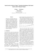
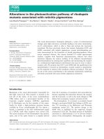
Fig. 1. Sequence alignments of BCCP (A) and BPL (B) from E. c oli and
A. aeolicus. Pairwise alignment was prepared using
CLUSTAL W
.(A)
The start residue of the BCCP-87 domain and the BCCP subtilisin
fragment are indicated (fl and Ñ, respectively). The start codon of the
BCCPD67 domain is shown (›), and the biotinylated lysine residue is
indicated (r). Secondary structural elements of the BCCP-87 domain
are shown and the ÔthumbÕ region is indicated (*). (B) Pairs of dis-
ordered surface loops which are close in space in the E. coli BirA
structure are shown ( and +) The trypsin cleavage sites of A. aeolicus
BPL are indicated ($) as is the site of subtilisin cleavage of E. coli BirA
(*).
1280 D. J. Clarke et al.(Eur. J. Biochem. 270) Ó FEBS 2003
expressed A. aeolicus BCCP lacking 67 residues from the
N-terminus (BCCPD67, Fig. 1) with an N-terminal His
6
-tag
(total length 96 aa). The homology scores between
A. aeolicus BCCPD67 and E. coli BCCP-87 (a domain
containing 87 C-terminal amino acids) are 51.9% identity
and 69.6% similarity (Fig. 1).
Cloning, expression and purification of BPL
The A. aeolicus bpl gene was amplified by PCR using
A. aeolicus genomic DNA as a template and cloned into
plasmid pCR2.1. DNA sequencing confirmed the previ-
ously published gene sequence, with the exception of a single
base change at position 325 (TfiC), which results in the
substitution of a cysteine residue with an arginine. Subse-
quently the bpl gene was cloned into a pET expression
vector for expression in various E. coli cells (DE3 lysogens);
we found optimum recovery of protein using the
BL21(DE3) strain. Cells were grown in shake flasks at
37 °C and expression induced with 1 m
M
IPTG (see
Experimental procedures).
The predicted pI of the A. aeolicus BPLis9.1andasthe
enzyme contains a high proportion of positively charged
residues, cation-echange chromatography was used to
purify it in a single step (Fig. 2, lanes 2–4). Initially the
crude lysate was incubated at 60 °C which resulted in the
precipitation of a significant quantity of E. coli proteins. It
was then necessary to dialyse the sample overnight (20 °C)
against 10 m
M
Hepes (pH 7.5) as immediate loading of an
untreated extract onto a ResourceS column resulted in very
poor binding (< 5%). It is unclear why this step was
necessary, but after dialysis binding to the cation-exchange
column approached 100%. BPL eluted from the column at
200 m
M
NaCl and we obtained the enzyme with a purity of
greater than 95% (as determined by SDS/PAGE). Electro-
spray mass spectrometry analysis gave the molecular mass
of the protein as 26636.8 ± 2.3 Da, consistent with the
post-translational removal of the N-terminal methionine
residue, and accurate to within experimental error of the
predicted value of 26634.6 Da. The final yield of BPL using
this method was > 10 mg per litre of cell culture and this
protein was used for all subsequent kinetic and cross linking
analysis.
Cloning, expression and purification of BCCPD67
We designed primers to clone a truncated domain of the
A. aeolicus bccp gene missing the first 201 bp, which encode
the N-terminal 67 amino acids of A. aeolicus BCCP (Fig. 1).
The truncated gene was amplified from genomic DNA
using PCR and cloned into the pCR2.1 vector. DNA
sequencing confirmed the expected gene sequence, and the
bccpD67 gene was subsequently cloned into a pET-derived
expression vector with an N-terminal His
6
-tag. E. coli
BL21(DE3) competent cells were used for recombinant
expression (described under Experimental procedures) and
the BCCPD67 cell lysate was first purified by nickel-affinity
chromatography (Fig. 2, lanes 6–8). The protein eluted with
200 m
M
imidazole and, as precipitation had been observed
at high concentrations of this eluant, it was immediately
diluted 1 : 1 with 10 m
M
Hepes (pH 7.5) and dialysed
against this buffer. SDS/PAGE analysis indicated
BCCPD67 to be > 90% pure but electrospray mass spectro-
metry revealed the presence of two distinct species. The first,
of molecular mass 10740.1 ± 1.1 Da, corresponded to the
predicted mass of apo-BCCPD67 (10739.6 Da) while the
second corresponding to the holo-form (biotinyated), with a
mass increase of 226.1 Da (10965.4 Da; predicted mass
10965.7 Da). This confirmed that the A. aeolicus BCCPD67
domain folded correctly, and was recognized and biotinyl-
ated by the host E. coli BirA. To separate the apo- and
holo-forms of BCCPD67 we employed anion exchange
chromatography in a similar way to that used for E. coli
BCCP-87 [27]. Fractions from the column were analysed by
electrospray mass spectrometry and the apo-protein eluted
at a slightly lower salt concentration than the holo-form
(160–240 m
M
NaCl vs. 240–320 m
M
NaCl). Approximately
80% of the apo-BCCPD67 was resolved from the holo-form
by collecting only the leading fractions of the protein peak.
The final yield of apo-BCCPD67 was 5–10 mg per litre of
cell culture and 1 mg per litre of the holo-form.
Cloning, expression and purification of BCCPD67 K117L
mutant
A mutant of the truncated bccpD67 gene, with the active
lysine residue (K117) replaced by a leucine residue, was
produced using the megaprimer method [21]. The mutation
was confirmed by DNA sequencing before the gene was
cloned into a pET-derived expression vector with an
N-terminal His
6
-tag and the resulting construct was then
transformed into E. coli BL21(DE3) cells for expression (as
described in Experimental procedures). The BCCPD67
K117L protein was purified using nickel-affinity chroma-
tography and the protein eluted with 200 m
M
imidazole
(Fig. 2, lanes 10–12). Protein-containing fractions were
immediately dialysed against 10 m
M
Hepes (pH 7.5). Fur-
ther purification on anion-exchange chromatography gave a
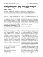
Fig. 2. Purification of A. aeolicus BPL, BCCPD67 and BCCPD67
K117L. Protein purification was analysed by SDS/PAGE under
reducing conditions. Lanes 1, 5 and 9, low molecular mass marker;
lane 2, BPL cell lysate; lane 3, BPL cell lysate after heat purifica-
tion;lane4,BPLafterResourseSpurification;lane6,BCCPD67 cell
lysate;lane7,BCCPD67 after Ni-affinity purification; lane 8, apo-
BCCPD67 after Mono-Q purification; lane 10, BCCPD67 K117L cell
lysate; lane 11, BCCPD67 K117L after Ni-affinity purification; lane 12,
BCCPD67 K117L after Mono-Q purification.
Ó FEBS 2003 Biotinylation in Aquifex aeolicus (Eur. J. Biochem. 270) 1281
single species with a mass of 10724.8 ± 1.1 Da, consistent
with the predicted mass of apo-BCCPD67 K117L of
10724.6 Da. A species was not present at +226 Da, an
indication that in vivo biotinylation had not occurred. The
yield of the apo-BCCPD67 K117L mutant was 15 mg of
protein per litre of cell culture.
Biochemical properties of BPL
Activity assays were performed with BPL by measuring
the incorporation of [
14
C]biotin into the purified apo-
BCCPD67 biotin-accepting domain [22]. In initial experi-
ments we observed optimal enzyme activity at pH 8.5,
and magnesium ions, ATP, biotin and apo-BCCPD67
were all required for activity. The activity of the enzyme
was also measured at varying temperatures, with optimal
activity at 70 °C. Activity was seen to decrease by
roughly 50% for every 10 °C drop in temperature, and
increasing the temperature above 70 °Cresultedin
enzyme precipitation, together with a dramatic loss in
activity (data not shown). The tolerance of BPL for
other nucleotide sources was measured by replacing ATP
with UTP, GTP or CTP. No BPL activity was detected
for any of these three substrates, suggesting that the
enzyme is completely dependent on ATP for its nucleo-
tide supply (data not shown).
In assays performed with BCCPD67 K117L as the biotin
acceptor no biotinylation was observed, verifying K117 as
the active residue and demonstrating the specificity of the
BPL catalysed reaction.
Kinetic analysis of BPL
The kinetic constants for
D
-biotin, MgATP and apo-
BCCPD67 were determined using steady-state kinetics
(Fig. 3). The K
m
for
D
-biotin was determined to be
440 ± 70 n
M
.TheK
m
values for BPLs from other species
range from low nanomolar to low micromolar; 67 ± 11 n
M
(Saccharomyces cerevisiae BPL), 300 n
M
(E. coli BirA),
130 n
M
(Arabidopsis thaliana HCS) and 3.3 m
M
(chicken
liver HCS1) [28–31]. The K
m
for MgATP was
15.1 ± 1.5 l
M
, which is similar to that determined for the
S. cerevisiae BPL (20.9 ± 3 l
M
)andA. thaliana HCS
(4.4 l
M
). In contrast, the K
m
for MgATP for E. coli BirA
is around 300 l
M
. It should be noted that the kinetic
analyses for each BPL were performed under slightly
different reactions conditions, for example an elevated
temperature was used in the study presented here. Finally,
the K
m
for apo-BCCPD67 was 160 ± 32 l
M
.Arangeof
biotinylation substrates have been used in assays of BPL
activity with cross-species reactivity frequently observed,
e.g. S. cerevisiae BPL has a K
m
of 11.1 ± 1 m
M
for E. coli
BCCP-87. However, we could not test E. coli BCCP-87 as a
substrate for BPL because the rate of biotinylation at 37 °C
was outside the lower limit of detection in our assay.
As shown in Scheme 1 the first step in all biotinylation
reactions studied thus far involves the synthesis of a
biotinyl-5¢-AMP intermediate and the release of PP
i
.This
molecule is the substrate for biotin transfer to BCCP and is
also the corepressor of E. coli BirA. To prove that
A. aeolicus BPL synthesises biotinyl-5¢-AMP we incubated
BPL with biotin and [
14
C]MgATP at 70 °C and used
streptavidin-coated membranes to capture radioactive bio-
tinyl-5¢-[
14
C]AMP (data not shown). Furthermore, we
noted that biotinylation was inhibited by the addition of
NaCl in concentrations above 200 m
M
.
Proteolysis of BPL
We subjected BPL to limited proteolysis in the presence and
absence of biotin and MgATP (Fig. 4). Digestion with both
trypsin and chymotrypsin resulted in formation of a
fragment of 21 kDa. Chymotrypsin digestion also pro-
duced an array of smaller peptide fragments. We found that
only 34% of total BPL remained after trypsin cleavage in
the absence of substrates. However, preincubation of BPL
with saturating amounts of biotin or MgATP separately
increased its resistance to digestion (50% and 63%
remained, respectively). Moreover, preincubation with both
substrates dramatically increased the resistance of BPL to
proteolysis with trypsin (98.9% remained). Comparative
analysis with chymotrypsin showed that 11% of BPL
remained intact after digestion. Preincubation of the enzyme
with MgATP afforded little protection (13% of BPL
remaining), whereas 34% and 92% BPL remained after
preincubation with biotin and biotin and ATP. Taken
together these results suggest that the binding of the
substrates and/or the formation of the intermediate,
biotinyl-5¢-AMP, plays a role in protecting BPL from
protease cleavage.
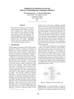
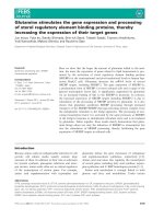
Fig. 3. Steady-state kinetic analysis of BPL substrate binding. The activity of A. aeolicus BPL was measured under steady-state conditions at 70 °C.
Two substrates were kept at constant saturating levels while the concentration of the third substrate was varied over the ranges shown above in the
graphs. From the curves, K
m
values for biotin (A), MgATP (B) and apo-BCCPD67 (C) were determined (see Experimental procedures).
1282 D. J. Clarke et al.(Eur. J. Biochem. 270) Ó FEBS 2003
LC-MS analysis of the peptide fragment produced from
BPL after treatment with trypsin revealed the presence of
two distinct species of mass 215549.5 ± 2.6 Da and
21678.6 ± 5.9 Da. Primary structure analysis of BPL
established these masses corresponded to trypsin cleavage
between R44 and K45, and K45 and W46 adjacent to the
proposed catalytic centre and biotinyl-5¢-AMP binding site.
Chemical crosslinking of BPL and BCCP
Although structures of E. coli BirA and both apo- and holo-
BCCP-87 have been determined, our goal was to isolate a
BPL:BCCP complex for biochemical and structural studies.
Previous work in our laboratory used the chemical
crosslinking agent EDC to isolate an E. coli flavodoxin–
flavodoxin reductase complex, so we used this reagent to
crosslink BPL and various forms of BCCPD67 [32]. Initially
we incubated BPL and apo-BCCPD67 in the presence of
excess EDC at room temperature with and without
saturating amounts of biotin and MgATP, but we did not
observe any crosslinked species of predicted molecular mass
36 kDa on SDS/PAGE (data not shown). However, a
species was observed when the incubation was carried out at
elevated temperatures, with 60 °C being the optimum
(Fig. 5A). The presence of the substrates had no observable
effect on crosslinking. Interestingly, when BPL was incuba-
ted with holo-BCCPD67 and EDC the amount of cross-
linked species generated was significantly reduced compared
to the apo form (Fig. 5B). Moreover, the incubation of BPL
with the BCCPD67 K117L mutant led to the formation of
crosslinked complex in comparable amounts to that using
apo-BCCPD67 (Fig. 5C). Purification of the BPL:
BCCPD67 complex from unreacted proteins was achieved
using size exclusion chromatography, which resolved the
mixture into three peaks (Fig. 6). We noted that both BPL,
BCCPD67 and the complex eluted from the size exclusion
column at retention volumes different to that predicted by
their molecular masses (45, 35 and 70 kDa, respectively).
However, analysis by SDS/PAGE revealed that the
BPL:BCCPD67 complex eluted from the column first and
had a molecular mass of 37 kDa (Fig. 6, inset). Electrospray
analysis of the complex gave a molecular mass of
37 200 ± 200 Da which agrees well with the predicted
mass of a 1 : 1 heterodimer.
Discussion
The attachment of biotin to the specific lysine residue of the
apo- forms of biotin-requiring enzymes is a complex,
multistep reaction. The BPL enzyme (also known as
holocarboxylase synthetase, HCS) catalysing this process
first activates biotin as biotinyl-5¢-AMP then transfers the
biotin to a specific lysine of the BCCP domain. The BPLs
and BCCPs from a diverse range of organisms including
E. coli (BirA), yeast, human and plant have been isolated
and it has been shown that the BPL from one organism can
biotinylate the BCCP domain from another [28]. This
suggests some degree of structural homology between these
proteins and primary structure analysis reveals there is a
high degree of amino acid sequence similarity throughout
the catalytic domain of the BPL family and the biotinyl
domain of BCCPs [33]. An understanding of the protein–
Fig. 5. SDS/PAGE analysis of chemical crosslinking assays. Gel A, crosslinking of BPL and apo-BCCPD67. Gel B, crosslinking of BPL and holo-
BCCPD67. Gel C, crosslinking of BPL and BCCPD67 K117L. Lanes 1–5 of each gel, assay after 0, 5, 10, 15 and 30 min respectively. Gel A, lanes 6
and 7, control assays with BCCPD67 alone and BPL alone.
Fig. 4. Proteolysis of A. aeolicus BPL. A. aeolicus BPL was treated
with trypsin or chymotrypsin either with or without equilibrating the
enzyme with 1 m
M
MgATP and/or 50 l
M
biotin. Lanes 1–4, Trypsin
digest; lane 1 BPL; lane 2, BPL + MgATP; lane 3, BPL + biotin;
lane 4, BPL + MgATP and biotin. Lanes 5–8 Chymotrypsin digest;
lane 5, BPL; lane 6, BPL + MgATP; lane 7, BPL + biotin; lane 8,
BPL + MgATP and biotin.
Ó FEBS 2003 Biotinylation in Aquifex aeolicus (Eur. J. Biochem. 270) 1283
protein interactions that mediate this highly specific reaction
requires three dimensional structures of each of the com-
ponents. The structure of the E. coli BirA monomer in
complex with biotinyl-lysine revealed details of the protein–
substrate interactions but several loops within the active site
were disordered [9]. More recently, the structure of the BirA
dimer has provided insights into how the ligase also acts as a
transcriptional repressor by binding to the E. coli biotin
operon operator [12]. The structures of the apo- and holo-
forms of E. coli BCCP-87, determined by X-ray and NMR,
are virtually identical and showed that the biotinyl-lysine
residue is located at an exposed b-turn, flanked by
important, highly conserved methionine residues [13,15].
A more recent NMR study, combined with results from site-
directed and random mutagenesis [29,34,35], allowed mod-
elling of the elusive E. coli BPL:BCCP-87 complex and it
appears that its formation is dependent on subtle, compet-
ing protein–protein interactions [36].
Analysis of the complete genome of the hyperthermophile
A. aeolicus revealed the presence of BPL and BCCP
homologues (Fig. 1). The A. aeolicus BPL enzyme belongs
to the class I group of BPLs since it lacks the DNA-binding
domain found in BirA and is the smallest characterized thus
far. Eukaryotic BPLs also lack predicted DNA-binding
domains but have large N-terminal extensions with
unknown functions [33]. The full-length A. aeolicus BCCP
has a C-terminus showing high sequence homology to the
biotin domains of biotin-carboxylases and contains the
eight amino acid ÔthumbÕ motif found in E. coli BCCP
[33,37,38]. The N-terminus has a large proportion of
charged residues, and displays little similarity to any other
BCCPs.
Using recombinant proteins isolated from E. coli we have
characterized the full-length BPL and BCCP biotinylation
domain BCCPD67 (with a His
6
N-terminal tag) from a
hyperthermophile. We have gained insight into this
extremely specific post-translational modification reaction
at high temperatures and used features of the two A. aeo-
licus proteins to capture a BPL:BCCP complex. We found
A. aeolicus BPL to be monomeric, and thus competing
homodimerization interactions found in E. coli BirA are
not present. We isolated a mixture of apo- and holo-forms
of A. aeolicus BCCPD67 and so conclude that it must be a
substrate for E. coli BPL in vivo. Biotinylation in hyper-
thermophiles proceeds via the two-step reaction sequence
found in other organisms (Scheme 1). Isolated A. aeolicus
BPL could biotinylate apo-BCCPD67 at temperatures up to
70 °C albeit at a slow rate. It is interesting to compare the
A. aeolicus BPL:BCCPD67 biotinylation reaction with that
of a mutant E. coli BirA lacking the N-terminal DNA
binding domain (BirA65-321) and E. coli BCCP-87. The
BirA65-321 mutant could synthesize biotinyl-5¢-AMP and
transfer biotin to apo-BCCP-87 at the same rate as wild-
type BirA. However, the affinity of BirA65-321 mutant for
biotin and biotinyl-5¢-AMP was decreased 100-fold and
1000-fold, respectively [39]. This suggested that in BirA, the
N-terminal domain is somehow involved in tight-binding of
the two ligands. In future, it would be interesting to study a
BPL:BirA chimera by fusing the DNA-binding domain at
the N-terminus of A. aeolicus BPL.
Substrate K
m
valuesforBPLsfromanumberofspecies
have been shown to range from the low nanomolar to low
millimolar. In steady-state kinetic assays at 70 °C, the
A. aeolicus BPL bound biotin, MgATP and apo-BCCPD67
with affinites of 440 n
M
,15.1l
M
and 160 l
M
, respectively.
The kinetic constant for MgATP suggests that A. aeolicus
BPL resembles those from eukaryotic biotin auxotrophs
(low micromolar). In contrast, E. coli BirA binds MgATP
with a K
m
in the low millimolar range which reflects its dual
function as both repressor of biotin biosynthesis and biotin
ligase. It is interesting to note that A. aeolicus contains all
thegenesrequiredtoconvertpimelatetobiotin(bioW, bioF,
bioA, bioDandbioB) suggesting it can synthesize this
vitamin but the in vivo concentration within A. aeolicus cells
is unknown. The K
m
for the apo-BCCPD67 domain used in
this study is high compared to others but this may reflect the
factthatthefirst67aminoacidresidues,whichcontaina
high number of charged residues, could play an important
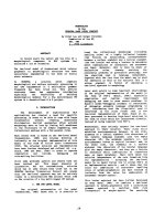
Fig. 6. Purification of the chemically crosslinked BPL:apo-BCCPD67 complex by size-exclusion chromatography. The chromatogram above was
obtained when the cross-linking reaction was applied to a Superdex 75 column. The three peaks correspond to the crosslinked complex (7–8 mL),
BPL (10 mL) and BCCPD67 (11–12 mL). Insert: SDS/PAGE analysis of the column fractions. Lane 1, cross-linking reaction before purification.
Lane 2–11, 1 mL fractions eluting between 6 and 15 mL.
1284 D. J. Clarke et al.(Eur. J. Biochem. 270) Ó FEBS 2003
role in tight binding to BPL. Most biochemical studies use
these truncated BCCP domains and future work using full
length BCCPs should elucidate the role of the N-terminal
interaction with BPL. It is also possible that the addition of
the His
6
-tag to the protein has altered its kinetic properties
and may contribute to the abnormally high K
m
for
BCCPD67. The calculated k
cat
/K
m
for biotin of
1.7 ± 0.1 · 10
4
M
)1
Æs
)1
is 300-, 100- and 35-fold smaller
than the E. coli BirA, yeast and A. thaliana BPL enzymes,
respectively [28,30,40] but reflects the fact that the A. aeo-
licus BPL k
cat
is low at 70 °C(cf. A. aeolicus grows
optimally at 95 °C).
Limited proteolysis with trypsin produced two fragments
of 20 kDa, differing in length by only one residue (Fig. 4).
Mass spectrometry revealed that cleavage had occurred
after residues R44 and K45 which, by comparison with
E. coli BirA, are predicted to lie near the putative inter-
mediate binding site (Fig. 1). Treatment of BPL with trypsin
and chymotrypsin in the presence of biotin or MgATP
decreased the susceptibility to cleavage by a small but
noticeable amount. However, incubation of the enzyme in
the presence of both substrates rendered A. aeolicus BPL
protease-resistant. The same region is protease-sensitive in
S. cerevisiae BPL and is also protected by incubation with
bothbiotinandMgATP[28].TheE. coli BirA structure
contains five surface loops, four of which are in the central
domain with loop regions (110–128, 212–233) and (140–146,
193–199) close together in three-dimensional space [6]. The
region containing 110–128 in E. coli BirA is highly analog-
ous to residues 32–50 in A. aeolicus BPL whereas the other
loop regions have low pairwise sequence homology. A
protease-sensitive site has been reported between residues
217 and 218 of BirA. In contrast, A. aeolicus BPL is not
cleaved at this site but is cleaved in the adjacent loop region
(32–50). This suggests that this highly conserved region
forms an exposed loop near the biotinyl-5¢-AMP binding
site (Fig. 1). These flexible, unstructured regions are also
involved in BCCP binding and are believed to become more
rigid upon substrate-binding [6,34].
A recent combined mutagenesis/biological selection
approach identified two single glutamate residues E119
and E147 of E. coli BCCP-87 that appear to interact with
BPL [22]. A BCCP-87 E119K mutant is inactive as a
substrate for BirA, whereas the E147K protein could be
biotinylated, albeit poorly. It is presumed that these acidic
BCCP-87 residues interact with basic BirA counterparts and
mutation of BirA residues K277 and R317 were found to
effect biotinylation and ATP-binding, respectively. This
surprising result suggested that the C-terminal domain of
BirA, which had been ascribed no biochemical function,
also plays a significant role in apo-BCCP and substrate
recognition [29].
It has been shown that ion pair networks are a common
feature in heat-resistant proteins and are believed to play
important roles in their increased thermal stability [17,41].
As both the A. aeolicus BPL and BCCP contain a large
number of charged residues and we observed inhibition of
biotinylation at high salt concentrations, we presume that
ionic interactions are involved in the formation of the
hyperthermophilic BPL:BCCPD67 complex. To investigate
the formation of the BPL:BCCPD67 heterodimer we used
the chemical cross-linking agent EDC to capture a
BPL:BCCP complex for the first time. The zero-length
EDC reagent activates acidic residues on one protein to
form an unstable urea derivative [42]. This derivative then
reacts with a nucleophile (such as lysine) on another protein
to form an amide link between the two proteins. Incubation
of BPL and apo-BCCPD67 in the presence of EDC led to
the time-dependent appearance of a species of 37 kDa on
SDS/PAGE gels (Fig. 5A), which is in agreement with the
predicted mass of a 1 : 1 complex of BPL and apo-
BCCPD67. We noticed that BPL, BCCPD67 and the
complex eluted earlier than predicted from the size-exclu-
sion column. Future studies will analyse the proteins by
equilibrium sedimentation experiments in a similar way to
that described for the BCCP-87 and BCCP [25]. Neverthe-
less, the complex was easily separated from the unreacted
proteins using this procedure (Fig. 6) and allowed us to
confirm its mass by electrospray mass spectrometry. Inter-
estingly, the complex was not formed between BPL and
holo-BCCPD67 (Fig. 5B) suggesting that biotinylation had
either caused a conformational change in BCCPD67 such
that it no longer bound to BPL or that the biotin moiety had
somehow blocked residues that react with the EDC reagent.
Furthermore, a complex was formed between the BCCPD67
K117L mutant and BPL both in the absence and presence
of saturating amounts of biotin and MgATP (Fig. 5C). This
demonstrates that the active lysine residue does not take
part in the cross-linking reaction and saturating amounts of
both substrates do not inhibit complex formation.
Although the published 3D structures of the apo- and
holo- forms of BCCP-87 show no major structural differ-
ences, some structural studies (both NMR and X-ray) have
concluded that the lack of any major differences between
them might not be wholly reflected in their behaviour in
solution [15]. NMR titration experiments were carried out
with BirA and apo-BCCP-87 and, in light of our data, it
would be interesting to repeat this work with BirA and holo-
BCCP-87 to determine if any differences arise. Recent
elegant studies by Cronan and Solbiati et al. highlight a
difference in the stability of apo-BCCP-87 and holo-BCCP-
87 to proteolysis and stress the importance of the essential
so-called ÔthumbÕ domain of BCCP-87 (residues 91–100)
which had previously been shown to interact with the ureido
ring of the attached biotin moiety [37,43]. Studies using
chemically biotinylated BCCP-87 recently confirmed that
this increased stability is an inherent property of holo-
BCCP-87 and not due to a conformational change imparted
by BPL. Furthermore, thumbless holo-BCCP-87 mutants
exhibit little increased stability over their apo- counterparts,
implying the majority of this increased stability is due to the
thumb–biotin interaction. The authors conclude that the
more protease sensitive apo- BCCP has a more dynamic
form than the holo- protein. The A. aeolicus BCCPD67 also
contains a well-conserved thumb domain (Fig. 1) and we
are currently producing thumbless BCCPD67 mutants for
analysis by EDC cross-linking with BPL (D. Clarke and
D. Campopiano, unpublished results).
A recent study suggested that the C-terminal domain of
BirA is essential for the catalytic activity of the enzyme and
plays a role in ATP and BCCP binding [29]. Also, a model
of the E. coli BirA:holo-BCCP-87 complex has been
suggested based upon structural studies, sequence analysis,
mutagenesis and limited proteolysis experiments [36]. The
Ó FEBS 2003 Biotinylation in Aquifex aeolicus (Eur. J. Biochem. 270) 1285
model (PDB code 1K67) was built using the coordinates of
the BirA dimer in the presence of biotin (PDB code 1HXD)
and holo-BCCP-87 (PDB 1BIA). Residues in both E. coli
proteins thought to be responsible for BirA:BCCP-87
complex formation are conserved in A. aeolicus BPL and
BCCPD67 (Fig. 1). A current goal is to identify the charged
residues taking part in the EDC-mediated crosslinking
reaction and A. aeolicus BPL and BCCPD67 mutants are
currently being studied using high-temperature in vitro
biotinylation and chemical crosslinking assays.
Acknowledgements
We wish to thank Profs. K. Stetter and R. Huber (University of
Regensburg) for the gift of A. aeolicus chromosomal DNA. The
Nuffield Foundation Bursary Scheme is acknowledged for its support
of (J. C.). This work was supported by the Biotechnology and
Biological Sciences Research Council, UK, and the University of
Edinburgh.
References
1. Campbell, J.W. & Cronan, J.E.J. (2002) Bacterial fatty acid
biosynthesis: Targets for antibacterial drug discovery. Ann. Rev.
Microbiol. 55, 305–332.
2. Cronan, J.E. Jr & Waldrop, G.L. (2002) Multi-subunit acetyl-
CoA carboxylases. Prog. Lipid Res. 41, 407–435.
3. Samols, D., Thornton, C.G., Murtif, V.L., Kumar, G.K., Haase,
F.C. & Wood, H.G. (1988) Evolutionary conservation among
biotin enzymes. J. Biol. Chem. 263, 6461–6464.
4. Knowles, J. (1989) The mechanism of biotin-dependent enzymes.
Annu. Rev. Biochem. 58, 195–221.
5. Perham, R.N. (2000) Swinging arms and swinging domains in
multifunctional enzymes: catalytic machines for multistep reac-
tions. Annu. Rev. Biochem. 69, 961–1004.
6. Chapman-Smith, A. & Cronan, J.E. Jr (1999) The enzymatic
biotinylation of proteins: a post-translational modification of
exceptional specificity. Trends Biochem. Sci. 24, 359–363.
7. Beckett, D. & Matthews, B.W. (1997) Escherichia coli repressor of
biotin biosynthesis. Methods Enzymol. 279, 362–377.
8. Cronan, J.E. Jr (1989) The E. coli bio operon: transcriptional
repression by an essential protein modification enzyme. Cell 58,
427–429.
9. Wilson, K.P., Shewchuk, L.M., Brennan, R.G., Otsuka, A.J. &
Matthews, B.W. (1992) Escherichia coli biotin holoenzyme syn-
thetase/bio repressor crystal structure delineates the biotin- and
DNA-binding domains. Proc Natl Acad. Sci. USA 89, 9257–9261.
10. Brennan, R.G., Vasu, S., Matthews, B.W. & Otuska, A.J. (1989)
Crystallization of the bifunctional biotin operon repressor. J. Biol.
Chem. 264,5.
11. Kwon, K. & Beckett, D. (2000) Function of a conserved sequence
motif in biotin holoenzyme synthetases. Protein Sci. 9, 1530–1539.
12. Weaver, L.H., Kwon, K., Becket, D. & Matthews, B.W. (2001)
Corepressor-induced organisation and assembly of the biotin
repressor: a model for allosteric activation of a transcriptioal
regulator. Proc. Natl Acad. Sci. USA 98, 6045–6050.
13. Athappilly, F.K. & Hendrickson, W.A. (1995) Structure of the
biotinyl domain of acetyl-coenzyme A carboxylase determined by
MAD phasing. Structure 3, 1407–1419.
14. Yao, X., Wei, D., Soden, C.J., Summers, M.F. & Beckett, D.
(1997) Structure of the carboxy-terminal fragment of the apo-
biotin carboxyl carrier subunit of Escherichia coli acetyl-CoA
carboxylase. Biochemistry 36, 15089–15100.
15. Roberts, E.L., Shu, N., Howard, M.J., Broadhurst, R.W.,
Chapman-Smith, A., Wallace, J.C., Cronan, J.E. Jr & Perham,
R.N. (1999) solution structures of apo and holo biotinyl domains
from acetyl coenzyme A carboxylase of Escherichia coli
determined by triple-resonance nuclear magnetic resonance spec-
troscopy. Biochemistry 38, 5045–5053.
16. Deckert, G., Warren, P.V., Gaasterland, T., Young, W.G., Lenox,
A.L.,Graham,D.E.,Overbeek,R.,Snead,M.A.,Keller,M.,
Aujay, M., Huber, R., Feldman, R.A., Short, J.M., Olsen, G.J. &
Swanson, R.V. (1998) The complete genome of the hyper-
thermophilic bacterium Aquifex aeolicus. Nature 392, 353–358.
17. Hough, D.W. & Danson, M.J. (1999) Extremozymes. Curr. Op. In
Chem. Biol. 3, 39–46.
18. Rothschild, L.J. & Mancinelli, R.L. (2001) Life in extreme
environments. Nature 409, 1092–1101.
19. Mukhopadhyay, B., Purwantini, E., Kreder, C.L. & Wolfe,
R.S. (2001) Oxaloacetate synthesis in the methanarchaeon
Methanosarcina barkeri: pyruvate carboxylase genes and a puta-
tive Escherichia coli-type bifunctional biotin protein ligase gene
(bpl/birA) exhibit a unique organization. J. Bacteriol. 183, 3804–
3810.
20. Sambrook, J., Maniatis, T. & Fritsch, E.F. (1989) Molecular
Cloning: a Laboratory Manual, 2nd edn. Cold Spring Harbor
Laboratory Press, Cold Spring Harbor, New York, USA.
21. Sarker, G. & Summer, S.S. (1990) The Megaprimer Method of
Site-directed Mutagenesis. Biotechniques 8, 404–407.
22. Chapman-Smith, A., Morris, T.W., Wallace, J.C. & Cronan, J.E.
Jr (1999) Molecular recognition in a post-translational modifica-
tion of exceptional specificity. J. Biol. Chem. 274, 1449–1457.
23. Lee, H.J. & Wilson, I.B. (1971) Enzymic parameters: measurement
of V and K
m
. Biochim. Biophys. Acta. 242, 519–522.
24. Thompson, J.D., Higgins, D.G. & Gibson, T.J. (1994) Clustal W:
Improving the sensitivity of progressive multiple sequence
alignment through sequence weighting, positions-specific gap
penalties and weight matrix choice. Nucleic Acids Res. 22, 4673–
4680.
25. Nenortas, E. & Beckett, D. (1996) Purification and characterisa-
tion of intact and truncated forms of Escherichia coli biotin car-
boxyl carrier subunit of acetyl-CoA carbioxylase. J. Biol. Chem.
271, 7559–7567.
26. Stolz, J., Ludwig, A. & Sauer, N. (1998) Bacteriophage lambda
surface display of a bacterial biotin acceptor domain reveals the
minimal peptide size required for biotinylation. FEBS Letts 440,
213–217.
27. Chapman-Smith, A., Turner, D.L., Cronan, J.E., Jr, Morris, T.W.
& Wallace, J.C. (1994) Expression, biotinylation and purification
of a biotin-domain peptide from the biotin carboxy protein of
Escherichia coli acetyl-CoA carboxylase. Biochem. J. 302, 881–887.
28. Polyak, S.W., Chapman-Smith, A., Brautigan, P.J. & Wallace,
J.C. (1999) Biotin protein ligase from Sacchacromyces cerevisiae.
J. Biol. Chem. 274, 32847–32854.
29. Chapman-Smith, A., Mulhern, T.D., Whelan, F., Cronan, J.E. Jr
& Wallace, J.C. (2001) The C-terminal domain of biotin protein
ligase from E. coli is required for catalytic activity. Protein Sci. 10,
2608–2617.
30.Tissot,G.,Pepin,R.,Job,D.,Douce,R.&Alban,C.(1998)
Purification and properties of the chloroplastic form of biotin
holocarboxylase synthetase from Arabidopsis thaliana over-
expressed in Escherichia coli. Eur. J. Biochem. 258, 586–596.
31. Murthy, P.N.A. & Mistry, S.P. (1974) In vitro synthesis of pro-
pionyl-CoA holocarboxylase by a partially purified mitochondrial
preparation from biotin-deficient chicken liver. Can. J. Biochem.
52, 800–803.
32. McIver, L., Leadbeater, C., Campopiano, D.J., Baxter, R.L.,
Daff, S., Chapman, S.K. & Munro, A.W. (1998) Characterisation
of flavodoxin NADP
+
oxidoreductase and flavodoxin; key
components of electron transfer in Escherichia coli. Eur. J. Bio-
chem. 257, 577–585.
1286 D. J. Clarke et al.(Eur. J. Biochem. 270) Ó FEBS 2003
33. Chapman-Smith, A. & Cronan, J.E. Jr (1999) Molecular biology
of biotin attachment to proteins. J. Natr. 129, 477S–484S.
34. Reche, P.A., Howard, M.J., Broadhurst, R.W. & Perham, R.N.
(2000) Heteronuclear NMR studies of the specificity of the post-
translational modification of biotinyl domains by biotinyl protein
ligase. FEBS Lett. 479, 93–98.
35. Polyak, S.W., Chapman-Smith, A., Mulhern, T.D., Cronan, J.E.
Jr & Wallace, J.C. (2001) Mutational analysis of protein substrate
presentation in the post-translational attachment of biotin to
biotin domains. J. Biol. Chem. 276, 3037–3045.
36. Weaver, L.H., Kwon, K., Beckett, D. & Matthews, B.W. (2001)
Competing protein:protein interactions are proposed to control
the biological switch of the E. coli biotin repressor. Protein Sci. 10,
2618–2622.
37. Cronan, J.E. Jr (2001) The biotinyl domain of Esherichia coli
acetyl-CoA carboxlyase. J. Biol. Chem. 276, 37355–37364.
38. Cronan, J.E. Jr (2002) Interchangable enzyme molecules. J. Biol.
Chem. 277, 22520–22527.
39. Xu, Y. & Beckett, D. (1996) Evidence for interdomain interaction
in the Escherichia coli repressor of biotin biosynthesis from studies
of an N-terminal domain deletion mutant. Biochemistry 35, 1783–
1792.
40. Xu, Y. & Beckett, D. (1997) Biotinyl-5¢-adenylate synthesis cata-
lysed by Escherichia coli repressor of biotin biosynthesis. Methods
Enzymol. 279, 405–421.
41. Petsko, G.A. (2001) Structural basis of thermostablity in hyper-
thermophilic proteins, or ÔThereÕs more than one way to skin a
cat’. Methods Enzymol. 334, 469–478.
42. Grabarek, Z. & Gergely, J. (1990) Zero-length cross-linking pro-
cedure with the use of active esters. Anal. Biochem. 185, 131–135.
43. Solbiati, J., Chapman-Smith, A. & Cronan, J.E. Jr (2002) Stabi-
lization of the biotinoyl domain of Escherichia coli acetyl–CoA
carboxylase by interactions between the attached biotin and the
protruding ÔthumbÕ structure. J. Biol. Chem. 277, 21604–21609.
Ó FEBS 2003 Biotinylation in Aquifex aeolicus (Eur. J. Biochem. 270) 1287