Báo cáo khoa học: Structure, linkage mapping and expression of the heart-type fatty acid-binding protein gene (fabp3 ) from zebrafish (Danio rerio) pot
Bạn đang xem bản rút gọn của tài liệu. Xem và tải ngay bản đầy đủ của tài liệu tại đây (512.43 KB, 12 trang )
Structure, linkage mapping and expression of the heart-type fatty
acid-binding protein gene (
fabp3
) from zebrafish (
Danio rerio
)
Rong-Zong Liu
1
, Eileen M. Denovan-Wright
2
and Jonathan M. Wright
1
1
Department of Biology and
2
Department of Pharmacology, Dalhousie University, Halifax, Nova Scotia, Canada
We have determined the cDNA nucleotide sequence,
deduced the amino acid sequence and defined the gene
structure for the cellular heart-type (H-FABP) or fatty acid-
binding protein 3 (FABP3) from zebrafish. The zebrafish
FABP3 exhibited the greatest amino acid sequence identity
to fish and mammalian heart-type FABPs. 3¢ RACE and 5¢
RLM-RACE mapped two alternative polyadenylation sites
and three transcription start sites, respectively. Southern
blot and hybridization analysis indicated that a single fabp3
gene exists in the zebrafish genome. The zebrafish fabp3 gene
consists of four exons interrupted by three introns with
identical exon/intron structure and coding capacity with that
of orthologous mammalian H-FABP genes. Radiation
hybrid mapping assigned the zebrafish fabp3 gene to linkage
group 19 of the zebrafish genome. Comparative genomic
analysis revealed conserved syntenies of the zebrafish fabp3
gene and the orthologous human and mouse fabp3 genes.
Northern blot analysis detected an mRNA transcript of 780
nucleotides. Insituhybridization of the zebrafish fabp3-
specific oligonucleotide probe to tissue sections of adult
zebrafish revealed that the fabp3 mRNA was localized in the
ovary and liver, but not in the heart, muscle or brain as
reported for the mammalian fabp3 gene transcript. RT-
PCR, however, detected zebrafish fabp3 mRNA in all
the tissues examined. Emulsion autoradiography further
revealed that the zebrafish fabp3 mRNA was most abundant
in primary growth stage (stage I) oocytes and decreased
during the oocyte growth phase. The fabp3 mRNA levels
were reduced and restricted to the ooplasm of cortical
alveolus stage (stage II) oocytes, and nearly undetectable in
stage III and matured oocytes. Inspection of the 5¢ upstream
sequence of the zebrafish fabp3 gene revealed a number of
cis elements that may be involved in the expression of
the zebrafish fabp3 gene in oocytes and liver.
Keywords: FABP gene; oocyte; tissue-specific expression; cis
element; linkage mapping.
Intracellular fatty acid-binding proteins (FABP) and the
related cellular retinol and retinoic acid binding proteins
(CRBP and CRABP, respsectively) are low molecular mass
( 15 kDa) polypeptides encoded by a multigene family,
hereafter collectively referred to as the intracellular lipid-
binding protein (ILBP) family [reviewed in 1–3]. Based on
X-ray crystallography and protein modelling studies, all
ILBPs investigated have a similar clamshell-shaped, three-
dimensional conformation comprised of two orthogonal
b-sheets with two a-helices. These proteins have been shown
to bind hydrophobic ligands with high affinity in vitro, but
until recently their role(s) in vivo remained the source of
speculation. Compelling evidence for their physiological
role(s), however, has been provided by work with knockout
mice [4–6]. Based on this work, there is now direct evidence
that some of the proteins of this multigene family play an
important role in the uptake and transport of long-chain
fatty acids, fuel utilization and the interaction with other
transport and enzyme systems. Indirect evidence suggests
that FABPs may be involved in the regulation of transcrip-
tion of specific genes during early development and
neurogenesis, and in diseased states [1,7,8].
Fourteen members of this multigene family have been
identified in mammals and named according to the initial
site of isolation, e.g. adipocyte fatty acid-binding protein
(A-FABP), brain (B-FABP), epidermal (E-FABP), heart
(H-FABP), intestinal (I-FABP), liver (L-FABP), etc.
Nomenclature based on the initial site of isolation or
patterns of tissue-specific expression has given rise to
multiple names for the same proteins, which is, on occasion,
confusing. For instance, the H-FABP was named mam-
mary-derived growth inhibitor and muscle-type FABP due
to its presence in mammary gland and skeletal muscle [9,10].
In addition, tissue-distribution or function, or both, of
proteins encoded by orthologous genes from different species
may have different physiological functions. Hertzel and
Bernlohr [2] have suggested therefore an alternative nomen-
clature for ILBPs, which we have followed in this report.
Schleicher et al. [11] speculate that the ILBP multigene
family has undergone at least 14 gene duplications. The
liver/intestinal/ileal FABP clade emerged from the heart/
adipose/myelin P2 FABP lineage some 700 million years
Correspondence to J. M. Wright, Department of Biology,
Dalhousie University, Halifax, Nova Scotia, Canada, B3H 4H7.
Fax: + 1 902 494 3736, Tel.: + 1 902 494 6468,
E-mail:
Abbreviations: FABP, fatty acid binding protein; FABP3, heart-type
fatty acid-binding protein; CRBP, cellular retinol binding protein;
CRABP, cellular retinoic acid binding protein; ILBP, intracellular
lipid binding protein; RLM-RACE, RNA ligase-mediated RACE;
CIP, calf intestinal phosphatase; TAP, tobacco acid pyrophosphatase;
RH, radiation hybrid; mya, million years ago;
EST, expressed sequence tag.
(Received 17 March 2003, revised 30 May 2003, accepted 5 June 2003)
Eur. J. Biochem. 270, 3223–3234 (2003) Ó FEBS 2003 doi:10.1046/j.1432-1033.2003.03705.x
ago (mya), prior to the vertebrate/invertebrate divergence.
The CRBP and CRABP members seem to have diverged
from the liver/intestinal FABP clade about 500 mya. The
mammalian CRBPI and CRBPII are thought to have arisen
by gene duplication after the split with the amphibia, as
Xenopus has a single CRBP gene [12].
Not only has the primary amino acid sequence of
members of this multigene family been conserved over
hundreds of millions of years, so has their gene structure. All
ILBP genes studied to date consist of four exons of almost
identical coding capacity interrupted by three introns of
varying size. The muscle-type FABP gene from the desert
locust is the sole exception in that it lacks intron 2 [13].
While the coding sequence and structure of the ILBP genes
have been conserved, following duplication of the ancestral
gene the regulatory elements in their gene promoters have
not, giving rise to specific temporal and spatial patterns of
expression for members of this multigene family.
Lipid storage and utilization differs in various taxa. For
example, mammals store lipids subcutaneously and in
adipose tissue, whereas fish deposit and store lipids in several
tissues including mesenteric fat, liver, dark muscle [14] and
oocytes [15 and references therein]. Tissue-specific patterns
of ILBP gene expression in nonmammalian species may
therefore differ from that observed in mammals. Few studies
have focused on the tissue-specific patterns of ILBP expres-
sion in fishes, the largest and most evolutionary diverse
group of vertebrates. FABPs have been detected in the white
heart muscle of ocean pout and sea raven, the liver of nurse
shark, elephant fish, lamprey and catfish, and the aerobic
muscle of striped bass. More recently, cDNAs have been
characterized for an H-FABP from rainbow trout and two
distinct isoforms of H-FABP from the ventricle of four
Antarctic fishes (reviewed in [1]). We have recently reported
the nucleotide sequence of cDNAs encoding three FABPs
(I-, B-, and Lb-type FABP) and a related CRBPII, and their
tissue-specific expression in adult zebrafish [16–19]. Here we
describe an H-FABP (fabp3) gene from zebrafish. While high
amino acid sequence similarity, identical gene structure and
coding capacity, and conserved syntenic relationship to the
mammalian H-FABP suggest that it is an orthologous
H-FABP gene (or fabp3), expression patterns of the zebrafish
and mammalian fabp3 genes, as well as their 5¢cis regulatory
elements were strikingly different. This zebrafish FABP
mRNA was detected in abundance in primary oocytes and in
the liver by both tissue section in situ hybridization and RT-
PCR, but the mRNA was only detected in the heart and
other tissues of adult zebrafish by RT-PCR. Messenger
RNA levels for the zebrafish fabp3 decreased during the first
phase of oogenesis, the growth phase, to the point that it was
undetectable in mature oocytes. We propose that differences
in tissue expression patterns of the fabp3 genes from
mammals and zebrafish were a result of evolutional diver-
gence of transcriptional regulation.
Materials and methods
Growth and maintenance of zebrafish adults
and embryos
Zebrafish were purchased from a local aquarium store
and cultured in filtered, aerated water at 28.5 °Cin35L
aquaria. Fish were maintained on a 24-h cycle of 14 h light
and 10 h darkness. Fish were fed with a dry fish feed,
TetraMin Flakes (TetraWerke, Melle, Germany), in the
morning and hatched brine shrimp (Artemia cysts from
INVE, Grantsville, UT, USA) in the afternoon. Fish
breeding and embryo manipulation was conducted accord-
ing to established protocols [20]. Animal protocols were
reviewed by the University Committee on Laboratory
Animals and conducted in accordance with the Canadian
Council on Animal Care’s Ethics of Animal Investigation.
Cloning of
fabp3
cDNAs from zebrafish
3¢ Rapid amplification of cDNA ends (3¢ RACE) was
employed to clone a full length coding sequence for the
zebrafish fabp3 using primers based on a GenBank zebrafish
EST (accession number, AW077983). First strand cDNA
was synthesized from total RNA of adult zebrafish with a 3¢
adaptorprimer(5¢-GGCCACGCGTCGACTAGTACT
17
-3¢)
and reverse transcriptase Superscript II (GibcoBRL, Bur-
lington, Ontario, Canada). Using the reverse-transcription
reaction as template, the cDNA was amplified by PCR
using the antisense primer complementary to the 3¢ adaptor
and a 5¢ sense primer (5¢-TCAGCTCAAACATGGCA
GAC-3¢, Fig. 1B). The PCR product was separated by
electrophoresis in low melting point agarose and purified
from the gel using the Qiaquick gel extraction kit (Qiagen,
Mississauga, Ontario, Canada). The minor band (Fig. 2)
was reamplified by PCR and repurified. The purified DNA
fragments were cloned into the plasmid vector, pGEM-T
(Promega, Madison, WI, USA), and three of the major
band and one of the minor band clones were sequenced
(University of Toronto Sequencing Facility, Toronto,
Ontario, Canada) in both directions. The deduced amino
acid sequence of the FABP was determined using the
algorithm in
GENE RUNNER
v. 3.05 (Hastings Software,
Inc.). Nucleotide and amino acid sequences were aligned
using
CLUSTALW
[21].
RT-PCR
Total RNA was extracted from adult zebrafish tissues using
Trizol reagent and the protocol recommended by the
supplier (GibcoBRL, Burlington, Ontario, Canada). One
microgram of total RNA from each sample was used as the
template for the synthesis of first strand cDNA by reverse
transcriptase (SuperScript II, GibcoBRL). For PCR ampli-
fication, oligonucleotide primers were synthesized based on
the cDNA sequence shown in Fig. 1B, forward primer,
5¢-TCAGCTCAAACATGGCAGAC-3¢ and reverse pri-
mer, 5¢-TTGATGAGGACGGATTGAGG-3¢. PCR was
carried out in 25 lL volumes containing 1.25 U of Taq
DNA polymerase, 1.5 m
M
MgCl
2
,200l
M
of each dNTP,
0.4 l
M
of each primer, and 1 lL cDNA template from the
reverse transcription reaction. Following an initial denatur-
ationstepat94 °C for 2 min, the reaction was subjected to 28
cycles of amplification at 94 °C for 30 s, 57 °Cfor30s,
72 °C for 1 min, and a final extension for 5 min. Fifteen
microlitres of each reaction was size-fractionated by 1%
(w/v) agarose gel electrophoresis, the gel stained with
ethidium bromide and photographed under UV light. As a
positive control in RT-PCR experiments, the constitutively
3224 R Z. Liu et al.(Eur. J. Biochem. 270) Ó FEBS 2003
expressed mRNA for the receptor for activated C kinase 1
(RACK1) was RT-PCR amplified in tandem with experi-
mental samples from all RNA samples assayed using
forward (5¢-ATCCAACTCCATCCACCTTC-3¢)and
reverse (5¢-ATCAGGTTGTCAGTGTAGCC-3¢)primers
[22]. The RT-PCR conditions employed for detection of
RACK1 mRNA were the same as RT-PCR of fabp3 mRNA
(see above). As a negative control, reactions contained all
RT-PCR components and specific primers for either fabp3
or RACK1 mRNA, but lacked the cDNA template.
Southern blot and hybridization
Genomic DNA was isolated from adult zebrafish by
standard methodology [23]. Eight microgram aliquots
of genomic DNA were digested individually with HincII,
HaeIII, MboIorRsaI. The digested DNA was size-
fractionated on a 0.8% (w/v) agarose gel and transferred
to a nylon membrane in an alkaline transfer solution
containing 0.4
M
NaOH and 1
M
NaCl. A hybridization
probe specific for the coding region of the fabp3 gene was
generated by RT-PCR using the primers described above
and total RNA from adult zebrafish, and radiolabelled in
a subsequent PCR with [a
32
P]dATP (Amersham Pharma-
cia Biotech, Baie d’Urfe
´
, Quebec, Canada). The mem-
brane was prehybridized at 68 °C for 2 h in a solution
containing 5· SSPE (1· SSPE: 180 m
M
NaCl, 10 m
M
NaH
2
PO
4
,1m
M
EDTA, pH 7.4), 5· Denhardt’s solution
[1· Denhardt’s: 0.1% (w/v) polyvinylpyrrolidone, 0.1%
(w/v) bovine serum albumin, 0.1% w/v Ficoll],
Fig. 1. Nucleotide sequences of the cDNA and the 5¢ upstream region of zebrafish fabp3 gene. (A) The 5¢ upstream region of zebrafish fabp3 gene. The
coding sequence of the first exon is shown in uppercase letters and underlined and the deduced amino acid sequence indicated below. 5¢ upstream
sequence of the initiation sites and the 5¢ UTR of the first exon are shown in lowercase letters. The multiple transcription start sites, mapped by 5¢
RLM-RACE, are marked by stars and the major transcription start site is numbered as +1. The core sequence of a potential TATA box upstream
of the transcription initiation sites and two GC boxes are boxed. The external and internal antisense primer sequences used for promoter cloning are
in bold. GenBank accession number: AY246558. (B) Nucleotide and deduced amino acid sequences of the zebrafish FABP3 cDNA. cDNA clones
for the zebrafish FABP3 were generated by 3¢ RACE using a sense PCR primer based on an EST in GenBank (accession number AW077983). Both
strands of the cDNA clones were sequenced, aligned and the amino acid sequence for the zebrafish fabp3 was deduced. The coding nucleotides are
shown in uppercase and the 5¢ and 3¢ UTRs are in lowercase. Numbers on the right correspond to the nucleotides in the cDNA sequence. Two
polyadenylation signals are double underlined and an alternative polyadenylation site is marked with a star. Underlined sequences correspond to
primers used in PCR and RT-PCR experiments (see Materials and methods). A single nucleotide difference in one of the cDNA clones is shown
above the nucleotide sequence at position 104. The antisense probe used for tissue section in situ hybridization and emulsion autoradiography is
boxed. The positions of introns in the coding region are marked by an inserted symbol Ô.Õ and the nucleotide sequences of the 5¢ splice donor and
3¢ splice acceptor for each exon/intron junction are shown in boxes, with the intron residues (gt/ag) adjoining the splice junctions in bold. The
GenBank accession number of the zebrafish FABP3 cDNA is AF448057.
Ó FEBS 2003 fabp3 gene from zebrafish (Eur. J. Biochem. 270) 3225
100 lgÆmL
)1
yeast tRNA and 0.5% (w/v) SDS. The
hybridization probe was added to the solution at
5 · 10
5
cpmÆmL
)1
andhybridizationwasallowedtopro-
ceed for approximately 16 h. The blot was washed twice at
room temperature for 5 min in 2· NaCl/Cit (1· NaCl/Cit:
0.15
M
NaCl, 0.015 sodium citrate, pH 7.0) and 0.1% SDS,
twice at 68 °Cfor15minin0.2· NaCl/Cit, 0.1% SDS, and
the membrane exposed to X-ray film at )70 °C for 48 h.
Cloning of the zebrafish
fabp3
promoter
The 5¢ upstream sequence of the zebrafish fabp3 gene was
cloned using linker-mediated PCR as described previously
[24]. Briefly, zebrafish genomic DNA was digested with the
restriction enzyme BglII and the digest was then ligated to a
double-stranded DNA linker. Nested PCR was performed
to amplify the promoter sequence using two sense primers
designed based on the DNA linker sequence and two
antisense primers complementary to the 5¢ end sequence
of cDNA (external: 5¢-TGCTCTCCTTCAAGTTCCA
CG-3¢;internal:5¢-AATGAGAGCGAGAGCAGATGG-3¢,
Fig. 1A
). The amplified product was fractionated by gel
electrophoresis, purified, cloned and sequenced.
5¢ RNA ligase-mediated rapid amplification
of cDNA ends (5¢ RLM-RACE)
5¢ RNA ligase-mediated RACE (RLM-RACE) was
employed to determine the initiation site for transcription
of the zebrafish fabp3 gene using methodology previously
described [23]. cDNA for 5¢ RLM-RACE was prepared
using the Ambion RLM-RACE kit following the supplier’s
instructions. Briefly, 10 lg of total RNA was treated with
calf intestinal phosphatase (CIP) and divided into two
aliquots. One aliquot was then treated with tobacco acid
pyrophosphatase (TAP) to remove the 5¢ 7-methyl guanine
cap of intact, mature mRNA molecules. RNA molecules
that had 5¢ phosphate groups, including degraded or
unprocessed mRNAs lacking a 5¢ cap, structural RNAs
and traces of contaminating genomic DNA, were dephos-
phorylated by CIP treatment and were therefore not ligated
to the adapter primer sequence. The two preparations of
RNA (plus and minus TAP treatment) were incubated
with a 45-base RNA adapter (5¢-GCUGAUGGCGAU
GAAUGAACACUGCGUUUGCUGGCUUUGAUGA
AA-3¢) and T4 RNA ligase. A random-primed reverse
transcription reaction was performed to synthesize cDNA.
The 5¢ end of the fabp3 gene transcript using two nested
sense primers corresponding to the RNA adapter
sequence and two nested antisense primer specific to
mRNA (outer: 5¢-TTGATGAGAGCGGATTGAGG-3¢;
inner: 5¢-ATTGGCAACTTGACGCGTG-3¢,Fig.1B).
PCR conditions were the same as previously described
[23]. The PCR product was size-fractionated by 2.5%
agarose gel-electrophoresis. Three bands of 250 bp,
200 bp and 180 bp in the TAP+ reaction were excised
from the gel and purified using the Qiaquick gel extraction
kit (Qiagen). The two minor bands ( 250 bp and
180 bp) were reamplified by one more round of PCR.
The purified PCR products were cloned and sequenced.
The transcription start sites of the zebrafish fabp3 gene
were defined by aligning the 5¢ RLM-RACE sequences
with the fabp3 gene sequence.
Linkage analysis by radiation hybrid mapping
Radiation hybrids of the LN54 panel [25] were used to map
the fabp3 gene to a specific zebrafish linkage group by PCR.
DNA (100 ng) from each of the 93 mouse–zebrafish cell
hybrids was used as template to amplify part of the coding
and 3¢ UTR sequence of the fourth exon of the zebrafish
fabp3 gene, using a pair of zebrafish fabp3 gene-specific
primers (forward: 5¢-ACTTGGCGACATCGTCTCC-3¢;
reverse: 5¢-TCTGGAGGTTTGGAAGTTGG 3¢,Fig.1B).
The reactions contained 1· PCR buffer (MBI Fermentas),
1.5 m
M
MgCl
2
,0.25l
M
each forward and reverse primer,
200 l
M
each dNTP and 1 U of Taq DNA polymerase. The
PCR templates for the three controls contained 100 ng of
DNA from a 1 : 10 mixture of zebrafish/mouse DNA
(zebrafish cell line AB9 and mouse cell line B78, respect-
ively). Following an initial denaturation at 94 °Cfor4min,
the DNA was subjected to 32 cycles of amplification at
94 °C for 30 s, 55 °C for 30 s, 72 °C for 30 s and a final
extension at 72 °C for 7 min. Fifteen microlitres of the
reaction was fractionated by gel electrophoresis in 2% (w/v)
agarose. The radiation hybrid panel was scored based
on the absence (0) or presence (1) of the expected 169 bp
DNA fragment, or an ambiguous result (2), to generate the
RH vector and analyzed according to the directions at
http://zfin.org [25].
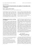
Fig. 2. Cloning of the zebrafish FABP3 cDNA by 3¢ RACE. Agarose
gel electrophoretic separation of 3¢ RACE products for zebrafish
FABP3 cDNA cloning. Both the major band of 650 bp and the
minor band of 570 bp were excised, cloned and sequenced. M:
100 bp DNA ladder (with molecular sizes shown on the left of the
panel). Lane 1: 3¢ RACE product of FABP3 cDNA; lane 2: negative
control without template.
3226 R Z. Liu et al.(Eur. J. Biochem. 270) Ó FEBS 2003
Detection of
fabp3
mRNA by Northern blot
and hybridization
Fifteen micrograms of total RNA from adult zebrafish was
size-fractionated by 2% (w/v) agarose gel electrophoresis in
a Mops buffer (40 m
M
Mops, 10 m
M
sodium acetate, 1 m
M
EDTA, pH 7.2) and 0.2
M
formaldehyde. The resolved
RNA was transferred to Hybond-N
+
membrane (Amer-
sham Pharmacia Biotech, Baie d’Urfe
´
, Quebec, Canada)
according to standard methods [22]. The fabp3 cDNA
generated by RT-PCR from total RNA of adult zebrafish
waslabelledwith[a
32
P]dATP during the reaction and used
as a hybridization probe. The membrane was prehybridized
in 5 mL of ExpressHyb
TM
solution (BD Biosciences Clon-
tech, Franklin Lakes, New Jersey, USA) at 68 °Cfor
30 min, and then hybridized to the denatured probe in the
same solution at 68 °C for 1 h. The blot was rinsed three
times at room temperature with 2· NaCl/Cit and 0.05%
SDS (w/v), washed once at room temperature for 20 min
with the same solution, and then washed at 50 °Cfor1hin
0.1· NaCl/Cit/0.1% SDS. The membrane was then
exposed to X-ray film at )70 °C for 48 h.
In situ
hybridization to tissue sections and emulsion
autoradiography
Oligonucleotides corresponding to nucleotides 426–456
(antisense) and nucleotides 393–422 (sense) of the fabp3
cDNA sequence (Fig. 1B) were synthesized and used as
probes in tissue section in situ hybridization and emulsion
autoradiography to localize the fabp3 mRNA at the tissue
and cellular level in adult zebrafish. Insituhybridization was
performed as described previously [26]. Briefly, 20 lm
cryostat sections obtained from fresh-frozen adult zebrafish
were mounted onto Superfrost Plus glass slides (Fisher
Scientific, Nepean, Ontario, Canada), fixed in 4% parafor-
maldehyde for 5 min, in 1 NaCl/P
i
twiceforthreemin,and
then washed in 1· NaCl/Cit for 20 min. The fixed tissue
sections were immersed in a hybridization buffer containing
50% formamide, 5· NaCl/Cit, 1· Denhardt’s solution,
20 m
M
sodium phosphate (pH 6.8), 0.2% (w/v) SDS,
5m
M
EDTA, 10 lgÆmL
)1
poly(A)
n
, 10% dextran sulfate,
and 5· 10
6
cpmÆmL
)1
of [a
33
P]dATP 5¢ end-labelled oligo-
nucleotide probe. Hybridization was performed at 42 °Cfor
16 h. The slides were washed sequentially in 1· NaCl/Cit
(four times for 15 min), 0.5· NaCl/Cit (four times for
15 min) and 0.25· NaCl/Cit (twice for 15 min) at 55 °C,
and then once in 0.25· NaCl/Cit for 60 min at room
temperature. The sections were exposed to Kodak Biomax
single emulsion film at room temperature for 5 days, and
then dipped in Kodak NTB2 nuclear track emulsion and
exposed for 14 days at 4 °C. The tissue sections were then
developed, counter-stained with cresyl violet and viewed
under bright-field and dark-field illumination [26].
Results
Nucleotide and deduced amino acid sequence
of cDNAs coding for the zebrafish FABP3
Three cDNA clones derived from the major product and
one clone from the minor product of 3¢ RACE (Fig. 2) for
the zebrafish FABP3 were 636 bp and 528 bp in length,
respectively, not including the poly(A) tail. Both sequences
contain the same open reading frame for a polypeptide of
133 amino acids (Fig. 1B). The sequence also contains an
11 bp 5¢ UTR, a 223 bp (major) or 115 bp (minor)
transcript of the 3¢ UTR with an alternative polyadenyla-
tion signal of AATAAA at nucleotides 616–621 (major
transcript) or 509–514 (minor transcript) of the cDNA
sequence (Fig. 1B). A single nucleotide difference, a C-to-T
transition, in one of the 3¢ RACE cDNA clones was seen at
position 104 in the coding region. This nucleotide difference,
however, did not change the encoded amino acid at this site.
The nucleotide difference may be due to either an artefact
during RT-PCR cloning of the FABP3 cDNA, or, more
likely, this nucleotide difference represents an allelic variant
of the zebrafish fabp3 gene. The latter conclusion is
supported by the finding that independently cloned EST
sequences (GenBank accession numbers AI617818,
AW077983, AW281103, AW778251, BI672083, BI673099,
BM082353, BQ481071, BQ481317) contain a C rather than
a T at this position, while some other ESTs (accession
numbers BQ480714, BM186245, BM005090, BM025130,
BI867082, BM186677, BQ260001, BI868173) contain a T
rather than a C.
The deduced amino acid sequence of the zebrafish
FABP3 was aligned with FABP sequences from zebrafish
and six other species using
CLUSTALW
[21] (Fig. 3). The
zebrafish FABP3 exhibits highest sequence identity with
rainbow trout H-FABP (80%) and mammalian H-FABPs
(72–74%). The zebrafish FABP3, similar to mammalian
H-FABPs, had high sequence identity to B-FABPs from
zebrafish (Fig. 3) and mammals (data not shown). Amino
acid identity between the zebrafish FABP3 and two
paralogous zebrafish FABPs, I-FABP and liver basic-type
FABP (Lb-FABP), was 28% and 26%, respectively.
Fig. 3. Comparison of the zebrafish FABP3 to H-FABPs from six dif-
ferent species and paralogues of other zebrafish FABPs. The deduced
aminoacidsequenceofthezebrafishfabp3 (ZF FABP3; Swiss-Prot
and TrEMBL accession number: Q8UVG7) was compared to the
sequences of H-FABPs from rainbow trout (TR H-FABP; O13008),
rat (RT H-FABP; P07483), human (HM H-FABP; P05413), mouse
(MS H-FABP; P11404), pig (PG H-FABP; O02772), cow (CW
H-FABP; P10790) and the zebrafish FABP paralogues, brain-type (ZF
B-FABP; Q9I8N9), intestinal-type (ZF I-FABP; Q9PRH9) and the
basic liver-type (ZF Lb-FABP; Q9I8L5). Dots indicate amino acid
identity. Dashes have been introduced to maximize alignment. The
percentage amino acid sequence similarities between the zebrafish
FABP3 and other FABPs are shown at the end of the sequences.
Ó FEBS 2003 fabp3 gene from zebrafish (Eur. J. Biochem. 270) 3227
Southern analysis of the zebrafish
fabp3
gene
Using a pair of primers flanking the entire coding region
and a portion of the 3¢ UTR for the zebrafish FABP3
cDNA sequence (Fig. 1B), a radiolabelled hybridization
probe from adult zebrafish total RNA was generated by
RT-PCR for Southern blot and hybridization analysis. The
cDNA probe hybridized to restriction fragments of zebra-
fish genomic DNA of 4.7 kb and 7.2 kb in HincII-digested
DNA, 2.6 kb and 5.1 kb in HaeIII-digested DNA, 2.4 kb
and 5.0 kb MboI-digested DNA, and 1.7 kb and 5.0 kb in
RsaI-digested DNA (Fig. 4). Of the four restriction endo-
nucleases used in the Southern blot, RsaIandHincII have
two and one recognition sites, respectively, while HaeIII and
MboI have no recognition sites within the FABP3 cDNA
sequence. The most parsimonious explanation for the
simple hybridization seen in the Southern blot is that the
fabp3 gene exists as a single copy in the zebrafish genome.
We were unable to detect the predicted 0.1 kb RsaI
restriction fragment in the Southern blot, presumably due
to the low hybridization signal from such a small DNA
fragment or it may have migrated off the end of the gel
during electrophoresis. As both the HaeIII and MboI-
digested DNA samples generated two fragments in the
Southern blot hybridization, and neither site is present in the
cDNA sequence, we predict that single recognition sites for
each of these restriction endonucleases is present in one of
the introns of the zebrafish gene.
DNA sequence and structure of the zebrafish
fabp3
gene
Using the zebrafish FABP3 cDNA sequence to search the
zebrafish genome sequence database of the Wellcome Trust
Sanger Institute, we retrieved four DNA traces containing
the coding sequence of exon 1 (zfishG-a1962b06.q1c), exon
2 (Z35725-a5890c08.q1c), exon 3 (Z35724-a1164g06.q1c)
and exon 4 (Z35725-a1576b09.p1c), respectively. The
zebrafish FABP3 cDNA sequence was identical to the
DNA trace sequences, except for a T-to-C substitution in
the sequence of DNA trace Z35724-a1164g06.q1c in exon 3,
which corresponds to the sequence of some FABP3 ESTs
reported in GenBank. The zebrafish fabp3 gene consists
of four exons encoding 24, 58, 34 and 17 amino acids,
respectively, interrupted by three introns (Fig. 1B). All
exon/intron splice junctions of the zebrafish fabp3 gene
conform to the GT/AG rule [27]. Comparison of the
structure of the zebrafish fabp3 gene with that of the
orthologous human and mouse genes revealed an identical
exon/intron organization, conserved splice junction
sequence, and coding capacity for each exon (data not
shown).
Multiple transcription start sites for the zebrafish
fabp3
gene
Using 5¢ RLM-RACE, we determined the 5¢ end of the
capped and complete zebrafish fabp3 gene transcripts. A
major band of approximately 200 bp, and two minor bands
of 250 bp and 180 bp, were observed after agarose gel
electrophoresis of nested PCR products from the CIP/TAP-
treated RNA. No specific band was detected for the
negative control using an RNA sample that was not treated
with TAP (Fig. 5). Thus, these RACE products probably
represent the 5¢ ends of the mature fabp3 gene transcripts.
By aligning the sequence of the 5¢ RLM-RACE products
with the fabp3 gene sequence, the transcription start sites of
the zebrafish fabp3 gene were mapped to 29 bp, 61 bp and
116 bp upstream of the initiation codon (Fig. 1A). Multiple
transcription start sites have also been reported for mam-
malian fabp3 genes [9,28].
Assignment of the zebrafish
fabp3
gene to linkage
group 19
A PCR product of the predicted size (229 bp) was generated
for positive mouse–zebrafish hybrid cell lines of the LN54
panel [ 25] using primers specific to the fourth exon of the
zebrafish fabp3 gene (original mapping data can be provided
on request). A PCR product of the predicted size was also
generated in the two positive controls, a 1 : 10 mixture of
zebrafish and mouse genomic DNA, and zebrafish genomic
DNA. No band was derived from the negative control
containing mouse genomic DNA only. Online analysis of
the radiation hybrid mapping data assigned the zebrafish
fabp3 gene to linkage group 19 at 365.69 cR (LN54 panel)
or 67.51 cM (ZMAP panel) in the zebrafish genome with a
LOD score of 14.6. Comparison of the syntenic relationship
of fabp3 gene on zebrafish linkage group 19 with that of the
human and mouse orthologous gene revealed conserved
syntenies (Table 1). Five mapped genes syntenic to the
zebrafish fabp3 gene on LG 19 are located on human
Fig. 4. Southern blot analysis of zebrafish genomic DNA using the fabp3
cDNA as a hybridization probe. Eight microgram aliquots of zebrafish
genomic DNA were digested with one of the following restriction
endonculeases: RsaI(lane1),MboI(lane2),HaeIII (lane 3) and HincII
(lane 4). The size-fractionated DNA was transferred to nylon mem-
branes and hybridized to fabp3 cDNA. Molecular mass markers in kb
are shown on the left of the panel.
3228 R Z. Liu et al.(Eur. J. Biochem. 270) Ó FEBS 2003
chromosome 1, and four of them were syntenic to the mouse
fabp3 gene on mouse chromosome 4. Four other genes
syntenic to the fabp3 gene in zebrafish, however, reside on
human chromosome 6 at 6p21.3 and mouse chromosome
17. The syntenic genes in the zebrafish LG 19 are distributed
at two distinct chromosomal locations in the human and
mouse genomes suggesting that this region has undergone
interchromosomal rearrangements after the divergence of
fish and mammals.
Tissue-specific expression of the
fabp3
gene in zebrafish
In a Northern blot and hybridization of total RNA from
adult zebrafish to a [a
32
P]dATP-labelled zebrafish FABP3
cDNA,wedetectedabroadbandofRNAof 780
nucleotides (Fig. 6A). Detection of fabp3 gene transcripts
with two different 3¢ polyadenylation sites and three
different 5¢ transcription start sites for the zebrafish fabp3
mRNA could give rise to six potential transcripts of 741,
686, 654, 654, 620, 575 and 543 nucleotides in length, not
including the poly(A) tail. The relative abundance of each
of these transcripts in different tissues has not yet been
determined.
An oligonucleotide antisense probe was used to detect the
distribution of the fabp3 gene transcript in adult tissue
sections by in situ hybridization. The zebrafish fabp3
mRNA was exclusively localized to liver (Fig. 6B1) and
ovary (Fig. 6C1). No hybridization to adult tissue sections
was observed with the oligonucleotide sense probe. The
signal emanating from a region of the eye and skin
(Fig. 6B2,C2) has been observed by us with many sense
probes, indicating a nonspecific interaction between the
DNA probe and components of these tissues. We conclude
therefore that the fabp3 gene transcript detected by in situ
hybridization is localized to the liver and ovary of adult
zebrafish.
In order to resolve further the tissue and cellular
localization of the fabp3 mRNA in adult zebrafish, we
performed emulsion autoradiography on tissue sections.
The hybridization signal, corresponding to the location of
the zebrafish fabp3 gene transcript, was most intense and
uniform in primary growth stage (stage I) oocytes,
including both ooplasm and germinal vesicle (Fig. 7A;
white granules in the emulsion). Stages of oocyte matur-
ation in zebrafish are described in [15]. The hybridization
signal was less intense and restricted to the ooplasm of
cortical alveolus stage (stage II) oocytes. In stage III
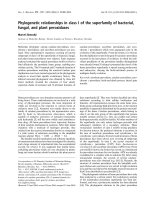
Fig. 5. Agarose gel electrophoresis of 5¢ RLM-RACE product and PCR
identification of the corresponding clones. (A) Total RNA from whole
adult zebrafish was sequentially treated with calf intestinal alkaline
phosphatase, tobacco acid pyrophosphatase and then ligated to a
designated RNA adapter. Following two rounds of nested PCR, one
major PCR-amplified product of approximately 200 bp and two
minor products of 180 bp and 250 bp were size-fractionated by gel
electrophoresis in 2.5% agarose (lane 1). RNA treated to the same
experimental regime, but with tobacco acid pyrophosphatase digestion
omitted, did not generate a product (lane 2). A ladder of 100 bp
molecular mass markers (MBI Fermentas) is shown in lane M, with
sizes indicated to the left. (B) Positive colonies from transformants of
the three 5¢ RLM-RACE reactions were of 200 bp (lane 1), 250 bp
(lane 2) and 180 bp (lane 3). The correct size of DNA insert was
confirmed by colony PCR. Lane M: 100 bp DNA ladder, with
molecular sizes shown on the left of the panel.
Table 1. Conserved syntenic relationship of the zebrafish fabp3 gene in human and mouse genome.
Zebrafish
a
Human
b
Mouse
b
Gene location Gene location Gene location
fabp3 19 67.51 c
M
FABP3 1p33-p32 Fabp3 4 61.0 c
M
oprd1 19 8.42 c
M
OPRD1 1p36.1-p34.3 Oprd1 4 64.8 c
M
rpl11 19 70.21 c
M
RPL11 1p36.1-35 Rpl11 4D3
fuca1 19 48.02 c
M
FUCA1 1p34 Fuca1 4 65.7 c
M
mycl1 19 50.99 c
M
(T51) MYCL1 1p34.3 Mycl1 4 65.7 c
M
ifl2 19 56.41 c
M
(LN54) IFL2 1q21.1 Ifl2 3F2
bing1 19 38.6 c
M
BING1 6p21.3 Bing1 17 B1
daxx 19 38.6 c
M
DAXX 6p21.3 Daxx 17 B1/17.0 c
M
psmb9 19 42.56 c
M
PSMB9 6p21.3 Psmb9 17 18.59 c
M
rxre 19 43.76 c
M
(LN54) RXRB 6p21.3 Rxrb 17 18.49 c
M
a
ZMAP (http://zfin.org).
b
LocusLink( NCBI.
Ó FEBS 2003 fabp3 gene from zebrafish (Eur. J. Biochem. 270) 3229
oocytes, hybridization to the fabp3 mRNA was almost
undetectable under the conditions employed here. No
hybridization signal was seen in follicular cells surround-
ing the oocytes in the ovary. A moderate, but uniform,
hybridization signal was seen over the hepatocytes of the
liver (Fig. 7B–D). No hybridization signal was evident in
adult zebrafish heart, muscle (Fig. 7B,C) or brain (data
not shown), tissues where mammalian and other fish
H-FABPs are known to be expressed [reviewed in 7,14].
The in situ hybridization and emulsion autoradiography
was repeated three times using two different antisense
oligonucleotide probes to the fabp3 gene transcript with
the same result (data not shown).
To verify the tissue-specific distribution of the zebrafish
fabp3 gene transcript, RT-PCR was performed on total
RNA extracted from ovary, liver, heart, muscle, brain,
intestine, skin and testis. As a positive control, RT-PCR was
employed to amplify the constitutively expressed zebrafish
receptor for activated C kinase 1 (RACK1) [21]. RT-PCR
product of the correct size for the fabp3 mRNA was
detected in all the tissues examined (Fig. 8), indicating a
wide tissue-distribution for the zebrafish fabp3 transcript
similar to that reported for mRNA expression of the fabp3
gene in mammals [7,29]. Although the zebrafish fabp3
mRNA was detected in a wide range of tissues by RT-PCR,
the mRNA must be of such low abundance in most of these
tissues, with the exception of primary oocytes and liver, that
it is below the sensitivity of detection by the technique of
Fig. 6. fabp3 Gene expression in adult zebrafish. (A) Northern blot
analysis of total RNA isolated from whole adult zebrafish using the
fabp3 cDNA as a hybridization probe detected a single transcript of
780 bases. (B and C) Insituhybridization of sense and antisense fabp3
specific oligonucleotides to mRNA in transverse tissue sections of
adult zebrafish. The arrows indicate specific hybridization of the
antisense probe to the fabp3 mRNA in liver (B1) and ovary (C1). No
specific hybridization was evident when the sense oligonucleotide was
used as a hybridization probe (B2 and C2).
Fig. 7. Autoradiographic emulsion of zebrafish sections hybridized to the
fabp3 antisense probe. The zebrafish sections that hybridized to the
fabp3 probe were exposed to autoradiographic emulsion, cresyl violet
counter-stained and viewed under bright and dark field illumination
(panels on the left and right, respectively). (A) Silver grains corres-
ponding to the fabp3 mRNA were visualized by dark field illumination
in different stages of zebrafish oocytes. Abundant silver grains were
observed throughout stage I oocytes (I in bright field). In stage II
oocytes (II in bright field) the density of silver grains diminished rel-
ative to stage I oocytes and was restricted to the ooplasm. No silver
grains were observed in stage III (III in bright field) and matured
oocytes. (B–D) Silver grains were detected over hepatocytes of the liver
(L), but not in heart (H, panel B), muscle (M, panel C), or intestine
(I, panel D).
3230 R Z. Liu et al.(Eur. J. Biochem. 270) Ó FEBS 2003
tissue section in situ hybridization and emulsion auto-
radiography employed here.
Potential 5¢
cis
regulatory elements of the zebrafish
fabp3
gene
Inspection of the 5¢ upstream sequence of the zebrafish
fabp3 gene revealed a typical cellular and viral TATA box
element, with a matrix sequence of 5¢-ttaTAAAtcagccag-3¢.
The core sequence (TAAA) of this TATA box is located
60 bp upstream of the major transcription start site. This
TATA box in the zebrafish fabp3 gene differs from the
common TATA box element (TTTAAA) found in the
fabp3 gene promoter sequence of mouse [9]), rat [30] and
pigs [31]. Two adjacent GC boxes () 102, ) 112) are located
further upstream in the proximal promoter of the zebrafish
fabp3 gene (Fig. 1A).
Numerous transcription factor responsive elements were
predicted by computer analysis of the 1220 bp 5¢ flanking
sequence of fabp3 gene, and some of these may be associated
with the tissue-specific expression of this gene in zebrafish
(Table 2). A Yin Yang 1 (YY1) transcription element was
identified in the 5¢ flanking sequence that may be associated
with oocyte-specific expression of the zebrafish fabp3 gene.
Studies in tissue culture suggest that YYI may play a role in
controlling the expression of developmentally regulated
genes. Recently, it has been reported that YY1 is abundant
in the oocytes of mouse [32] and Xenopus [33]. Fabp3 might
be one of the targeted genes of YY1 in zebrafish oocytes.
Two additional types of cis elements for the transcription
factors E2F and GATA-2 were found in the 5¢ upstream
region of the zebrafish fabp3 gene. These transcription
factors are expressed during Drosophila and Xenopus
oogenesis [34,35].
The presence of the hepatocyte nuclear factor 1 (HNF1)
elements in the 5¢ upstream region of the zebrafish fabp3
gene may be relevant to the expression of this gene in the
zebrafish liver. In a recent report, the expression of L-FABP
was markedly reduced in HNF1a-null mice. HNF1a
elements were found in the 5¢ flanking sequence of the
mouseL-FABPgeneandHNF1a was shown to be required
for transactivation of the L-FABP promoter [36]. This
finding indicated an important role of HNF1a in control of
expression of the L-FABP gene in mouse liver. Conceivably,
expression of the zebrafish fabp3 gene in liver may be
regulated by HNF1a.
There are differences in the number and location of cis
regulatory elements in the 5¢ upstream sequences of the
fabp3 gene between zebrafish and mammals. The wide-
spread E-box elements in rodent fabp3 genes [9,30] were not
found in the 5¢ flanking sequence of the zebrafish fabp3
gene. Moreover, the DR-1 element, a binding site for the
retinoic acid receptor, retinoid X receptor and peroxisome
proliferator-activated receptor (PPAR), identified in the 5¢
upstream region of rodent fabp3 genes [9,30], is absent in the
5¢ upstream sequence of the zebrafish fabp3 gene. In
contrast, the abundant POU elements distributed through-
out the 5¢ upstream sequence of the zebrafish fabp3 gene
Fig. 8. Zebrafish fabp3 mRNA in adult tissues detected by RT-PCR.
Zebrafish fabp3 mRNA-specific primers amplified a product from
total RNA extracted from ovary (O), liver (liver), skin (S), intestine (I),
brain (B), heart (H), testis (T) and muscle (M). An RT-PCR product
was generated from RNA in all samples for the constitutively
expressed receptor for activated C kinase (RACK1), used as a positive
control. A negative control (–) lacking an RNA template generated no
RT-PCR product.
Table 2. Potential 5¢ cis regulatory elements of the zebrafish fabp3 gene.
Name
of family/matrix Further information Position Strand
Core
similarity
Matrix
similarity Sequence
TBPF/TATA.01 cellular and viral TATA box elements )53 (+) 1.000 0.925 ttaTAAAtcagccag
AP2F/AP2.01 activator protein 2 )94 (+) 0.976 0.924 ccCCCCcaggcc
AP1F/AP1.01 AP1 binding site )902 (–) 0.881 0.954 gtgaATCAa
SP1F/SP1.01 stimulating protein 1 SP1 )100 (–) 1.000 0.896 ggggGGCGgatgg
HNF1/HNF1.01 hepatic nuclear factor 1 )297 (+) 1.000 0.830 cGTTAattagttttt
HNF1/HNF1.02 hepatic nuclear factor 1 )891 (–) 0.806 0.774 tGATAataaatgtgaat
GATA/GATA1.05 GATA-binding factor 1 )333 (+) 1.000 0.966 ttaGATAaaa
GATA/GATA1.03 GATA-binding factor 1 )635 (+) 1.000 0.949 ccctGATAaatta
GATA/GATA1.02 GATA-binding factor 1 )671 (+) 1.000 0.966 tgctgGATAagtgg
GATA/GATA2.01 GATA-binding factor 2 )889 (–) 1.000 0.945 aatGATAata
SORY/SOX5.01 Sox-5 )363 (+) 1.000 0.862 atgaCAATga
SORY/SOX5.01 Sox-5 )955 (–) 1.000 0.861 tataCAATct
YY1F/YY1.01 Yin and Yang 1 )760 (–) 1.000 0.839 atatggCCATttagtttatt
ECAT/NFY.02 nuclear factor Y )974 (–) 1.000 0.928 catCCAAtcgc
ECAT/NFY.02 nuclear factor Y )1099 (–) 1.000 0.946 catCCAAtcac
E2FF/E2F.01 E2F, involved in cell cycle regulation )1082 (+) 0.750 0.777 tgcacggGGAAaatg
E2FF/E2F.02 E2F, involved in cell cycle regulation )1198 (+) 1.000 0.849 gcacCAAA
Ó FEBS 2003 fabp3 gene from zebrafish (Eur. J. Biochem. 270) 3231
(data not shown) has not been reported for the mammalian
fabp3 genes. However, elements for transcription factors,
AP1 and NF1, are present in the 5¢ upstream region of both
zebrafish and mammalian fabp3 genes [9,30].
Discussion
Among the members of the ILBP multigene family,
H-FABP shows the widest range of tissue-specific distri-
bution. Mammalian H-FABP is found in heart, skeletal
and smooth muscle, specific regions of the brain, distal
tubule cells of the kidney, stomach parietal cells, lactating
mammary gland, ovary, testis and placenta [reviewed in
7,29], but is absent in the liver, white fat and intestine
[29,37–43]. The zebrafish fabp3 gene, described in the
present study, showed highest amino acid sequence
similarity, identical gene structure and coding capacity,
and conserved genomic syntenies to mammalian H-
FABPs. However, the zebrafish fabp3 gene displayed a
different pattern of tissue distribution to that of the
orthologous mammalian H-FABPs. Steady-state mRNA
level of the H-FABP in adult tissues of five different fish
species has been analyzed by Northern blot hybridization.
In the four Antarctic teleost fish species, both H-FABP
isoforms exhibited similar expression patterns to the
mammalian H-FABP, i.e. high mRNA level in the heart,
muscle and brain, but absent in the liver [14]. In the
mummichog (Fundulus heteroclitus), the H-FABP mRNA
level was most abundant in the male (female tissues were
not examined) liver, gills and gonads, which is more
similar to the tissue-distribution pattern of the zebrafish
fabp3 reported here [44]. By in situ hybridization and
emulsion autoradiography, we detected the fabp3 gene
transcript in ovary and liver (Figs 6 and 7). Only by the
highly sensitive technique of RT-PCR were we able to
detect the fabp3 gene transcript in the other tissues such as
brain, heart, intestine, muscle, skin and testis of adult
zebrafish (Fig. 8). Comparison of the 5¢ upstream
sequence of the zebrafish fabp3 gene and the orthologous
mammalian genes revealed numerous differences in cis
elements. Together, the differences between zebrafish and
mammals in cis elements and the expression pattern of the
fabp3 gene suggests that, while primary amino acid, gene
structure and syntenic relationships have been conserved,
cis elements in the 5¢ upstream regions of these genes have
apparently evolved following divergence of fish and
mammals leading to altered patterns of gene expression.
This is not the case for the zebrafish B-FABP (fabp7)
gene, which shows conservation in both expression pattern
(brain-specific) and regulatory elements with its mamma-
lian orthologs [24].
Although at least 15 paralogous members of the FABP
multigene family have been characterized in various
species, the precise in vivo physiological functions of each
of these proteins are still not well understood. In
mammalian cardiac and skeletal muscle, H-FABP is
thought to participate in fatty acid a-oxidation and
energy production (reviewed in [2,45]). Studies using
H-FABP knockout mice demonstrated that H-FABP is
required for efficient uptake, intracellular transportation
and utilization of fatty acids in cardiac muscle [4,5].
However, H-FABP is also abundant in tissues that do
not use fatty acids as an energy source such as mammary
gland and developing brain [9,46]. Similarly, the detection
of high steady-state levels of the fabp3 gene transcript in
zebrafish oocytes and liver, tissues which do not use fatty
acids primarily as energy sources, indicates that FABP3
participates in a-oxidation in muscle and lipogenesis in
other tissues in fish and mammals. FABP3 (H-FABP)
may play a general and fundamental role in fatty acid
transportation in tissues exhibiting anabolic and catabolic
lipid metabolism.
During development of the animal oocyte, large
quantities of mRNAs, rRNAs and tRNAs are synthes-
ized, some mRNAs are translated immediately into
protein while others are stored in an inactive form,
ribosomes and mitochondria accumulate, and quantities
of polysaccharides and lipids are synthesized. In addition
to the metabolic activity of the oocyte, proteins, lipids and
carbohydrates enter into the cytoplasm from outside the
cell [48]. During stage III of oocyte development or
vitellogenesis, much of the stored lipid and protein is
packed into yolk granules that accumulate in all animal
oocytes except mammals [15,47]. Oocyte development in
oviparous species (birds, fish, amphibians and reptiles) is,
indeed, dependent on the uptake of nutrients and their
storage as yolk, whose constituents are subsequently used
by the embryo during early stages of development. These
yolk granules often comprise 95% of the cytoplasmic
volume that accounts for the relatively large size of eggs
of oviparous species compared to eggs of mammalian
species [47]. The abundance of the fabp3 mRNA prior to
the vitellogenic (III) stage seen in the zebrafish (Fig. 7A)
correlates in time with accumulation of fatty acids within
the oocyte. The accumulation of fatty acids may be
sequestered by FABP to prevent cytotoxicity to the cell,
and transported by FABP to sites of triglyceride synthesis
and/or storage within the cytoplasm. As antibodies to the
zebrafish fabp3 are not currently available, we are unable
to assess the stage of oocyte maturation at which the
fabp3 gene transcript is translated into protein. We
suspect, however, that the FABP3 protein is most
abundant immediately prior to and during the vitellogenic
(III) stage of oocyte development.
In Antarctic teleost fishes, the mRNA of two distinct
heart-type FABP isoforms has been detected in cardiac
tissue [14]. It is likely that there is a duplicated fabp3 gene in
zebrafish, which may play a role in muscular a-oxidation.
Based on preliminary analysis, we have cloned a cDNA and
identified the corresponding gene in the zebrafish genome
sequence database (Wellcome Trust Sanger Instititute) that
may be expressed in the zebrafish heart. Characterization of
this newly discovered gene may provide clues to the
expression and function of FABPs in zebrafish cardiac
tissue.
Acknowledgements
This work was supported by a research grant from the Natural Sciences
and Engineering Research Council of Canada (to J. M. W.), a research
grant from the Canadian Institutes of Health Research (to E. D W.)
and an Izaak Walton Killam Memorial Scholarship (to R Z. L.). We
wish to thank Mukesh Sharma and Steve Mockford for their assistance
and helpful comments during the experimental stages of this work.
3232 R Z. Liu et al.(Eur. J. Biochem. 270) Ó FEBS 2003
References
1. Stewart, J.M. (2000) The cytoplasmic fatty-acid-binding proteins:
thirty years and counting. Cell Mol. Life Sci. 57, 1345–1359.
2. Hertzel, A.V. & Bernlohr, D.A. (2000) The mammalian fatty acid-
binding protein multigene family: molecular and genetic insights
into function. Trends Endocrinol. Metab. 11, 175–180.
3. Schaap, F.G., van der Vusse, G.J. & Glatz, J.F. (2002) Evolution
of the family of intracellular lipid binding proteins in vertebrates.
Mol. Cell Biochem. 239, 69–77.
4. Binas, B., Danneberg, H., McWhir, J., Mullins, L. & Clark, A.J.
(1999) Requirement for the heart-type fatty acid binding protein in
cardiac fatty acid utilization. FASEB J. 13, 805–812.
5. Schaap,F.G.,Binas,B.,Danneberg,H.,vanderVusse,G.J.&
Glatz, J.F.C. (1999) Impaired long-chain fatty acid utilization by
cardiac myocytes isolated from mice lacking the heart-type fatty
acid binding protein gene. Circ. Res. 85, 329–337.
6. Ribarik Coe, N., Simpson, M.A. & Bernlohr, D.A. (1999) Tar-
geted disruption of the adipocyte lipid-binding protein (aP2 pro-
tein) gene impairs fat cell lipolysis and increases cellular fatty acid
levels. J. Lipid Res. 40, 967–972.
7. Veerkamp, J.H. & Maatman, G.H.J. (1995) Cytoplasmic fatty
acid-binding proteins: their structure and genes. Prog. Lipid Res.
34, 17–52.
8. Bo
¨
rchers, T. & Spenser, F. (1994) Fatty acid binding proteins.
Curr. Top. Membranes 40, 261–226.
9. Treuner.M.,Kozak,C.A.,Gallahan,D.,Grosse,R.&Muller,T.
(1994) Cloning and characterization of the mouse gene encoding
mammary-derived growth inhibitor/heart-fatty acid-binding pro-
tein. Gene 147, 237–242.
10. Prinsen, C.F. & Veerkamp, J.H. (1996) Fatty acid binding and
conformational stability of mutants of human muscle fatty acid-
binding protein. Biochem. J. 314, 253–260.
11. Schleicher, C.H., Co
´
rdoba, O.L., Santome
´
, J.A. & Dell’Angelica,
E.C. (1995) Molecular evolution of the multigene family of
intracellular lipid-binding proteins. Biochem. Mol. Bio. Int. 36,
1117–1125.
12. Matarese, V., Stone, R.L., Waggoner, D.W. & Bernlohr, D.A.
(1989) Intracellular fatty acid trafficking and the role of cytosolic
lipid binding proteins. Prog. Lipid Res. 28, 245–272.
13. Zhang, J. & Haunerland, N.H. (1998) Transcriptional regulation
of FABP expression in flight muscle of the desert locust, Schisto-
cerca gregaria. Insect Biochem. Mol. Biol. 28, 683–691.
14. Vayda, M.E., Londravelli. R.L., Cashon, R.E., Costello, L. &
Sidell, B.D. (1998) Two distinct types of fatty acid-binding protein
are expressed in heart ventricle of Antarctic teleost fishes. Biochem.
J. 330, 375–382.
15. Selman, K., Wallace, R.A., Sarka, A. & Qi, X. (1993) Stages of
oocyte development in the zebrafish, Brachydanio rerio. J. Morph.
218, 203–224.
16. Denovan-Wright, E.M., Pierce, M. & Wright, J.M. (2000) Nu-
cleotide sequence of cDNA clones coding for a brain-type fatty
acid binding protein and its tissue-specific expression in adult
zebrafish (Danio rerio). Biochim. Biophys. Acta 1492, 221–226.
17. Pierce, M., Wang, Y., Denovan-Wright, E.M. & Wright, J.M.
(2000) Nucleotide sequence of a cDNA clone coding for an
intestinal-type fatty acid binding protein and its tissue-specific
expression in zebrafish (Danio rerio). Biochim. Biophys. Acta
1490, 175–183.
18. Denovan-Wright, E.M., Pierce, M., Sharma, M.K. & Wright,
J.M. (2000) cDNA sequence and tissue-specific expression of a
basic liver-type fatty acid binding protein in adult zebrafish (Danio
rerio). Biochim. Biophys. Acta 1492, 227–232.
19. Cameron, M.C., Denovan-Wright, E.M., Sharma, M.K. &
Wright, J.M. (2002) Cellular retinol-binding protein type II
(CRBPII) in adult zebrafish (Danio rerio). cDNA sequence, tissue-
specific expression and gene linkage analysis. Eur. J. Biochem. 269,
4685–4692.
20. Westerfield, M. (1995) TheZebrafishBook:aGuideforthe
Laboratory Use of Zebrafish (Danio rerio). University of Oregon
Press, Eugene, USA.
21. Thompson, J.D., Higgins, D.G. & Gibson, T.J. (1994) CLUSTAL
W: improving the sensitivity of progressive multiple sequence
alignment through sequence weighting, position-specific gap
penalties and weight matrix choice. Nucleic Acids Res. 22,
4673–4680.
22. Hamilton, L.C. & Wright, J.M. (1999) Isolation of com-
plementary DNAs coding for a receptor for activated C kinase
(RACK1) from zebrafish (Danio rerio) and tilapia (Oreochromis
niloticus): constitutive developmental and tissue expression. Mar.
Biotechnol. 1, 279–285.
23.Sambrook,J.,Fritsch,E.F.&Maniatis,T.(1989)Molecular
Cloning: A Laboratory Manual, 2nd edn. Cold Spring Harbor
Laboratory, Cold Spring Harbor, NY, USA.
24. Liu, R Z., Denovan-Wright, E.M. & Wright, J.M. (2003) Struc-
ture, mRNA expression and linkage mapping of the brain-type
fatty acid-binding protein gene (fabp7) from zebrafish (Danio
rerio). Eur. J. Biochem. 270, 715–725.
25. Hukriede, N.A., Joly, L., Tsang, M., Miles, J., Tellis, P.,
Epstein, J.A., Barbazuk, W.B., Li, F.N., Paw, B., Postlethwait,
J.H., Hudson, T.J., Zon, L.I., McPherson, J.D., Chevrette, M.,
Dawid, I.B., Johnson, S.L. & Ekker, M. (1999) Radiation
hybrid mapping of the zebrafish genome. Proc.NatlAcad.Sci.
USA 96, 9745–9750.
26. Denovan-Wright, E.M., Newton, R.A., Armstrong, J.N., Babity,
J.M. & Robertson, H.A. (1998) Acute administration of cocaine,
but not amphetamine, increases the level of synaptotagmin IV
mRNA in the dorsal striatum of rat. Mol. Brain Res. 55, 350–354.
27. Breathnach, R. & Chambon, P. (1981) Organization and expres-
sion of eucaryotic split genes coding for proteins. Annu. Rev.
Biochem. 50, 349–383.
28. Qian, Q., Kuo, L., Yu, Y.T. & Rottman, J.N. (1999) A concise
promoter region of the heart fatty acid-binding protein gene dic-
tates tissue-appropriate expression. Circ. Res. 84, 276–289.
29. Heuckeroth, R.O., Birkenmeier, E.M., Levin, M.S. & Gordon, J.I.
(1987) Analysis of the tissue-specific expression, developmental
regulation, and linkage relationships of a rodent gene encoding
heart fatty acid binding protein. J. Biol. Che.
30. Zhang, J., Rickers-Haunerland, J., Dawe, I. & Haunerland, N.H.
(1999) Structure and chromosomal location of the rat gene
encoding the heart fatty acid-binding protein. Eur. J. Biochem.
266, 347–351.
31. Gerbens, F., Rettenberger, G., Lenstra, J.A., Veerkamp, J.H. & te
Pas, M.F. (1997) Characterization, chromosomal localization, and
genetic variation of the porcine heart fatty acid-binding protein
gene. Mamm. Genome. 8, 328–332.
32. Donohoe, M.E., Zhang, X., McGinnis, L., Biggers, J., Li, E. &
Shi, Y. (1999) Targeted disruption of mouse Yin Yang 1 tran-
scription factor results in peri-implantation lethality. Mol. Cell
Biol. 19, 7237–7244.
33. Ficzycz, A. & Ovsenek, N. (2002) The Yin Yang, 1 transcription
factor associates with ribonucleoprotein (mRNP) complexes in the
cytoplasm of Xenopus oocytes. J. Biol. Chem. 277, 8382–8387.
34. Myster, D.L., Bonnette, P.C. & Duronio, R.J. (2000) A role
for the DP subunit of the E2F transcription factor in axis
determination during Drosophila oogenesis. Development 127,
3249–3261.
35. Partington, G.A., Bertwistle, D., Nicolas, R.H., Kee, W.J., Pizzey,
J.A. & Patient, R.K. (1997) GATA-2 is a maternal transcription
factor present in Xenopus oocytes as a nuclear complex which is
maintained throughout early development. Dev. Biol. 181,
144–155.
Ó FEBS 2003 fabp3 gene from zebrafish (Eur. J. Biochem. 270) 3233
36. Akiyama, T.E., Ward, J.M. & Gonzalez, F.J. (2000) Regula-
tion of the liver fatty acid-binding protein gene by hepatocyte
nuclear factor 1alpha (HNF1alpha). Alterations in fatty acid
homeostasis in HNF1alpha-deficient mice. J. Biol. Chem. 275,
27117–27122.
37. Chrisman, T.S., Claffcy, K.P., Saouat, R., Hanspal, J. & Brecher,
P. (1987) Measurement of rat heart fatty acid binding protein by
ELISA. Tissue distribution, developmental changes and sub-
cellular distribution. J. Mol. Cell. Cardiol. 19, 423–431.
38. Claffey, K.P., Herrera, V.L., Brecher, P. & Ruiz-Opazo, N. (1987)
Cloning and tissue distribution of rat heart fatty acid binding
protein mRNA: identical forms in heart and skeletal muscle.
Biochemistry 26, 7900–7904.
39. Miller, W.C., Hickson, R.C. & Bass, N.M. (1988) Fatty acid
binding proteins in the three types of rat skeletal muscle. Proc. Soc.
Exp. Biol. Med. 189, 183–188.
40. Bass, N.M. & Manning, J.A. (1986) Tissue expression of three
structurally different fatty acid binding proteins from rat heart
muscle, liver, and intestine. Biochem. Biophys. Res. Commun. 137,
929–935.
41. Hittel, D. & Storey, K.B. (2001) Differential expression of adipose-
and heart-type fatty acid binding proteins in hibernating ground
squirrels. Biochim. Biophys. Acta 1522, 238–243.
42. Kurtz, A., Zimmer, A., Schnu
¨
tgen, F., Bru
¨
ning,G.,Spener,F.&
Mu
¨
ller, T. (1994) The expression pattern of a novel gene encoding
brain-fatty acid binding protein correlates with neuronal and glial
cell development. Development 120, 2637–2649.
43.Paulussen,R.J.A.,vanMoerkerk,H.T.B.&Veerkamp,J.H.
(1990) Immunochemical quantitation of fatty acid-binding pro-
teins. Tissue distribution of liver and heart FABP types in human
and porcine tissues. Int. J. Biochem. 22, 393–398.
44. Bain, L.J. (2002) cDNA cloning, sequencing, and differential
expression of a heart-type fatty acid-binding protein in the mum-
michog (Fundulus heteroclitus). Mar. Environ. Res. 54, 379–383.
45. Glatz, J.F. & Vusse, G.J. (1996) Cellular fatty acid-binding pro-
teins: Their function and physiological significance. Prog. Lipid
Res. 35, 243–282.
46.Sellner,P.A.,Chu,W.,Glatz,J.F.&Berman,N.E.(1995)
Developmental role of fatty acid-binding proteins in mouse brain.
Brain Res. Dev. Brain Res. 89, 33–46.
47. Wolfe, S.L. (1993) Molecular and Cellular Biology.Wadsworth
Publishing Co, Belmont, CA, USA.
3234 R Z. Liu et al.(Eur. J. Biochem. 270) Ó FEBS 2003