Báo cáo khoa học: Fidelity and spatio-temporal control in MAP kinase (ERKs) signalling Delivered on 24 October 2002 at the 28th FEBS Meeting in Istanbul potx
Bạn đang xem bản rút gọn của tài liệu. Xem và tải ngay bản đầy đủ của tài liệu tại đây (275.06 KB, 9 trang )
THE SIR HANS KREBS LECTURE
Fidelity and spatio-temporal control in MAP kinase (ERKs) signalling
Delivered on 24 October 2002 at the 28th FEBS Meeting in Istanbul
Jacques Pouysse
´
gur and Philippe Lenormand
Institute of Signaling, Developmental Biology and Cancer Research, CNRS-UMR 6543, Centre Antoine Lacassagne, Nice, France
The mitogen activated protein (MAP) kinase module:
(Raf fi MEK fi ERKs) is central to the control of cell
growth, cell differentiation and cell survival. The fidelity of
signalling and the spatio-temporal activation are key deter-
minants in generating precise biological responses. The
fidelity is ensured by scaffold proteins – protein kinase
ÔinsulatorsÕ – and by specific docking sites. The duration and
the intensity of the response are in part controlled by the
compartmentalization of the signalling molecules. Growth
factors promote rapid nuclear translocation and persistent
activation of p42/p44 MAP kinases, respectively and
ERK2/ERK1, during the entire G1 period with an extinc-
tion during the S-phase. These features are exquisitely con-
trolled by the temporal induction of the MAP kinase
phosphatases, MKP1–3. MKP1 and 2 induction is strictly
controlled by the activation of the MAP kinase module
providing evidence for an auto-regulatory mechanism. This
negative regulatory loop is further enhanced by the capacity
of p42/p44 MAPK to phosphorylate MKP1 and 2. This
action reduces the degradation rate of MKPs through the
ubiquitin–proteasomal system. Whereas the two upstream
kinases of the module (Raf and MEK) remain cytoplasmic,
ERKs (anchored to MEK in the cytoplasm of resting cells)
rapidly translocate to the nucleus upon mitogenic stimula-
tion. This latter process is rapid, reversible and controlled by
the strict activation of the MAPK cascade. Following long-
term MAPK stimulation, p42/p44 MAPKs progressively
accumulate in the nucleus in an inactive form. Therefore we
propose that the nucleus represents a site for ERK action,
sequestration and signal termination. With the generation of
knockdown mice for each of the ERK isoforms, we will
illustrate that besides controlling cell proliferation the ERK
cascade also controls cell differentiation and cell behaviour.
Keywords: MAP kinases; MAPK-phosphatases; scaffolding
proteins; nucleus; growth control; cell signalling.
Introduction
It is a great privilege for me to be invited to give this
lecture in honour of one of the most emblematic and
unique figures in Biochemistry. I first started out studying
the regulation of metabolism of ÔexoticÕ sugars (Hexuro-
nates) in Escherichia coli at a time when prokaryotic
genetics was ÔexplodingÕ, confirming the extraordinary
accuracy and complexity of metabolic pathways. Unfor-
tunately, I never had the opportunity to meet Hans
Krebs, however, I had the immense pleasure of starting
my first postdoctoral training in 1971 with one of Krebs’s
prominent students, Hans Kornberg (Professor of Bio-
chemistry, Leicester at that time). I then turned my
interest to growth control in mammalian cells, studying
successively, cell surface glycopropteins, anaerobic glyco-
lysis, pHi molecular control, growth factor action and
MAP kinase signalling.
p42/p44 MAP kinases (ERKs) belong to a major signal-
ling module, conserved throughout evolution, that is
activated in mammalian cells via stimulation of receptor
tyrosine kinases, G-protein coupled receptors and integrins
[1].These cell surface signals converge towards activation of
the small G-protein, Ras that recruits the serine/threonine
kinase, Raf to the membrane where it is fully activated by
largely unknown mechanisms [2]. The signal is amplified via
two downstream kinases, MAPKK or ERK kinase (MEK)
and extracellular regulated kinase (ERK), that are activated
uniquely via phosphorylation. MEK is phosphorylated on
two serine residues by Raf, and then ERKs are dually
phosphorylated on a tyrosine and threonine residue by
MEK (sequence TEY). Amplification via this signalling
cascade is such that activation of 5% of Ras molecules is
sufficient to induce full activation of ERK [3].
In 1993, we were the first to report that the long-term
activation of p42/44 MAP kinases is mandatory for cell
cycle entry [4]. ERK activation provides an integrated
pleiotypic response: it activates the transcription of many
genes, via phosphorylation of transcription factors and
Correspondence to J. Pouysse
´
gur, Institute of Signaling, Develop-
mental Biology and Cancer Research, CNRS-UMR 6543, Centre
Antoine Lacassagne, 33 Avenue de Valombrose, 06189 Nice, France.
Fax: + 33 492 03 1225, Tel.: + 33 492 03 1222,
E-mail:
Abbreviations: CDK, cyclin dependent kinase; Crm1, chromosomal
region maintenance 1; EGF, epithelial growth factor; ERK, extra-
cellular regulated kinase; JNK, c-jun N-terminal kinase; KSR, kinase
suppressor of Ras; MAPK, mitogen activated protein kinase;
MEK, MAPK of ERK kinase; MKP, MAP kinase phosphatase;
MNK1, mitogen and stress kinase1; MP1, MEK partner 1;
NGF, nerve growth factor; PEA15, phosphoprotein enriched in
astrocytes 15 kDa; PI3K, phosphatidyl inositol 3 kinase;
PP2A, protein phosphatase 2 A; Ste5, sterile 5.
(Received 17 May 2003, accepted 6 June 2003)
Eur. J. Biochem. 270, 3291–3299 (2003) Ó FEBS 2003 doi:10.1046/j.1432-1033.2003.03707.x
chromatin; it induces cyclin D1, a rate-limiting component
of the G1 phase [5,6]; it stimulates protein synthesis via
MNK1 and increases nucleotide synthesis (review in [7]).
In a single cell, activation of the ERK pathway can lead
to induction of antagonistic effects, e.g. in PC12 cells, both
differentiation and cell proliferation require ERK activation
[following nerve growth factor (NGF) or expithelial growth
factor (EGF) stimulation, respectively]. In these cells, EGF
causes transient activation of ERK, whereas NGF causes
sustained activation, thus the duration of ERK activation
specifies signal identity [8]. Similarly, we have observed in
fibroblasts a correlation between the strength of mitogenic
signalling and the duration of ERK stimulation. We have
shown that non-mitogenic factors induce transient activa-
tion of ERK (<60 min) that does not lead to cell cycle
entry, whereas mitogens induce cell proliferation and
concomitant long-term stimulation of ERKs (up to 6 h)
[9]. Similarly, it has been shown that very potent ERK
activation protects cells from apoptosis induced by anchor-
age and serum removal [10], whereas moderate ERK
activation is required to permit apoptosis induced by
anchorage and serum removal [11].
Clearly, the ERK pathway must be tightly controlled in
its duration of activation and subcellular localization to
ensure the proper outcome of integrated biological
responses such as cell proliferation, differentiation and
survival. In this lecture, I will address some of the key
questions of ERK signalling: (a) how is fidelity of p42/p44
MAPK signalling ensured? (b) how is ERK activity
controlled in time and space and (c) why are there two
ERK isoforms and do they have overlaping functions?
Fidelity in signalling
Scaffolding and docking sites provide the solution
MAP kinase modules have evolved by gene duplication and
several closely related modules, delivering specific biological
responses, are coexpressed in a particular cell. This is well
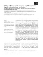
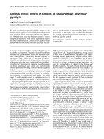
illustrated in Saccharomyces cerevisiae (Fig. 1). There is
a high degree of homology between mammalian MAPK
modules, both in their general organization and at the
protein level, with a high percentage of similarity in the
primary sequence of the different MAPKs (60% amino acid
identity between ERKs, JNKs and p38 MAPKs). Further-
more, the substrates of the three main mammalian MAPKs:
ERK, p38 and JNK display similar phosphorylation
consensus motifs: (T/S)P. How does the cell succeed in
delivering specific biological responses, limiting therefore
inappropriate crosstalk between the parallel MAPK mod-
ules? How is signal fidelity built within these modules? Two
mechanistic devices have emerged to enforce specificity.
Scaffold proteins. These scaffolds create multienzyme
complexes that bring together components of a single
kinase cascade (review by [12]). These complexes insulate
the module from activation by irrelevant stimuli and favour
the rapid transmission of the signal through the cascade.
Second, specific docking sites on MAPKs that serve for the
binding of substrates, activators and regulators increase the
fidelity and the efficiency of the enzymatic reactions.
The most studied MAPK scaffolding protein is Ste5, from
the yeast S. cerevisiae (reviewed in [13]). Through distinct
regions, Ste5 binds simultaneously to Ste11 (MAPKKK),
Ste7 (MAPKK) and Fus3 (one of the two MAPKs) but
Ste5 binds weakly to the other MAPK, Kss1. Fus3 is
preferentially implicated in the mating pheromone response,
whereas Kss1 is primarily involved in the filamentous
response. Fus3 and Kss1 share the same activators, Ste11
and Ste7, however, it seems that Kss1 may be better at
transmitting low-level, long-duration, scaffold independent
signalling, whereas Fus3 preferentially transmits scaffold-
associated signalling (review by [14]). Contrary to that
previously thought, scaffolded complexes are not stably
assembled. During vegetative growth, recent work showed
that the upstream activators Ste11 and Ste7 are predomin-
antly cytoplasmic, while the scaffold Ste5 and the MAPK
Fus3 are located both in the nucleus and in the cytoplasm
and shuttle permanently between these two cellular com-
partments [15]. In pheromone-treated cells, Ste11, Ste7 and
Fus3 are colocalized with Ste5 to tips of mating projections.
However, subsequently activated-Fus3 dissociates rapidly
from this multiprotein complex to translocate to the
nucleus. The role of the scaffolding protein, Ste5 in this
signalling pathway is essential, as Ste5 that cannot transit
via the nucleus is unable to localize to the cell periphery and
is unable to activate the pathway [16]. This novel regulatory
scheme may ensure that cytoplasmic Ste5 does not activate
downstream kinases in the absence of pheromone.
In mammalian cells, the identification and the role of
scaffolding proteins, in particular for the ERK module, is
not as well advanced. First, a two-hybrid screen, using
MEK1 as a bait, identified MP1 (MEK Partner 1) as a
scaffold protein that specifically binds MEK1 and ERK1 to
the exclusion of MEK2 and ERK2, thereby enhancing the
activation of ERK1 [17]. A partner of MP1, p14 was
discovered recently and the MP1-p14 complex scaffolds
MEK1 and ERK1 to the cytoplasmic surface of late
endosomes lysosomes where P14 is localized [18]. Reduc-
tion of MP1 or p14 protein levels by short interfering
(si)RNA results in defective signal transduction [19].
Another and presumably more central scaffold protein
for the ERK pathway is the KSR protein (kinase suppressor
of Ras). KSR was first identified by genetic screening in
Drosophila melanogaster and Caenorhabditis elegans as an
activator of the Ras pathway as mutations in KSR resulted
Fig. 1. MAPK modules and their associated functions in Saccharomyces
cerevisiae.
3292 J. Pouysse
´
gur and P. Lenormand (Eur. J. Biochem. 270) Ó FEBS 2003
in attenuation of Ras-mediated signalling (reviewed in
[20,21]). A mammalian homologue has been isolated that
interacts directly with MEK and ERK via distinct domains
while interaction with Raf appears to be indirect. KSR1
translocates from the cytoplasm to the cell membrane in
response to growth factor treatment. This process is
controlled by the serine/threonine kinase, C-TAK1 that
phosphorylates KSR1 at a site that confers 14-3-3 binding,
thus sequestering the KSR1 complex in the cytoplasm in the
absence of stimulation [22]. In response to growth factors,
the KSR1 S392 site is dephosphorylated by an unknown
phosphatase, and KSR1 is liberated from 14-3-3 binding
and translocates to the plasma membrane where it brings
MEK and ERK in close vicinity to the active Raf signalling
complex. Therefore, KSR1 seems to act as a scaffold protein
to maintain specificity and ensure signalling through the
ERK cascade. This notion has been beautifully demonstra-
ted by siRNA-mediated KSR knockdown in Drosophila
melanogaster [23]. For the JNK module, an interesting set of
JIP proteins (JNK-Interacting-Protein) has been identified.
These JIP proteins function by aggregating components of a
JNK module (including MLK, MKK7, and JNK) [24].
Interestingly JIP-1 has recently been shown to also bind to
the MAP kinase phosphatase MKP-7 indicating that JIP-1
scaffold protein modulates JNK signalling via association
with both protein kinases and protein phosphatases [25].
Therefore, from the unique properties of Ste5, JIP and
KSR, an emerging concept arises: scaffolding proteins are
not only insulators between homologous signalling mod-
ules, but they play an important role as regulators of the
subcellular localization and modulation of the kinase signal
intensity.
Docking sites. All MAPK members phosphorylate their
substrates on the consensus (T/S)P sequence and many
potential substrates contain this minimal motif (review by
[26]). Therefore MAPKs, presumably like all enzymes, have
acquired specific docking sites to specify interactions with
relevant substrates. These docking sites also contribute to
increase the local concentration of the kinase, hence
favouring substrate phosphorylation. The key residue of
the ERK docking site is composed of a cluster of acidic
residues on the C-terminus of the kinase that is remarkably
conserved from C. elegans to humans. This acidic cluster,
also called CD (for common docking) is not only found in
ERKs but in all MAPK members [27,28]. Data from the
three-dimensional structure of ERK indicate that the
common docking site is localized on the opposite side of
the kinase respective to the catalytic cleft, thus, substrates
must dissociate from the docking site to be phosphorylated,
indicating that association via kinase docking sites is highly
dynamic [29]. Docking sites on ERK interacting proteins
have been identified on substrates, activators, scaffolding
proteins and phosphatases. On interactor proteins, docking
sites are constituted by a cluster of positively charged amino
acids (D-domain), that interact on the same negatively
charged ERK docking site. This implies that interaction of
these proteins with ERK are mutually exclusive, thereby
providing a molecular mechanism for the sequential
activation and inactivation of ERK.
The specificity of the ERK interaction with proteins may
not be determined solely by the negatively charged ERK
motif, as interchanging (by mutation) of the docking site
present on ERK by the docking site present on p38 MAPK
still allows the binding of MEK to ERK while no binding of
MKK6 (the upstream activator of p38 MAPK) can be
detected [27]. It is now thought that the docking region on
ERK is contained in a docking groove, where several
interacting motifs cooperate to confer strong and specific
binding for each MAPK-interacting molecules [28,29]. The
spacing and organization of these different motifs on the
different MAPK interacting proteins is a feature that may
account for the differential MAPK specificities observed
[30]. Furthermore, in yeast, both the MKK–MAPK dock-
ing interaction and binding to the scaffolding protein, Ste5
make mutually reinforcing contributions to efficiently
conduct mating pheromone signalling [31].
Moreover, the phosphorylation state of partners can also
modulate the affinity of the interaction. For example, the
association of ERK with its substrate, Elk1, is enhanced
upon ERK activation [32], whereas, interaction of ERK
with its activator, MEK, is reduced upon activation of the
signalling cascade [33]. Interestingly, crosstalk with other
signalling pathways can be mediated by regulating docking
interactions. On the matter of protein substrates, there
are two classes of docking sites, the D-domain (cluster
of positively charged amino acids) and the FXFP motif,
whose binding pocket on ERK remains to be determined. A
systematic study of docking sites on Elk1 indicates that the
D-domain, and the FXFP motif form a flexible modular
system that has two functions [34]. First, the affinity of a
substrate for ERK can be regulated by the number, type,
position and arrangement of these docking sites. Second,
docking sites can direct phosphorylation of specific (S/T)P
residues [29,34].
The discovery of these kinase docking sites has provided
new tools to deregulate the ERK signalling cascade. The
first example based on these interactions was the trapping of
active ERKs in the cytoplasm by overexpression of an
inactive form of MKP3, which possesses a specific ERK
docking site. MKP3 is a cytoplasmic MAPK phosphatase,
therefore, its overexpression was able to retain ERKs in
the cytoplasm upon mitogenic stimulation [35]. A second
example was provided by microinjection into the nucleus of
a peptide corresponding to the ERK binding site of MEK.
This action led to the disruption of the association of
ERK/MEK in the nucleus hence significantly inhibiting
the MEK driven export of ERK out of the nucleus [36].
Similarly, microinjection into the nucleus of a peptide
corresponding to the ERK binding site on p90rsk, has been
shown to disrupt interaction between ERK and nuclear
phosphatases thus increasing active ERK in the nucleus
[37]. This latter experiment confirms that several interacting
proteins act via highly homologous docking sites as a
peptide corresponding to the sequence of an ERK substrate
can impede ERK association with phosphatases.
Spatio-temporal control of ERK activity
Schematically, mitogenic stimulation of G0-arrested cells
elicits biphasic ERK activation. After an initial burst of
activation (30–60 min) that varies with the cell type and the
strength of the stimulus, there is a prolonged activation
peaking from 2–h poststimulation, finally this activation
Ó FEBS 2003 MAPK (ERK) signalling module (Eur. J. Biochem. 270) 3293
gradually diminishes and ERK activity is reduced almost to
basal levels at the end of the G1 phase of the cell cycle. This
activity remains very low along the S phase, whereas, a burst
of ERK activity appears at the G2/M transition [38].
Considering that dephosphorylation of either the threonine
or the tyrosine residue within the ERK activation loop TEY
motif is sufficient for total kinase inactivation [39], numer-
ous phosphatases could be implicated in the two phases of
inactivation: the rapid initial phase and the slower and
delayed one.
The serine/threonine specific phosphatase, PP2A has
been implicated in the first inactivation of ERK observed
within minutes of NIH-3T3 cell stimulation [40] and of
Xenopus oocytes stimulation [41]. The remaining phospho-
tyrosine residue must be removed by a constitutive
phosphatase. Several related tyrosine specific phosphatases
such as PTP-SL, STEP, He-PTP/LC-PTP show a good
specificity towards ERKs [30,42,43]. However, these
cytosolic tyrosine phosphatases present a restricted expres-
sion pattern and thus the ubiquitously expressed phos-
phatase(s) that may play the same role in most cells is
(are) not yet identified. Interestingly, a cytosolic Droso-
phila tyrosine phosphatase, PTP-ER, related to the
tyrosine phosphatases mentioned above, plays an import-
ant role in down-regulating ERK activation during
Drosophila eye development [44]. It is not known,
however, if PTP-ER plays a major role in the inactivation
of the first peak of ERK activation.
The delayed phase of ERK inactivation is dependent on
protein synthesis, indicating that neosynthesized phospha-
tases are required [40,45]. Furthermore, these phosphatases
have a tyrosine specificity as they are inhibited by vanadate
treatment [37,40,41]. The phosphatases that fullfil these
criteria are the MAPK phosphatases (MKPs). MKPs
belong to the dual specificity phosphatases family (DUSP)
as they are capable of dephosphorylating both the tyrosine
and the threonine residues of MAPKs (reviewed in [46,47]).
Indeed, we have demonstrated that MKPs are good
candidates for setting the low steady-state activity of ERKs.
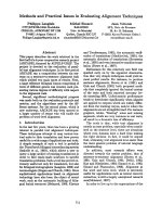
In fact, as shown in Fig. 2, ERK activity itself induces an
autocontrol mechanism. We first showed that exclusive
activation of p42/p44MAPKs is sufficient to induce the
immediate early genes, mkp1 and mkp2 [45]. Second, we
established that MKP1 and MKP2 proteins are direct
substrates of ERKs and third that these MKPs, when
phosphorylated, are less sensitive to rapid degradation by
the ubiquitine-proteasomal system [48]. Indeed, MKP1 is
phosphorylated on serine 359 and serine 364 by ERK [48],
which does not modify phosphatase activity, but increases
their half life, reinforcing the negative feed back autocontrol
(Fig. 2). Finally, MKP3 [49] and MKP1 [50] are catalyti-
cally activated upon ERK binding to their N-terminal
non-catalytic moiety. Catalytic activation of MKP1 and of
MKP3 occurs by binding of ERK via the classical docking
site. Hence, substrate-specificity is ensured by two means:
protein–protein interaction and catalytic activation of the
phosphatase.
The precise role of each MKPs in vivo is not yet
understood. Clearly, expression of some MKPs is restricted
to specific subcellular compartments, cytoplasm (MKP3) or
nucleus (MKP1 and 2) that must impinge on the range of
available substrates. It is probable that there is some degree
of redundancy between MKPs as invalidation of the mkp1
gene did not affect mouse physiology [51]. More work is
required to assess the functional role of individual MKPs
in vivo.
Temporal compartimentalization of the ERK module
ERK nuclear translocation is a key event in signalling. In
resting cells, Raf, MEK and ERKs are cytoplasmic.
Following mitogenic stimulation, intracellular redistribution
of ERK occurs in two phases. First, there is an immediate
ERK nuclear translocation that can be visualized, in
particular with antibodies specific for the phosphorylated
and active form of ERKs, by immunofluorescence as soon
as 2 min [37]. The pool of ERK protein progressively
accumulates in the nucleus after several hours of mitogenic
stimulation (3–6 h depending of the cell type), depleting the
cytoplasmic compartment. This process of ERK nuclear
accumulation is reversible and follows the time-course of
ERK inactivation. If the activation of the ERK module is
maintained (ER-Raf construct, activatable by tamoxifen),
ERKs remain in the nucleus [52]. Non-mitogenic stimuli
induce the initial nuclear entry but fail to trigger the nuclear
accumulation of ERKs [52,53]. Similarly, when the fate of
cells is differentiation, only differentiating signals trigger the
nuclear accumulation of ERK observed after several hours
of stimulation [8]. This nuclear translocation is a key event
in ERK signalling. This was demonstrated by an experiment
designed to retain active ERK in the cytoplasm [35]; under
these conditions, fibrobasts fail to replicate their DNA.
Alternatively, forcing an active form of ERK into the
nucleus of fibroblasts promotes oncogenic transformation
[54]. Physiologically, the cytoplasmic retention of ERK may
play a critical role in maintaining a differentiated phenotype
in some cell types. For example, increased expression of
the protein, Phosphoprotein Enriched in Astrocytes 15kD
(PEA15) traps ERK in the cytoplasm of astrocytes and
Fig. 2. Schematic model illustrating the auto-regulation of the ERK
module. MAP kinase phosphatase 1 and 2 (MKP1/2), products of the
immediate early genes mkp1 and mkp2, are directly induced via the
activation of p42/p44 MAP kinases, providing a progressive retro-
inhibition of ERKs. In addition, MKP1/2 are directly phosphorylated
by ERKs, increasing their stability and therefore reinforcing the
retrocontrol [48].
3294 J. Pouysse
´
gur and P. Lenormand (Eur. J. Biochem. 270) Ó FEBS 2003
blocks cell proliferation, whereas, genetic deletion of
PEA15 increases astrocyte proliferation [55]. Regulation
of ERK signalling by cytoplasmic trapping may be a
frequent phenomenon as it was shown recently that
b-arrestin associates with ERK and enhances the ERK
cytoplasmic activity while inhibiting ERK mediated tran-
scription [56].
ERK nuclear accumulation and inactivation. This ERK
translocation process produced a surprise when we
double-labelled cells either with anti-ERK protein or with
antibodies directed against active ERKs (phosphoERK-
antibodies). Clearly, this double-labelling revealed that the
ERK protein pool that accumulated in the nucleus
became inactivated with time [37]. As seen in Fig. 3, at
the peak of ERK nuclear accumulation (3 h in hamster
fibroblasts), virtually no active phospho-ERK was detect-
able in the nucleus. We demonstrated by an independent
approach that the capacity of nuclear ERKs to phos-
phorylate nuclear substrates (HIF-1a)atthistimeof
stimulation (3 h of FCS) was severely blunted [37].
However, short inhibition of tyrosine phosphatases with
vanadate, fully reactivated phospho-ERK in the nucleus
and maximally phosphorylated the nuclear substrate,
HIF-1a [37]. When activation of the ERK pathway is
transient, ERKs rapidly exit out of the nucleus, however,
during sustained activation of the module, ERKs remain
in the nucleus in an inactive form as shown in Fig. 3
[37,57]. The nuclear accumulation of ERK in the nucleus
requires the ERK-dependent transcriptional induction of
short-lived nuclear anchoring proteins [57]. The identity of
these nuclear anchors remains elusive, however, the use
of anti-phospho-ERK antibodies provided new clues
in understanding this nuclear accumulation of ERK.
MKP1 and MKP2 are the best candidates for inactiva-
tion of ERK in the nucleus as: (a) they are induced by
activation of the ERK pathway; (b) they are localized in the
nucleus; (c) they possess ERK docking sites and (d) they are
inhibited by tyrosine phosphatase specific inhibitors [46,47].
Furthermore, MKP1 and MKP2 may participate in the
nuclear anchoring of ERKs as these proteins present all
the characteristics of ERK nuclear anchors, MKP1/2 are
induced by ERK activation [45], and are short-lived nuclear
proteins whose expression is virtually abolished within 1 h
upon traductional or translational block [48]. The use of
RNA interference to abrogate expression of each MKP
isoforms, may help to provide quick answers to these
questions.
The mechanisms of ERK nuclear import and export are
still largely unknown. These protein kinases do not possess
any of the common nuclear import sequences (NLS) and
previous work has shown that ERKs cross the nucleopore
by passive diffusion [58]. It has been shown that ERK
associates with MEK in the cytoplasm of resting cells via
their docking sites, an interaction that is reduced dramati-
cally upon activation of the MEK/ERK signalling pathway,
thus allowing ERK to translocate to the nucleus [36].
Clearly, activation of the pathway is essential as blocking
MEK activity abrogates ERK nuclear translocation [52];
however, phosphorylation mutants of ERK still translocate
to the nucleus in response to cell stimulation [53,59,60].
Phosphorylation-dependent dimerization of ERK has also
Fig. 3. Long-term activation of p42/p44 MAPKs induces their nuclear accumulation in a dephosphorylated and inactive form. Resting CCL39 hamster
fibrobasts (left panel) were stimulated for 3 h with 10% fetal bovine serum (FCS) (middle and right panels).The green immunoflurescence indicates
the location of the proteins, ERK1 and 2, whereas the red immunofluorescence indicates the dual phosphorylated and active forms of ERK1 and 2.
The right panel shows confocal images. Reproduced from reference [37].
Ó FEBS 2003 MAPK (ERK) signalling module (Eur. J. Biochem. 270) 3295
been proposed to explain ERK nuclear entry. Indeed, ERK-
b-galactosidase fusion proteins are unable to enter the
nucleus when ERK dimerization motifs are mutated [58].
However, ERK1 dimerization mutants expressed in erk1
null mouse fibroblasts present the same time course of
nuclear translocation as wild type ERK1 (P, Lenormand
and J, Pouyssegur, unpublished results). As discussed
previously, recent work on the scaffolding protein, KSR
indicates that KSR participates in the regulation of the
subcellular localization of kinase cascade components.
However, the contribution of KSR in the release of ERK
from the cytoplasmic complex has not yet been established.
Recent work in Drosophila may provide clues in under-
standing this phenomenon. Indeed, deletion or mutations in
the Drosophila importin a homologue, DIM-7 or mutations
in the importin b homologue Ketel, reduce the nuclear
localization of D-ERK, the ERK Drosophila homologue.
Interestingly, DIM-7 associates with phosphorylated
D-ERK which should allow a better understanding of
how ERK can interact with the active import machinery
while lacking a classical NLS [61]. Another point of interest
is the demonstration of direct binding of ERK to nucleo-
pore complex [60]. In that case, ERK transport across the
nucleopore would be propelled by Brownian motion. It has
been shown in permeabilized mammalian cells that ERK
associates directly with the nucleopore complex and trans-
locates to the nucleus independently of soluble factors and
ATP. Furthermore, ERK binds in vitro to an FG repeat
region of nucleoporin CAN/Nup214.
Altogether, the relative contribution of these different
mechanisms in conducting ERK across the nuclear mem-
brane remains to be determined.
Several studies have clearly established the continuous
shuttling of ERK between the cytoplasm and nucleus.
When quiescent cells are treated with leptomycin B, that
blocks Crm1-dependent nuclear export, ERK appears
within minutes in the nucleus [37]. This occurs in the total
absence of ERK activation as it is not impeded by prior
treatment with the MEK inhibitor, U0126 [52]. The export
of ERK from the nucleus has remained as enigmatic as the
import as ERK1 and ERK2 protein sequences do not show
motifs homologous to a nuclear export sequence (NES).
However, blocking active nuclear export with leptomycin B
triggers the nuclear accumulation of ERK and MEK
[37,62]. In the presence of leptomycin B, addition of growth
factors for 5 min is sufficient to mobilize the entire
cytoplasmic pool of ERK in the nucleus. This result stresses
the rapid and constant ERK cytoplasmic/nuclear shuttling.
We believe that MEK, with its built-in export sequence,
might be at the heart of this shuttling mechanism. Although
MEK always appears in the cytoplasm, due to its very
efficient NES, MEK is the Ôdriving exporting-forceÕ of
nuclear inactivated ERKs. In summary, ERKs oscillate
between two high affinity complexes in separate cellular
compartments. In resting cells ERKs are associated with the
Ôactivating centerÕ, the Raf-MEK cytoplasmic complex.
Upon long mitogenic treatment, ERKs are sequestered in
the nucleus, closely associated to the neosynthesized ÔMKP-
inactivating centerÕ away from the site of activation. By this
mechanism we propose that mammalian cells operate the
termination of the MAPK signal, a condition required to
trigger the appropriate biological response.
Knockdown of
erk1
and
erk2
genes in mice
In previous experiments in which the biological functions of
p42/p44 MAP kinases have been addressed (antisense or
expression of dominant-negative MEK or ERK), both
isoforms have been inactivated [63]. So far, the pharmaco-
logical inhibition of ERK1 and ERK2 relies on MEK
specific inhibitors that invariably blunt the activation of
ERK1 and ERK2 [64]. Therefore, the specific role of the
two ERK isoforms is still an entirely open question. In
mammals, ERK1 and ERK2 are expressed ubiquitously,
although the expression level could vary in different tissues.
These two protein kinases are highly similar (overall 84%
identity at the amino acid level, and up to 90% identity
when the short N-terminal stretch is not taken into account)
and, in vitro, both isoforms present apparently the same
substrate specificity and the same time course of activation.
Interestingly however, ERK1 and ERK2 do not share an
identical pattern of compartimentalization as illustrated
from the work of HuberÕs group [19]. Thus, a pressing
question is what are their specializations and do they have
overlaping functions? A way to address this issue is to
produce single ERK invalidating mutations in mice. From
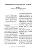
our published work, and work in progress, it is clear that
isoform-specific invalidation in mice provides contrasting
results (Fig. 4). First ERK1
–/–
mice are viable, fertile and of
normal size [63]. Clearly, in these animals, in which we only
found a defect in thymocyte terminal differentiation, ERK2
can compensate for most of the functions of ERK1. On the
contrary, disruption of the Erk2 locus leads to embryonic
lethality early in mouse development after the implantation
stage. Erk2 mutant embryos failed to form the ectoplacental
cone and extraembryonic ectoderm, which give rise to
mature trophoblast derivatives in the foetus (Sylvain
Meloche, Institute of Clinical Research, Montreal; personal
communication). In these embryos, ERK1 cannot compen-
sate for the loss of ERK2, thus, specific functions of these
isoforms remain to be discovered. Alternatively, ERK1 is
not expressed in some cells or at such low levels compared to
ERK2 that it cannot provide the strength of activation
required for embryonic survival.
Finally, during the course of these studies it became
apparent that mice disrupted in the Erk1 locus were more
actively displaying facilitated striatal-mediated learning and
memory [65]. This unexpected behaviour revealed that
ERK1 ablation led to an increase in the temporal activation
of ERK2. The exact mechanism of the interplay between the
two isoforms is not understood, but this finding indicates
Fig. 4. Phenotypes of ERK1 and ERK2 null mice.
3296 J. Pouysse
´
gur and P. Lenormand (Eur. J. Biochem. 270) Ó FEBS 2003
that a little alteration in the intensity and temporal
activation of ERKs could have a profound effect in animal
physiology.
In conclusion, the ERK MAP kinase module, reported in
early1990,hasbeenshowntoplayacentralrolein
signalling growth, differentiation and survival from inver-
tebrates to humans. A sophisticated autocontrol mechanism
associated with nuclear/cytoplasmic shuttling ensure the
intensity and temporal modulation of ERK activity in
response to various hormonal, growth factor and extracel-
lular matrix stimuli. Cancer and many other diseases are
simply the reflection of alterations in this fine tuning
mechanisms.
Despite intense research efforts worldwide, questions
concerning this ERK module remain unanswered: are
there other scaffolding proteins in mammalian cells? What
are the exact roles of KSR1 and KSR2? Do they represent
the basis for two separate and competing modules for
MEK1/MEK2 and ERK1/ERK2? What is the identity of
the ERK nuclear anchoring complex and which MAPK
phosphatases are essential in terminating the ERK signal?
We anticipate that the siRNA knockdown approach with
inducible vectors will greatly facilitate the investigation of
these questions.
Acknowledgements
We thank Drs Gilles Page
`
s, Fergus McKenzie, Anne Brunet, Jean-
Marc Brondello and the regretted Veronique Volmat for their unique
contribution to the MAP kinase project and all the members of the
laboratory for helpful discussion. I am particularly grateful to Dr
C. Brahimi-Horn for carefull reading of the manuscript.
References
1. Widmann, C., Gibson, S., Jarpe, M.B. & Johnson, G.L. (1999)
Mitogen-activated protein kinase: conservation of a three-kinase
module from yeast to human. Physiol. Rev. 79, 143–180.
2. Kerkhoff, E. & Rapp, U.R. (2001) The Ras-Raf relationship: an
unfinished puzzle. Adv. Enzyme Regul. 41, 261–267.
3. Hallberg, B., Rayter, S.I. & Downward, J. (1994) Interaction of
Ras and Raf in intact mammalian cells upon extracellular stimu-
lation. J. Biol Chem. 269, 3913–3916.
4. Page
`
s, G., Lenormand, P., L’Allemain, G., Chambard, J C.,
Me
´
loche, S. & Pouysse
´
gur, J. (1993) Mitogen-activated protein
kinases p42mapk and p44mapk are required for fibroblast cell
proliferation. Proc. Natl. Acad. Sci. USA 90, 8319–8323.
5. Lavoie, J.N., L’Allemain, G., Brunet, A., Muller, R. & Pouysse-
gur, J. (1996) Cyclin D1 expression is regulated positively by the
p42/p44MAPK and negatively by the p38/HOGMAPK pathway.
J. Biol Chem. 271, 20608–20616.
6. Lavoie, J.N., Rivard, N., L’Allemain, G. & Pouyssegur, J. (1996)
A temporal and biochemical link between growth factor-activated
MAP kinases, cyclin D1 induction and cell cycle entry. Prog. Cell
Cycle Res. 2, 49–58.
7. Whitmarsh, A.J. & Davis, R.J. (2000) A central control for cell
growth. Nature. 403, 255–256.
8. Traverse,S.,Seedorf,K.,Paterson,H.,Marshall,C.,Cohen,P.&
Ullrich, A. (1994) EGF triggers neuronal differentiation of PC12
cells that overexpress the EGF receptor. Curr. Biol. 4, 694–701.
9. Kahan, C., Seuwen, K., Me
´
loche, S. & Pouysse
´
gur, J. (1992)
Coordinate, biphasic activation of p44 mitogen activated protein
kinase and S6 kinase by growth factors in hamster fibroblasts.
J. Biol. Chem. 267, 13369–13375.
10. Le Gall, M., Chambard, J.C., Breittmayer, J.P., Grall, D.,
Pouyssegur, J. & Van Obberghen-Schilling, E. (2000) The p42/p44
MAP kinase pathway prevents apoptosis induced by anchorage
and serum removal. Mol. Biol. Cell. 11, 1103–1112.
11. Zugasti, O., Rul, W., Roux, P., Peyssonnaux, C., Eychene, A.,
Franke, T.F., Fort, P. & Hibner, U. (2001) Raf-MEK-Erk cascade
in anoikis is controlled by Rac1 and Cdc42 via Akt. Mol. Cell
Biol. 21, 6706–6717.
12. Whitmarsh, A.J. & Davis, R.J. (1998) Structural organization of
MAP-kinase signaling modules by scaffold proteins in yeast and
mammals. Trends Biochem. Sci. 23, 481–485.
13. Elion, E.A. (1995) STE5: a meeting place for MAP kinases and
their associates. Trends Cell Biology 5, 322–327.
14. Pryciak, P.M. (2001) MAP kinases bite back. Dev. Cell. 1,
449–451.
15. van Drogen, F., Stucke, V.M., Jorritsma, G. & Peter, M. (2001)
MAP kinase dynamics in response to pheromones in budding
yeast. Nat. Cell Biol. 3, 1051–1059.
16. Mahanty, S.K., Wang, Y., Farley, F.W. & Elion, E.A. (1999)
Nuclear shuttling of yeast scaffold Ste5 is required for its recruit-
ment to the plasma membrane and activation of the mating
MAPK cascade. Cell 98, 501–512.
17. Schaeffer, H.J., Catling, A.D., Eblen, S.T., Collier, L.S., Krauss,
A. & Weber, M.J. (1998) MP1: a MEK binding partner that
enhances enzymatic activation of the MAP kinase cascade.
Science. 281, 1668–1671.
18. Wunderlich, W., Fialka, I., Teis, D., Alpi, A., Pfeifer, A., Parton,
R.G., Lottspeich, F. & Huber, L.A. (2001) A novel 14-kilodalton
protein interacts with the mitogen-activated protein kinase scaf-
fold mp1 on a late endosomal/lysosomal compartment. J. Cell
Biol. 152, 765–776.
19. Teis, D., Wunderlich, W. & Huber, L.A. (2002) Localization of
the MP1-MAPK scaffold complex to endosomes is mediated by
p14 and required for signal transduction. Dev Cell. 3, 803–814.
20. Therrien, M., Chang, H.C., Solomon, N.M., Karim, F.D., Was-
sarman, D.A. & Rubin, G.M. (1995) KSR, a novel protein kinase
required for RAS signal transduction. Cell 83, 879–888.
21. Morrison, D.K. (2001) KSR: a MAPK scaffold of the Ras path-
way? J. Cell Sci. 114, 1609–1612.
22. Muller, J., Ory, S., Copeland, T., Piwnica-Worms, H. & Morrison,
D.K. (2001) C-TAK1 regulates Ras signaling by phosphorylating
the MAPK scaffold, KSR1. Mol. Cell. 8, 983–993.
23. Roy, F., Laberge, G., Douziech, M., Ferland-McCollough, D. &
Therrien, M. (2002) KSR is a scaffold required for activation of
the ERK/MAPK module. Genes Dev. 16, 427–438.
24. Yasuda, J., Whitmarsh, A.J., Cavanagh, J., Sharma, M. & Davis,
R.J. (1999) The JIP group of mitogen-activated protein kinase
scaffold proteins. Mol. Cell Biol. 19, 7245–7254.
25. Willoughby, E.A., Perkins, G.R., Collins, M.K. & Whitmarsh,
A.J. (2003) The JNK-interacting protein-1 scaffold protein targets
MAPK phosphatase-7 to dephosphorylate JNK. J. Biol. Chem.
278, 10731–10736.
26. Lewis, T.S., Shapiro, P.S. & Ahn, N.G. (1998) Signal transduction
through MAP kinase cascades. Adv. Cancer Res. 74, 49–139.
27. Tanoue, T., Adachi, M., Moriguchi, T. & Nishida, E. (2000) A
conserved docking motif in MAP kinases common to substrates,
activators and regulators. Nat. Cell Biol. 2, 110–116.
28. Bardwell, L. & Thorner, J. (1996) A conserved motif at the amino
termini of MEKs might mediate high affinity interaction with the
cognate MAPKs. Trends in Biochem. Sci. 21, 373–374.
29. Tanoue, T., Maeda, R., Adachi, M. & Nishida, E. (2001) Identi-
fication of a docking groove on ERK and p38 MAP kinases
that regulates the specificity of docking interactions. EMBO J. 20,
466–479.
30. Tarrega, C., Blanco-Aparicio, C., Munoz, J.J. & Pulido, R.
(2002) Two clusters of residues at the docking groove of
Ó FEBS 2003 MAPK (ERK) signalling module (Eur. J. Biochem. 270) 3297
mitogen-activated protein kinases differentially mediate their
functional interaction with the tyrosine phosphatases PTP-SL and
STEP. J. Biol. Chem. 277, 2629–2636.
31. Sharrocks, A.D., Yang, S.H. & Galanis, A. (2000) Docking
domains and substrate-specificity determination for MAP kinases.
Trends Biochem. Sci. 25, 448–453.
32. Yang, S.H., Yates, P.R., Whitmarsh, A.J., Davis, R.J. & Shar-
rocks, A.D. (1998) The Elk-1 ETS-domain transcription factor
contains a mitogen-activated protein kinase targeting motif. Mol.
Cell. Biol. 18, 710–720.
33. Fukuda, M., Gotoh, Y. & Nishida, E. (1997) Interaction of
MAP kinase with MAP kinase kinase: its possible role in the
control of nucleocytoplasmic transport of MAP kinase. EMBO J.
1901–08.
34. Fantz, D.A., Jacobs, D., Glossip, D. & Kornfeld, K. (2001)
Docking sites on substrate proteins direct extracellular signal-
regulated kinase to phosphorylate specific residues. J. Biol. Chem.
276, 27256–27265.
35. Brunet, A., Roux, D., Lenormand, P., Dowd, S., Keyse, S. &
Pouyssegur, J. (1999) Nuclear translocation of p42/p44 mitogen-
activated protein kinase is required for growth factor-induced gene
expression and cell cycle entry. EMBO J. 18, 664–674.
36. Fukuda, M., Gotoh, I., Gotoh, Y. & Nishida, E. (1996) Cyto-
plasmic localization of MAP kinase kinase directed by its
N-terminal, leucin-rich amino acid sequence, which acts as a
nuclear export signal. J. Biol. Chem. 271, 20024–20028.
37. Volmat, V., Camps, M., Arkinstall, S., Pouysse
´
gur, J. &
Lenormand, P. (2001) The nucleus, a site for signal termination by
sequestration and inactivation of p42/p44 MAP kinases. J. Cell.
Sci. 114, 3433–3443.
38. Roberts, E.C., Shapiro, P.S., Nahreini, T.S., Pages, G., Pouysse-
gur, J. & Ahn, N.G. (2002) Distinct cell cycle timing requirements
for extracellular signal-regulated kinase and phosphoinositide
3-kinase signaling pathways in somatic cell mitosis. Mol. Cell Biol.
22, 7226–7241.
39. Posada, J. & Cooper, J.A. (1992) Requirements for phosphory-
lation of MAP kinase during meiosis in xenopus oocytes. Science.
255, 212–215.
40. Alessi, D., Gomez, N., Moorhead, G., Lewis, T., Keyse, S.M. &
Cohen, P. (1995) Inactivation of p42 MAP kinase by protein
phosphatase 2A and a protein tyrosine phosphatase, but not
CL100, in various cell lines. Curr. Biol. 5, 283–295.
41. Sohaskey, M.L. & Ferrell, J.E. Jr (1999) Distinct, constitutively
active MAPK phosphatases function in Xenopus oocytes:
implications for p42 MAPK regulation in vivo. Mol. Biol. Cell. 10,
3729–3743.
42. Oh-hora, M., Ogata, M., Mori, Y., Adachi, M., Imai, K., Kosugi,
A. & Hamaoka, T. (1999) Direct suppression of TCR-mediated
activation of extracellular signal-regulated kinase by leukocyte
protein tyrosine phosphatase, a tyrosine-specific phosphatase.
J. Immunol. 163, 1282–1288.
43. Pettiford, S.M. & Herbst, R. (2000) The MAP-kinase ERK2 is a
specific substrate of the protein tyrosine phosphatase HePTP.
Oncogene. 19, 858–869.
44. Karim, F.D. & Rubin, G.M. (1999) PTP-ER, a novel tyrosine
phosphatase, functions downstream of Ras1 to downregulate
MAP kinase during Drosophila eye development. Mol. Cell. 3,
741–750.
45. Brondello, J.M., Brunet, A., Pouyssegur, J. & McKenzie, F.R.
(1997) The dual specificity mitogen-activated protein kinase
phosphatase-1 and – 2 are induced by the p42/p44MAPK cascade.
J. Biol. Chem. 272, 1368–1376.
46. Camps, M., Nichols, A. & Arkinstall, S. (2000) Dual specificity
phosphatases: a gene family for control of MAP kinase function.
Faseb J. 14, 6–16.
47. Keyse, S.M. (2000) Protein phosphatases and the regulation of
mitogen-activated protein kinase signalling. Curr. Opin. Cell Biol.
12, 186–192.
48. Brondello, J.M., Pouyssegur, J. & McKenzie, F.R. (1999)
Reduced MAP kinase phosphatase-1 degradation after p42/
p44MAPK-dependent phosphorylation. Science. 286, 2514–2517.
49. Camps, M., Nichols, A., Gillieron, C., Antonsson, B., Muda, M.,
Chabert, C., Boschert, U. & Arkinstall, S. (1998) Catalytic acti-
vation of the phosphatase MKP-3 by ERK2 mitogen-activated
protein kinase. Science. 280, 1262–1265.
50. Slack, D.N., Seternes, O.M., Gabrielsen, M. & Keyse, S.M. (2001)
Distinct binding determinants for ERK2/p38alpha and JNK map
kinases mediate catalytic activation and substrate selectivity of
map kinase phosphatase-1. J. Biol Chem. 276, 16491–16500.
51. Dorfman, K., Carrasco, D., Gruda, M., Ryan, C., Lira, S.A. &
Bravo, R. (1996) Disruption of the erp/mkp-1 gene does not affect
mouse development: normal MAP kinase activity in ERP/MKP-
1-deficient fibroblasts. Oncogene. 13, 925–931.
52. Volmat, V. & Pouyssegur, J. (2001) Spatiotemporal regulation of
the p42/p44 MAPK pathway. Biol. Cell. 93, 71–79.
53. Lenormand, P., Sardet, C., Page
`
s, G., L’Allemain, G., Brunet, A.
& Pouysse
´
gur, J. (1993) Growth Factors Induce Nuclear Trans-
location of MAP Kinases (p42maPk and p44mapk) but not of
Their Activator MAP Kinase Kinase (p45mapkk) in Fibroblasts.
J. Cell. Biol. 122, 1079–1089.
54. Robinson, M.J., Stippec, S.A., Goldsmith, E., White, M.A. &
Cobb, M.H. (1998) A constitutively active and nuclear form of the
MAP kinase ERK2 is sufficient for neurite outgrowth and cell
transformation. Curr. Biol. 8, 1141–1150.
55. Formstecher, E., Ramos, J.W., Fauquet, M., Calderwood, D.A.,
Hsieh, J.C., Canton, B., Nguyen, X.T., Barnier, J.V., Camonis, J.,
Ginsberg, M.H. & Chneiweiss, H. (2001) PEA-15 mediates cyto-
plasmic sequestration of ERK MAP kinase. Dev. Cell. 1, 239–250.
56. Tohgo, A., Pierce, K.L., Choy, E.W., Lefkowitz, R.J. & Luttrell,
L.M. (2002) beta-Arrestin scaffolding of the ERK cascade
enhances cytosolic ERK activity but inhibits ERK mediated
transcription following angiotensin AT1a receptor stimulation.
J. Biol. Chem. 277, 9429–9436.
57. Lenormand, P., Brondello, J.M., Brunet, A. & Pouyssegur, J.
(1998) Growth factor-induced p42/p44 MAPK nuclear translo-
cation and retention requires both MAPK activation and neo-
synthesis of nuclear anchoring proteins. J. Cell Biol. 142, 625–633.
58. Adachi, M., Fukuda, M. & Nishida, E. (1999) Two co-existing
mechanisms for nuclear import of MAP kinase: passive diffusion
of a monomer and active transport of a dimer. EMBO J. 18,
5347–5358.
59. Khokhlatchev, A.V., Canagarajah, B., Wilsbacher, J., Robinson,
M., Atkinson, M., Goldsmith, E. & Cobb, M.H. (1998) Phos-
phorylation of the MAP kinase ERK2 promotes its homo-
dimerization and nuclear translocation. Cell. 93, 605–615.
60. Matsubayashi, Y., Fukuda, M. & Nishida, E. (2001) Evidence
for existence of a nuclear pore complex-mediated, cytosol-
independent pathway of nuclear translocation of ERK MAP
kinase in permeabilized cells. J. Biol. Chem. 276, 41755–41760.
61. Lorenzen, J.A., Baker, S.E., Denhez, F., Melnick, M.B., Brower,
D.L. & Perkins, L.A. (2001) Nuclear import of activated D-ERK
by DIM-7, an importin family member encoded by the gene
moleskin. Development. 128, 1403–1414.
62. Adachi, M., Fukuda, M. & Nishida, E. (2000) Nuclear export of
MAP kinase (ERK) involves a MAP kinase kinase (MEK)-
dependent active transport mechanism. J. Cell Biol. 148, 849–856.
63. Page
`
s, G., Guerin, S., Grall, D., Bonino, F., Smith, A., Anjuere,
F., Auberger, P. & Pouyssegur, J. (1999) Defective thymocyte
maturation in p44 MAP kinase (Erk 1) knockout mice. Science.
286, 1374–1377.
3298 J. Pouysse
´
gur and P. Lenormand (Eur. J. Biochem. 270) Ó FEBS 2003
64. Kohno, M. & Pouyssegur, J. (2003) Pharmacological inhibitors of
the ERK signaling pathway: application as anticancer drugs.
Prog.CellCycleRes. 5, 219–224.
65. Mazzucchelli, C., Vantaggiato, C., Ciamei, A., Fasano, S.,
Pakhotin,P.,Krezel,W.,Welzl,H.,Wolfer,D.P.,Pages,G.,
Valverde,O.,Marowsky,A.,Porrazzo,A.,Orban,P.C.,
Maldonado, R., Ehrengruber, M.U., Cestari, V., Lipp, H.P.,
Chapman, P.F., Pouyssegur, J. & Brambilla, R. (2002) Knockout
of ERK1 MAP kinase enhances synaptic plasticity in the striatum
and facilitates striatal-mediated learning and memory. Neuron. 34,
807–820.
Ó FEBS 2003 MAPK (ERK) signalling module (Eur. J. Biochem. 270) 3299