Báo cáo khoa học: In vitro characterization of a plastid terminal oxidase (PTOX) pot
Bạn đang xem bản rút gọn của tài liệu. Xem và tải ngay bản đầy đủ của tài liệu tại đây (361.23 KB, 8 trang )
In vitro
characterization of a plastid terminal oxidase (PTOX)
Eve-Marie Josse, Jean-Pierre Alcaraz, Anne-Marie Laboure
´
and Marcel Kuntz
Universite
´
J. Fourier and CNRS (Plastes et Diffe
´
renciation Cellulaire, UMR 5575), Grenoble, France
The plastid terminal oxidase (PTOX) encoded by the Ara-
bidopsis IMMUTANS gene was expressed in Escherichia coli
cells and its quinone/oxygen oxidoreductase activity moni-
tored in isolated bacterial membranes using NADH as an
electron donor. Specificity for plastoquinone was observed.
Neither ubiquinone, duroquinone, phylloquinone nor ben-
zoquinone could substitute for plastoquinone in this assay.
However, duroquinol (fully reduced chemically) was an
accepted substrate. Iron is also required and cannot be
substituted by Cu
2+
,Zn
2+
or Mn
2+
. This plastoquinol
oxidase activity is independent of temperature over the
15–40 °C range but increases with pH (from 5.5 to 9.0).
Unlike higher plant mitochondrial alternative oxidases, to
which PTOX shows sequence similarity (but also differences,
especially in a putative quinone binding site and in cysteine
conservation), PTOX activity does not appear to be regu-
lated by pyruvate or any other tested sugar, nor by AMP. Its
activity decreases, however, with increasing salt (NaCl or
KCl) concentration. Various quinone analogues were tested
for their inhibitory activity on PTOX. Pyrogallol analogues
were found to be inhibitors, especially octyl gallate
(I
50
¼ 0.4 l
M
) that appears far more potent than propyl
gallate or gallic acid. Thus, octyl gallate is a useful inhibitor
for future in vivo or in organello studies aimed at studying
the roles of PTOX in chlororespiration and as a cofactor
for carotenoid biosynthesis.
Keywords: alternative oxidase; immutans; iron center;
plastoquinone; quinol oxidase.
The cloning of the Arabidopsis gene, IMMUTANS,has
identified a new important plastid located enzyme [1,2].
Inactivation of this gene first indicated that it was a cofactor
for early steps in carotenoid biosynthesis, namely phytoene
desaturation. Reduced phytoene desaturation leads to
reduced carotenoid content, which in turn leads to photo-
oxidative damage visible as a variegated phenotype com-
prising white and green sectors in the immutans mutant. The
tomato ghost mutant is impaired in the corresponding gene
[3] and its phenotype resembles immutans in leaves while
fruit do not redden and accumulate phytoene.
The IMMUTANS gene product shows limited similarity
with mitochondrial alternative oxidases (AOX, [4]) that are
ubiquinol oxidases catalyzing a cyanide-resistant reduction
of oxygen to water in the respiratory electron transfer chain
(recently reviewed in [5]). Sequence comparison between
AOXs and the IMMUTANS gene product allowed a
reassessment of the amino acids that are likely to have
functional involvement in AOXs [6] and suggested that the
IMMUTANS gene product may function as a terminal
oxidase located within plastids (PTOX, [3]). These data are
reinforced by the demonstration that electrons provided by
photosytem II can be diverted at a significant rate towards a
chloroplast quinol oxidase in Chlamydomonas [7]. Based on
the similarity of immunological and pharmacological
properties between the IMMUTANS encoded PTOX in
Arabidopsis and the plastoquinol oxidizing activity in
Chlamydomonas, the involvement of PTOX in chlororespi-
ration was proposed [7]. Furthermore, the Arabidopsis
PTOX was functional when expressed in tobacco and
strongly accelerated the nonphotochemical re-oxidation of
quinones [8]. During the dark to light induction phases of
photosynthesis at low irradiances, PTOX drives electron
flow to O
2
.
The terminal oxidase activity of the IMMUTANS gene
product could also be monitored after expression in E. coli
[3]. Both in vitro and in vivo activity [8,9] are sensitive to nPG
and to a lesser extent to SHAM. Both compounds are also
known inhibitors of the mitochondrial AOX [10,11]. Thus,
AOXs and PTOX share similar structural and catalytic
features. However, a number of established characteristics
of AOXs, such as regulatory mechanisms [12–15] and the
use of iron as a cofactor [16–19], have not been assessed for
PTOX. Thus, we set out to define, more fully, the properties
of PTOX in vitro. In this report, we produced the protein in
E. coli and studied its substrate preferences, the factors
influencing its activity and the nature of suitable metal
cofactors and inhibitors in vitro.
Materials and methods
Chemicals
The following chemicals were purchased from Sigma: 1,4-
benzoquinone, duroquinone (2,3,5,6-tetramethyl-1,4-benzo-
quinone), decyl-ubiquinone (2,3-dimethoxy-5-methyl-6-
decyl-1,4-benzoquinone), vitamin K1 (2-methyl-3-phytyl-1,
4-naphtoquinone), decyl-plastoquinone (2,3-methyl-5-
Correspondence to M. Kuntz, Universite
´
J. Fourier, CERMO,
BP53, 38041 Grenoble cedex 9, France.
Fax: + 33 4 76 51 43 36, Tel.: + 33 4 76 51 44 92,
E-mail:
Abbreviations: AOX, (mitochondrial) alternative oxidases; PTOX,
plastid terminal oxidase; dPQ, decyl-plastoquinone; nPG, n-propyl
gallate; DQ, duroquinone; VK1, vitamin K1; BQ, benzoquinone;
dUQ, decyl-ubiquinone.
(Received 11 April 2003, revised 23 July 2003,
accepted 25 July 2003)
Eur. J. Biochem. 270, 3787–3794 (2003) Ó FEBS 2003 doi:10.1046/j.1432-1033.2003.03766.x
decyl-1,4-benzoquinone), DBMIB (2,5-dibromo-3-methyl-
6-isopropyl-1,4-benzoquinone, n-propyl gallate (3,4,5-tri-
hydroxy-benzoic acid-n-propyl ester). The following
chemicals were from Aldrich: pyrogallol (1,2,3-trihydroxy-
benzene), octyl gallate (3,4,5-trihydroxybenzoic acid-n-octyl
ester). The following chemicals were from Acros Organics:
chloranilic acid (2,5-dichloro-3,6-dihydroxy-1,4-benzoqui-
none, p-chloranil (2,3,5,6-tetrachloro-1,4-benzoquinone),
gallic acid (3,4,5-trihydroxy-benzoic acid).
Heterologous protein production
The portion of the Arabidopsis IMMUTANS cDNA coding
for the entire mature peptide was in-frame inserted in the
E. coli expression vector, pQE31 (Qiagen), as described [3].
E. coli cells (strain XL-1 Blue) were grown in M9/glycerol
medium until D
600
¼ 0.3. Isopropyl thio-b-
D
-galactoside
was then added (final concentration 40 l
M
) to induce
expression of the recombinant gene during 3 h. The control
strain was grown in parallel. After sonication and elimin-
ation of the debris, membranes were recovered upon
centrifugation at 100 000 g for 1 h.
Measurement of oxygen consumption
Pelleted membranes were resuspended in 0.2
M
Tris/HCl
pH 7.5, 0.75
M
sucrose. Oxygen consumption was meas-
ured in a Clark O
2
electrode chamber (Hansatech, Oxylab).
A typical assay contained 100 lg membrane protein in the
following buffer: Tris-maleate 50 m
M
pH 7.5, 0.2 m
M
decyl-plastoquinone, 10 m
M
KCl, 5 m
M
MgCl
2
,1m
M
EDTA. For experiments at pH 8.5–9.0, a 50 m
M
glycine/
NaOH buffer was used. Unless otherwise stated, tempera-
ture was 25 °C. For experiments with different tempera-
tures, the apparatus was recalibrated for each temperature.
A typical O
2
traceisshownin[3,9].Setsofdataare
compiled as histograms in the present study.
Immunodetection
Protein samples were fractionated by SDS/PAGE and
electroblotted onto nitrocellulose. Immunodetection was
performed using either the HRP-conjugate substrate kit
(BioRad) or the ECL Western blotting kit (Amersham) as
recommended by the suppliers. Production of polyclonal
anti-PTOX was as described previously [9].
Results
Substrate requirement and factors influencing PTOX
activity
in vitro
Following expression of the Arabidopsis IMMUTANS
cDNA in E. coli, bacterial membranes were isolated.
NADH was used as an electron donor allowing the
bacterial respiratory complex I to reduce quinones; this in
turn drives O
2
consumption via either the cytochrome
pathway (in the absence of KCN) or PTOX activity (in the
presence of KCN).
As quinones are potentially rate-limiting, decyl-plasto-
quinone (dPQ) is a routinely present component of the
reaction mix described by Josse et al. [3]. In fact, O
2
consumption could be detected without exogenously added
quinones in both PTOX-containing membranes (Fig. 1A)
and control membranes (from cells not expressing PTOX;
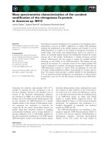
Fig. 1. Monitoring of Arabidopsis PTOX activity in isolated E. coli
membranes in the presence of various added quinones. Electron transport
was initiated by NADH addition (1 m
M
final concentration). Various
quinones were then added (0.2 m
M
final concentration), namely decyl-
plastoquinone (dPQ), or decyl-ubiquinone (dUQ), duroquinone (DQ),
vitamin K1 (VK1) or benzoquinone (BQ). Sequential addition of
KCN (inhibitor of the E. coli cytochrome b-dependent O
2
consump-
tion; 2 m
M
final concentration) and then n-propyl gallate (nPG;
inhibitor of PTOX-dependent O
2
consumption) followed. O
2
con-
sumption was recorded and expressed as nmol O
2
Æmin
)1
Æmg protein
)1
.
Aliquots of the same membrane samples were used for further
experiments during a week: a progressive decline in overall respiratory
activity was observed but experiments remained reproducible.
Experiments were also reproducible from one sample preparation to
another, although some variation in quantitative levels of respiratory
activity was observed. Typical and representative experiments are
summarized here: (A) experiment performed using membranes from a
PTOX expressing strain; (B) experiment performed using membranes
from a control strain; (C) Ratio of O
2
consumption in (A) over (B).
3788 E M. Josse et al. (Eur. J. Biochem. 270) Ó FEBS 2003
Fig. 1B). However, O
2
consumption was stimulated
strongly in both membrane types (Fig. 1A,B) in the
presence of 0.2 m
M
of ubiquinone and, to some extent, of
dPQ, duroquinone or vitamin K1 (phylloquinone). In
contrast, addition of benzoquinone did not lead to such
changes (Fig. 1A,B): O
2
consumption rates were similar to
controls without added quinones (not shown). This suggests
that quinones are rate-limiting in this assay prior to KCN
addition.
When KCN was added to PTOX-containing membranes,
O
2
consumption rates decreased to background levels in the
assays containing ubiquinone, vitamin K1 or benzoquinone
(Fig. 1A), as they did without quinone addition (not
shown). In contrast, an O
2
consumption rate attributed to
PTOX activity was observed in the presence of dPQ. This
activity, as we have demonstrated previously [3,7,9], can be
identified by its specific inhibition by nPG. The fact that O
2
consumption is maintained above background level in the
presence of duroquinone and KCN cannot be linked to
PTOX, as nPG does not inhibit it. In the latter case, nPG
stimulates O
2
consumption rate for a reason that is as yet
unclear. This is also the case in control membranes
(Fig. 1B). It shall be noted that, as expected, the cyanide-
resistant O
2
consumption is not detected in these control
membranes (Fig. 1B).
Figure 1C presents the ratio of O
2
consumption in
PTOX-containing membranes relative to control mem-
branes devoid of PTOX. This ratio is close to one for all
tested quinones except dPQ. In this latter case, the ratio rises
only after KCN addition, which is consistent with our
previous observation [3] that PTOX activity is only engaged
in this assay when the cytochrome pathway is blocked. It
should also be mentioned that, when quinones were added
after KCN, only the addition of dPQ, not the other
quinones, allowed a restoration of O
2
consumption by the
PTOX-containing membrane (not shown). All these data
indicate that the activity of PTOX is dependent on the
presence of dPQ in this assay.
However, because of the above mentioned difficulty
encountered using duroquinone, it was necessary to exam-
ine whether duroquinol added directly as an electron donor
can be a substrate for PTOX. In a modified assay (NADH
and dPQ being omitted), a cyanide-resistant O
2
consump-
tion was observed (not shown) upon addition of fully
reduced duroquinol (in vitro borohydride-reduced duroqui-
none; final concentration 0.2 m
M
). This activity was totally
inhibited by nPG. This activity was absent in control
membranes.
In all subsequent assays (using NADH as an electron
donor), dPQ was routinely added. A potential pH depend-
ence of PTOX activity was first examined. As shown in
Fig. 2A, prior to KCN addition, the highest oxygen
consumption rate was observed at pH 6.0 and 6.5, with a
decline at higher pH especially at pH 9.0. In contrast, after
KCN addition the O
2
consumption rate attributed to
PTOX increased progressively in the tested pH range. This
increase, visible in Fig. 2A, might be underestimated due to
the lower respiratory activity of E. coli membranes at the
highest pH. One can also note that, after nPG addition, a
certain increase in O
2
consumption occurs at the highest pH.
However, this is a nonspecific effect also observed in control
membranes (Fig. 2B). This prevents us from calculating the
real PTOX activity (O
2
consumption rate after KCN
addition minus rate after nPG addition). In order to
circumvent this problem, Fig. 2C presents PTOX activities
recalculated from each value obtained after KCN addition
minus the averaged values after nPG addition (excluding
those at the highest pH).
We then examined the effect of temperature on PTOX
activity (Fig. 3). As expected for respiratory activity of
E. coli membranes, prior to KCN addition, an increase in
O
2
consumption (of approximately fourfold) was observed
with a temperature rise from 15 to 40 °C. A positive effect of
temperature rise was also observed after KCN addition.
This is, however, probably the consequence of the increase
in the overall quinone reducing activity of the membranes
and not a direct effect on PTOX activity. This is corrobor-
ated by the fact that the ratio of O
2
consumption before and
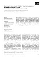
Fig. 2. pH influence on PTOX activity. Experiments were performed
as in Fig. 1, except that dPQ was added in all cases together with
NADH. (A) Experiment performed using membranes from a PTOX-
expressing strain. (B) Experiment performed using membranes from a
control strain. (C) PTOX activity calculated as O
2
consumption in
presence of KCN minus averaged O
2
consumption in presence of nPG
(see text).
Ó FEBS 2003 Plastid terminal oxidase (Eur. J. Biochem. 270) 3789
after KCN addition does not change significantly with
temperature (not shown). It is also noticeable that PTOX
activity (calculated as the rate after KCN minus rate after
nPG) increases approximately fourfold with temperature
from 15 to 40 °C, in a reasonably linear manner up to
40 °C. This indicates that PTOX is active over a wide
temperature range and suggests PTOX could help protect-
ing plants against temperature stress. However, tempera-
tures below 15 °C could not be tested, as respiratory activity
of E. coli membrane is too low under those conditions.
PTOX exhibits differences with the mitochondrial
alternative oxidase
Despite the presence of conserved amino acids [3,4,19],
AOX and PTOX sequence alignments also show striking
differences, for example in between two highly conserved
regions that are probably involved in iron binding ([6],
Fig. 4). Interestingly, this region matches a consensus
sequence [aliphatic-(X)3-H-(X)2/3-L/T/S] proposed by
Fisher and Rich [20] for quinone binding sites. Although
speculative at this stage, this consensus sequence was found
in various quinone-binding proteins including AOX [20,21].
It should be mentioned that we position this potential site in
a different polypeptide region than originally proposed in
AOX [20], the reason being that the originally proposed
region is not a highly conserved one in PTOX sequences.
Furthermore, a quinone binding site would be expected to
be close to iron binding amino acids, which is the case here
([6], Fig. 4). This potential quinone-binding site appears to
be conserved in PTOX sequences (including a homologous
sequence from the cyanobacterium Nostoc) on one hand
and in numerous AOX sequences on the other. Thus, the
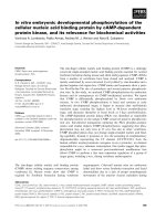
Fig. 3. Temperature influence on PTOX activity. Experiments were
performed as in Fig. 2, except that the pH was 7.5. (A) Experiment
performed using membranes from a PTOX-expressing strain. (B)
PTOX activity calculated as O
2
consumption in presence of KCN
minus O
2
consumption in presence of nPG.
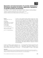
Fig. 4. Comparison of a putative quinone-binding site in PTOX and AOX sequences. The potential Q site is shaded and arrows indicate the conserved
residues matching a consensus sequence proposed by Fisher and Rich [20]. Neighbouring amino acids probably involved in iron binding are shown
(*) in a conserved LET and NERMHL region [6]. Highly conserved positions are boxed in black (identical amino acids) or grey (similar amino
acids). Sequences used: AthaPTOX: Arabidopsis thaliana (accession number CAA06190, position 130–195); CannPTOX: Capsicum annuum
(AAG02288, 132–197); LescPTOX: Lycopersicum esculentum (AAG02286, 141–206); TaesPTOX: Triticum aestivum (AAG0045, 52–117);
OsatPTOX: Oryza sativa (AF085174, 116–181); CreiPTOX: Chlamydomonas reinhardtii (AAM12876, 272–388); NostPTOX: Nostoc sp. PCC 7120
(NP-486136, 23–88); NtabAOX: Nicotiana tabacum (Q41224, 176–240); OsatAOX: O. sativa (BAA28772, 155–219); TbruAOX: Trypanosoma
brucei (BAB72245, 117–181; NcraAOX: Neurospora crassa (EAA29895, 157–221).
3790 E M. Josse et al. (Eur. J. Biochem. 270) Ó FEBS 2003
sequences of this site are grouped into separate classes for
PTOX and AOX.
Another striking sequence difference is the lack of
conserved Cys in the N-terminal domain of PTOX [3]
whilst a conserved Cys in AOX is involved in the
stimulation of AOX activity by a-keto acids [13,14,22].
When examining the effect of pyruvate on PTOX activity,
no detectable effect was observed when pyruvate was added
to the reaction mixture in the range of 1–8 m
M
(data
not shown). It should also be mentioned that other
carbohydrates, namely glyceraldehyde-3-phosphate or
3-phospho-glycerate (that play a more central role in plastid
metabolism) had no effect either (data not shown).
As some fungal AOX, not stimulated by pyruvate, are
induced by purine nucleotides, such an activation mechan-
ism was examined for PTOX. However, addition of AMP
had no effect on PTOX activity (data not shown).
Millenaar et al. [23] reported on the in vivo inhibitory
effect of sugars and organic acids (citrate) on the mito-
chodrial AOX activity and proposed that the effect of
organic acids could be due to the fact that they chelate metal
cations. We did observe an inhibitory effect of both citrate
and malate on PTOX activity in vitro. However, a similar
inhibitory effect was also observed upon addition of NaCl
at a concentration identical to its presence in the malate and
citrate solutions (Table 1). It is therefore probable that, in
our assay, inhibition of PTOX activity in the presence of
these organic acids is due to the increasing salt concentra-
tion (possibly affecting membrane association of PTOX),
rather than to a metal chelating activity. This is confirmed
by the fact that KCl also showed an inhibitory effect on
PTOX activity to a similar extent as NaCl (not shown).
Therefore, KCl (which was routinely present in the reaction
mixture) should be omitted for optimized PTOX activity.
Iron requirement for PTOX activity
To examine the role of iron and other metals, PTOX
expressing bacteria and control bacteria were grown in the
presence of 50 l
M
o-phenanthroline, a chelator of divalent
cations [18], which was added at the time PTOX expression
was induced. This led to a slow-down in bacterial growth.
However, as shown in Fig. 5A, this treatment had no
influence on O
2
consumption of membranes isolated
subsequently from control cells. However, it dramatically
abolished PTOX activity in PTOX-expressing cells. In
contrast, when FeSO
4
(125 l
M
) was added to the medium
together with o-phenanthroline, PTOX activity was
observed. CuSO
4
, MnSO
4
or ZnSO
4
addition could not
restore PTOX activity (not shown).
A complementary experiment was performed by adding
iron at the end of the culture phase of o-phenanthroline-
treated bacteria, after washing away the chelator by
Table 1. Effect of citrate, malate and NaCl on PTOX activity in vitro.
Assays were performed in the presence of NADH, decyl-plastoqui-
none and KCN as described in the figure legends. Citrate or malate
was added to PTOX-containing membranes to the given concentra-
tions from a 0.1
M
stock-solution (buffered by the addition of NaOH
to a 0.2
M
final concentration). The concentration of NaCl was also
increased in a separate assay. PTOX activity is given as percentage of
starting O
2
consumption (before addition of these compounds). The
latter activity (approximately 65 nmolÆmin
)1
Æmg protein
)1
)wascal-
culated from the O
2
consumption in the presence of KCN minus
residual O
2
consumption after n-propyl gallate addition.
[Cit] or [mal] (m
M
)
PTOX activity (%) in
presence of:
[NaCl] (m
M
)
Cit Mal NaCl
0 100 100 100 0
18995802
577837710
10 61 73 70 20
Fig. 5. Influence of metal cations on PTOX activity and integration in
E. coli membranes. (A) Monitoring of PTOX activity using assays
performed as described in Fig. 2. Membrane samples were from the
following bacterial cultures: C, untreated control; CO, treated with
50 l
M
o-phenanthroline; P, untreated PTOX-expressing; PO, expres-
sing PTOX and treated with o-phenanthroline; POF, expressing
PTOX and treated with both o-phenanthroline and 125 l
M
FeSO
4
.(B)
The upper panel shows the immunodetection of PTOX (arrow) using
polyclonal antibodies [3,9] on blots of 50 lg protein samples extracted
from the bacterial cultures mentioned in (A) and separated by PAGE.
Lower panel shows a Coomassie staining of the gel. (C) As in (B), using
20 lg protein samples from purified membranes. The strong immu-
nodetected band below PTOX corresponds to an abundant protein
visible by Ponceau staining in all lanes of the gel (lower panel).
Ó FEBS 2003 Plastid terminal oxidase (Eur. J. Biochem. 270) 3791
centrifugation and resuspension of the bacteria. In this case,
PTOX activity could be partially restored (not shown). We
failedtoobtainsuchare-activationwhenFeSO
4
was added
directly to isolated membranes.
Protein gel blots were probed with an anti-PTOX Ig to
examine whether PTOX was actually present after treat-
ment. As shown in Fig. 5B, while PTOX was not detected
in control cells, it is detected in similar amounts in
nontreated, o-phenanthroline-treated and o-phenanthroline
plus FeSO
4
-treated PTOX expresssing cells. Figure 5C
shows that PTOX is present in similar amounts in the
membrane fraction isolated from PTOX-containing cells
whether iron has been chelated or not. Thus, iron depriva-
tion does not seem to prevent membrane association of
PTOX.
Inhibitors of PTOX activity
in vitro
It is worth mentioning that when halogenated quinone
analogues were tested, namely dibromo-methyl-isopropyl-
benzoquinone (DBMIB), chloranilic acid or p-chloranil, no
inhibitory effect on PTOX could be demonstrated (not
shown).
We then examined the effect of n-propyl-gallate (nPG)
analogues (Fig. 6). We first checked the noncarboxylic
derivative, pyrogallol and found it had no effect on PTOX
activity up to 1 m
M
, with only a slight inhibition evident at
2m
M
(not shown). We then checked gallic acid, the
nonesterified derivative of nPG. This compound had no
effect on the O
2
consumption rate of control membranes
when added in the range of 0.1–2 m
M
. In contrast, a
concentration-dependent inhibition was observed on PTOX
activity (Fig. 7A). An inhibition of approximately 50% was
observed at 0.9 m
M
, which is a ninefold higher concentra-
tion than for nPG [9]. In contrast, when n-octyl-gallate, a
gallic acid derivative esterified with a long-chain acyl group,
was used, a clear inhibition of PTOX activity was observed
(100% at 5 l
M
). An inhibition of approximately 50% was
observed at 0.4 l
M
, which is a 250-fold lower concentration
than for nPG (Figs 6,7).
Discussion
The in vivo functions of PTOX are obviously complex as
they are linked to both carotenoid biosynthesis and
chlororespiration [4]. To obtain further knowledge of this
enzyme, we have used an in vitro assay based on E. coli
membranes which have incorporated PTOX and which uses
NADH as an electron donor. In this assay, we observed a
specificity for plastoquinone for PTOX activity (plasto-
quinol oxidase). The ability of PTOX to use plastoquinol as
a substrate was not unexpected as it is the major quinone in
plastids. However, the fact that reduced quinones, such as
vitamin K1, are not used by PTOX was not an obvious
feature, as it is also found in plastids. The inability of PTOX
to use ubiquinol in our E. coli membrane-based assay is also
unexpected as it is the homologous quinone in this electron
transfer chain and, out of all tested quinones, it is the one
which most efficiently incorporated into it (judging from its
strong stimulation of respiratory rates; Fig. 1). It is there-
fore unlikely that the inability of PTOX to use ubiquinone is
an artefact of this assay (which relies on the ability of the
membranes to reduce quinones). Therefore, we propose that
the preference of PTOX for plastoquinol over ubiquinol
may be linked to a steric compatibility/incompatibility of
these quinones with the PTOX quinone-binding site. This
suggestion needs to be tested experimentally.
It should be noted (Fig. 1) that benzoquinone was the
only quinone tested that did not stimulate O
2
consumption
in E. coli membranes (prior to KCN addition). Thus,
benzoquinone is not likely to be integrated in this electron
transport chain. Whether a benzoquinone derivative with
an allylic/terpenic side chain would be functional and
possibly a substrate for PTOX remains to be tested.
We also observed that addition of duroquinone could not
substitute for plastoquinone in the NADH-based assay.
However, duroquinol (when added in its fully reduced form)
can serve as an electron donor to PTOX. In the former case,
duroquinone is incorporated into the electron transfer chain
(judging from its stimulatory effect on E. coli membrane
respiration; Fig. 1), but it is certainly not present in a fully
reduced form. It is not currently known whether the
apparent discrepancy between these assays is due to a strong
preference of PTOX for plastoquinol over duroquinol, or to
a lower concentration of duroquinol in the NADH-based
assay.
Fig. 6. Chemical structures of plastoquinone compared to quinone ana-
logues. Inhibitor concentration leading to 50% inhibition of PTOX
activity are given in brackets, except for pyrogallol for which this value
could not be determined (n.d.) DBMIB, which does not inhibit PTOX
activity, is shown for comparison.
Fig. 7. Sensitivity of PTOX activity to gallic acid (A) or n-octyl gallate
(B). Experiments were performed as described in Fig. 2, except that
n-propyl gallate was substituted by increasing concentration of the
given inhibitors. Activity before addition of inhibitor is set at 100
(absolute amount were calculated as explained in Table 1).
3792 E M. Josse et al. (Eur. J. Biochem. 270) Ó FEBS 2003
Nevertheless, our data describe two useful assays for
monitoring PTOX activity, namely a simple one (in which
all added components, namely plastoquinone and NADH,
are commercially available), and one in which the concen-
tration of the (artificial, in vitro reduced) electron donor can
be changed in a controlled way.
Comparison of sequences of both PTOX and AOX
identifies a putative quinone-binding site (Fig. 4) that
clearly differs between PTOX and AOX. The consensus
sequence is aliphatic-SVL-H-M/L-YE-T/S for PTOX and
GML-L/R-H-L/C-XSL for AOX. This might explain their
different substrate preferences. A fuller understanding of
quinone–protein interactions in this class of enzyme may be
achieved by testing the effect of sequence modification on
substrate specificity.
The influence of temperature (Fig. 3) or pH increase
(Fig. 2) on PTOX activity observed in this report can
only presently be considered as valid for the present
assay. As it has been shown that PTOX, when over-
expressed in tobacco, can accelerate quinol re-oxidation
in photosynthetic membranes [8], it will be interesting to
examine whether pH also regulates its activity in planta.
It is known that stromal alkalinization does occur in
light and allows optimal functioning of photosynthetic
carbon reduction cycle enzymes [24–26]. It should also be
mentioned that Mg-protoporphyrin-IX monomethylester
cyclase (a di-iron enzyme) activity also has a pH value
optimum of 9.0 [27]. Although it is difficult to know
precisely what the pH values are in the microenviron-
ment of the stromal side of thylakoids, reported stromal
pH changes are in the same range as that which
increases PTOX enzyme activity in vitro.Also,the
amount of PTOX enzyme increases during light periods
and decreases during dark periods [28].
Our observation (Table 1) that increasing salt concentra-
tions inhibits PTOX activity in the present assay was
unexpected in the light of the opposite effect of salt on AOX
activity [29]. It will be necessary to examine the effect of salt
on PTOX in homologous plastid membranes.
Sequence comparison between PTOX and AOX [3,4]
revealed that PTOX does not possess the conserved Cys
present in the N-terminal domain of higher plant AOXs
which is involved in the activation of its enzyme activity by
pyruvate [12–14]. A number of other conserved Cys in
PTOX [3,4] could potentially substitute for the one which is
active in AOX. However, our present data suggest that such
an activation mechanism does not operate for PTOX. This
is also the case for AOX from lower eukaryotes [30] which
are all lacking this conserved Cys.
Our data show that PTOX activity is dependent on iron,
which cannot be substituted by other divalent cation metals
(Fig. 5). That AOX is a di-iron carboxylate protein has been
demonstrated recently by EPR studies [17]. Therefore, it
seems reasonable to conclude that the proposed iron
binding sites in PTOX sequences [3,6,19] are part of a
di-iron carboxylate centre.
The available PTOX null mutants (immutans, ghost),
as well as over-expression lines, are useful tools to check
for in vivo roles of PTOX [1–3,8]. However, specific and
potent inhibitors are also of interest as they would allow
inactivation of PTOX at a certain time during plant
development without pleiotropic effects that can be
associated with genetic mutants. When examining poten-
tial inhibitors, we observed that none of the tested
quinone-like structures with substitutions on all carbons
of the ring (e.g. DBMIB; Fig. 6) inhibited PTOX
activity. In contrast, pyrogallol derivatives (that all
possess nonsubstituted carbons) all showed inhibitory
activity. This suggests that it is the core of the quinone-
like structures (the pyrogallol or gallic acid moiety) that
is actually inhibiting PTOX activity (provided this core
structure is sterically compatible with the quinone
binding site). However, gallic acid itself is poorly
inhibitory (Fig. 7) without a nonpolar side chain. The
latter may favour positioning of the gallic acid moiety in
a hydrophobic environment. In this respect, PTOX does
not seem to differ from AOX [11,31]. This information
can be used to design new specific inhibitors. Gallic acid
analogues, with even longer hydrophobic side chains, and
possibly with another group substituting for an alcohol
group, are potential candidates. Currently, octyl gallate,
which is a far better inhibitor than nPG, can conveni-
ently replace nPG for in organello or in vivo studies.
Acknowledgements
WethankDrsP.Carol,A.J.Dorne,J.Gaffe
´
and I. Prieur-Lavoue
´
for
helpful discussions, E. Charpentier and L. Zekraoui for technical
assistance.
References
1. Carol, P., Stevenson, D., Bisanz, C., Breitenbach, J., Sandmann,
G., Mache, R., Coupland, G. & Kuntz, M. (1999) Mutations in
the Arabidopsis gene IMMUTANS cause a variegated phenotype
by inactivating a chloroplast terminal oxidase associated with
phytoene desaturation. Plant Cell 11, 57–68.
2. Wu,D.,Wright,D.A.,Wetzel,C.,Voytas,D.F.&Rodermel,S.
(1999) The IMMUTANS variegation locus of Arabidopsis defines
a mitochondrial alternative oxidase homolog that functions dur-
ing early chloroplast biogenesis. Plant Cell 11, 43–55.
3. Josse, E.M., Simkin, A.J., Gaffe
´
, J., Laboure
´
, A.M., Kuntz, M. &
Carol, P. (2000) A plastid terminal oxidase associated with caro-
tenoid desaturation during chromoplast differentiation. Plant
Physiol. 123, 1427–1436.
4. Carol, P. & Kuntz, M. (2001) A plastid terminal oxidase comes to
light: implications for carotenoid biosynthesis and chlororespira-
tion. Trends Plant Sci. 6, 31–36.
5. Moore, A.L., Albury, M.S., Crichton, P.G. & Affourtit, C. (2002)
Function of the alternative oxidase: is it still a scavenger? Trends
Plant Sci. 7, 478–481.
6. Berthold, D.A., Andersson, M.E. & Nordlund, P. (2000) New
insight into the structure and function of the alternative oxidase.
Biochim. Biophys. Acta 1460, 241–254.
7. Cournac, L., Redding, K., Ravenel, J., Rumeau, D., Josse, E.M.,
Kuntz, M. & Peltier, G. (2000) Electron flow between photo-
system II and oxygen in chloroplasts of photosystem I-deficient
algae is mediated by a quinol oxidase involved in chlororespira-
tion. J. Biol. Chem. 275, 17256–17262.
8. Joe
¨
t,T.,Genty,B.,Josse,E.M.,Kuntz,M.,Cournac,L.&Peltier,
G. (2002) Involvement of a plastid terminal oxidase in plasto-
quinone oxidation as evidenced by expression of the Arabidopsis
thaliana enzyme in tobacco. J. Biol. Chem. 277, 31623–31630.
9. Cournac, L., Josse, E.M., Joe
¨
t, T., Rumeau, D., Redding, K.,
Kuntz, M. & Peltier, G. (2000) Flexibility in photosynthetic elec-
tron transport: a newly identified chloroplast oxidase involved in
Ó FEBS 2003 Plastid terminal oxidase (Eur. J. Biochem. 270) 3793
chlororespiration. Philos. Trans. R. Soc. Lond. B Biol Sci. 355,
1447–1454.
10. Siedow, J.N. & Girvin, M.E. (1980) Alternative respiratory
pathway: its role in seed respiration and its inhibition by propyl
gallate. Plant Physiol. 65, 669–674.
11. Siedow, J.N. & Bickett, D.M. (1981) Structural features required
for inhibition of cyanide-insensitive electron transfer by propyl
gallate. Arch. Biochem. Biophys. 207, 32–39.
12. Millenaar, F.F., Benschop, J.J., Wagner, A.M. & Lambers, H.
(1998) The role of the alternative oxidase in stabilizing the in vivo
reduction state of the ubiquinone pool and the activation state of
the alternative oxidase. Plant Physiol. 118, 599–607.
13. Rhoads, D.M., Umbach, A.L., Sweet, C.R., Lennon, A.M.,
Rauch, G.S. & Siedow, J.N. (1998) Regulation of the cyanide-
resistant alternative oxidase of plant mitochondria. Identification
of the cysteine residue involved in alpha-keto acid stimulation
and intersubunit disulfide bond formation. J. Biol. Chem. 273,
30750–30756.
14. Vanlerberghe, G.C., McIntosh, L. & Yip, J.Y. (1998)
Molecular localization of a redox-modulated process regulating
plant mitochondrial electron transport. Plant Cell 10, 1551–
1560.
15. Affourtit, C., Albury, M.S., Crichton, P.G. & Moore, A.L. (2002)
Exploring the molecular nature of alternative oxidase regulation
and catalysis. FEBS Lett. 510, 121–126.
16. Siedow,J.N.,Umbach,A.L.&Moore,A.L.(1995)Theactivesite
of the cyanide-resistant oxidase from plant mitochondria contains
a binuclear center. FEBS Lett. 362, 10–14.
17. Berthold, D.A., Voevodskaya, N., Stenmark, P., Graslund, A. &
Nordlund, P. (2002) EPR studies of the mitochondrial alternative
oxidase. Evidence for a diiron carboxylate center. J. Biol. Chem.
277, 43608–43614.
18. Ajayi, W.U., Chaudhuri, M. & Hill, G.C. (2002) Site-directed
mutagenesis reveals the essentiality of the conserved residues in the
putative diiron active site of the trypanosome alternative oxidase.
J. Biol. Chem. 277, 8187–8193.
19. Berthold, D.A. & Stenmark, P. (2003) Membrane-bound diiron
carboxylate proteins. Annu. Rev. Plant Biol. 54, 497–517.
20. Fisher, N. & Rich, P.R. (2000) A motif for quinone binding sites
in respiratory and photosynthetic systems. J. Mol. Biol. 296,
1153–1162.
21. Dupuis, A., Prieur, I. & Lunardi, J. (2001) Towards a character-
ization of the connecting module of Complex I. J. Bionerg.
Biomembr. 33, 159–168.
22. Umbach, A.L., Gonzalez-Meler, M.A., Sweet, C.R. & Siedow,
J.N. (2002) Activation of the plant mitochondrial alternative
oxidase: insights from site-directed mutagenesis. Biochim. Biophys.
Acta 1554, 118–128.
23. Millenaar, F.F., Gonzalez-Meler, M.A., Siedow, J.N., Wagner,
A.M. & Lambers, H. (2002) Role of sugars and organic acids in
regulating the concentration and activity of the alternative oxidase
in Poa annua roots. J. Exp. Bot. 53, 1081–1088.
24. Werdan, K., Heldt, H.W. & Milovancev, M. (1975) The role of
pH in the regulation of carbon fixation in the chloroplast stroma.
Studies on CO2 fixation in the light and dark. Biochim. Biophys.
Acta 396, 276–292.
25. Wu, W. & Berkowitz, G.A. (1992) Stromal pH and photosynthesis
are affected by electroneutral K+ and H+ exchange through
chloroplast envelope ion channels. Plant Physiol. 98, 666–672.
26. Hauser, M., Eichelmann, H., Oja, V., Heber, U. & Laisk, A.
(1995) Stimulation by light of rapid ph regulation in the chlor-
oplast stroma in vivo as indicated by CO
2
solubilization in leaves.
Plant Physiol. 108, 1059–1066.
27. Walker, C.J., Castelfranco, P.A. & Whytte, B.J. (1991)
Enzymological properties of the Mg-protoporphyrin IX mono-
methyl ester oxidative cyclase system. Biochem. J. 276, 691–697.
28. Simkin, A.J., Laboure
´
, A.M., Kuntz, M. & Sandmann, G. (2003)
Comparison of carotenoid content, gene expression and enzyme
levels in tomato (Lycopersicon esculentum) leaves. Z. Naturforsch.
58c, 371–380.
29. Kay, C. & Palmer, J.M. (1985) Solubilization of the alternative
oxidase of cuckoo-pint (Arum maculatum) mitochondria. Stimu-
lation by high concentration of ions and effects of specific
inhibitors. Biochem. J. 228, 309–318.
30. Umbach, A.L. & Siedow, J.N. (2000) The cyanide-resistant
alternative oxidases from the fungi Pichia stipitis and Neurospora
crassa are monomeric and lack regulatory features of the plant
enzyme. Arch. Biochem. Biophys. 378, 234–245.
31. Hoefnagel, M.H., Wiskich, J.T., Madgwick, S.A., Patterson, Z.,
Ottmeier,W.&Rich,P.R.(1995)Newinhibitorsoftheubiquinol
oxidase of higher plant mitochondria. Eur. J. Biochem. 233,
531–537.
3794 E M. Josse et al. (Eur. J. Biochem. 270) Ó FEBS 2003