Báo cáo khoa học: Thioredoxin reductase from the malaria mosquito Anopheles gambiae Comparisons with the orthologous enzymes of Plasmodium falciparum and the human host pdf
Bạn đang xem bản rút gọn của tài liệu. Xem và tải ngay bản đầy đủ của tài liệu tại đây (365.17 KB, 10 trang )
Thioredoxin reductase from the malaria mosquito
Anopheles gambiae
Comparisons with the orthologous enzymes of
Plasmodium falciparum
and the human host
Holger Bauer
1
, Stephan Gromer
1
, Andrea Urbani
2
, Martina Schno¨ lzer
2
, R. Heiner Schirmer
1
and Hans-Michael Mu¨ ller
3
1
Biochemie Zentrum, Universita
¨
t Heidelberg, Heidelberg, Germany;
2
Deutsches Krebsforschungszentrum, Heidelberg, Germany;
3
European Molecular Biology Laboratory, Heidelberg, Germany
The mosquito, Anopheles gambiae, is an important vector of
Plasmodium falciparum malaria. Full genome analysis
revealed that, as in Drosophila melanogaster,theenzyme
glutathione reductase is absent in A. gambiae and func-
tionally substituted by the thioredoxin system. The key
enzyme of this system is thioredoxin reductase-1, a homo-
dimeric FAD-containing protein of 55.3 kDa per subunit,
which catalyses the reaction NADPH + H
+
+ thio-
redoxin disulfide fi NADP
+
+ thioredoxin dithiol. The
A. gambiae trxr gene is located on chromosome X as a
single copy; it represents three splice variants coding for two
cytosolic and one mitochondrial variant. The predominant
isoform, A. gambiae thioredoxin reductase-1, was recomb-
inantly expressed in Escherichia coli and functionally com-
pared with the wild-type enzyme isolated in a final yield
of 1.4 UÆml
)1
of packed insect cells. In redox titrations,
the substrate A. gambiae thioredoxin-1 (K
m
¼ 8.5 l
M
,
k
cat
¼ 15.4 s
)1
at pH 7.4 and 25 °C) was unable to oxidize
NADPH-reduced A. gambiae thioredoxin reductase-1 to
the fully oxidized state. This indicates that, in contrast to
other disulfide reductases, A. gambiae thioredoxin reduc-
tase-1 oscillates during catalysis between the four-electron
reduced state and a two-electron reduced state. The thio-
redoxin reductases of the malaria system were compared.
A. gambiae thioredoxin reductase-1 shares 52% and 45%
sequence identity with its orthologues from humans and
P. falciparum, respectively. A major difference among the
three enzymes is the structure of the C-terminal redox cen-
tre, reflected in the varying resistance of catalytic inter-
mediates to autoxidation. The relevant sequences of this
centre are Thr–Cys–Cys–SerOH in A. gambiae thioredoxin
reductase, Gly–Cys–selenocysteine–GlyOH in human thio-
redoxin reductase, and Cys–X–X–X–X–Cys–GlyOH in the
P. falciparum enzyme. These differences offer an interesting
approach to the design of species-specific inhibitors.
Notably, A. gambiae thioredoxin reductase-1 is not a
selenoenzyme but instead contains a highly unusual redox-
active Cys–Cys sequence.
Keywords: Anopheles gambiae; Drosophila melanogaster;
Diptera; insect redox metabolism; Plasmodium falciparum.
The mosquito Anopheles gambiae is of importance as a
vector of tropical malaria caused by the protozoan organism
Plasmodium falciparum. The genome of A. gambiae is the
second insect genome – after the distantly related model
organism Drosophila melanogaster [1,2] – that has been
completely sequenced [3]. Annotation of the nucleotide
sequences allows access to the genetic background of a
disease-transmitting dipteran insect and, furthermore, offers
the opportunity of comparative sequence analyses from
single genes up to genomic organization [4]. Another aspect is
highlighted in this report: only on the basis of the full genome
sequence does it become possible to exclude the presence of a
given protein function in all cells and all developmental stages
of an organism. A case in point is the absence of the enzyme
glutathione reductase (GR) in Diptera [5].
Our focus is the redox metabolism of insects [6]. Being
present at millimolar concentrations, the tripeptide gluta-
thione (GSH) is the most abundant antioxidative thiol
compound in most cell compartments. Its redox state
determines the intracellular redox environment [7]. Thus,
GSH is the major redox buffer compound and essential
for the detoxification of free radicals and xenobiotics [8,9].
In the majority of pro- and eukaryotic organisms, the
oxidized form of glutathione (glutathione disulfide,
GSSG) is reduced to the mono-thiol form (GSH) by the
Correspondence to R. H. Schirmer, Biochemie Zentrum, Im Neuen-
heimer Feld 504, D-69120 Heidelberg, Germany.
Fax: + 49 6221 545586, Tel.: + 49 6221 544165,
E-mail: or H M. Mu
¨
ller, European
Molecular Biology Laboratory, Meyerhofstr. 1, D-69117 Heidelberg,
Germany. Fax: + 49 6221 387306, Tel.: + 49 6221 387440,
E-mail:
Abbreviations:EH
2
, enzyme in a two-electron reduced state; EH
4
,
enzyme in a four-electron reduced state; E
ox
,enzymeinanoxidized
state; EST, expressed sequence tag; GR, glutathione reductase;
GSH/GSSG, reduced/oxidized glutathione; Sec, selenocysteine;
Trx, thioredoxin; TrxR, thioredoxin reductase.
(Received 14 July 2003, revised 29 August 2003,
accepted 1 September 2003)
Eur. J. Biochem. 270, 4272–4281 (2003) Ó FEBS 2003 doi:10.1046/j.1432-1033.2003.03812.x
NADPH-dependent flavoenzyme GR, which catalyses the
following reaction: NADPH + GSSG + H
+
fi NADP
+
+
2GSH [10–12]. D. melanogaster cells exhibit a high 2[GSH]/
[GSSG] ratio but have been shown to lack a typical GR [5].
As reported here, this is also true for A. gambiae.An
important candidate able to functionally substitute for
GR is the thioredoxin (Trx) system [13], which compri-
ses NADPH, thioredoxin reductase(s) (TrxR) and Trxs
[14,15].
Trxs are small, ubiquitous thiol proteins with a relative
molecularmassof 12 kDa and a redox active cysteine
pair represented in a WCGPC sequence motif. Therefore,
they cycle between a disulfide (TrxS
2
) and a dithiol
[Trx(SH)
2
]form.
Trxs were first described as electron-donating substrates
for ribonucleotide reductase [16], but they cleave disulfide
bonds in a number of other proteins equally well. Thus, Trxs
take part in the redox control of numerous processes such
as protein folding, signalling and transcription [14,17–19].
Trx reduction is catalysed by the flavin-dependent oxido-
reductase TrxR, as follows: NADPH + TrxS
2
+H
+
fi
NADP
+
+Trx(SH)
2
. TrxRs belong to a disulfide reduc-
tase superfamily that includes enzymes such as GR,
trypanothione reductase, lipoamide dehydrogenase, and
mercuric ion reductase. These homodimeric proteins are
structurally, as well as mechanistically, closely related [20].
Evolution has produced two classes of TrxRs: small
TrxRs (found in bacteria, plants and fungi) and large
TrxRs (present in other eukaryotes) [21]. In contrast to
GRs and low molecular weight TrxRs, large TrxRs
possess an additional redox centre located in the
C-terminal extension which is necessary for the interaction
with the substrate Trx. In mammalian enzymes this redox
centre is represented by a neighboring cysteine–seleno-
cysteine pair [22] and in the TrxR of P. falciparum it is a
cysteine pair separated by a spacer sequence of four
amino acids [23]. D. melanogaster TrxR was described as
the first member of a third type of large TrxRs. It is
characterized by two adjacent cysteines preceding the
C-terminus [5,24].
High molecular mass TrxRs exhibit a rather broad
substrate spectrum that includes a number of natural and
also artificial disulfide compounds, such as 5,5¢-dithiobis(2-
nitrobenzoate). Glutathione disulfide, however, is not a
substrate. In the fruit fly it was shown that GSSG reduction
can occur in a dithiol–disulfide exchange reaction
with reduced Trx [5]. At physiological concentrations of
GSSG and Trx, this system allows GSSG fluxes of
> 100 l
M
Æmin
)1
[25].
In this report we introduce the TrxR of the malaria
mosquito A. gambiae. The protein could be isolated from
whole insects and from cultured insect cells. With the
progress of the Anopheles genome project it was possible to
identify the complete sequence of the gene and its organi-
zation. We cloned, recombinantly expressed and character-
ized the enzyme. Our data support the assumption that the
substitution of the Trx system for GR, as well as the
mechanistic particularities of the TrxR, are a common
principle in dipteran insects. In the context of the malarial
system, this implies that the TrxRs of insect vector, parasite,
and human host differ in their cellular roles as well as their
enzyme mechanisms.
Experimental procedures
D. melanogaster Trx-2, A. gambiae Trx-1 and P. falciparum
Trx-1 were prepared and purified as previously described
[25,26]. PCR chemicals and restriction enzymes were
purchased from MBI Fermentas and Applied Biosystems,
precast polyacrylamide gels from Bio-Rad and molecular
mass standards from Amersham Pharmacia Biotech. Anti-
biotics, substrates for enzyme assays, and other chemicals
were from BioMol, Fluka, or Sigma. All compounds were
of the highest available purity.
Purification of authentic
A. gambiae
TrxR
from insect cells
A. gambiae cells (cell line 4a-2s4) were cultured in
Schneider’s medium and harvested as described previ-
ously [27]. A 0.3-mL volume of lysis buffer (50 m
M
Tris/
HCl, 3 m
M
EDTA, 2.5 m
M
phenylmethanesulfonyl
fluoride, 5 l
M
pepstatin and 5 l
M
cystatin, pH 7.6) was
added per mL of frozen cell pellet. The pellets were
thawed at 37 °C in a water bath, fresh phenyl-
methanesulfonyl fluoride (ad 500 l
M
) was added, and
the cells were disintegrated by ultrasound. All subsequent
steps were carried out at 4 °C. The suspension was
centrifuged for 1 h at 26 000 g. The supernatant was set
aside, and the pellet was resuspended in lysis buffer and
centrifuged as described above. The combined supernatants
were mixed with two volumes of TE buffer (50 m
M
Tris/HCl,
1m
M
EDTA, pH 7.6) and slowly loaded onto a cooled 2¢,5¢-
ADP–Sepharose column (1.5 mL per 10 mL of cell pellet)
equilibrated with TE buffer. The column was washed with
two column volumes of TE buffer, 1.5 column volumes of
100 m
M
KCl in TE buffer, three column volumes of 1 : 3
diluted TE buffer, 1.5 column volumes of 1 m
M
NADH in
1 : 3 diluted TE buffer, and two column volumes of TE
buffer. TrxR activity was then eluted with 2 m
M
NADP
+
in
TE buffer. Fractions containing significant amounts of
activity were pooled, concentrated and washed with TE
buffer in a 10-kDa Amicon concentrator. Purity was
analyzed by SDS/PAGE and silver staining. GR activity
was not detected in the crude extract or in any column
fraction, even when the column was washed with 1
M
KCl
and 1 m
M
NADPH in TE buffer. The combined and
concentrated washing solutions were stored at )80 °Casa
source of other NADPH-dependent enzymes of A. gambiae.
TrxR assay
TrxR assays were conducted at 25 °C with a reaction volume
of 1 mL consisting of buffer T (100 m
M
potassium phos-
phate, 2 m
M
EDTA, pH 7.4) and 100 l
M
NADPH. For
determination of the K
m
values of Trxs, Trx concentrations
were varied from 3 to 50 l
M
[25]. The assays were started
by the addition of 10 milliunits of A. gambiae TrxR-1
(1 unit ¼ 1 lmol of NADPH consumption per minute under
substrate saturation), and the Trx-dependent NADPH
oxidation was followed spectrophotometrically at 340 nm
applying an e-value of 6.22 m
M
)1
Æcm
)1
. K
m
and V
max
values
were obtained by applying the Michaelis–Menten equation.
GR activity was determined using established protocols
[28,29].
Ó FEBS 2003 Thioredoxin reductase from Anopheles gambiae (Eur. J. Biochem. 270) 4273
Trx-dependent GSSG-reduction assay
The Trx-dependent GSSG-reduction assay was conducted
as described previously [25,26]. The mixture contained
100 l
M
NADPH and A. gambiae Trx-1 in concentrations
from 5 to 50 l
M
. The assay was started by the addition of
1UofA. gambiae TrxR-1, and NADPH oxidation was
followed at 340 nm. After reduction of Trx was complete,
1m
M
GSSG was added, and GSSG reduction was followed
by further NADPH consumption. The composition of the
assay mixture guarantees that > 98% of Trx is present in
the reduced form.
L
-Dehydroascorbate reduction assay for TrxR
L
-Dehydroascorbate (dimer; Sigma-Aldrich) was studied as
a substrate in the range of 50 l
M
to 5 m
M
in TrxR assay
mixture containing 200 l
M
NADPH and 300 n
M
A. gamb-
iae TrxR subunits; NADPH consumption was determined
spectroscopically at 340 nm. Purified human TrxR served
as a positive control.
Protein determination
The protein concentration in crude fractions was estimated
assuming an absorption of 10 at 280 nm for a 1% solution.
For determining the exact concentration of TrxR, flavin
absorbance was measured at 450 nm after denaturation
of the enzyme sample by 0.1% SDS and heating to 80 °C;
the FAD released by this procedure has an e-value of
11.3 m
M
)1
Æcm
)1
.
Sequence studies on authentic
A. gambiae
TrxR
A10lg sample of purified authentic A. gambiae TrxR was
applied per lane in reducing SDS/PAGE. After electro-
phoresis, one lane was Coomassie-stained and the putative
TrxR band was subjected to tryptic digestion (see below).
Another lane was blotted [in 50 m
M
sodium borate, 20%
(v/v) methanol, pH 9.0, at 150 mA] overnight onto a
poly(vinylidene difluoride) membrane and stained with
0.1% amido-black in 2% acetic acid. This yielded a protein
band of 58 kDa. An additional minor band of 62 kDa
appeared when phenylmethanesulfonyl fluoride was present
during all steps of the protein isolation. Both bands were
excised and subjected to Edman degradation.
Selenium analysis
Ten microgram samples of purified authentic A. gambiae
TrxR were subjected to atomic absorption spectrometry for
selenium determination (Dr Muntean, Labor Seelig, Karls-
ruhe, Germany). A negative control (TE buffer) and a
positive control (10 lg of human TrxR in TE buffer) were
analysed in parallel.
Cloning of
A. gambiae
TrxR-1
The A. gambiae trxr-1 gene was PCR cloned from genomic
DNA as well as from the cDNA of adult insects. In the case
of the amplification from genomic DNA, the bases coding
for the first five amino acids, located on the first exon, were
included in the primer. For the cloning of genomic DNA,
the following primers were applied: forward, 5¢-CGCAG
GATCCGCGCCATTGAATCAGGAAAACTATGAGT
ACGATCTGGTG-3¢ (containing a BamHI restriction
site); and reverse, 5¢-TCCTAAGCTTCTAGCTGCAG
CAGGTCGCCGGCGTCG-3¢ (containing a HindIII
restriction site). Dimethylsulfoxide [5% (v/v)] was added
to the PCR mixture to improve amplification of the GC-rich
gene. PCR conditions were as follows: 94 °C for 60 s; 35
cycles of 30 s at 94 °C, 30 s at 68 °Cand90 sat72°C; then
10 min at 72 °C. The PCR fragment was cloned into the
expression vector, pQE-60 (Qiagen), and Escherichia coli
NovaBlue cells (Novagen) were transformed with the
plasmid. The insert was verified by sequencing.
Protein expression
Transformed E. coli NovaBlue cells were grown overnight
at 34 °Cin2· YT medium containing 50 lgÆmL
)1
carbeni-
cillin. A. gambiae TrxR-1 expression was then induced with
0.3 m
M
isopropyl-b-
D
-thiogalactopyranoside for 4 h at
34 °C. After centrifugation (3000 g,10min,4°C), cells
were resuspended in 25 m
M
TE buffer and treated with
lysozyme (0.2 mgÆmL
)1
) and DNase (0.02 mgÆmL
)1
)for
20 min at room temperature. Phenylmethanesulfonyl fluo-
ride (100 l
M
), pepstatin (3 l
M
)andcystatin(80n
M
)were
added as protease inhibitors and the cells were disintegrated
by ultrasound. The homogenate was centrifuged at 38 000 g
for 30 min at 4 °C and the supernatant was applied to a
2¢,5¢-ADP–Sepharose column equilibrated with 50 m
M
TE
buffer. The column was washed with five volumes of 25 m
M
TE buffer and one volume of 50 m
M
TE buffer. A. gambiae
TrxR-1 was eluted by 2 m
M
NADP
+
in 50 m
M
TE, the
final yield being 40 mgÆL
)1
of protein culture. SDS/
PAGE, using a 10% gel, showed a single band of the
expected size, the purity being > 95% as judged by silver
staining.
MS of tryptic peptides
Protein bands were excised from SDS/PAGE, and their
cysteine residues were reduced and alkylated with iodoacet-
amide. The samples were then digested with porcine trypsin
(Promega) in 40 m
M
ammonium bicarbonate at 37 °Cfor
6–8 h. The reaction was stopped by freezing. Tryptic
peptides were extracted by ZipTip C18 reverse phase
material (Millipore), chromatographed, and taken up in a
saturated solution of a-cyano-4-hydroxycinnamonic acid in
50% (v/v) acetonitrile/water.
MALDI mass spectra were recorded in the positive ion
mode with delayed extraction on a Reflex IV time-of-flight
instrument equipped with an MTP multiprobe inlet and a
337-nm nitrogen laser. Mass spectra were obtained by
averaging 50–200 individual laser shots. Calibration of the
spectra was internally performed by a two-point linear fit
using the autolysis products of trypsin at m/z 842.50 and m/z
2211.10.
The peptide masses were screened against the NCBInr
database using the peptide search algorithm
MASCOT
(Matrix Science). Fragments generated by postsource decay
experiments were analysed using the database search
algorithm
MS
-
TAG
().
4274 H. Bauer et al. (Eur. J. Biochem. 270) Ó FEBS 2003
Results
When this project started, only one Trx (A. gambiae Trx-1)
had been described as a part of the Trx-based redox
metabolism in the malaria mosquito A. gambiae [25].
Complementary studies on the fruit fly, D. melanogaster
[5], suggested investigating, in detail, the redox homeostasis
in a disease-transmitting insect. The characterization of
A. gambiae TrxR allows the comparison of three different
mechanisms of Trx reduction in the P. falciparum malaria
system, i.e. that in the parasite, the human host, and the
insect vector.
Isolation of
A. gambiae
TrxR from insect cells
From 8 mL of pelleted A. gambiae cells, 19 U of A. gamb-
iae TrxR activity was extracted. Approximately 60% TrxR
was recovered from the 2¢,5¢-ADP–Sepharose affinity
column. No GR activity was detected either in the dialyzed
crude extract or in any fraction eluted from the column.
SDS/PAGE analysis of the TrxR fraction revealed two
major bands representing apparent molecular masses of
62 kDa and 58 kDa (inset Fig. 1A). We observed a decrease
in intensity of the heavy band when the cell extract was
ageing. As this process could be prevented by repeated
addition of phenylmethanesulfonyl fluoride, it was conclu-
ded that proteolytic cleavage of the 62 kDa protein yielded
a product co-migrating with the 58 kDa band.
Sequence analysis by Edman degradation
and mass spectral analysis
Edman degradation of the 58 kDa band resulted in the
N-terminal sequence of 17 residues given in Fig. 1B; the
62 kDa protein resisted Edman degradation.
For further sequence information on A. gambiae TrxR,
we conducted mass spectral analyses of the two bands from
the SDS/PAGE gel. To achieve this, the proteins were
subjected to tryptic digestion. The patterns of high-yield
peptides (Fig. 1B) were indistinguishable, which suggests
that the two electrophoretic bands represent splice isoforms
of A. gambiae TrxR (see below). Furthermore, sequence
comparison of the peptides with D. melanogaster TrxR-1
confirmed that we had indeed isolated a homologue of the
Drosophila enzyme [5].
Lack of detectable selenium
A particular point of interest was whether TrxR from
Anopheles is a selenoprotein (like its relatives from humans
and other mammals) [30,31] or whether A. gambiae TrxR is
a selenium-free ÔDrosophila-typeÕ enzyme. In mammalian
TrxR, the C-terminal redox centre is formed by a Cys–
selenocysteine (Sec) pair [22,32]. No significant selenium –
less than 0.01 mol per mol of A. gambiae TrxR subunit
compared with 0.94 mol per mol of human TrxR subunit –
was determined by atomic absorption spectrometry. The
absence of a catalytic Sec residue in A. gambiae TrxR was
corroborated by the finding that
L
-dehydroascorbate –
which is a substrate of the selenium-dependent TrxRs of
mammals [33] – was not a substrate at concentrations up to
5m
M
.
Genomic organization of the
A. gambiae
TrxR gene
The genome sequence analysis of A. gambiae, reported
previously [3], enabled us to address the genomic organiza-
tion of the gene (Fig. 2). A. gambiae trxr occurs in three
different splicing variants (AJ459821, AJ549084, AJ549085)
as a single-copy gene on chromosome X. In contrast to
D. melanogaster TrxR-1 – where the coding sequence is
interrupted by three introns – the A. gambiae TrxR coding
sequences were found to be separated by a single intron
corresponding to the proximal intron in D. melanogaster
TrxR-1 (Fig. 2).
The identification of three types of expressed sequence
tags (ESTs), varying in the sequence of the first exon only,
suggests that three alternative transcription start sites are
operative (exons 1–3 of A. gambiae trxr in Fig. 2). Exon 1 is
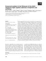
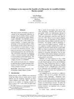
Fig. 1. Characterization of authentic Anopheles gambiae thioredoxin
reductase-1 (TrxR-1) by physicochemical analyses. (A) The absorption
spectra of 6.6 l
M
TrxR-1 in the oxidized form E
ox
(dashed curve) and
in the four-electron reduced form (EH
4
) (solid curve), which was
obtained by adding 33 l
M
NADPH to the E
ox
sample. In the EH
4
sample, the absorption at wavelengths below 400 nm is largely a result
of excess NADPH. The inset shows A. gambiae TrxR species in a
silver-stained gel after SDS/PAGE. In lane 1, the two variants of
A. gambiae TrxR isolated from cultured insect cells can be distin-
guished; lane 2 shows recombinant A. gambiae TrxR-1, and the outer
lane marker proteins. (B) The results of peptide analyses. The DNA-
deduced sequence of A. gambiae TrxR-1 is shown in standard script.
Tryptic peptides of authentic enzyme that were identified by MS are
underlined and marked in bold. These peptides were found in both
protein bands shown on lane 1 in the inset. The N-terminal sequence
(17 residues in bold italics) was identified by Edman degradation of the
protein isolated from the major band of the SDS/PAGE gel. The
minor band of 62 kDa resisted Edman degradation.
Ó FEBS 2003 Thioredoxin reductase from Anopheles gambiae (Eur. J. Biochem. 270) 4275
represented in the NCBI database by 14 ESTs that overlap
with exon 4. Joining of exon 1 (encoding the five amino
acids MAPLN) with exon 4 (encoding QENYEY and
further 491 amino acids), leads to a protein of 502 residues
which is the orthologue of D. melanogaster TrxR-1 (Figs 1B
and 3). This protein is introduced here as A. gambiae TrxR-1
(CAD30858).
Two alternative 5¢ exons–exon2andexon3–were
identified, each represented by a single EST. The N-terminal
segment contributed by exon 2 (MATAVLARPARS
LINVVQCVRL
IRTQATVMFA) shows the properties of
a mitochondrial targeting sequence containing the predicted
cleavage site between IRT and QAT. The last four amino
acids (VMFA) are not encoded by EST BM603316, but the
correct overlap with exon 4 was proven by sequencing a
PCR product amplified with an oligonucleotide pair specific
for exon 1 and exon 4 (data not shown). The deduced
N-terminal sequence of the putative mitochondrial enzyme
is thus QATVMFA|KENEY, the change from Q to K in
position 5 resulting from splicing.
Exon 3 occurs in EST BM583435, which extends into
exon 4. The resulting N-terminal sequence – MAAATAAE|
QENYEY – probably represents a second cytosolic species
of TrxR.
As judged by the number of EST sequences and by
Edman degradation of enzyme isolated from insect cells,
we can state that A. gambiae TrxR-1 is the major isoform
in vivo. Similarly to the fruit fly, no GR-like sequence could
be identified in the mosquito genome [34]. This is consistent
with the absence of detectable genuine GR activity in
Anopheles cell extracts.
Cloning and characterization of
A. gambiae
TrxR-1
The A. gambiae trxr-1 gene was cloned and recombinantly
expressed in E. coli. In SDS/PAGE, this protein co-migrates
with wild-type A. gambiae TrxR-1 – isolated from cultured
Anopheles cells or whole insects – at a position representing
a molecular mass of 58 kDa (Fig. 1A). The discrepancy
between this value and the molecular mass of 54.5 kDa
deduced from the amino acid sequence has also been
observed in other disulfide reductases; they all show a
subunit molecular mass (deduced by SDS/PAGE) that is
overestimated by 7–10%. The molecular basis for this
electrophoretic behaviour is unknown [35].
The identity between authentic and recombinant
A. gambiae Trx-1 is supported by the concurrence of
deduced and experimentally determined amino acid
sequences (Fig. 1B). Thus, TrxR-1 from A. gambiae con-
tains 502 amino acids per subunit; the calculated molecular
mass is 54.5 kDa per subunit for the apoprotein and
2 · 55.3 kDa for the FAD-containing homodimeric holo-
enzyme. The protein shares 69% sequence identity with its
orthologue from D. melanogaster (Fig. 3). Both insect
enzymes have sequence elements that are typical for large
TrxRs, including the flavin-near redox-active Cys–Val–
Asn–Val–Gly–Cys motif, as well as a C-terminally located
redox centre (Fig. 3). In the sequence of A. gambiae TrxR-1
and D. melanogaster TrxR-1 a sequentially adjacent cys-
teine pair (Cys500¢ and Cys501¢) is present. Thus, the two
insect Trxs known, to date, are typical members of the large
TrxR enzyme class characterized by an additional redox
centre. However, in contrast to mammalian TrxRs or TrxR
from P. falciparum, this part of the active site is structurally
distinct in the insect enzymes. Indeed a redox centre formed
by two sequential Cys residues is highly unusual [24].
A. gambiae
TrxR-1 as a Trx-reducing enzyme
TrxR-1 was tested with Trxs from A. gambiae (A. gambiae
Trx-1), D. melanogaster (D. melanogaster Trx-2), and
P. falciparum (P. falciparum Trx-1) as oxidizing substrates
(Table 1). The catalytic efficiency of A. gambiae TrxR-1 is
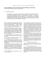
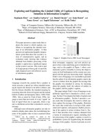
Fig. 2. Genomic organization of the Anopheles gambiae thioredoxin
reductase-1 (TrxR-1)geneincomparisonwiththeDrosophila melano-
gaster TrxR gene. In both insects the trxr locusislocatedonchro-
mosome X. Numbered boxes represent exons within the gene. Coding
regions are shown in black, untranslated sequences are shaded in grey.
Scale bar divisions are in kilobases. A. gambiae trxr occurs in three
possible splice isoforms (AJ459821, AJ549084, AJ549085) that differ in
the first exon (exon 1, 2 or 3) joined to exon 4. The orthologous
Drosophila locus, shown below, is similarly organized, except that the
sequence corresponding to exon 4 of A. gambiae trxr is interrupted by
two short introns, which results in exons 4–6. Exons 1, 2 or 3 joined to
exons 4–6 yield transcripts CG2151-RA, CG2151-RB and CG2151-
RC, respectively. In both insect species, exon 1 encodes the N-terminal
sequence of an abundant cytosolic, exon 2 of a mitochondrial, and
exon 3 of a minor cytosolic TrxR form.
Table 1. Kinetic parameters of Anopheles gambiae thioredoxin reductase-1 (TrxR-1) with different thioredoxins and 5,5¢-dithiobis(2-nitrobenzoic acid)
(DTNB). All values were determined in 100 m
M
potassium phosphate buffer, 2 m
M
EDTA, pH 7.4. As expected, A. gambiae Trx-1 was the best
substrate of A. gambiae TrxR-1, but the k
cat
/K
m
value was only marginally better than for thioredoxin-2 (Trx-2) from Drosophila melanogaster.
Plasmodium falciparum thioredoxin-1 (Trx-1) showed the highest k
cat
value, but the K
m
was significantly lower than for the insect thioredoxins.
DTNB was included as an artificial disulfide substrate which is reduced by most high molecular mass thioredoxin reductases.
Substrate K
m
(l
M
) V
max
(UÆmg
)1
) k
cat
(s
)1
) k
cat
/K
m
(
M
)1
Æs
)1
)
A. gambiae Trx-1 8.5 ± 1.5 16.9 ± 1.6 15.4 ± 1.5 1.81 · 10
6
D. melanogaster Trx-2 9.0 ± 1.0 15.7 ± 1.3 14.3 ± 1.2 1.58 · 10
6
P. falciparum Trx-1 33 ± 5 17.2 ± 1.2 15.7 ± 1.1 0.48 · 10
6
DTNB 700 ± 200 6.0 ± 0.7 5.5 ± 0.6 7.9 · 10
3
4276 H. Bauer et al. (Eur. J. Biochem. 270) Ó FEBS 2003
comparable with the value previously determined for
D. melanogaster TrxR-1 [5,25]. Expectedly, with a K
m
value
of 8.5 l
M
and a k
cat
of 15.4 s
)1
, A. gambiae Trx-1 is the best
substrate of the enzyme but D. melanogaster Trx-2, i.e. the
orthologue of A. gambiae Trx-1 in Drosophila,isanalmost
equally good substrate. The K
m
value for the reducing
substrate NADPH was found to be 5.0 l
M
.
By comparison with other TrxRs [21,24,36], we can
delineate the pathways of electrons in A. gambiae TrxR-1
during catalysis (Figs 3 and 4). The reducing equivalents
flow from the nicotinamide of NADPH via the flavin and
the pair Cys57/Cys62 to the redox centre Cys500’/Cys501’
of the other subunit, and hence to the disulfide of the
substrate Trx. The thiolate of Cys62 in A. gambiae
TrxR-1 forms a charge transfer complex with the
reoxidized flavin during catalysis [20,24,37]. This charge
transfer gives rise to the absorption band at 530 nm
(Fig. 1A) and thus to the orange/red colour of stable
catalytic intermediates that contain both oxidized flavin
and Cys62 as a thiolate. Unexpectedly, freshly prepared
A. gambiae TrxR was found to be orange/red, which
indicated the presence of reduced Cys62. Auto-oxidation
of the A. gambiae TrxR-1 preparations was very slow and
proceeded over a range of hours to days, finally producing
the typical yellow colour of oxidized enzyme E
ox
(Fig. 1A). The freshly isolated enzyme also resisted
oxidation by its native substrate, A. gambiae Trx. In
contrast, most disulfide reductases, when present in
reduced forms, can be easily oxidized by their native
substrates [20]. Consequently, we conducted redox
titration experiments on A. gambiae TrxR-1, starting out
with the oxidized form, E
ox
, and monitoring the absorbance
Fig. 3. Multiple sequence alignment (
CLUSTAL W
) of high molecular mass thioredoxin reductases (TrxR). The search was conducted (NCBI accession
numbers in parentheses) with the enzymes from Anopheles gambiae (AgTrxR-1, CAD30858), Drosophila melanogaster (DmTrxR-1, AAG25639),
humans (hTrxR, AAB35418), and from the malaria parasite Plasmodium falciparum (PfTrxR-1, CAA60574). The enzyme of Anopheles shares
69%, 52%, and 45% sequence identity with the TrxRs of D. melanogaster, humans, and P. falciparum, respectively. The sequences of the redox-
active centres are shaded in grey; U (residue 498) in the human enzyme represents selenocysteine (Sec).
Ó FEBS 2003 Thioredoxin reductase from Anopheles gambiae (Eur. J. Biochem. 270) 4277
at 530 nm. As shown in Fig. 1A, the absorption coefficient
at 530 nm was 0.4 m
M
)1
Æcm
)1
for the oxidized enzyme, E
ox
,
1.6 m
M
)1
Æcm
)1
after addition of one equivalent NADPH,
leading to the two-electron reduced enzyme species EH
2
,
and 3.0 m
M
)1
Æcm
)1
for the enzyme reduced with two or
more equivalents NADPH, giving rise to the four-electron
reduced enzyme species EH
4
. After reoxidation with 100 l
M
A. gambiae Trx-1, the e-value fell to 1.6 m
M
)1
Æcm
)1
, indi-
cative of a two-electron reduced enzyme species (EH
2
). In
contrast, reoxidation of EH
4
with 125 l
M
potassium
ferricyanide, as described previously [16,24], led to the E
ox
species with an e-value of 0.4 m
M
)1
Æcm
)1
at 530 nm.
These data confirm that the native substrate does not
reoxidize the enzyme to E
ox
but only to the EH
2
state where
the redox-active Cys residues 57, 62, 500’, and 501’ are
present partly as thiols so that the thiolate of Cys62 can still
form a charge transfer complex with flavin. For catalysis,
this implies that the very first catalytic cycle is primed by two
NADPH molecules, which results in the four-electron
reduced state. Oxidation with TrxS
2
then leads to the two-
electron reduced state, where the two disulfide bridges
are partially reduced, i.e.: priming reaction, E
ox
+
2NADPH + 2H
+
fi EH
4
+ 2NADP
+
; catalytic cycle,
EH
4
+TrxS
2
fi EH
2
+Trx(SH)
2
;EH
2
+NADPH+
H
+
fi EH
4
+NADP
+
.TrxS
2
+NADPH+H
+
fi
Trx(SH)
2
+NADP
+
. This balance reaction of the cata-
lytic cycle, of course, represents the net reaction catalyzed by
TrxR.
Discussion
The isolation and characterization of A. gambiae TrxR
contributes to the understanding of the redox metabolism in
Diptera. The principles of redox homeostasis that were
tentatively postulated for the fruit fly can be extended to a
disease-transmitting insect. In short, a GR is absent,
although GSH is a key compound of the redox networks
also in insects [38]. The nonenzymatic reduction of GSSG
by reduced Trx is probably a major pathway for GSH
reduction in these organisms [5]. Thus, TrxR indirectly
substitutes for the function of GR. As described previously,
A. gambiae Trx-1 is a highly expressed protein in vivo [25].
The efficiency of GSSG reduction by A. gambiae Trx-1 is
similar to its orthologue (Trx-2) in D. melanogaster and
probably sufficient to maintain physiological needs.
In the Anopheles mosquito, one TrxR gene is present
which occurs in three splice variants. Alternative use of first
exons was previously reported for mammalian and Droso-
phila TrxR genes [39]. In D. melanogaster, three alternative
transcripts have been identified: CG2151-RA is the ortho-
logue of A. gambiae TrxR-1; CG2151-RB corresponds to a
mitochondrial TrxR form; and CG2151-RC encodes a
second cytosolic TrxR. Transcripts coding for the latter
form are rare, as only two ESTs corresponding to CG2151-
RC have been identified (compared with more than 80
CG2151-RA ESTs). Thus, the trxr loci in Anopheles and
in Drosophila are structurally organized in a similar way:
there are three alternative first exons, coding probably for a
major cytosolic, a mitochondrial, and a minor cytosolic
form (Fig. 2).
Unlike in A. gambiae, a second TrxR gene, trxr-2,was
identified in the genome of D. melanogaster.However,a
D. melanogaster TrxR-1 null mutant leads to death, at the
latest during the second larval instar [40], and both cytosolic
and mitochondrial TrxR-1 forms have been shown to be
necessary for survival [41]. Thus, the putative activity of
TrxR-2 is not sufficient to compensate for the lack of either
the cytosolic or the mitochondrial TrxR-1.
The A. gambiae TrxR preparation from insect cells
results in two enzyme species that can be distinguished by
SDS/PAGE (Fig. 1). The predominant band represents the
cytosolic variant A. gambiae TrxR-1, and the 62 kDa band
is possibly the mitochondrial precursor variant. This
assumption is supported by the size of the protein and by
the observation that it is stabilized by protease inhibitors.
A. gambiae TrxR-1 shares 69% sequence identity with
D. melanogaster TrxR-1, including the important redox-
active Cys–Cys motif on the C-terminal extension (Figs 3
and 4). For the Drosophila enzyme it was shown that both
cysteines are essential for the interaction with the natural
substrate Trx [5,24]. In the case of rat TrxR, which is a
selenoprotein with a Cys–Sec–sequence instead, the
Sec fi Cys exchange results in a mutant with less than
1% catalytic activity when compared with the wild-type
enzyme [30,42]. The main difference between the insect
enzymes and the TrxR from rat concerns the amino acid
residues adjacent to the cysteines. In mammalian
TrxRs, including the human orthologue, we find a Gly–
Cys–Cys–Gly sequence, whereas in A. gambiae TrxR-1, it is
Thr–Cys–Cys–Ser and in D. melanogaster TrxR-1 it is Ser–
Cys–Cys–Ser. There is evidence that the hydroxyl functions
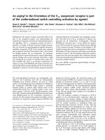
Fig. 4. Sketch of homodimeric Anopheles gambiae in the four-electron
reduced (EH
4
)form.The dimer interface is shown as a diagonal line
with a black circle at the centre. This circle represents the molecular
two-fold symmetry axis. The sketch shows the EH
4
form where all
four redox-active cysteines are present in the reduced form. The
thiolate 62 forms a charge transfer complex with oxidized flavin,
and Cys501¢ is ready to attack the disulfide bond of the substrate
thioredoxin disulfide. By analogy with other disulfide reductases,
the most probable scenario leading from oxidized enzyme (E
ox
)to
EH
4
is as follows. When NADPH binds to one subunit (upper
right), its reducing equivalents flow via the flavin to the disulfide
Cys62/Cys57. The resulting dithiol is reoxidized by exchange with
the disulfide bridge between residues 500¢ and 501¢ of the other
subunit. Subsequently, binding and oxidation of a second NADPH
molecule leads to re-reduction of the disulfide between Cys57 and
Cys62.
4278 H. Bauer et al. (Eur. J. Biochem. 270) Ó FEBS 2003
of the flanking amino acid residues are crucial for catalysis
(H. S. Gromer et al., unpublished data). It is interesting to
note that despite the evolutionary distance of 250 million
years [34] between the fruit fly and the mosquito, not only
the basic principles of redox metabolism and the genomic
organization of the TrxR gene, but also the mechanistic
peculiarities of these orthologous enzymes, have remained
highly conserved.
With a k
cat
of 15–22 s
)1
for Trx [5,24], the insect TrxRs
exhibit a somewhat lower turnover number than their
mammalian relatives ( 35–45 s
)1
) [30,43], but they have
the advantage of being independent of the rare trace element
selenium. This evolutionary adaptation is plausible because
TrxR is apparently the mainstay enzyme of the antioxidant
metabolism in insects, whereas mammals have a second,
GR-based system.
Redox processes also represent an interesting aspect of
parasite–host interaction. For human malaria it is well
known that disturbance of the antioxidative metabolism
results in an inhibition of parasitic growth in erythrocytes.
The most prominent example is glucose-6-phosphate-dehy-
drogenase (Glc6PDH) deficiency, an inherited disease also
known as favism [44,45]. A major effect of Glc6PDH
deficiency is an impaired NADPH production, which
affects the ability of the erythrocyte to resist oxidative
stress. The effects of Glc6PDH deficiency can be imitated by
GR-inhibitors such as carmustine [46] or isoalloxazines [47].
The current interpretation of Glc6PDH deficiency, as a
condition protecting from severe malaria, is based on the
observation that the anion channel protein of the erythro-
cyte membrane undergoes oxidative changes when ring-
stage parasites are present in the red blood cell. This
oxidized band-3 protein is recognized by a specific antibody
that initiates immunologic processes to eliminate the
parasitized cell [48,49].
With respect to the insect vector A. gambiae,ithasalso
been shown that nitrosative and oxidative stress imposed
by NO and peroxynitrite play a dominating role in the
host’s defence against the parasite [50,51]. The insect cells,
in turn, have to protect themselves against these reactive
agents. When Anopheles cells are challenged by oxidative
stress, transcription of numerous genes that are associated
with the Trx system are induced, prominently among
them the TrxR gene [52]. Interestingly, a similar response
occurs after exposing the cells to bacterial peptidoglycan.
Trx system-related genes are also transcribed in the
salivary glands of A. gambiae. It is assumed that the
corresponding proteins are especially important for pro-
tecting the glands from heme-driven free radical attack
[53]. Thus, redox processes play a major role in host–
parasite interactions, not only in human blood but also
inside the insect vector.
The differences between the enzyme systems involved in
antioxidative metabolism offer an interesting novel target
for the development of insect-specific TrxR inhibitors. The
potential of TrxR as a target for novel insecticides is
supported by the fact that TrxR-1 knockouts in Drosophila
are lethal in early embryonic stages [40]. Based on the
genomic data and comparative genome analyses, it can be
reasonably assumed that this is also true for Anopheles.The
development of novel insecticides is an important approach
in the fight against malaria which is becoming more and
more complicated, not least as a result of the occurrence of
insecticide resistance [54]. In this context it should be noted
that inhibitors of the Trx system have toxic effects
themselves but, in addition, they sensitize organisms for
other toxic agents [13,55,56]. Consequently, inhibitors of
A. gambiae TrxR-1 are expected to protect other insecti-
cides from the development of resistance.
Acknowledgements
We are indebted to Dr Stefan M. Kanzok for the early studies on the
Anopheles thioredoxin system and for his help in database searches. Our
work was supported by the Deutsche Forschungsgemeinschaft (Grants
B2 and C1 of SFB 544 ÔControl of Tropical Infectious DiseasesÕ to
R.H.S. and H.M.M., respectively, as well as grant GR2028/1-1 to S.G.)
and by the Fonds der Chemischen Industrie (Grant 161576) to R.H.S.
References
1. Adams, M.D., Celniker, S.E., Holt, R.A., Evans, C.A., Gocayne,
J.D., Amanatides, P.G., Scherer, S.E., Li, P.W., Hoskins, R.A.,
Galle, R.F., George, R.A., Lewis, S.E., Richards, S., Ashburner,
M., Henderson, S.N., Sutton, G.G., Wortman, J.R., Yandell,
M.D., Zhang, Q., Chen, L.X. et al. (2000)Thegenomesequenceof
Drosophila melanogaster. Science 287, 2185–2195.
2. Myers, E.W., Sutton, G.G., Delcher, A.L., Dew, I.M., Fasulo,
D.P., Flanigan, M.J., Kravitz, S.A., Mobarry, C.M., Reinert,
K.H., Remington, K.A., Anson, E.L., Bolanos, R.A., Chou,
H.H.,Jordan,C.M.,Halpern,A.L.,Lonardi,S.,Beasley,E.M.,
Brandon, R.C., Chen, L., Dunn, P.J. et al. (2000) A whole-genome
assembly of Drosophila. Science 287, 2196–2204.
3. Holt, R.A., Subramanian, G.M., Halpern, A., Sutton, G.G.,
Charlab, R., Nusskern, D.R., Wincker, P., Clark, A.G., Ribeiro,
J.M., Wides, R., Salzberg, S.L., Loftus, B., Yandell, M., Majoros,
W.H., Rusch, D.B., Lai, Z., Kraft, C.L., Abril, J.F., Anthouard,
V., Arensburger, P. et al. (2002) The genome sequence of the
malaria mosquito Anopheles gambiae. Science 298, 129–149.
4. Kanzok, S.M. & Zheng, L. (2003) The mosquito genome – a
turning point? Trends Parasitol. 19, 329–331.
5. Kanzok, S.M., Fechner, A., Bauer, H., Ulschmid, J.K., Mu
¨
ller,
H.M., Botella-Munoz, J., Schneuwly, S., Schirmer, R.H. & Becker,
K. (2001) Substitution of the thioredoxin system for glutathione
reductase in Drosophila melanogaster. Science 291, 643–646.
6. Hemingway, J. & Ranson, H. (2000) Insecticide resistance in insect
vectors of human disease. Annu. Rev. Entomol. 45, 371–391.
7. Schafer, F.Q. & Buettner, G.R. (2001) Redox environment of the
cell as viewed through the redox state of the glutathione disulfide/
glutathione couple. Free Radic. Biol. Med. 30, 1191–1212.
8. Flohe
´
, L. (2003) Lest I forget thee, glutathione. Biol. Chem. 384,
503–687.
9. Dolphin, D., Poulson, R. & Avramovic, O., (eds). (1989) Coen-
zymes and Cofactors – Glutathione,Vol.III.PartAandB.John
Wiley & Sons, New York.
10. Schirmer, R.H., Krauth-Siegel, R.L. & Schulz, G.E. (1989) Glu-
tathione reductase. In Coenzymes and Cofactors – Glutathione
(Dolphin,D.,Poulson,R.&Avramovic,O.,eds),Vol.III,pp.
553–596. John Wiley & Sons, New York.
11. Schirmer, R.H., Bauer, H. & Becker, K. (2002) Glutathione
reductase. In Wiley Encyclopedia of Molecular Medicine
(Creighton T.E., ed.), Vol. 5, pp. 1471–1475. John Wiley & Sons,
New York.
12. Sarma, G.N., Savvides, S.N., Becker, K., Schirmer, M., Schirmer,
R.H. & Karplus, P.A. (2003) Glutathione reductase of the
malarial parasite Plasmodium falciparum: crystal structure and
inhibitor development. J. Mol. Biol. 328, 893–907.
Ó FEBS 2003 Thioredoxin reductase from Anopheles gambiae (Eur. J. Biochem. 270) 4279
13. Gromer,S.,Urig,S.&Becker,K.(2004)Thethioredoxinsystem–
from science to clinic. Med. Res. Rev. in press.
14. Arne
´
r, E.S. & Holmgren, A. (2000) Physiological functions of
thioredoxin and thioredoxin reductase. Eur. J. Biochem. 267,
6102–6109.
15. Holmgren, A. (1985) Thioredoxin. Annu. Rev. Biochem. 54,
237–271.
16. Holmgren, A. (1989) Thioredoxin and glutaredoxin systems.
J. Biol. Chem. 264, 13963–13966.
17. Holmgren, A. (1984) Enzymatic reduction-oxidation of protein
disulfides by thioredoxin. Methods Enzymol. 107, 295–300.
18. Schenk, H., Klein, M., Erdbrugger, W., Dro
¨
ge,W.&Schulze-
Osthoff, K. (1994) Distinct effects of thioredoxin and antioxidants
on the activation of transcription factors NF-kappa B and AP-1.
Proc.NatlAcad.Sci.USA91, 1672–1676.
19. Follmann,H.&Ha
¨
berlein, I. (1995) Thioredoxins: universal, yet
specific thiol-disulfide redox cofactors. Biofactors 5, 147–156.
20. Williams, C.H. Jr (1992) Lipoamide dehydrogenase, glutathione
reductase, thioredoxin reductase, and mercuric ion reductase – a
family of flavoenzyme transhydrogenases. In Chemistry and
Biochemistry of Flavoenzymes (Mu
¨
ller F., ed.), Vol. 3, pp. 121–211.
CRC Press, Inc., Boca Raton, FL.
21. Williams, C.H., Arscott, L.D., Mu
¨
ller, S., Lennon, B.W., Ludwig,
M.L.,Wang,P.F.,Veine,D.M.,Becker,K.&Schirmer,R.H.
(2000) Thioredoxin reductase: two modes of catalysis have
evolved. Eur. J. Biochem. 267, 6110–6117.
22. Zhong, L., Arne
´
r, E.S. & Holmgren, A. (2000) Structure and
mechanism of mammalian thioredoxin reductase: the active site is
a redox-active selenolthiol/selenenylsulfide formed from the con-
served cysteine-selenocysteine sequence. Proc. Natl Acad. Sci.
USA 97, 5854–5859.
23. Gilberger, T.W., Bergmann, B., Walter, R.D. & Mu
¨
ller, S.
(1998) The role of the C-terminus for catalysis of the large
thioredoxin reductase from Plasmodium falciparum. FEBS Lett.
425, 407–410.
24. Bauer, H., Massey, V., Arscott, L.D., Schirmer, R.H., Ballou,
D.A. & Williams, C.H. Jr (2003) The mechanism of high M
r
thioredoxin reductase from Drosophila melanogaster. J. Biol.
Chem. 278, 33020–33028.
25. Bauer, H., Kanzok, S.M. & Schirmer, R.H. (2002) Thioredoxin-2
but not thioredoxin-1 is a substrate of thioredoxin peroxidase-1
from Drosophila melanogaster: isolation and characterization of a
second thioredoxin in D. melanogaster and evidence for distinct
biological functions of Trx-1 and Trx-2. J. Biol. Chem. 277, 17457–
17463.
26. Kanzok, S.M., Schirmer, R.H., Tu
¨
rbachova, I., Iozef, R. &
Becker, K. (2000) The thioredoxin system of the malaria parasite
Plasmodium falciparum: glutathione reduction revisited. J. Biol.
Chem. 275, 40180–40186.
27. Mu
¨
ller, H.M., Dimopoulos, G., Blass, C. & Kafatos, F.C. (1999)
A hemocyte-like cell line established from the malaria vector
Anopheles gambiae expresses six prophenoloxidase genes. J. Biol.
Chem. 274, 11727–11735.
28. Fa
¨
rber, P.M., Arscott, L.D., Williams, C.H. Jr, Becker, K. &
Schirmer, R.H. (1998) Recombinant Plasmodium falciparum
glutathione reductase is inhibited by the antimalarial dye methy-
lene blue. FEBS Lett. 422, 311–314.
29. Worthington, D.J. & Rosemeyer, M.A. (1976) Glutathione
reductase from human erythrocytes: catalytic properties and
aggregation. Eur. J. Biochem. 67, 231–238.
30. Zhong, L. & Holmgren, A. (2000) Essential role of selenium in the
catalytic activities of mammalian thioredoxin reductase revealed
by characterization of recombinant enzymes with selenocysteine
mutations. J. Biol. Chem. 275, 18121–18128.
31. Gladyshev, V.N. & Hatfield, D.L. (1999) Selenocysteine-contain-
ing proteins in mammals. J. Biomed. Sci. 6, 151–160.
32. Gladyshev, V.N., Jeang, K.T. & Stadtman, T.C. (1996) Seleno-
cysteine, identified as the penultimate C-terminal residue in human
T-cell thioredoxin reductase, corresponds to TGA in the human
placental gene. Proc. Natl Acad. Sci. USA 93, 6146–6151.
33. May, J.M., Mendiratta, S., Hill, K.E. & Burk, R.F. (1997)
Reduction of dehydroascorbate to ascorbate by the selenoenzyme
thioredoxin reductase. J. Biol. Chem. 272, 22607–22610.
34. Zdobnov, E.M., von Mering, C., Letunic, I., Torrents, D.,
Suyama, M., Copley, R.R., Christophides, G.K., Thomasova, D.,
Holt, R.A., Subramanian, G.M., Mueller, H.M., Dimopoulos,
G., Law, J.H., Wells, M.A., Birney, E., Charlab, R., Halpern,
A.L.,Kokoza,E.,Kraft,C.L.,Lai,Z.et al. (2002) Comparative
genome and proteome analysis of Anopheles gambiae and
Drosophila melanogaster. Science 298, 149–159.
35. Krauth-Siegel, R.L., Mu
¨
ller, J.G., Lottspeich, F. & Schirmer,
R.H. (1996) Glutathione reductase and glutamate dehydrogenase
of Plasmodium falciparum, the causative agent of tropical malaria.
Eur. J. Biochem. 235, 345–350.
36. Sandalova, T., Zhong, L., Lindqvist, Y., Holmgren, A. &
Schneider, G. (2001) Three-dimensional structure of a mammalian
thioredoxin reductase: implications for mechanism and evolution
of a selenocysteine-dependent enzyme. Proc. Natl Acad. Sci. USA
98, 9533–9538.
37. Rietveld, P., Arscott, L.D., Berry, A., Scrutton, N.S., Deonarain,
M.P., Perham, R.N. & Williams, C.H. Jr (1994) Reductive and
oxidative half-reactions of glutathione reductase from Escherichia
coli. Biochemistry 33, 13888–13895.
38. Sohal, R.S., Arnold, L. & Orr, W.C. (1990) Effect of age on
superoxide dismutase, catalase, glutathione reductase, inorganic
peroxides, TBA-reactive material, GSH/GSSG, NADPH/
NADP
+
and NADH/NAD
+
in Drosophila melanogaster. Mech.
Ageing Dev. 56, 223–235.
39. Sun, Q.A., Zappacosta, F., Factor, V.M., Wirth, P.J., Hatfield,
D.L. & Gladyshev, V.N. (2001) Heterogeneity within animal
thioredoxin reductases: evidence for alternative first exon splicing.
J. Biol. Chem. 276, 3106–3114.
40. Missirlis, F., Phillips, J.P. & Ja
¨
ckle, H. (2001) Cooperative action
of antioxidant defense systems in Drosophila. Curr. Biol. 11,
1272–1277.
41. Missirlis, F., Ulschmid, J.K., Hirosawa-Takamori, M., Gronke,
S., Scha
¨
fer, U., Becker, K., Phillips, J.P. & Ja
¨
ckle, H. (2002)
Mitochondrial and cytoplasmic thioredoxin reductase variants
encoded by a single Drosophila gene are both essential for viability.
J. Biol. Chem. 277, 11521–11526.
42. Lee, S.R., Bar-Noy, S., Kwon, J., Levine, R.L., Stadtman, T.C. &
Rhee, S.G. (2000) Mammalian thioredoxin reductase: oxidation of
the C-terminal cysteine/selenocysteine active site forms a thiosele-
nide, and replacement of selenium with sulfur markedly reduces
catalytic activity. Proc.NatlAcad.Sci.USA97, 2521–2526.
43. Gromer, S., Arscott, L.D., Williams, C.H. Jr, Schirmer, R.H. &
Becker, K. (1998) Human placenta thioredoxin reductase: iso-
lation of the selenoenzyme, steady state kinetics, and inhibition
by therapeutic gold compounds. J. Biol. Chem. 273, 20096–20101.
44. Golenser, J., Miller, J., Spira, D.T., Kosower, N.S., Vande Waa,
J.A. & Jensen, J.B. (1988) Inhibition of the intraerythrocytic
development of Plasmodium falciparum in glucose-6-phosphate
dehydrogenase deficient erythrocytes is enhanced by oxidants and
by crisis form factor. Trop. Med. Parasitol. 39, 273–276.
45. Ginsburg, H., Atamna, H., Shalmiev, G., Kanaani, J. & Krugliak,
M. (1996) Resistance of glucose-6-phosphate dehydrogenase
deficiency to malaria: effects of fava bean hydroxypyrimidine
glucosides on Plasmodium falciparum growth in culture and on the
phagocytosis of infected cells. Parasitology 113, 7–18.
46. Becker, K. & Schirmer, R.H. (1995) 1,3-Bis (2-chloroethyl)-1-
nitrosourea as thiol-carbamoylating agent in biological systems.
Methods Enzymol. 251, 173–188.
4280 H. Bauer et al. (Eur. J. Biochem. 270) Ó FEBS 2003
47. Scho
¨
nleben-Janas, A., Kirsch, P., Mittl, P.R., Schirmer, R.H. &
Krauth-Siegel, R.L. (1996) Inhibition of human glutathione
reductase by 10-arylisoalloxazines: crystalline, kinetic, and elec-
trochemical studies. J. Med. Chem. 39, 1549–1554.
48. Giribaldi, G., Ulliers, D., Mannu, F., Arese, P. & Turrini, F.
(2001) Growth of Plasmodium falciparum induces stage-dependent
haemichrome formation, oxidative aggregation of band 3, mem-
brane deposition of complement and antibodies, and phagocytosis
of parasitized erythrocytes. Br. J. Haematol. 113, 492–499.
49. Cappadoro, M., Giribaldi, G., O’Brien, E., Turrini, F., Mannu,
F., Ulliers, D., Simula, G., Luzzatto, L. & Arese, P. (1998) Early
phagocytosis of glucose-6-phosphate dehydrogenase (G6PD) -
deficient erythrocytes parasitized by Plasmodium falciparum may
explain malaria protection in G6PD deficiency. Blood 92, 2527–
2534.
50. Tahar, R., Boudin, C., Thiery, I. & Bourgouin, C. (2002) Immune
response of Anopheles gambiae to the early sporogonic stages of
the human malaria parasite Plasmodium falciparum. EMBO J. 21,
6673–6680.
51. Dimopoulos, G., Seeley, D., Wolf, A. & Kafatos, F.C. (1998)
Malaria infection of the mosquito Anopheles gambiae activates
immune-responsive genes during critical transition stages of the
parasite life cycle. EMBO J. 17, 6115–6123.
52. Dimopoulos, G., Christophides, G.K., Meister, S., Schultz, J.,
White, K.P., Barillas-Mury, C. & Kafatos, F.C. (2002) Genome
expression analysis of Anopheles gambiae: responses to injury,
bacterial challenge, and malaria infection. Proc. Natl Acad. Sci.
USA 99, 8814–8819.
53. Francischetti, I.M., Valenzuela, J.G., Pham, V.M., Garfield, M.K.
& Ribeiro, J.M. (2002) Toward a catalog for the transcripts and
proteins (sialome) from the salivary gland of the malaria vector
Anopheles gambiae. J. Exp. Biol. 205, 2429–2451.
54. Feyereisen, R. (1995) Molecular biology of insecticide resistance.
Toxicol. Lett. 82–83, 83–90.
55. Powis, G., Mustacich, D. & Coon, A. (2000) The role of the redox
protein thioredoxin in cell growth and cancer. Free Radic. Biol.
Med. 29, 312–322.
56. Rahlfs, S., Schirmer, R.H. & Becker, K. (2002) The thioredoxin
system of Plasmodium falciparum and other parasites. Cell. Mol.
Life Sci. 59, 1024–1041.
Ó FEBS 2003 Thioredoxin reductase from Anopheles gambiae (Eur. J. Biochem. 270) 4281