Báo cáo Y học: A nonphosphorylated 14-3-3 binding motif on exoenzyme S that is functional in vivo pot
Bạn đang xem bản rút gọn của tài liệu. Xem và tải ngay bản đầy đủ của tài liệu tại đây (243.74 KB, 9 trang )
A nonphosphorylated 14-3-3 binding motif on exoenzyme S
that is functional
in vivo
Maria L. Henriksson
1,2
, Matthew S. Francis
2
, Alex Peden
4
, Margareta Aili
2
, Kristina Stefansson
1
,
Ruth Palmer
3
, Alastair Aitken
4
and Bengt Hallberg
1
1
Department of Medical Biosciences/Pathology, Umea
˚
University, Sweden;
2
Department of Molecular Biology, Umea
˚
University,
Sweden;
3
Umea
˚
Center for Molecular Pathogenesis, Umea
˚
University, Sweden;
4
Membrane Biology Group, Division of Biomedical
and Clinical Laboratory Sciences, University of Edinburgh, Scotland
14-3-3 proteins play an important role in a multitude of
signalling pathways. The interactions between 14-3-3 and
other signalling proteins, such as Raf and KSR (kinase
suppressor of Ras), occur in a phospho-specific manner.
Recently, a phosphorylation-independent interaction has
been reported to occur between 14-3-3 and several proteins,
for example 5-phosphatase, p75NTR-associated cell death
executor (NADE) and the bacterial toxin Exoenzyme S
(ExoS), an ADP-ribosyltransferase from Pseudomonas
aeruginosa. In this study we have identified the amino acid
residues on ExoS, which are responsible for its specific
interaction with 14-3-3. Furthermore, we show that a
peptide derived from ExoS, containing the 14-3-3 interaction
site, effectively competes out the interaction between ExoS
and 14-3-3. In addition, competition with this peptide blocks
ExoS modification of Ras in our Ras modification assay. We
show that the ExoS protein interacts with all isoforms of the
14-3-3 family tested. Moreover, in vivo an ExoS protein
lacking the 14-3-3 binding site has a reduced capacity
to ADP ribosylate cytoplasmic proteins, e.g. Ras, and shows
a reduced capacity to change the morphology of infected
cells.
Keywords: ADP-ribosylation; coenzyme binding site; cyto-
toxicity; NAD-dependent; peptide inhibitor.
Members of the 14-3-3 family function as adaptor or
scaffold proteins and appear to interconnect different
proteins involved in signal transduction, cell cycle regulation
and apoptosis (reviewed in [1–3]). Studies in other model
systems have also shown that 14-3-3 proteins are essential
for Drosophila melanogaster and yeast cell proliferation and
survival [4–7]. 14-3-3 proteins have been shown to interact
with phosphoserine-containing peptides, within a defined
consensus-binding motif. Some well-described 14-3-3 bind-
ing partners include the protein kinases Raf-1 [8], kinase
suppressor of Ras-1 [9], Ask1 [10], mitogen-activated
protein kinase/extracellular signal-regulated kinase kinase
[11], Bcr [12] and protein kinase C [13]. In addition, 14-3-3
proteins also interact in a phospho-specific manner with the
pro-apoptotic protein Bad [14] and the transcription factor
Forkhead [15].
Analysis of the crystal structural of 14-3-3 proteins has
revealed that all isoforms of 14-3-3 exist as a dimer, which is
made up of a conserved concave surface, a so-called
amphipathic groove, and a more variable outer surface in
each monomer [16–19]. It has been verified by both
mutational analysis and crystal studies that the basic cluster
in the amphipathic groove is involved in mediating the
interaction of 14-3-3 with the phosphorylated residues in its
interaction partners [20,21].
In addition to the defined interaction of 14-3-3 proteins
with phosphoserine-containing motifs [22], there are also
several reports showing an interaction between 14-3-3 and
nonphosphorylated substrates [23–32]. It is presumed that
there are structural similarities between the phosphorylated
and nonphosphorylated 14-3-3 ligands. The best studied
nonphosphorylated ligand for 14-3-3 is R18, an artificial
peptide isolated from a phage display library as a 14-3-3
binding sequence, which assumes an extended conformation
in the amphipathic groove in a manner similar to that
observed for the phosphorylated peptides and interacts with
14-3-3 with high affinity [33].
14-3-3 has also been shown to interact with Exoenzyme S
(ExoS) in an unphosphorylated manner and recently we
have shown that 14-3-3 interacts with the C-terminal region
of ExoS [27–29]. ExoS is a bi-functional toxin, encoded by
the pathogen Pseudomonas aeruginosa. ExoS contains a
C-terminal ADP-ribosyltransferase activity, which blocks
receptor-stimulated Ras activation through a modification
of Ras in vivo [34–36]. It has also been reported to contain
an N-terminal Rho GTPase-activating protein (GAP)
activity in vitro [37] and in vivo [34].
Since the interaction between ExoS and 14-3-3 has been
suggested to be important for the ADP-ribosylation activity
of ExoS, and more intriguingly appears to be independent
of serine-phosphorylation, we wanted to define the amino
acid sequence required for the ExoS interaction with 14-3-3
and its resultant activity both in vitro and in vivo. We have
approached these questions by using deletion and substitu-
tion analysis of ExoS both in vitro and in vivo. Various
Correspondence to B. Hallberg, Department of Medical Biosciences/
Pathology, Umea
˚
University, S-901 87 Umea
˚
,Sweden.
Fax: +46 90 77 14 20, Tel.: +46 90 785 25 23,
E-mail:
Abbreviations: ExoS, Exoenzyme S; NADE, p75NTR-Associated
cell Death Executor; GAP, GTPase-activating protein;
HRP, horseradish peroxidase.
(Received 22 March 2002, revised 16 August 2002,
accepted 20 August 2002)
Eur. J. Biochem. 269, 4921–4929 (2002) Ó FEBS 2002 doi:10.1046/j.1432-1033.2002.03191.x
mutant ExoS proteins were tested for their capacity to
interact with 14-3-3 and subsequently for their ADP-
ribosylation potential using Ras as a substrate both in vitro
and in vivo.
Here we identify the binding site on ExoS for 14-3-3
interaction and show that it harbours a short amino acid
sequence (DALDL) with similarities to the peptide sequence
(WLDL) of R18. In addition, we also show that a peptide
containing the interaction determinant of ExoS acts as an
efficient competitor for both 14-3-3 : ExoS binding and the
resulting activation of 14-3-3-dependent ExoS ADP-ribosy-
lation activity. We show that the ExoS proteins interact with
all isoforms of the 14-3-3 family. Finally, we show that the
DALDL sequence is necessary for the ADP-ribosylation
activity and the cytotoxic action of ExoS in vivo.
MATERIAL AND METHODS
Cell cultures, cell lysis
HeLa cells were grown in RPMI 1640 supplemented with
10% (v/v) fetal bovine serum and 100 UÆmL
)1
penicillin.
Following bacterial infection (80 min) cells were washed in
ice-cold NaCl/P
i
and lysed on ice in lysis buffer [1% (v/v)
Triton X-100, 100 m
M
NaCl, 50 m
M
Tris/HCl pH 7.5,
1m
M
EDTA supplemented with protease inhibitors
(10 lgÆmL
)1
aprotinin, pepstatin and leupeptin)]. Lysates
were subsequently cleared by centrifugation at 14 000 r.p.m.
for 10 min at 4 °C. Lysates were precleared with purified
glutathione S-transferase (GST). Lysates were incubated
with GST-fusion proteins for 1 h prior to the addition of
Glutathione Sepharose (Amersham Pharmacia Biotech) for
30 min. After three washes in lysis buffer, samples were
boiled in SDS/PAGE sample buffer.
Western analysis, peptide and antibodies
Anti-14-3-3b was from Santa Cruz; monoclonal Ras
(R02120) was from Transduction Laboratories; anti-phos-
pho-Erk, phospho-Akt, pan-Erk were from Cell Signaling
Technology; epidermal growth factor (EGF) was from UBI.
Immunoblotting was performed according to the manufac-
turer’s instructions using secondary antibodies conjugated
to horseradish peroxidase (HRP) sheep anti-mouse or
rabbit antibodies (Pierce and ECL Plus, Amersham Phar-
macia Biotech). A synthetic peptide, purified by reverse-
phase HPLC and characterized by MS, corresponding to
the putative 14-3-3 binding domain of ExoS (QSGHSQG
LLDALDLASKP), was purchased from Agrisera AB
(Sweden).
Plasmids
pGEX-ExoS(SD), was derived from wild-type ExoS
(pTS103) [38] as follows. Primers were designed to introduce
flanking 5¢-ClaI site (shown in italic type), 5¢-CAGGTCCG
GAATCGATGTCAGCGG-3¢, at position 1101, 3¢-NdeI/
NotI restriction sites (shown in italic type) 5¢-CCCCTCGT
CTCACCGGTATACCGCCGGCGCGAG-3¢, at position
1251, 5¢-NotI/NheI(5¢–GCTCGCGGCCGCAGCTAGCA
AACCGGAACGTTCAGG-3¢), at position 1277, and
3¢-EcoRI (5¢-TACGACGAATTCGGCCAGATCAAG
GC-3¢), at position 1359 over the area to be substituted.
PCR from wild-type ExoS was carried out using these
primers. PCR products were subsequently inserted into a
pGEX-2TK-ExoS(88–453) opened with ClaI–EcoRI to
produce pGEX-ExoS(SD). pGEX-ExoS(SD)ismutated
at amino acid positions 419–423 from SQGLL to
MAAAA and deleted from amino acid 424 to 428.
The substitution mutants, pGEX-2TK-ExoS(S1), pGEX-
2TK-ExoS(S2) and pGEX-2TK-ExoS(S3), were then con-
structed by digesting pGEX-2TK-ExoS(SD)withNdeI/
NheI and insertion of oligomers corresponding to the
appropriate amino acid substitutions, as outlined in Fig. 1B.
All constructs were sequenced using the DYEnamic.ET
terminal cycle sequencing kit (Amersham-Pharmacia).
pGEX-2TK-ExoS(88–453), pGEX-2TK-ExoS(400–453),
pGEX-2TK-ExoS(366–453), pGEX-2T-14-3-3-zeta and
pRSET-Ha-Ras were expressed as described previously
[29,39,40].
Competition analysis
14-3-3 (250 n
M
) was preincubated for 30 min at 37 °Cwith
increasing amounts of peptide, and then transferred into a
mixture containing (in a final volume of 20 lL): 0.2
M
sodium acetate, pH 6.0 and 500 n
M
GST–ExoS (366–453).
After 1 h at 37 °C the reaction was put on ice and 5 lL
Glutathione Sepharose beads were added and tumbled for
1hat4°C. Complexes were washed three times with 1 mL
20 m
M
Hepes, 120 m
M
NaCl, 10% glycerol, 0.5% NP-40,
2m
M
EDTA pH 8.0 and then subjected to SDS/PAGE and
immunoblotting.
Immunoblotting analysis of the 14-3-3 isoforms pulled
down by GST–ExoS mutants
HeLa cell lysate (2.4 mg) was incubated with GST-fusion
protein (10 lg) for 1 h prior to the addition of Glutathione
Sepharose for 30 min After three washes in lysis buffer,
samples were boiled in SDS/PAGE sample buffer. Protein
samples were loaded onto a single wide lane of an SDS/
polyacrylamide gel. After electrophoresis the separated
proteins were transferred onto nitrocellulose membranes at
200 mA constant current for 1 h. The membranes were then
blocked for 1 h in 5% skimmed milk in TBS-Tween (20 m
M
Tris/HCl pH 7.5, 137 m
M
NaCl, 0.1% Tween). Longitudi-
nal strips of the membrane were exposed to a range of 14-3-
3 isoform-specific antisera (diluted in 5% skimmed milk in
TBS-Tween, see Table 1) using a Biometra
TM
slot blot
apparatus [41]. After washing the slots separately with TBS-
Tween, the nitrocellulose filters were probed with HRP-
conjugated goat anti-(rabbit Ig) (Bio-Rad) diluted 1 : 2000
and developed by enhanced chemiluminescence.
Construction of arabinose inducible ExoS derivatives
and infection of cells
To ensure protein stability of ExoS derivatives, mutant
alleles were coexpressed with orf1, encoding the cognate
nonsecreted chaperone of ExoS [38,42]. In all cases, DNA
was amplified by PCR using conditions described previously
[43]. pMF366 was constructed from amplified DNA from
pTS103 [38] harbouring wild-type orf1, which was cloned
into the NcoI/XhoI (shown in italic type) sites of pBAD/
Myc-His under the control of an arabinose inducible
4922 M. L. Henriksson et al. (Eur. J. Biochem. 269) Ó FEBS 2002
promoter using the orf1 specific primers porf1a (forward):
5¢-GCCGCCTCCATGGACTCGGAACACGCC-3¢ and
porf1b (reverse): 5¢-TCGCCCGACTCGAGTCAGCGTA
GCTCTTC-3¢. Wild-type exoS sequence was cloned into
the XhoI/KpnI (shown in italic type) sites of pMF366 to
generate the plasmid pMF384, using DNA amplified from
pTS103 with the exoS specific primer pair pexoSa (forward):
5¢-CGGAGAAACTCGAGGAGAAGGCAACCATC-3¢,
pexoSb (reverse): 5¢-GTCTTTCTGGTACCACCGGTCA
GGCCAGA-3¢. pMF419 and pMF420 were obtained by
replacing the C-terminal ClaI/KpnI fragment from pMF384
with DNA amplified and restriction enzyme cut with ClaI/
KpnI from pGEX-2TK-ExoS(SD) and pGEX-2TK-ExoS
(S3), respectively, using the exoS specific primers, pexoSseq3
(position 973–991; forward): 5¢-AAGTGATGGCGCTTG
GTCT-3¢ and pexoSd (reverse): 5¢-ATGCATGGTACCTC
AGGCCAGATCAAGGCCGCG-3¢. All constructs were
confirmed by sequence analysis. Stable induction of protein
expression in strains grown in the presence of 0.02%
L
(+)
arabinose was confirmed by Western analysis as described
previously [44], using polyclonal rabbit anti-ExoS [38].
Bacterial infection of cells was performed in the presence of
0.1%
L
(+)arabinose as described previously [45].
RESULTS
Amino acids 420–429 of ExoS are important
for interaction with 14-3-3
Numerous observations have revealed that 14-3-3 binds
phosphorylated ligands [3]. The binding of 14-3-3 proteins
to nonphosphorylated partners is, however, far less defined.
As the interaction between ExoS and 14-3-3 has been
suggested to be important for the ADP-ribosylation activity
of ExoS, and more intriguingly appears to be independent
of serine phosphorylation, we decided to define the amino
acid sequence required for the ExoS interaction with 14-3-3.
To date, the best-studied example of a nonphosphorylated
interaction with 14-3-3 is with an artificial peptide, named
R18, which was isolated from a phage display library as
having high affinity for 14-3-3 proteins. A motif in R18
10
WLDLE
14
, was found in a similar position as the
phosphorylated residues in the 14-3-3 binding phosphopep-
tides, with negatively charged Asp12 and Glu14 making
contacts similar to those of phosphoserine [3]. The hydro-
phobic residues in the R18 peptide make contact with the
hydrophobic side of the amphipathic groove of 14-3-3,
implying that the R18 peptide interacts with 14-3-3 in a
manner very similar to phosphorylated ligands [33,46]. In an
earlier study we have shown that a C-terminal deletion in
which the extreme 26 amino acids of ExoS were removed
was unable to bind 14-3-3 (Fig. 1A, lane 4, and see [29]). On
further inspection we noted that ExoS contains a DALDL
sequence, at amino acid position 424–428, which is similar
to the WLDLE of R18 (Table 2).
To address the question of whether the DALDL
sequence is a determinant of the 14-3-3 : ExoS interaction,
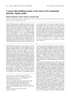
Fig. 1. GST–ExoS mutant analysis of interaction with endogenous
14-3-3 proteins. HeLa cells were harvested, and lysates were subjected
to pull-down analysis with 5 lg of various GST-fusion proteins.
Samples were separated by SDS/PAGE on a 12.5% gel. (A) Upper
panel: HeLa cell lysates were subjected to affinity precipitation with a
series of GST–ExoS mutants. Lanes correspond to schematic repre-
sentations of the constructs illustrated in (B). Lower panel: Commassie
blue stained SDS/PAGE, shows purified GST-fusion proteins purified
from Escherichia coli used in this study. Lanes correspond to schematic
representations of the constructs illustrated in (B). Lane 1 represents
2 lg of whole HeLa cell lysate. 14-3-3 proteins were detected by
immunoblotting with anti-14-3-3 antibodies. (B) Schematic diagram
detailing the various GST-fusion protein constructs of ExoS used in
the present study. Important amino acids for 14-3-3 interactions of
ExoS are indicated between amino acids 418 and 429. The region of
interest in ExoS and the limited similarity towards other nonphos-
phorylated 14-3-3 partners is shown in Table 2.
Table 1. Summary of isoform specific 14-3-3 antibodies used.
Isoform Antibody Epitope Position Dilution
bbT Ac-MDKSELV 1–7 1 : 3000
ff1002 Ac-MDKNELVQKAC 1–10 1 : 3000
ss197 Ac-MEKTELIQKAC 1–10 1 : 3000
rr789 Ac-MERASLIQKAC 1–10 1 : 3000
ee2025 Ac-MDDREDLVYQAKC 1–12 1 : 3000
gg2043 Ac-GDREQLLQRARC 2–12 1 : 3000
cc1006 Ac-VDREQLVQKAC 2–11 1 : 6000
Ó FEBS 2002 Identification of a 14-3-3 binding motif on ExoS (Eur. J. Biochem. 269) 4923
a set of deletion and substitution variants of ExoS were
generated for use in protein pull-down experiments (see
Fig. 1B). We constructed an ExoS deletion protein depicted
in Fig. 1B, named GST–ExoS(SD), as well as three over-
lapping substitution mutants between amino acids 419–429
[named GST–ExoS (S1 to S3)] (see Fig. 1B).
HeLa cell lysates were precleared with GST–agarose
beads prior to incubation with the various GST–ExoS
fusion proteins, as indicated. Samples were subsequently
washed and separated by SDS/PAGE, followed by immu-
noblotting with anti-14-3-3 Igs. As we have previously
shown, GST–ExoS(88–453) clearly interacts with 14-3-3,
whereas GST–ExoS(88–426) does not (Fig. 1A, compare
lanes 3 and 4). Precipitation of 14-3-3 proteins could also be
seen with GST–ExoS(S1) and (S2), although less 14-3-3
proteins were precipitated when compared with GST–
ExoS(88–453) (Fig. 1, compare lanes 6 and 7 with lane 3).
However, fusion proteins GST–ExoS(SD)andGST–
ExoS(S3) failed to interact with and precipitate 14-3-3
proteins (Fig. 1A, lane 5 and 8).
The E-son peptide blocks the ExoS : 14-3–3 interaction
We thus reasoned that 14-3-3 proteins may interact with
ExoS through residues within this region. However, the
possibility exists that mutation or deletion of ExoS may
cause conformational changes elsewhere in ExoS which are
responsible for the observed loss of ExoS : 14-3-3 binding
activity. To exclude this possibility and to investigate further
the interaction between 14-3-3 and ExoS we decided to
perform a peptide competition analysis. From our analysis
of the binding of 14-3-3 proteins to the ExoS deletion and
substitution mutants we synthesized a 18-mer peptide
spanning the area of interest from amino acid 415–432 of
ExoS (QSGHSQGLLDALDLASKP), which we have
denoted ÔE-sonÕ. As controls in our experiments we used
the previously published peptide; R18 in our analysis (for
details see Table 2 and [47]). In in vitro assays we observed
that the E-son peptide was able to competitively block the
ExoS : 14-3-3 interaction in a dose-dependent manner
(Fig. 2B). In fact, a 10-fold excess of the E-son peptide
was sufficient to compete out 90% of the interaction
between 14-3-3 and ExoS (Fig. 2B). We also noted that the
phage display peptide R18 was able to disrupt the interac-
tion between 14-3-3 and ExoS within a similar concentra-
tion range (Fig. 2A). These results therefore provide strong
evidence that 14-3-3 proteins do indeed interact with ExoS
through amino acid residues 415–432, containing the
DALDL sequence.
E-son blocks modification of Ras by ExoS
Having defined the sequences in ExoS required for
ExoS : 14-3-3 binding, we next wished to address the
question of whether these residues are of importance for
ExoS activity. This question can be approached by using an
in vitro Ras modification assay, where ADP-ribosylation of
Ras by ExoS is reflected by a gel mobility shift of Ras on
SDS/PAGE [35]. Incubation of Ha-Ras, 14-3-3, NAD and
GST alone does not alter the mobility of Ras proteins
(Fig. 3, lane 1). However, when ExoS is also included Ras
modification is readily observed by a change in mobility on
SDS/PAGE (Fig. 3, lane 2). When either the E-son or the
R18 peptide were preincubated with 14-3-3 prior to addition
of Ras, NAD and ExoS, no change in Ras shift due to
ADP-ribosylation of Ras by ExoS was observed (Fig. 3,
lanes 3 and 4 compared with lane 2). Thus, we are able to
show that both E-son and R18 are capable of inhibiting
ExoS activity efficiently, resulting in an observed inhibition
of the modification of Ras in vitro.
ExoS interacts with all isoforms of the 14-3-3 family
ExoS interacts with 14-3-3 proteins in the C-terminal part
and this interaction is necessary for the ADP ribosylation
Table 2. Protein interacting with 14-3-3 in a nonphosphorylated man-
ner. A literature search for nonphosphorylated 14-3-3 interacting
partners reveals five binding partners. References are indicated in
brackets after each interacting protein name and putative interaction
amino acid residues are marked in bold.
E-son (315–432) (this study) QSGHSQGLLDALDLASKP
R18 [33] FHCVPRDLSWLDLEANMCLP
GPIb-a (593–610) [30] QDLLSTVSIRYSGHSL
IP5-Pase(359–371) [24] ELVLRSESEEKVV
NADE (81–100) [23] EEMREIRRKLRELQLRNCLR
CLIC4 (145–161) [50] LKTLQKLDEYLNSPLPG
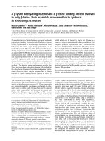
Fig. 2. E-son disrupts the binding between ExoS and 14-3-3. Recom-
binant 14-3-3 (250 n
M
)wasmixedwiththeindicatedamountofpep-
tide for 30 min at 37 °C, prior to incubation for 1 h with 500 n
M
purified GST–ExoS(366–453), followed by GST-bead precipitation,
washing and separation by SDS/PAGE and immunoblotting with
anti-14-3-3 antibodies. The R18 peptide (A) and E-son peptide (B)
were used as competitor peptides.
4924 M. L. Henriksson et al. (Eur. J. Biochem. 269) Ó FEBS 2002
activity of ExoS (see above and [29]). However we have no
indication as to whether ExoS interacts with all, or a subset
of the 14-3-3 family members (Table 1). To explore this
further we used pull-down assays using different GST–ExoS
deletion proteins. In our assay we used the following ExoS
constructs: ExoS(88–453) (see above), which harbours the
ability to bind 14-3-3 and to ADP-ribosylate endogenous
cellular targets both in vivo and in vitro [29]; GST–
ExoS(400–453), a construct that we have shown to bind
to 14-3-3 proteins but lacks both the GAP and the ADP-
ribosylation domains of ExoS [29]. In addition, we have also
made use of ExoS(88–426), which lacks the ability to
interact with 14-3-3 and shows a dramatically reduced
ADP-ribosylation activity both in vivo or in vitro (see above
and [29]). All 14-3-3 isoforms appear to be expressed in
HeLa cells, although 14-3-3 r and s are not as pronounced
as the other isoforms (Fig. 4A). The ExoS(88–453) protein
is able to affinity precipitate all 14-3-3 isoforms from HeLa
lysates (Fig. 4B), as is the ExoS(400–453) protein, although
ExoS(400–453) appears to have a reduced ability to interact
with 14-3-3 r (Fig. 4C) and ExoS(88–453) appears to have
a reduced ability to interact with 14-3-3 s (Fig. 4B). As
expected, GST–ExoS(88–426) does not affinity precipitate
14-3-3 proteins from HeLa whole cell lysates (Fig. 4D).
From this analysis we conclude that the full-length ExoS
protein does indeed have the capacity to interact with all
members of the 14-3-3 family.
ExoS mutants lacking the 14-3-3 binding site do not
modify Ras
in vivo
Most importantly we wished to test the significance of the
in vitro determined amino acid sequence for the interaction
between ExoS and 14-3-3 in a biological system in vivo.We
approached this question through the utilization of two
different assays. Firstly, we exploited the ADP-ribosylation
activity of ExoS towards an important endogenous target –
namely the small G-protein Ras – as a readout [35].
Secondly, we employed a cytotoxicity assay, since the ADP-
ribosylation activity of ExoS mediates a marked change in
cell morphology and has a lethal activity upon translocation
into the host cell in vivo [38,48,49].
In our earlier studies we have shown that Ras is modified
by ExoS expressed and delivered into the eukaryotic cells by
a genetically defined Yersinia pseudotuberculosis strain,
devoid of endogenous toxins, and also by several different
clinically relevant parental P. aeruginosa strains ([35] and
data not shown). Y. pseudotuberculosis strain, YPIII/
pIB251, can express and deliver heterogenous ExoS protein
(YPIII/pTS103) with high efficiency, at levels substantially
greater than parental P. aeruginosa 388 and PAK (MLH
and BH, unpublished results). To reduce the expression and
translocation of ExoS from the bacteria to the cell we
constructed a Y. pseudotuberculosis strain which expresses
and translocates ExoS and various ExoS mutants under the
control of an arabinose inducible promoter located on
pBAD/Myc-His [44]. Thus, by growing the bacteria in the
presence of 0.1% arabinose in the culture media we could
induce a reduced expression and translocation of ExoS into
eukaryotic cells compared to YPIII/pTS103, to increase the
sensitivity of our assay. In this study we measured the
Fig. 3. E-son blocks the modification of Ras by ExoS in vitro.
Recombinant Ha-Ras (10 l
M
) was incubated with 500 n
M
GST (lane
1), 500 n
M
GST–ExoS(88–453) fusion proteins (lanes 2–4) together
with recombinant 14-3-3 (250 n
M
)and1.25m
M
NAD
+
for 10 min at
37 °C. Samples were separated by SDS/PAGE, followed by immuno-
blotting with anti-Ras monoclonal antibody. E-son (100 l
M
;lane3)or
R18 (100 l
M
; lane 4) was preincubated with recombinant 14-3-3 for
30 min at 37 °C prior to addition of NAD
+
, Ha-Ras and GST-fusion
protein.
Fig. 4. Pull down of 14-3-3 isoforms with GST–ExoS mutants. HeLa
cells were harvested, and lysates were subjected to pull-down analysis
with various GST-fusion proteins as indicated. Cell lysates and the
eluates from the GST–ExoS pull downs were analysed for the presence
of 14-3-3 isoforms by immunoblotting. Eluted protein was subjected to
12% (w/v) SDS/PAGE. The separated proteins were then transferred
onto nitrocellulose and immunoblotted with 14-3-3 antisera specific for
the seven isoforms (b, f, s, r, e, g and c)usingaBiometra
TM
slot blot
apparatus. A summary of these antisera is shown in Table 1 and [41].
(A) Whole HeLa cell lysate. HeLa cell lysates were subjected to affinity
precipitation with (B) GST–ExoS(88–453) (C) GST–ExoS(400–453)
(D) GST–ExoS(88–426) and (E) GST-fusion protein. The position of
the 30 kDa marker proteins is indicated.
Ó FEBS 2002 Identification of a 14-3-3 binding motif on ExoS (Eur. J. Biochem. 269) 4925
modification of Ras in vivo as a reflection of ExoS ADP-
ribosyltransferase activity. HeLa cells were infected for
80 min with Y. pseudotuberculosis, which had been induced
to express and translocate ExoS, ExoS(SD) and ExoS(S3).
After stimulation with EGF for 2 min, cells were harvested
and the resultant lysate was separated on SDS/PAGE
followed by immunoblotting with anti-Ras, antiphospho-
Akt and antiphospho-Erk Igs (Fig. 5A). Stimulation of the
uninfected cells with EGF caused the phosphorylation of
both Erk and PKB/Akt (Fig. 5A, compare lanes 1 and 2).
The expected modification of Ras and its subsequent
inability to signal downstream to Erk and Akt was observed
in cells infected with bacteria expressing wild-type ExoS but
not in stimulated uninfected cells or in mock infected cells
(Fig. 5A, compare lane 4 with that of lanes 2 and 3).
However, this inhibition of the activation of Ras, Erk and
Akt was abrogated when the cells were infected with
bacteria producing ExoS mutants unable to interact with
14-3-3, e.g. ExoS(SD) and ExoS(S3) (Fig. 5A, lanes 5 and
6), thus indicating that mutation of the 14-3-3 binding motif
in ExoS results in an inactive ExoS molecule in vivo.
Cell morphology is not affected by an ExoS mutant
lacking the 14-3-3 binding site
It has previously been demonstrated that delivery of ExoS
into HeLa cells results in a change in cell morphology,
concommitent with a disruption of actin microfilaments,
which is followed by cell death, the latter also being
correlated to the ADP-ribosylation activity of ExoS
[35,38,49]. Here we wished to address whether infection of
HeLa cells with Y. pseudotuberculosis strain, YPIII/pIB251,
pregrown in 0.1% arabinose to induce expression of ExoS
mutants lacking the 14-3-3 binding site, could induce a
morphological change of HeLa cells. To achieve this we
infected cells with bacteria, which translocated either the
wild-type ExoS, ExoS(SD), or ExoS(S3).
The extent of cytotoxicity as visualized by a distinct
change in cell morphology in vivo was examined in HeLa
cells taken at 80 min postinfection. As control, the trans-
location efficiencies of ExoS, ExoS(SD) and ExoS(S3)
proteins were compared by immunoblot analysis to ensure
that the effects observed were not caused by decreased
translocation of protein (Fig. 5C).
As expected, intracellular wild-type ExoS induced a rapid
cytotoxic response toward infected HeLa cells, consistent
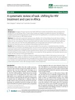
Fig. 5. Morphological and protein effects of ExoS infection on cells
in vivo and on EGF receptor signalling components downstream of Ras.
(A) Upper panel: phosphorylation of PKB/Akt and Erk was examined
in nonstimulated (–) (lane 1) and EGF stimulated (+) (lanes 2–6) cells.
Cells were infected for 80 min as follows: uninfected (lanes 1 and 2),
infected with Y. pseudotuberculosis, YPIII(pIB251) alone (mock
infected, lane 3), or YPIII(pMF384), expressing ExoS wild-type (lane
4), YPIII(pMF419), expressing ExoS(SD) (lane 5) or YPIII(pMF420),
expressing ExoS(S3) (lane 6). Whole cell lysates were subjected to SDS/
PAGE followed by immunoblotting with anti-phosphospecific Erk
(a-P-Erk) and PKB/Akt (a-P-PKB/Akt) Igs, as indicated. Middle
panel: the membrane was stripped and reprobed with anti-pan Erk
antibodies, as indicated. Lower panel: the same membrane was im-
munoblotted with anti-Ras antibodies. (B) Morphological changes
caused by different variants of ExoS preinduced in 0.1% arabinose.
HeLa cells, also in the presence of 0.1% arabinose, were infected with
YPIII(pMF384), expressing wild-type ExoS (3), YPIII(pMF419),
expressing ExoS(SD) (4), or YPIII(pMF420), expressing ExoS(S3) (5).
As controls we used uninfected HeLa cells (1) or HeLa cells infected
with YPIII(pIB251) (2) for mock infection. (C) Translocation of ExoS
variants by Y. pseudotuberculosis into HeLa cells. Bacteria were pre-
induced with 0.1% arabinose, allowed to infect HeLa cells for 80 min
prior to cold washing of the cells and harvest. ExoS was immuno-
precipitated from cell lysates with Sepharose G-coupled ExoS anti-
bodies, and analysed by immunoblotting using ExoS antibodies.
YPIII(pMF384), expressing ExoS (lane 4), YPIII(pMF419), expres-
sing ExoS(SD) (lane 5), or YPIII(pMF420), expressing ExoS(S3) (lane
6). Twenty lg whole cell lysate from ExoS(SD) infected cells (lane 1),
uninfected HeLa cells (lane 2) or HeLa cells infected with
YPIII(pIB251) (lane 3) for mock infection were used as controls.
4926 M. L. Henriksson et al. (Eur. J. Biochem. 269) Ó FEBS 2002
with published reports (Fig. 5B(3) and [38,48]). However,
HeLa cells infected with the bacteria expressing the ExoS
mutants: ExoS(SD), ExoS(S3), or mock infected, were
essentially indistinguishable, indicating no evidence of a
cytotoxic response (Fig. 5B, compare 3 with 1, 2, 4 and 5).
Using our new arabinose inducible strains, which translo-
cate a more physiological level of ExoS, we observe no GAP
domain induced cytotoxicity, however, a cytotoxic effect
can be seen when the ADP-ribosylation domain is complete.
We have also observed that there is no decrease or increase
in the Rho GAP activity between wild-type and SD of ExoS
constructs in vitro (M Aili and B Hallberg, data not shown).
In summary, the lack of Ras modification and inhibition
together with the loss of cytotoxic effect in ExoS mutant for
the 14-3-3 binding motif clearly points to an important
function for the 14-3-3:ExoS interaction in vivo.
DISCUSSION
In this study we have focused our attention on defining the
amino acids on ExoS important for its interaction with
14-3-3 both in vitro and in vivo. This is an important
consideration since the interactions between 14-3-3 and
many cellular proteins have been described to occur in a
phospho-specific manner [21,25]. However, the interaction
between 14-3-3 and ExoS has been reported to occur in a
phosphorylation-independent manner. We have shown that
the ExoS sequence between amino acid 424 and 428
(DALDL) is critical for the interaction between 14-3-3 and
ExoS. Furthermore, sequences flanking this DALDL
sequence also contribute to binding of 14-3-3 proteins.
Further evidence for a specific phosphorylation–independent
interaction between 14-3-3 and ExoS is provided by competi-
tion experiments utilizing the E-son peptide, corresponding
to the amino acids 415–432 of ExoS. Firstly, E-son efficiently
inhibits the formation of the 14-3-3:ExoS complex, with
similar kinetics as seen earlier with the R18 peptide [20]. It
seems that the interaction between 14-3-3 and ExoS is of a
specific and tight-binding nature. Secondly, and more
importantly, E-son is an efficient competitor for the 14-3-3
dependent ExoS ADP-ribosylation activity, as measured by
modification of the small GTPase protein Ras in vitro.
Considering the large number of 14-3-3 isoforms together
with the large number of putative target proteins for 14-3-3
within the cell, we asked which of the 14-3-3 isoforms were
able to interact with ExoS. It has been suggested that homo-/
hetero-dimer combinations of 14-3-3 may confer specificity,
which would mean that there are differences in specificity
towards the 14-3-3 partners [39]. It is also possible that
specific interactions occur as a result of particular subcellular
localizations or transcriptional regulation of isoforms rather
than of differences in their ability to bind to a specific target.
From our analysis we have strong evidence that full-length
ExoS(88–453) has the ability interact with all members of
the 14-3-3 family, although there may be a reduction in
the affinity of ExoS for 14-3-3 s (Fig. 4). Interesting, the
ExoS(400–453) construct, which lacks a GAP domain, did
not appear to interact with 14–3-3 r, in contrast with
ExoS(88–453). This discrepancy may be worthy of future
investigation but has not been approached here.
Comparisons of amino acid sequences in the published
nonphosphorylated interaction partners for 14-3-3 are
shown in Table 2. The identified 14-3-3 binding sequence
in ExoS, DALDL, shows similarities to the artificial
unphosphorylated peptide (R18) isolated from a phage
display library, which contains the sequence WLDLE (10–
14), and has been suggested to bind to the conserved
amphipathic groove of 14-3-3 [33]. It has been proposed that
negatively charged amino acids, such as glutamic and
aspartic acid residues are able to mimic a phosphorylated
serine motif of Raf-1, which would perhaps explain the
binding of 14-3-3 proteins to these motifs [33]. Furthermore,
it has been proposed that the motif RSESEE of the 43 kDa
inositol polyphosphate 5-phosphatase binds 14-3-3 proteins
due to the appearance of multiple negatively charged amino
acids (Table 2 and [24]). Another 14-3-3 binding protein is
GPIb-a, which contains a reported interaction domain [30].
This domain harbours the motif, QDLLSTVS, which shows
a weak resemblance to ExoS and R18. A motif that weakly
resembles this (ELQLRN) can be found at residues 90–112
of the p75NTR-associated cell death executor (NADE),
within the domain, which has recently been reported to
interact with 14-3-3 [23]. CLIC4, an ion channel protein,
also binds 14-3-3 proteins and harbours a sequence which
resembles a negatively charged motif, DEYLN, at residues
152–156 [50].
From this comparison of 14-3-3 nonphosphorylated
motif sequences it is not clear which amino acids within
the motif are important for the interaction between 14-3-3
and its nonphosphorylated ligand. However, it is clear that a
more thorough dissection is needed for the hypothesis that
negatively charged amino acids can substitute phosphoryl-
ated serine/threonine residues. Numerous reports have
clarified the importance of 14-3-3 proteins as a factor that
activates ExoS [27–29,35]. It has been proposed that the
dimeric structure of the 14-3-3 proteins allows it to bind two
ligands simultaneously, as the ligand-binding grooves run in
opposite directions in each monomer of the molecule
[3,21,51]. It is possible that the interaction between 14-3-3
and ExoS creates a conformational change in the structure
of ExoS, thereby changing ExoS from nonactive protein to
an active protein with ADP-ribosylation activity. Thus
14-3-3 proteins may have two functions, firstly as an
activator of ExoS and secondly to localize ExoS to a specific
domain within the cell.
Most importantly in this study we wished to test the
significance of the in vitro determined amino acid sequence
for the interaction between ExoS and 14-3-3 in vivo.We
have shown earlier that Ras (and its deactivation of
downstream targets such as Erk and PKB/Akt), and many
other small GTPases are modified by ExoS, expressed and
translocated into the eukaryotic cells by a genetically
defined Y. pseudotuberculosis strain and also by several
different Pseudomonas aeruginosa strains [34,35]. The Yer-
sinia strain expresses and translocates ExoS protein with
high efficiency, at levels greater than that observed in strains
such as P. aeruginosa 388 and PAK. For this reason we
have engineered a Yersinia strain to express wild-type ExoS
and two different mutants of ExoS under the control of an
arabinose-inducible promoter so that considerably lower
levels of ExoS proteins were translocated into infected cells.
We observed the expected phosphorylation of both PKB/
Akt and of Erk 1/2 after stimulation by EGF in HeLa cells.
As reported previously, infection of HeLa cells for 80 min
with bacteria expressing the wild-type ExoS caused the
ADP-ribosylation of Ras and inhibited the EGF mediated
Ó FEBS 2002 Identification of a 14-3-3 binding motif on ExoS (Eur. J. Biochem. 269) 4927
phosphorylation of both Erk and PKB/Akt. This effect was
not seen upon infection with bacteria expressing the ExoS
S3 constructs where the DALDL sequence has been
mutated or with the substitution/deletion ExoS(SD). These
results suggest that an ExoS construct lacking the DALDL
sequence, which we suggest to be the binding motif in ExoS
for 14-3-3, does not abrogate the activation of Ras, Erk or
PKB/Akt upon stimulation with EGF and thus is nonfunc-
tional in vivo. In addition, no modification of endogenous
Ras can be observed if 14-3-3 has lost its ability to interact
with ExoS, as is the case with ExoS(SD).
ExoS is known to cause infected cells to round up and
detach from the underlying surface, which correlates with
disruption of the actin microfilament structure within the
cell [38,48]. We observed that an ExoS protein lacking the
residues important for 14-3-3 binding motif is unable to
elicit the changes in cell morphology routinely observed
with wild-type ExoS. Thus, the 14-3-3 binding motif of
ExoS ) DALDL ) appears to be necessary for both the
ADP-ribosylation activity and the cytotoxic action of ExoS
in vivo.
In this report we have firstly identified the residues on
ExoS responsible for its specific interaction with 14-3-3,
both in vitro and in vivo. Secondly, we have shown that an
amino acid peptide derived from ExoS, containing the
important 14-3-3 interaction site, effectively competes out
the interaction between ExoS and 14-3-3. Thirdly, compe-
tition with this peptide blocks ExoS modification of Ras in
our in vitro Ras modification assay. Fourthly, we show that
the full-length ExoS proteins interact with all isoforms of the
14-3-3 family. Finally, in vivo an ExoS protein lacking the
14-3-3 binding site is unable to ADP ribosylate cytoplas-
matic proteins, e.g. Ras, and is impaired in its capacity to
change the morphology of infected cells.
ACKNOWLEDGEMENTS
Financial support for this work was from the MRC, UK (A. A), the
Wellcome trust (A. A), Swedish Cancer Society, Riksfo
¨
rbundet Cystisk
Fibros Forskningsfond, Sven Jerring foundation, Kungliga Vetenskap-
sakademin, Elsa and Folke Sahlbergs minnesfond, and the Swedish
Natural Science Council.
REFERENCES
1. Aitken, A. (1995) 14-3-3 proteins on the MAP. Trends Biochem.
Sci. 20, 95–97.
2. Aitken, A., Jones, D., Soneji, Y. & Howell, S. (1995) 14-3-3 pro-
teins: biological function and domain structure, Biochem. Soc.
Trans. 23, 605–611.
3. Fu, H., Subramanian, R.R. & Masters, S.C. (2000) 14-3-3 pro-
teins: structure, function, and regulation. Annu. Rev. Pharmacol
Toxicol. 40, 617–647.
4. Su, T.T., Parry, D.H., Donahoe, B., Chien, C.T., O’Farrell, P.H.
& Purdy, A. (2001) Cell cycle roles for two 14-3-3 proteins during
Drosophila development. J. Cell Sci. 114, 3445–3454.
5. Broadie, K., Rushton, E., Skoulakis, E.M. & Davis, R.L. (1997)
Leonardo, a Drosophila 14-3-3 protein involved in learning, regu-
lates presynaptic function. Neuron 19, 391–402.
6. Ford, J.C., al-Khodairy, F., Fotou, E., Sheldrick, K.S., Griffiths,
D.J. & Carr, A.M. (1994) 14-3-3 protein homologs required for the
DNA damage checkpoint in fission yeast. Science 265, 533–535.
7. van Heusden, G.P., Griffiths, D.J., Ford, J.C., Chin, A.W.T.F.,
Schrader, P.A., Carr, A.M. & Steensma, H.Y. (1995) The 14-3-3
proteins encoded by the BMH1 and BMH2 genes are essential in
the yeast Saccharomyces cerevisiae and can be replaced by a plant
homologue. Eur. J. Biochem. 229, 45–53.
8. Fantl, W.J., Muslin, A.J., Kikuchi, A., Martin, J.A., MacNicol,
A.M., Gross, R.W. & Williams, L.T. (1994) Activation of Raf-1
by 14-3-3 proteins. Nature 371, 612–614.
9. Xing, H., Kornfeld, K. & Muslin, A.J. (1997) The protein kinase
KSR interacts with 14-3-3 protein and Raf. Curr. Biol. 7, 294–300.
10. Zhang, L., Chen, J. & Fu, H. (1999) Suppression of apoptosis
signal-regulating kinase 1-induced cell death by 14-3-3 proteins.
Proc. Natl Acad. Sci. USA 96, 8511–8515.
11. Fanger, G.R., Widmann, C., Porter, A.C., Sather, S., Johnson,
G.L. & Vaillancourt, R.R. (1998) 14-3-3 proteins interact with
specific MEK kinases. J. Biol. Chem. 273, 3476–3483.
12. Reuther, G.W., Fu, H., Cripe, L.D., Collier, R.J. & Pendergast,
A.M. (1994) Association of the protein kinases c-Bcr and Bcr-Abl
with proteins of the 14-3-3 family. Science 266, 129–133.
13. Hausser, A., Storz, P., Link, G., Stoll, H., Liu, Y.C., Altman, A.,
Pfizenmaier, K. & Johannes, F.J. (1999) Protein kinase C mu is
negatively regulated by 14-3-3 signal transduction proteins. J. Biol.
Chem. 274, 9258–9264.
14. Zha, J., Harada, H., Yang, E., Jockel, J. & Korsmeyer, S.J. (1996)
Serine phosphorylation of death agonist BAD in response to
survival factor results in binding to 14-3-3 not BCL-X (L). Cell
87, 619–628.
15. Brunet, A., Bonni, A., Zigmond, M.J., Lin, M.Z., Juo, P., Hu,
L.S., Anderson, M.J., Arden, K.C., Blenis, J. & Greenberg, M.E.
(1999) Akt promotes cell survival by phosphorylating and
inhibiting a Forkhead transcription factor. Cell 96, 857–868.
16. Liu, D., Bienkowska, J., Petosa, C., Collier, R.J., Fu, H. &
Liddington, R. (1995) Crystal structure of the zeta isoform of the
14-3-3 protein. Nature 376, 191–194.
17. Xiao, B., Smerdon, S.J., Jones, D.H., Dodson, G.G., Soneji, Y.,
Aitken, A. & Gamblin, S.J. (1995) Structure of a 14-3-3 protein
and implications for coordination of multiple signalling pathways.
Nature 376, 188–191.
18. Wu,K.,Lu,G.,Sehnke,P.&Ferl,R.J.(1997)Theheterologous
interactions among plant 14-3-3 proteins and identification of
regions that are important for dimerization. Arch. Biochem. Bio-
phys. 339,2–8.
19. Jones,D.H.,Martin,H.,Madrazo,J.,Robinson,K.A.,Nielsen,
P.,Roseboom,P.H.,Patel,Y.,Howell,S.A.&Aitken,A.(1995)
Expression and structural analysis of 14-3-3 proteins. J. Mol. Biol.
245, 375–384.
20. Wang, H., Zhang, L., Liddington, R. & Fu, H. (1998) Mutations
in the hydrophobic surface of an amphipathic groove of 14-3-3zeta
disrupt its interaction with Raf-1 kinase. J. Biol. Chem. 273,
16297–16304.
21. Yaffe, M.B., Rittinger, K., Volinia, S., Caron, P.R., Aitken, A.,
Leffers, H., Gamblin, S.J., Smerdon, S.J. & Cantley, L.C. (1997)
The structural basis for 14-3-3: phosphopeptide binding specifi-
city. Cell. 91, 961–971.
22. Tzivion, G. & Avruch, J. (2002) 14-3-3 proteins: active cofactors in
cellular regulation by serine/threonine phosphorylation. J. Biol.
Chem. 277, 3061–3064.
23. Kimura, M.T., Irie, S., Shoji-Hoshino, S., Mukai, J., Nadano, D.,
Oshimura, M. & Sato, T.A. (2001) 14-3-3 is involved in p75
neurotrophin receptor-mediated signal transduction. J. Biol.
Chem. 276, 17291–17300.
24. Campbell, J.K., Gurung, R., Romero, S., Speed, C.J., Andrews,
R.K., Berndt, M.C. & Mitchell, C.A. (1997) Activation of the 43
kDa inositol polyphosphate 5-phosphatase by 14-3-3zeta. Bio-
chemistry 36, 15363–15370.
25. Muslin, A.J. & Xing, H. (2000) 14-3-3 proteins: regulation of
subcellular localization by molecular interference. Cell Signal. 12,
703–709.
4928 M. L. Henriksson et al. (Eur. J. Biochem. 269) Ó FEBS 2002
26. Seimiya, H., Sawada, H., Muramatsu, Y., Shimizu, M., Ohko, K.,
Yamane, K. & Tsuruo, T. (2000) Involvement of 14-3-3 proteins in
nuclear localization of telomerase. EMBO J. 19, 2652–2661.
27. Masters,S.C.,Pederson,K.J.,Zhang,L.,Barbieri,J.T.&Fu,H.
(1999) Interaction of 14-3-3 with a nonphosphorylated protein
ligand, exoenzyme S of Pseudomonas aeruginosa. Biochemistry 38,
5216–5221.
28. Fu, H., Coburn, J. & Collier, R.J. (1993) The eukaryotic host
factor that activates exoenzyme S of Pseudomonas aeruginosa is a
member of the 14-3-3 protein family. Proc. Natl. Acad. Sci. USA
90, 2320–2324.
29. Henriksson, M.L., Troller, U. & Hallberg, B. (2000) 14-3-3 pro-
teins are required for the inhibition of Ras by exoenzyme S. Bio-
chem. J. 349, 697–701.
30. Gu, M.&., X. (1998) A novel ligand-binding site in the zeta-form
14-3-3 protein recognizing the platelet glycoprotein Ibalpha and
distinct from the c-Raf-binding site. J. Biol. Chem. 273, 33465–
33471.
31. Calverley, D.C., Kavanagh, T.J. & Roth, G.J. (1998) Human
signaling protein 14-3-3zeta interacts with platelet glycoprotein Ib
subunits Ibalpha and Ibbeta. Blood 91, 1295–1303.
32. Andrews, R.K., Harris, S.J., McNally, T. & Berndt, M.C. (1998)
Binding of purified 14-3-3 zeta signaling protein to discrete amino
acid sequences within the cytoplasmic domain of the platelet
membrane glycoprotein Ib-IX-V complex. Biochemistry 37, 638–
647.
33. Petosa, C., Masters, S.C., Bankston, L.A., Pohl, J., Wang, B.,
Fu, H. & Liddington, R.C. (1998) 14-3-3zeta binds a phos-
phorylated Raf peptide and an unphosphorylated peptide via
its conserved amphipathic groove. J. Biol. Chem. 273, 16305–
16310.
34. Henriksson, M.L., Sundin, C., Jansson, A.L., Forsberg, A.,
Palmer, R.H. & Hallberg, B. (2002) Exosenzyme S show selective
ADP-ribosylation and GAP activities towards small GTPases
in vivo. Biochem. J.,inpress.
35. Henriksson, M.L., Rosqvist, R., Telepnev, M., Wolf-Watz, H. &
Hallberg, B. (2000) Ras effector pathway activation by epidermal
growthfactorisinhibitedinvivobyexoenzymeSADP-ribosy-
lation of Ras. Biochem. J. 347, 217–222.
36. McGuffie, E.M., Frank, D.W., Vincent, T.S. & Olson, J.C. (1998)
Modification of Ras in eukaryotic cells by Pseudomonas aerugi-
nosa exoenzyme S. Infect Immun. 66, 2607–2613.
37. Goehring, U.M., Schmidt, G., Pederson, K.J., Aktories, K. &
Barbieri, J.T. (1999) The N-terminal domain of Pseudomonas
aeruginosa exoenzyme S is a GTPase- activating protein for Rho
GTPases. J. Biol. Chem. 274, 36369–36372.
38. Frithz-Lindsten, E.Y., Rosqvist, R. & Forsberg, A. (1997) Intra-
cellular targeting of exoenzyme S of Pseudomonas aeruginosa via
type III-dependent translocation induces phagocytosis resistance,
cytotoxicity and disruption of actin microfilaments. Mol. Micro-
biol. 25, 1125–1139.
39. Dubois, T., Rommel, C., Howell, S., Steinhussen, U., Soneji, Y.,
Morrice, N., Moelling, K. & Aitken, A. (1997) 14-3-3 is phos-
phorylated by casein kinase I on residue 233. Phosphorylation at
this site in vivo regulates Raf/14-3-3 interaction. J. Biol. Chem. 272,
28882–28888.
40. Rodriguez-Viciana, P., Warne, P.H., Khwaja, A., Marte, B.M.,
Pappin,D.,Das,P.,Waterfield,M.D.,Ridley,A.&Downward,J.
(1997) Role of phosphoinositide 3-OH kinase in cell transforma-
tion and control of the actin cytoskeleton by Ras. Cell 89, 457–
467.
41. Wiltfang, J., Otto, M., Baxter, H.C., Bodemer, M., Steinacker, P.,
Bahn, E., Zerr, I., Kornhuber, J., Kretzschmar, H.A., Poser, S.,
Ruther, E. & Aitken, A. (1999) Isoform pattern of 14-3-3 proteins
in the cerebrospinal fluid of patients with Creutzfeldt-Jakob dis-
ease. J. Neurochem. 73, 2485–2490.
42. Yahr, T.L., Goranson, J. & Frank, D.W. (1996) Exoenzyme S of
Pseudomonas aeruginosa is secreted by a type III pathway. Mol.
Microbiol. 22, 991–1003.
43. Francis, M.S. & Wolf-Watz, H. (1998) YopD of Yersinia pseudo-
tuberculosis is translocated into the cytosol of HeLa epithelial cells:
evidence of a structural domain necessary for translocation. Mol.
Microbiol. 29, 799–813.
44. Francis, M.S., Aili, M., Wiklund, M.L. & Wolf-Watz, H. (2000) A
study of the YopD–lcrH interaction from Yersinia pseudotuber-
culosis reveals a role for hydrophobic residues within the amphi-
pathic domain of YopD. Mol. Microbiol. 38, 85–102.
45. Francis, M.S., Lloyd, S.A. & Wolf-Watz, H. (2001) The type III
secretion chaperone LcrH co-operates with YopD to establish a
negative, regulatory loop for control of Yop synthesis in Yersinia
pseudotuberculosis. Mol. Microbiol. 42, 1075–1093.
46. Masters, S.C. & Fu, H. (2001) 14-3-3 proteins mediate an essential
anti-apoptotic signal. J. Biol. Chem. 276, 45193–45200.
47. Muslin, A.J., Tanner, J.W., Allen, P.M. & Shaw, A.S. (1996)
Interaction of 14-3-3 with signaling proteins is mediated by the
recognition of phosphoserine. Cell 84, 889–897.
48. Sundin, C., Henriksson, M.L., Hallberg, B., Forsberg, A. &
Frithz-Lindsten, E. (2001) Exoenzyme T of Pseudomonas aerugi-
nosa elicits cytotoxicity without interfering with Ras signal trans-
duction. Cell. Microbiol. 3, 237–246.
49. Kaufman,M.R.,Jia,J.,Zeng,L.,Ha,U.,Chow,M.&Jin,S.
(2000) Pseudomonas aeruginosa mediated apoptosis requires the
ADP-ribosylating activity of exoS. Microbiology 146, 2531–2541.
50. Suginta, W., Karoulias, N., Aitken, A. & Ashley, R.H. (2001)
Chloride intracellular channel protein CLIC4 (p64H1) binds
directly to brain dynamin I in a complex containing actin, tubulin
and 14-3-3 isoforms. Biochem. J. 359, 55–64.
51. Rittinger, K., Budman, J., Xu, J., Volinia, S., Cantley, L.C.,
Smerdon, S.J., Gamblin, S.J. & Yaffe, M.B. (1999) Structural
analysis of 14-3-3 phosphopeptide complexes identifies a dual role
for the nuclear export signal of 14-3-3 in ligand binding. Mol. Cell
4, 153–166.
Ó FEBS 2002 Identification of a 14-3-3 binding motif on ExoS (Eur. J. Biochem. 269) 4929