Báo cáo khoa học: Domains of ERRcthat mediate homodimerization and interaction with factors stimulating DNA binding potx
Bạn đang xem bản rút gọn của tài liệu. Xem và tải ngay bản đầy đủ của tài liệu tại đây (318.71 KB, 12 trang )
Domains of ERRc that mediate homodimerization and interaction
with factors stimulating DNA binding
Moritz Hentschke, Ute Su¨ sens and Uwe Borgmeyer
Zentrum fu
¨
r Molekulare Neurobiologie Hamburg (ZMNH), Universita
¨
t Hamburg, Germany
The estrogen receptor-related receptor c (ERRc/ERR3/
NR3B3) is an orphan member of the nuclear receptor
superfamily closely related to the estrogen receptors. To
explore the DNA binding characteristics, the protein–DNA
interaction was studied in electrophoretic mobility shift
assays (EMSAs). In vitro translated ERRc binds as a
homodimer to direct repeats (DR) without spacing of the
nuclear receptor half-site 5¢-AGGTCA-3¢ (DR-0), to exten-
ded half-sites, and to the inverted estrogen response element.
Using ERRc deletion constructs, binding was found to be
dependent on the presence of sequences in the ligand binding
domain (LBD). A far-Western analysis revealed that ERRc
forms dimers even in the absence of DNA. Two elements,
located in the hinge region and in the LBD, respectively, are
necessary for DNA-independent dimerization. DNA bind-
ing of bacterial expressed ERRc requires additional factors
present in the serum and in cellular extracts. Fusion proteins
of the germ cell nuclear factor (GCNF/NR6A1) with ERRc
showed that the characteristic feature to be stimulated by
additional factors can be transferred to a heterologous
protein. The stimulating activity was further characterized
and its target sequence narrowed down to a small element in
the hinge region.
Keywords: orphan nuclear receptor; transcription factor;
estrogen receptor-related; DNA binding; dimerization.
The nuclear receptors (NR) comprise a family of transcrip-
tional regulators involved in a wide variety of biological
processes, such as embryonic development, differentiation,
and homeostasis. This family includes ligand-dependent
transcription factors for steroid hormones, estrogens, thy-
roid hormones, retinoids, vitamin D, and other hydropho-
bic compounds [1]. In addition, several members are orphan
receptors for which ligands have yet to be identified [2,3].
Nuclear receptors exhibit a modular structure with func-
tionally separable domains (A/B, C, D and EF) [4]. The
most highly conserved region of these proteins is the DNA-
binding domain (DBD, C-domain), which contains two
zinc-binding modules that fold to form a single structural
domain [5]. They confer binding to a core recognition motif,
or a NR half-site, resembling the sequence 5¢-AGGTCA-3¢.
Most receptors bind as homodimers or heterodimers
to palindromes or to direct-repeated sequences of the
AGGTCA motif [6]. However, a subset of orphan receptors
bind an extended NR half-site with the core sequence
5¢-TCAAGGTCA-3¢as monomers. The C-terminal exten-
sion (CTE) of the DBD contributes to the specific interac-
tion by base specific contacts in the minor grove of the
DNA. The C-terminal domain (EF) has an intrinsic ligand-
binding function, a ligand-dependent transactivation func-
tion (AF-2), and a dimerization interface. The variable,
N-terminal domain (A/B) is important in transcriptional
regulation of some nuclear receptors, and a short variable
domain (D) with a nuclear localization motif is thought to
be the hinge between C and EF.
Based on the evolution of the conserved DBD and of
the ligand-binding domain (LBD), the superfamily has
been divided into six subfamilies and 26 groups of
receptors [7]. Subfamily 3 comprises three groups, the
estrogen receptors ERa and ERb [8,9], the estrogen
receptor-related receptors (ERRs) and one receptor each
for the three steroid hormone classes: glucocorticoids,
mineralocorticoids, progestin, and androgen [10]. ERRa
and ERRb were initially isolated because of their homo-
logy to ERa [11]. Although structurally related, no
natural ligand is known for the ERRs. Both receptors
bind to extended NR half-sites and to classical estrogen
receptor response elements (EREs), inverted repeats of the
NR half-site separated by three base pairs [12–14]. Both
types of sequence element function as response elements
of ERa as well, suggesting a functional relationship
between these receptors [15]. Putative common target
genes of ERs and ERRs, such as lactoferrin, aromatase
and osteopontin [15–19], and common coactivators [14]
further strengthen the view of a functional interference of
these receptors. Although monomeric binding of ERRa
has been suggested [12,20], homodimer binding was
demonstrated by cotranslation of ERRa and truncated
ERRa, generating an intermediate band in electrophoretic
mobility shift assay (EMSA) [13,15,19].
Transfection studies revealed ERR-dependent activation
of promoters with EREs or extended half-sites. Activation
of the reporter genes occurred in the absence of any exogen-
ous added ligand. Interestingly, studies by Vannacker et al.
Correspondence to U. Borgmeyer, ZMNH, Universita
¨
tsklinikum
Eppendorf, Martinistrasse 52, 20246 Hamburg, Germany.
Fax: + 49 40 42803 5101, Tel.: + 49 40 42803 6622,
E-mail:
Abbreviations: CTE, C-terminal extension; DBD, DNA binding
domain; DR, direct repeat; ERR, estrogen receptor-related receptor;
EMSA, electrophoretic mobility shift assay; ERE, estrogen response
element; GCNF, germ cell nuclear factor; GST, glutathione
S-transferase; LBD, ligand-binding domain, NR, nuclear receptor.
Note: a web site is available from
(Received 25 April 2002, revised 26 June 2002,
accepted 10 July 2002)
Eur. J. Biochem. 269, 4086–4097 (2002) Ó FEBS 2002 doi:10.1046/j.1432-1033.2002.03102.x
show the requirement of a serum factor for transcriptional
activation [13].
By several means, a novel nuclear receptor was isolated
from human and mouse cDNA libraries [21–24]. Because
sequence comparisons reveal high homology to ERRa and
ERRb, the receptor was given the systematic name NR3B3,
and the trivial names ERRc and ERR3. ERRc is much
more closely related to ERRb than to ERRa. However, the
DBDs of all ERRs are more than 90% conserved. In the
adult mouse, ERRc is highly expressed in heart, brain,
kidney and skeletal muscle [25]. We have previously
described its spatial pattern of expression during embryonic
development and in the mature mouse brain [26]. In the
adult brain, high transcript levels were observed in the
isocortex, the olfactory system, cranial nerve nuclei, and
major parts of the coordination centers, a pattern that is
established in the embryo. During development expression
is prominent in the nervous system [27]. The gene is
preferentially transcribed in already differentiating areas of
the nervous system establishing many features of the adult
expression pattern. This expression pattern suggests func-
tions of the receptor not shared with its two close
homologues. Different isoforms have been described in
mouse and human, differing in the length of their
N-terminal domains [24,25,28]. Binding to an extended
NR half-site has been performed with in vitro translated
ERRc2 [28]. The authors conclude that ERRc2 binds as a
monomer to extended half-sites. Hong et al. (1999) dem-
onstrated ERRc-dependent activation of reporter genes
controlled by estrogen response elements in the absence of
any added ligand. An AF-2 activation domain bound by the
coactivator GRIP1 primarily mediates the transcriptional
activation [24]. Recent studies demonstrated binding and
antagonistic function of the synthetic estrogen receptor
modulators 4-hydroxytamoxifen to ERRc [29,30]. The
crystal structure of the human ERRc LBD bound to the
SRC-coactivator peptide has been resolved. In the crystal,
the LBD adopts a transcriptionally active conformation
suggesting that putative steroidal ligands would function as
antagonist [31].
Here, we describe the binding characteristics of mERRc2.
The receptor binds to DR-0, extended half-sites, and to
classical EREs. Interestingly, efficient binding depends on
additional factors present in the serum and in cellular
extracts. We present a sequence in the hinge region as the
target site of these activities. ERRc binds as dimer to
DNA. Dimerization depends on sequence elements, pre-
sent in the DBD, in the hinge region and in the LBD. The
C-terminal dimerization motifs function independent of
DNA.
MATERIALS AND METHODS
Plasmid constructs
Full-length ERRc2 cDNA was amplified by PCR with Pfu
polymerase (Stratagene) from a mouse embryonic day 15
brain cDNA. The forward primer, c2-start (5¢-AAAG
CTTGCCGCCACC
ATGGATTCGGTAGAACTTTGC
CT-3¢), includes HindIII and NcoI restriction sites, a Kozak
consensus site [32], the translational start codon of ERRc2
(underlined) and additional 20 nucleotides of the coding
sequence. The reverse primer, c2-stop (5¢-GGAT
CC
TCAGACCTTGGCCTCCAGCATTTC-3¢), includes
a BamHI restriction site, the translational stop codon
(underlined) and 21 nucleotides complementary to the
coding sequence. The product was cloned into the SrfIsite
of pCMV-Script vector (Stratagene) to generate pCMV-
ERRc2. The correct integration was verified by sequencing.
The SalI linearized plasmid pCMV-ERRc2servedasa
template to generate epitope-tagged and truncated con-
structs of ERRc. All products were cloned into the
pGEM-T Easy vector for sequence verification. To generate
in vitro translation plasmids, the inserts were isolated and
cloned into pSPUTK vector (Stratagene) through either
NcoIandSalI, or NcoIandBamHI sites. Inserts of clones
with internal NcoIorBamHI sites were isolated by partial
digestion. For the N-terminal truncation, DN-ERRc,
c2-stop and the forward primer DN(5¢-ACC
ATGGTAG
ATCCCCAGACCAAGTGTGAA-3¢)wereusedinthe
amplification. It includes an NcoI restriction site, a new
translational start codon (underlined) and a 21-nucleotide
sequence coding for amino acids 111–117 of ERRc2(all
numbers according to GenBank accession number
AF117254). For the C-terminal truncations the start primer
c2-VSVG-start (5¢-ACC
ATGGAGTACACCGACATCG
AGATGAACAGGCTGGGCAAGGATTCGGTAGAA
CTTTGCCTGCCT-3¢ that includes a translational start
codon (underlined), a sequence coding for an epitope of the
vesicular stomatitis virus glycoprotein (VSV-G) and the
reverse primers: D10 5¢-AGTCGAC
TCAAAGTTTGT
GCATGGGCACTTTGCC-3¢ (ERRc-448), D50 5¢-AGT
CGAC
TCACATGTGCTGGCCAGCCTCGTAATC-3¢
(ERRc-408), D82 5¢-AGTCGAC
TCAATTAGCAAGAG
CTATTGCTTT-3¢ (ERRc-376), D127 5¢-AGTCGA
C
TCATATATAATCGTCTGCATAGAC-3¢ (ERRc-
331), D173 5¢-AGTCGAC
TCAATGTTTTGCCCATCCA
ATGAT-3¢ (ERRc-285), D240 5¢-AGTCGAC
TCAGTTC
TCAGCATCTATTCTGCGCTT-3¢ (ERRc-218), were
used in the amplification, respectively. The reverse primers,
named according to the extent of the resulting protein
truncation, contain SalI restriction sites, translational stop
codons (underlined) and 21–24 nucleotides complementary
to the ERRc coding sequence. The position of the
C-terminal amino acid of proteins derived from the
respective products is given in parentheses. Fusion proteins
GE-1, GE-2, and GE-3 of N-terminal parts of murine germ
cell nuclear factor (mGCNF) and C-terminal parts of
ERRc were generated by in vitro translation. The respective
NcoI/SalI- and SalI/BamHI-fragments were generated by
PCR and cloned in a double ligation reaction in the
pSPUTK vector, digested with NcoIandBamHI.
The following oligonucleotides were used:
GCNF-start 5¢-ACCATGGAGCGGGACGAACGGCC
ACCTAGC-3¢, c2-stop, G2r 5¢-A
GTCGACTTCTTCT
TCTGATATCTGGACTGG-3¢(GCNF 1–167), E2f 5¢-
A
GTCGACAGAATAGATGCTGAGAACAGCCCA-3¢
(ERRc 213–458), G3r 5¢-A
GTCGACCAGACTGTAG
GACTGAGGGTCCAG-3¢(GCNF 1–271), and E3f 5¢-
A
GTCGACCATTTGTTGGTGGCTGAACCAGAG-3¢
(ERRc 240–458). The SalI restriction sites are underlined
and the respective amino acids encoded by the amplified
fragment are given in parentheses.
For GE-1, NcoI/AflII- and AflII/BamHI-fragments were
generated and cloned into pSPUTK. The oligonucleotides
GCNF-start, c2-stop, G1r 5¢-A
CTTAAGCATGCCCA
Ó FEBS 2002 DNA binding of ERRc (Eur. J. Biochem. 269) 4087
TCTGGAGACACTTGAG-3¢ (GCNF 1–140), and E1f
5¢-A
CTTAAGGAAGGGGTCCGTCTTGACAGAGTG-3¢
(ERRc 196–458) were used for the amplification. The AflII
restriction sites are underlined. A schematic view of the
constructs is given in Fig. 4A.
Generation of antibodies
The peptide AcNH
2
-YDDCSSTIVEDPQTK-CONH
2
rep-
resenting amino acids 101–115 of ERRc2 was synthesized
and cross-linked via the C-terminal lysine to keyhole limpet
hemocyanin. Eurogentec performed all procedures, inclu-
ding the immunization of rabbits. The serum of the second
boost was used.
Bacterial expression of ERRc
The NcoI–HindIII insert of pCMV-ERRc2 containing the
whole coding sequence was cloned into pGEX-KG expres-
sion vector (Amersham Biosciences). The resulting plasmid
pGEX-KG-ERRc2 coding for a fusion protein of glutathi-
one S-transferase (GST) and ERRc was transformed into
Escherichia coli BL21. Cells were grown in 500 mL Lennox
L broth base containing 200 lgÆmL
)1
ampicillin to an D
600
of 0.8–1.0. Subsequently, cells were induced under constant
shaking with 1 m
M
isopropyl thio-b-
D
-galctoside for 3 h at
37 °C. The cells were harvested and resuspended in 10 mL
ice cold phosphate-buffered saline (NaCl/P
i
), lysed by
sonication and centrifuged at 4 °Cwith20000g for
15 min. The GST fusion protein was purified from the
supernatant using glutathione–Sepharose 4B beads accord-
ing to the manufacturer’s instructions (Amersham
Biosciences).
Electrophoretic mobility shift assays (EMSAs)
Single-stranded oligonucleotides were purchased (Meta-
bion) and annealed in 10 m
M
Tris/HCl, pH 7.5, 60 m
M
NaClandstoredat)20 °C. Double-stranded oligonucleo-
tides had 5¢ overhangs of four nucleotides on both strands.
For EMSAs, double-stranded oligonucleotides were labeled
using Klenow polymerase (Roche) with [a-
32
P]dATP
(Amersham Biosciences) and unincorporated nucleotides
were removed by gel filtration on Sephadex G25 spin
columns (Roche). Labeled oligonucleotides were stored at
4 °Cin10m
M
Tris/HCl, pH 7.5, 1 m
M
EDTA, 60 m
M
NaCl.
In vitro translation was performed using the SP6-
polymerase TNT Reticulocyte Lysate System (Promega)
according to the manufacturer’s instructions and stored at
)70 °C. Binding reactions were performed in a total volume
of 12 lLconsistingof20m
M
Hepes pH 7.4, 80 m
M
NaCl,
20 m
M
KCl, 2 m
M
dithiothreitol, 1 lg Cot-1 DNA and, if
not stated otherwise, 1 lL of reticulocyte lysate or cellular
extract. Complete Protease Inhibitor was added according
to the manufacturer specifications (Roche). Binding reac-
tions were incubated for 30 min followed by the addition of
2 lL of the labeled oligonucleotides and incubated further
for 30 min at room temperature. For the supershift and for
the analysis of the serum activity, 2 lL of serum diluted in
NaCl/P
i
was added before loading and incubated for an
additional 30 min. Complexes were resolved by nondena-
turing PAGE in 0.5 · Tris/borate/EDTA (45 m
M
Tris base,
45 m
M
boric acid, 1 m
M
EDTA) at 4 °Cat20VÆcm
)1
for
4 h. The gels were dried, analyzed with the Fujix BAS 2000
bioimaging system by the
TINA
TM
software (Raytest) and
exposed to BioMax MR film (Kodak).
Oligonucleotides used were as follows: SIS 5¢-ctaca
gaAGGTCAAGGTCAaatgaag-3¢; LFRE 5¢-gttgcaCCT
TCAAGGTCAtctgaac-3¢;DR-05¢-agcttcAGGTCAAGG
TCAgagagct-3¢;DR-0A5¢-agcttcACCTCAAGGTCAga
gagct-3¢;ERE5¢-gttcAGGTCActgTGACCTgacctg-3¢.
Sequences corresponding to half-sites are capitalized. The
sequence of one strand is shown after the fill-in reaction.
Serum treatment
The serum was stored at )20 °C. Aliquots were incubated
for 20 min at 22 °C, 65 °C, 70 °C, 75 °C, 80 °C, and 95 °C,
respectively. Samples were centrifuged for 10 min at
13 000 g and the supernatant was used in EMSA.
Treatment with 4 volumes of organic solvents was for
20 min at room temperature. Samples were centrifuged for
10 min at 13 000 g. The supernatant of the precipitation
with ethanol, methanol, isopropanol, and acetone was dried
in a speed-vac concentrator and suspended in 0.5 volumes
NaCl/P
i
. The precipitates were dried at room temperature
and resuspended in 1 volume of NaCl/P
i
. The organic phase
of the extraction with ethanol and with chloroform were
dried and resuspended in 0.5 volumes of NaCl/P
i
. Charcoal
treatment was overnight.
Cell lysates
Cells were grown to approximately 80% confluence on
92 mm tissue culture dishes, washed twice with NaCl/P
i
,
and harvested in 1.5 mL NaCl/P
i
by gently scraping with a
rubber policeman. Cells were centrifuged with 300 g and the
pellet was resuspended in lysis buffer (3 lLÆmg
)1
,20m
M
Tris/HCl, 100 m
M
NaCl). Cells were lysed by freeze–thaw,
centrifuged (16 000 g) and the supernatant was stored at
)80 °C.
Far-Western based protein–protein interaction
For the far-Western overlay binding assay 3 lLof
reticulocyte lysate programmed to synthesize the indicated
proteins was subjected to SDS/PAGE using 10% acryl-
amide and transferred by semidry electroblotting to
poly(vinylidene difluoride) (PVDF) membranes. Further
incubations were carried out on an orbital shaker. The
proteins were partially renatured by first incubating the
membrane in 6
M
guanidine/HCl, which was stepwise
diluted in buffer A (25 m
M
Hepes, pH 7.5, 25 m
M
NaCl,
5m
M
MgCl
2
,1m
M
dithiothreitol) to 0.187
M
.After
renaturation, the membrane was incubated at room
temperature for at least 2 h in buffer A with 0.05%
NP40 and 5% milk powder. The membranes were then
overlaid overnight at 4 °CwithERRc,synthesizedby
in vitro translation in the presence of [
35
S]methionine
(>1000 CiÆmmol
)1
; Amersham Biosciences) and diluted
1 : 400 in buffer B (20 m
M
Hepes, pH 7.5, 75 m
M
KCl,
0.1 m
M
EDTA, 2.5 m
M
MgCl
2
,1m
M
dithiothreitol, 1%
milk powder, 0.05% NP40). The membranes were then
washed three times in buffer B, each wash lasting at least
10 min. Signals were detected with a Fujix BAS 2000
4088 M. Hentschke et al. (Eur. J. Biochem. 269) Ó FEBS 2002
bioimaging analyzer and autoradiographed with Kodak
BioMax MR film.
RESULTS
Increased DNA binding of
in vitro
generated ERRc
in the presence of serum
In order to analyze the DNA interaction of ERRc, the full-
length cDNA coding for ERRc2 was cloned into an in vitro
translation vector and a rabbit antiserum was generated.
The antiserum, aERR, was directed against the peptide
AcNH
2
-YDDCSSTIVEDPQTK-CONH
2
, encoded by the
exon that also codes for the amino acids of the first zinc-
finger. Western blot analysis revealed that the antiserum
recognizes in vitro expressed ERRc (not shown). The
DNA-binding specificity was determined by incubation of
in vitro translated ERRc and incubated with the GCNF
response element SIS [33], and with the ERRa response
element LFRE [16], both sharing the core sequence
5¢-TCAAGGTCA-3¢, followed by an electrophoretic
mobility shift analysis (EMSA) (Fig. 1A,B). A weak
complex was observed on both elements in the absence
of the antiserum. Although the intensity of this complex
varied slightly in the presence of serum, the most remark-
able difference is a tremendous increase of two new
protein–DNA complexes in the presence of serum. Appar-
ently, these novel bands are ERRc–DNA complexes
bound by one and two antibodies, respectively. The
experiment offers three major conclusions. Firstly, two
elements, the DR-0 element of the human bPDGF
promoter (SIS) and an extended half-site of the lactoferrin
promoter (LFRE) are bound by ERRc. These elements
have previously been shown to be binding sites for GCNF,
and for both, ERRa and GCNF, respectively [16,33,34].
Secondly, the antiserum recognizes the native protein when
it is bound to DNA. Thirdly, the DNA binding activity is
promoted by the presence of aERR. To distinguish
between the effect of specific ERRc-antibodies and an
undefined function of the serum, binding was performed in
the presence or absence of the preimmune serum. Again,
an increase of binding was observed; however, as expected,
this was mainly due to an increase of the faint complex
present in the absence of serum (Fig. 1C). In the presence
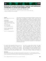
Fig. 1. Binding of ERRc is modulated by the presence of serum. EMSA of in vitro translated ERRc withSIS,aDR-0elementandwithLFREan
extended half-site. (A, B) Supershift of ERRc-SIS (A) and ERRc-LFRE (B) complexes by increasing amounts of antiserum a-ERR. Constant
amounts of the binding element and of in vitro generated ERRc were subjected to electrophoresis in the absence (lanes 1), and in the presence of
0.004 lL (lanes 2), 0.008 lL (lanes 3), 0.016 lL (lanes 4), 0.03 lL(lanes5),0.06lL (lanes 6), 0.13 lL (lanes 7), 0.25 lL(lanes8),0.5lL
(lanes 9), 1 lL (lanes 10), and 2 lL (lanes 11) of a-ERR. (C) Increasing amounts of the preimmune serum (PIS) result in an increase of the ERRc-
SIS complex. The binding reaction was subjected to electrophoresis in the absence (lane 1), and in the presence of ERRc (lanes 2–12) with
increasing amounts of PIS [0.004 lL(lane3)to2 lL (lane 12)]. (D) Binding was performed in the absence (lanes 1–7), and in the presence of 1 lL
PIS (lanes 8–12) with increasing amounts of ERRc (0.06 lL in lanes 1 and 7; 0.13 lL in lanes 2 and 8; 0,25 lL in lanes 3 and 9; 0.5 lLinlanes4
and 10; 1 lL in lanes 5 and 11; 2 lL in lanes 6 and 12). The ERRc–DNA complexes are marked by an arrow, the ERRc–DNA complexes bound
by a-ERR are indicated by arrowheads. SIS and LFRE indicate free DNA.
Ó FEBS 2002 DNA binding of ERRc (Eur. J. Biochem. 269) 4089
of constant amounts of serum, less in vitro translated
ERRc was necessary for DNA binding (Fig. 1D). Taken
together, these experiments reveal that although a specific
protein–DNA complex of in vitro generated ERRc is
formed in the absence of serum, lower ERRc-concentra-
tions are needed in the presence of serum.
Increased binding activity in the presence of serum
is heat sensitive
Having identified serum as a stimulating factor, we next
thought to elucidate the nature of this activity. To initiate
the characterization of the stimulating serum effect, its
sensitivity against heat was tested. Rabbit serum was treated
for 20 min at various temperatures, centrifuged, and the
supernatant was analyzed by EMSA (Fig. 2A). The stimu-
lating effect, still present at a temperature of 75 °C, was
absent after incubation at 80 °C. Precipitation of the
proteins was not observed at 70 °C, some precipitation
occurred at 75 °C, and massive precipitation was found at
higher temperatures. Hence, the stimulating factor in the
serum is either heat-sensitive, e.g. a protein, or associated
with the precipitate.
Characterization of the stimulating activity
For further characterization of the stimulating factor, the
serum was subjected to various treatments. Whereas a size
exclusion assay with a Bio-Gel P30 spin column of an
exclusion limit of about 40 000 Da demonstrated that the
activity was in the fraction of the large molecules, a
microdialysis with a nitrocellulose membrane with a pore
size of 0.025 lm did not diminish the effect (not shown).
After precipitation with ethanol, methanol, isopropanol or
acetone, the activity was detected in the precipitate (Fig. 2B).
Because the effect might be due to a small molecule tightly
associated with a protein, the serum was subjected to several
extraction methods. Extraction using ethanol, chloroform,
or ether could not separate the activity from the hydrophilic
phase. In addition, the activation factor did not quantita-
tively interact with charcoal (Fig. 2B). Hence, the factor in
the serum may be a protein, e.g. serum albumin, stabilizing a
conformation with a higher DNA affinity, or a small
molecule tightly associated with a protein.
To distinguish an indirect mechanism mediated by
constituents of the reticulocyte lysate from a direct effect
on ERRc, binding of bacterial expressed GST–ERRc
fusion protein was investigated. No binding of the affinity
purified fusion protein was detected in the absence of
serum. Again, addition of serum greatly enhanced binding
to DR-0, thereby excluding indirect mechanisms
(Fig. 3A). In addition, expression in E. coli allowed
analyzing of a possible direct effect of the reticulocyte
lysate on DNA binding. Indeed, the addition of lysate,
programmed to synthesize the unrelated protein luciferase,
stimulated binding of ERRc (Fig. 3A). Furthermore,
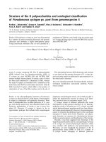
Fig. 2. Characterization of the activating func-
tion of the serum. (A) Binding of ERRc to SIS
was analyzed in the presence (lanes 1–10)
and in the absence of rabbit serum (lane 11).
Prior to binding, the serum was subjected to
increasing temperatures as indicated. (B)
Binding of ERRc toSISintheabsence(lane1)
and in the presence of bovine serum (lane
2–18). Serum was not treated (lanes 2, 18), or
precipitated with ethanol (lanes 3, 4), meth-
anol (lanes 5, 6), isopropanol (lanes 7, 8) or
aceton (lanes 9, 10), as indicated. The preci-
pitates(lanes3,5,7,9)andtherespective
supernatants (lanes 4, 6, 8, 10) were tested.
After organic extraction, with ethylacetate
(lanes 11, 12), chloroform (lanes 13, 14), and
diethylether (lanes 15, 16), the hydrophilic
(lanes 11, 13, 15) and the organic phase
(lanes 12, 14, 16) were analyzed. In lane 17 the
binding reaction was supplemented with
charcoal-treated serum. The ERRc–DNA
complexes are marked by arrows, f indicates
free DNA.
4090 M. Hentschke et al. (Eur. J. Biochem. 269) Ó FEBS 2002
bovine serum albumin and highly purified human serum
albumin, both activated DNA binding of the bacterial
expressed protein. (Fig. 3B). However, ovalbumin does
not enhance DNA binding, suggesting that the effect is
not a pure function of the protein concentration (data not
shown).
The detection of factors stimulating the DNA binding
activity in reticulocyte lysate suggests that cellular constit-
uents may have a stimulating activity. To address this issue,
we tested whole cell extract derived from CV-1 cells, NIH/
3T3 cells and P19 cells, respectively (Fig. 3C). All extracts
stimulated the binding activity GST–ERRc fusion proteins
suggesting a physiological function of the enhancement of
DNA binding.
A sequence element in the hinge region is essential
for the stimulating effect
As demonstrated above, limiting factors greatly enhanced
the formation of ERRc–DNA complexes. As a conse-
quence, it should be possible to map elements in the receptor
as targets of these factors.
To this end, the truncated protein ERRc-218 coding for
the first 218 amino acids, and two fusion construct of the
N-terminal part of GCNF with the C-terminal part of
ERRc, GE-2 and GE-3 (Fig. 4A), were tested. GE-2 covers
amino acids 1–167 of GCNF and 213–458 of ERRc,
whereas GE-3 covers amino acids 1–271 of GCNF and
240–458 of ERRc.ASalI restriction site at the fusion codes
for two additional amino acids, valine and aspartic acid. In
both fusion proteins DNA binding is mediated by GCNF.
The truncated in vitro translated protein ERRc-218, lacking
amino acids forming the LBD and the C-terminal part of
the hinge region, still binds to DNA, and the addition of
serum results in increased binding (Fig. 4B). Consequently,
the LBD is not necessary for the activating function of the
serum. Hence, an allosteric conformational switch by
binding of a steroid ligand bound to a carrier in the serum
is very unlikely. At least some of the target sequences must
be located either in the A/B domain, the DBD, or the hinge
region. As expected, binding of GCNF is not increased by
serum addition. The same is true for GE-3 in which most of
the LBD of GCNF is replaced by that of ERRc,further
demonstrating that the LBD is not involved in the
activation. We conclude that the LBD is neither essential
for the activation, nor does its fusion to a homologous
protein result in a transfer of the activity. However, the
binding of GE-2, in which the C-terminal part of the hinge
and the LBD of GCNF are replaced by the corresponding
domains of ERRc, is greatly stimulated by serum (Fig. 4B).
Accordingly, the ERRc-hinge region confers the activation.
ERRc-218 and GE-2, both affected by the addition of
serum, have a sequence overlap of six amino acids. These
results suggest a central role of the common sequence,
ÔNH
2
-RIDAEN-COOHÕ, in the stimulating effect. Three of
these amino acids are charged, further implying that the
stimulating effect is not induced by lipophilic ligand receptor
interaction. A comparison with the homologous receptors
ERRa,ERRb,ERa,andERb and a data base search in
the nonredundant protein data base revealed that the
ÔRIDAENÕ element is unique for ERRc.
ERRc binds as a homodimer to DNA
Dimerization is essential for the function of most nuclear
receptors. Previously, ERRc wasreportedtobindasa
monomer to DNA [28]. However, a recent report assumes
that ERRc binds also as a dimer to DNA [35]. For ERRa
and ERRb, monomeric and dimeric binding has been
demonstrated [3]. The repeat nature of the binding site, and
thefactthatERRc–DNA complexes have a mobility very
similar to a GCNF homodimer and to a PPARc/RXRc
heterodimer (not shown), suggest that ERRc binds to DNA
preferentially as a dimer.
To address the dimerization properties of ERRc in
solution, we constructed the mutant DN-ERRc in which the
entire N-terminal domain of ERRc is deleted (Fig. 4A).
This mutant still binds to DR-0 and forms protein–DNA
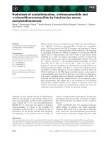
Fig. 3. Binding of a bacterial expressed GST–
ERRc fusion protein depends on factors present
in serum and in cellular extracts. The purified
GST–ERRc fusion protein was tested for
binding to the SIS element. Only the upper
half of the autoradiograph is shown. (A)
Binding without additional factors, and in the
presence of fetal bovine serum (FCS) and
reticulocyte lysate (RL), as indicated. (B)
Binding in the presence of bovine serum
albumin (BSA) and human serum albumin
(HSA), respectively. (C) Whole cell extracts of
the kidney derived cell line CV-1, of NIH/3T3
fibroblasts, and of the embryonal carcinoma
cell line P19, were incubated with the SIS ele-
ment in the absence and in the presence of the
GST–ERRc fusion protein, as indicated.
Ó FEBS 2002 DNA binding of ERRc (Eur. J. Biochem. 269) 4091
complexes with a mobility higher than that of the wild-type
receptor (Fig. 5, compare lanes 1 and 2). The mixing of
ERRc with DN-ERRc results in the formation of DNA-
bound ERRc/DN-ERRc heterodimers, which migrate with
a mobility intermediate between those of the homodimeric
ERRc and DN-ERRc complexes. (Fig. 5). Dimeriziation is
detected on DR-0, and also in weaker complexes formed on
the extended half-site DR-0 A, and on ERE, an inverted
repeat with a spacing of three base pairs, the classical
estrogen response element.
DNA binding of C-terminal deletion mutants
Dimerization motifs are commonly found in the DBDs
including the CTE and in the C-terminus of nuclear
receptors [36–38]. To identify sequence elements in the
LBD that contribute to DNA binding, a series of C-terminal
truncated ERRc polypeptides comprising the first 218–448
amino acids of the 458 amino acid full-length protein were
generated (Fig. 4A). An SDS/PAGE analysis of the
proteins generated by in vitro translation in the presence
of [
35
S]methionine demonstrated their synthesis in similar
amounts (not shown). Binding to DR-0 was tested in
comparison to the full-length protein, to DN-ERRc,to
GE-2, and to GE-3. ERRc-448, lacking the C-terminal nine
amino acids, the sequence harboring the H12 a helical
region still binds to DNA [31] (Fig. 6). Although the protein
migrates faster during denaturing gel electrophoresis, the
protein–DNA complex has a slightly reduced mobility when
compared to the full-length ERRc. This may be either due
to a conformational change or to differences in the surface
charge distribution of the truncated receptor. Further
truncation of additional 41 amino acids in mutant
ERRc-408 gives rise to a much weaker complex indicating
a reduced DNA affinity that may be the result of an
impaired folding or a reduced dimerization function. Again,
the complexes migrate slightly slower when compared to
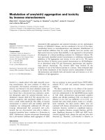
Fig. 5. ERRc binds as a homodimer to DNA. Binding of the full-length
ERRc (lanes 1, 4, 7), the N-terminal truncated protein DN-ERRc
(lanes 2, 5, 8) and a mixture of both proteins (lanes 3, 6, 9) were
subjectedtoanEMSAwiththeindicatedDNAelements(SIS:aDR-0
element of the bPDGF promoter; DR-0A: an extended half-site; ERE,
an estrogen response element of the vitellogenin promoter). The
position of the ERRc–DNA complexes (double arrow) of the
DN-ERRc–DNA complexes (arrow), and of the heterodimer
(arrowhead) are indicated.
Fig. 4. Localization of the ERRc domain involved in the enhanced DNA binding. (A) Schematic view of truncated ERRc andfusionproteinsof
GCNF and ERRc used in this study. The position of the N-terminal A/B-domain, the DBD (C-domain), the hinge region (D-
D
omain), and the
LBD (EF domain) are indicated. For the truncated protein, the first and last amino acid is indicated with respect to the full-length protein. In the
chimeras GE-1, GE-2, and GE-3 the numbering refers to the amino acids of GCNF and ERRc, respectively. (B) Binding of the truncated protein
ERRc-218, of the fusion proteins GE-2 and GE-3, and of GCNF to SIS in the presence and in the absence of a rabbit serum (RS), as indicated. The
positions of the complexes of SIS with ERRc-218 (double arrow), GE-2 (open arrowhead), GE-3 (filled arrowhead), and GCNF (arrow) are
indicated.
4092 M. Hentschke et al. (Eur. J. Biochem. 269) Ó FEBS 2002
full-length proteins. All three proteins form an additional
weak and faster migrating complex, apparently a monomer.
The intensity of this band is not affected by the truncations,
indicating that reduced binding of the dimer is due to
inefficient dimerization. The truncated ERRc-408 lacks the
a helices 10–12. Helices 9 and 10 have been implicated in
dimerization of various nuclear receptors. A crystal struc-
ture of the RXRa LBD revealed a dimer interface formed
mainly by helix 10 and, to a lesser extent, helix 9 and the
loop between helix 7 and helix 8 [39]. A weak dimer is
formed by ERRc-376, a truncated protein lacking helix 9.
Further truncation of helices 6–8 in ERRc-331, and helices
4–8 in ERRc-285 results in much smaller, weak complexes.
In contrast, the smallest truncated protein, ERRc-218,
lacking the whole LBD and part of the hinge region shows a
robust complex (Fig. 6). This protein consists of the DBD
and includes 25 amino acids of the D domain and therefore
the CTE. Several conclusions can be drawn from the
binding analysis. According to the conserved a helical
sandwich structure, as determined for ERa [40,41] and
more recently for ERRc [31], a dimerization function can be
assigned to a region containing ahelices 10 and 11. The
increase of binding by the additional deletion of the a helices
1–3 and of the C-terminal part of the hinge region (compare
ERRc-218 and ERRc-285) suggests that these elements
offer some steric hindrance for dimerization or DNA
binding. An additional dimerization function can be
assigned to the N-terminal 218 amino acids. In analogy to
other nuclear receptors, this function is proposed to be
located in the DBD including the CTE [36,37]. Taken
together, these results imply homophilic interaction of
ERRc on various NR response elements mediated by at
least two dimerization modules.
Dimerization function of ERRc
Two nuclear receptor dimerization interfaces have been
defined, one within the DBD and one within the LBD. A
two step-model for dimeric binding of RXR heterodimers
has been proposed. First, heterodimers would be formed
through their dimerization interfaces contained in the LBD,
and in a second step the DBDs would be able to bind with
high affinity to DNA [42]. In order to analyze to what extent
dimerization of the truncated proteins is impaired in the
absence of DNA, C-terminal deletion proteins ERRc-448 to
ERRc-218 were separated by SDS/PAGE and subjected to
a far-Western analysis, a method based on direct protein-
interaction. Only ERRc-448 was identified as binding
partner of the full-length protein labeled by incorporation
of [
35
S]methionine (Fig. 7A). Further truncation of 41
amino acids abolishes the homophilic interaction. This
result is in agreement with the DNA-binding analysis:
highly reduced binding of the truncated proteins is most
likely the result of the deletion of a dimerization function,
whichcanbelocatedtothea helical region 10–11. On the
other hand, the smallest deletion mutant tested, ERRc-218,
binds to DNA but does not dimerize with the full-length
protein under far-Western conditions (Fig. 7A). The dimer-
ization of this mutant may be dependent on the presence of
the DNA-response element. As in solution, the full-length
protein binds to DN-ERRc, the N-terminal truncated
protein. The chimeric protein GE-1 (GCNF1-140/
ERRc196–458) containing the 263 C-terminal amino acids
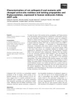
of ERRc is efficiently bound by labeled ERRc (Fig. 7B).
This interaction indicates that the dimerization motifs in the
C-terminus function independently of the motifs in the
DBD. Decreasing amounts of ERRc-specific residues in
GE-2 and GE-3 are accompanied by reduced and abolished
interaction, respectively. Hence, additional amino acids in
the D and helix 1 region are important for dimerization. For
DNA-independent dimerization, both elements, one located
between 219 and 239 and the second between amino acids
409 and 448 are necessary.
DISCUSSION
In this study, we show that ERRc binds to a DR-0 element,
but also to extended half-sites. ERRs have a conserved
DBD. Therefore, it is not surprising that they all bind
to elements with the extended half-site element
TCAAGGTCA. In addition, a weak complex was detected
on ERE, an inverted response element. There are conflicting
results in the literature as to whether ERRs bind as
monomers or dimers. Our results show that ERRc binds
preferentially as a dimer to all of these elements. This has
Fig. 6. Binding of truncated and chimeric receptors to a DR-0 element.
The full-length protein (lane 1), C-terminal deletions (lanes 2–7), the
N-terminal deletion DN-ERRc (lane 8), and fusion proteins GE-2 and
GE-3 (lanes 9 and 10) were tested in EMSA. Equal amounts of primed
reticulocyte lysate and labeled SIS-binding site were used in each lane.
The positions of the DNA complexes with ERRc (filled arrow),
ERRc-218 (open arrow), DN-ERRc (double arrow), GE-2 (open
arrowhead) and GE-3 (closed arrowhead)are indicated. The position
ERRc monomers bound to DNA in lanes 1–3 is indicated by the
bracket.
Ó FEBS 2002 DNA binding of ERRc (Eur. J. Biochem. 269) 4093
been demonstrated by mixing of an N-terminal truncated
protein with the full-length protein. An intermediate band in
an EMSA is confirmation of dimerization. Additionally,
protein–DNA complexes of the orphan receptor GCNF
that binds to DR-0 as a dimer show a very similar migration
[43]. An important future question is the identification of
functional binding sites and the analysis of a possible cross-
talk of receptors with a similar binding site specificity. To
further characterize functional domains of the protein,
binding of C-terminal truncated proteins to SIS, a DR-0
element of the bPDGF promoter was analyzed by EMSA.
Surprisingly, binding of some of the truncated protein gave
rise to a slower migrating complex. This phenomenon has
also been observed for truncated GCNF bound to DR-0
[44]. Because the analysis was performed under nondena-
turing conditions, a reasonable explanation is a less compact
structure of the truncated protein, or differences in the
surface charge distribution of the truncated receptor. A
faster migrating weak complex that appears to be a
monomer shows a similar behavior (Fig. 6, lanes 1–3).
However, in contrast to the dimer, the intensity of this band
is not affected by the deletion, suggesting a reduced
dimerization function. The truncated protein ERRc-218,
which contains the DBD including its C-terminal extension
binds to DNA, suggesting that ERRc has a DNA–
dependent dimerization interface. The weak complexes
formed by ERRc-408, ERRc-376, ERRc-331, and ERRc-
285 further strengthens the assumption that these trunca-
tions have a distorted DNA-independent dimerization.
As an independent approach we subjected various
deletion mutants and fusion proteins to a direct analysis
of protein–protein interaction by far-Western blots. The
interaction of the mutated proteins with the radioactive full-
length ERRc supported the results of the EMSA. It is
important to note that deletion of the N-terminal domain
does not influence the dimerization properties of the
receptor. However, the C-terminal LBD is important for
homophile interactions. The deletion ERRc-408 lacking the
helix 10/11 does not dimerize. The crystal structures of the
LBDs of hRXRa,hRARc,hTRa, and hERa show that this
dimerization is mediated mainly by helices 9 and 10
[39,40,45–47]. A recent analysis of the ERRc LBD shows
that it adopts a canonical three-layered a helical sandwich
structure and superimposes well with the hER LBD [31].
In addition, the analysis allowed the study of the
interaction of a fusion protein with a heterologous DBD.
Although ERRc does not bind to GCNF, the fusion protein
GE-1, composed of the N-terminal GCNF portion with the
DBD and the C-terminal portion of ERRc is bound by full-
length ERRc. Therefore, the dimerization function in the
C-terminus works independently of the dimerization func-
tion in the DBD. In addition, the analysis shows that both,
the C-terminal (Fig. 7A, compare lanes 2 and 3), and the
N-terminal truncation of the C-terminus (Fig. 7B, compare
Fig. 7. A Far-Western analysis deciphers the DNA binding-independent dimerization function of ERRc. (A) C-terminal deletion mutants of ERRc
were separated by SDS/PAGE, blotted to a membrane filter, and probed for interaction with
35
S-radiolabeled ERRc (lanes 2–7). The probe,
separated on the same gel is shown in lane 1. The arrow indicates the position of ERRc-448. (B) The full-length protein (lane 1), the proteins GE-1,
GE-2, GE-3 (lanes 2–4), DN-ERRc (lane 5), and GCNF as a negative control (lane 6) were separated by SDS/PAGE and subjected to a
Far-Western analysis as described in A. The arrow indicates the position of ERRc. (C) Schematic representation of ERRc. The position of the
dimerization motifs is indicated by black bars, the numbers refer to the amino acids important for dimerization.
4094 M. Hentschke et al. (Eur. J. Biochem. 269) Ó FEBS 2002
lanes 3 and 4) abolish dimerization. Therefore, at least two
dimerization interfaces in the C-terminus exist, one located
between amino acid 213 and 239, and the second between
amino acids 409 and 448. The C-terminal interface includes
helix 10, whose function in dimerization is well established
for several receptors. For the further N-terminal located
interface, the presence of amino acids in the hinge region up
to helix 1 in the LBD is important: the CTE is not essential.
Interestingly, Tetel et al. reported that the minimal fragment
mediating progesterone receptor homodimerization was the
hinge-LBD construct [48]. In addition, GST pull down
experiments reveal the importance of the
D
-
D
omain of the
thyroid hormone receptor for homodimerization and hete-
rodimerization with RXR. However, in the same experi-
mental design, the EF domain of the RXR formed
heterodimers with the thyroid hormone receptor [49]. The
His-tagged ERRc LBD forms dimers in solution [31].
The discrepancy could be due to the fact that in our study
the binding partner is immobilized, the GE-3 starts 11
amino acids further to the C-terminus, or that the GCNF
fusion affects dimerization. On the other hand it is possible
that the His-tag influences protein interaction [50].
The serum effect is very surprising because in vivo,ERRc
should never be in a direct contact with the serum. However,
it is possible that a serum factor enters the cell. Co-transfec-
tion with ERRs and a reporter gene also suggest a function
of serum in transcription activation [13]. Because we
achieved activation of binding by purified serum albumin,
it appears more likely that the endogenous activators differ
from the serum factor. Preliminary results in our laboratory
(M. Hentschke, unpublished observations) show that at
least two active fractions can be separated by ion exchange
chromatography and by gel filtration chromatography of
crude P19 cell extracts. The identification of the active
components in these fractions will be an important prere-
quisite to analyze the mechanism underlying the phenom-
enon. A specific effect should be dependent on the presence
of sequence elements present in ERRc but not in GCNF.
Therefore, we have focussed on the target protein, ERRc.
Indeed, the C-terminal deletions reveal that even the binding
of the smallest protein analyzed is activated by additional
factors. However, neither binding of GCNF, nor of the
chimera GE-3 is influenced by additional factors. However,
binding of GE-2 with 27 additional amino acids is clearly
stimulated by additional factors. Taken together these
experiments reveal that amino acids 1–218, and amino acids
213–458 fused to GCNF can mediate this increase in DNA
binding. Although, the importance for efficient binding of
additional factors has been shown for additional nuclear
receptors, to our knowledge this is the first example where a
short sequence with a central function in mediating this
effect has been identified for ERRs.
The question arises as to whether there are other
receptors whose binding depends on additional proteins.
Indeed, there are several reports about cellular extracts,
necessary for efficient binding of steroid hormone receptors
[51,52]. The function of the high-mobility group box
proteins, HMG-1 and HMG-2, members of the nonhistone
chromatin proteins, has been analyzed in more detail. They
are recruited to DNA by steroid hormone receptors and
although very abundant, subsequently led to an increase in
transcriptional activity in transient transfection assays
[53–56], but have no effect on binding of several nonsteroid
hormone receptors [54]. HMG-1/-2 appear to act by
facilitating receptor interaction with target DNA sites [56].
The HMG box contacts the DNA in the minor groove
introducing a strong bend [57]. Therefore, the HMG box
proteins have been proposed to substitute for the lack of a
minor groove-interacting surface in the DBD of the steroid
hormone receptors [54,56]. However, they do not result in
the supershift of the retarded bands that would be expected
if HMGs were present in the complex. A deletion analysis of
the androgen receptor indicated that that HMG-1 needs at
least part of the CTE and of the hinge region for the
stimulation of receptor DNA binding [58]. Whether the
observed effect on DNA binding of ERRc can be mediated
by HMG box proteins is presently unknown. HMG-1 is a
very conserved and abundant protein, which interacts with
many apparently unrelated proteins [59]. The recent iden-
tification of SRY, a nuclear HMG box-containing protein
as an interaction partner of the androgen receptor suggests
that additional differential expressed HMG box proteins
may be identified as interaction partner of nuclear receptors
[60]. The analysis of the ERRc LBD structure revealed that
the ligand free conformation is the transcriptionally active
form suggesting that alternative mechanisms may be
important to regulate the activity of this true orphan [31].
A systematic approach will be necessary to identify the most
efficient interaction partners and to understand how these
additional proteins succeed to increase DNA binding of
ERRc and therefore modulate the activity of this orphan
receptor.
ACKNOWLEDGEMENTS
We thank Prof Schaller for the support of this work. This project was
supported by a fellowship to M. H. through the Graduiertenkolleg 255
and is part of his doctoral thesis. Special thanks go to Drs Irm
Hermans-Borgmeyer and Sabine Hoffmeister-Ullerich for the fruitful
discussion throughout the project, to Simon Hempel for help with the
figures and to Cornelia Meyer, Mirja Bernhardt and Anja Nitzsche for
assistance during their practical training.
REFERENCES
1. Mangelsdorf, D.J., Thummel, C., Beato, M., Herrlich, P., Schu
¨
tz,
G.,Umesono,K.,Blumberg,B.,Kastner,P.,Mark,M.,
Chambon, P. & Evans, R.M. (1995) The nuclear receptor super-
family: the second decade. Cell 83, 835–839.
2. Enmark, E. & Gustafsson, J.A. (1996) Orphan nuclear receptors –
the first eight years. Mol. Endocrinol. 10, 1293–1307.
3. Gigue
`
re, V. (1999) Orphan nuclear receptors: from gene to func-
tion. Endocrinol. Rev. 20, 689–725.
4. Green, S. & Chambon, P. (1988) Nuclear receptors enhance our
understanding of transcription regulation. Trends Genet. 4, 309–
314.
5. Renaud, J.P. & Moras, D. (2000) Structural studies on nuclear
receptors. Cell. Mol. Life Sci. 57, 1748–1769.
6. Khorasanizadeh, S. & Rastinejad, F. (2001) Nuclear–receptor
interactions on DNA-response elements. Trends Biochem. Sci. 26,
384–390.
7. Laudet, V. (1997) Evolution of the nuclear receptor superfamily:
early diversification from an ancestral orphan receptor. J. Mol.
Endocrinol. 19, 207–226.
8. Green, S., Walter, P., Kumar, V., Krust, A., Bornert, J.M., Argos,
P. & Chambon, P. (1986) Human oestrogen receptor cDNA:
sequence, expression and homology to v-erb-A. Nature 320, 134–
139.
Ó FEBS 2002 DNA binding of ERRc (Eur. J. Biochem. 269) 4095
9. Kuiper, G.G.J.M., Enmark, E., Pelto-Huikko, M., Nilsson, S. &
Gustafsson, J.A. (1996) Cloning of a novel estrogen receptor
expressed in rat prostate and ovary. Proc. Natl Acad. Sci. USA 93,
5925–5930.
10. Beato, M. & Klug, J. (2000) Steroid hormone receptors: an
update. Hum. Reprod. Update 6, 225–236.
11. Gigue
`
re, V., Yang, N., Segui, P. & Evans, R.M. (1988) Identifi-
cation of a new class of steroid hormone receptors. Nature 331,
91–94.
12. Bonnelye, E., Vanacker, J.M., Dittmar, T., Begue, A.,Desbiens, X.,
Denhardt, D.T., Aubin, J.E., Laudet, V. & Fournier, B. (1997) The
ERR-1 orphan receptor is a transcriptional activator expressed
during bone development. Mol. Endocrinol. 11, 905–916.
13. Vanacker, J M., Bonnelye, E., Chopin-Delannoy, S., Delmarre,
C., Cavaille
`
s, V. & Laudet, V. (1999) Transcriptional activities of
the orphan nuclear receptor ERR alpha (estrogen receptor-related
receptor-alpha). Mol. Endocrinol. 13, 764–773.
14. Zhang, Z. & Teng, C.T. (2001) Estrogen receptor alpha and
estrogen receptor-related receptor alpha1 compete for binding and
coactivator. Mol. Cell. Endocrinol. 172, 223–233.
15. Vanacker, J M., Pettersson, K., Gustafsson, J A. & Laudet, V.
(1999) Transcriptional targets shared by estrogen receptor-related
receptors (ERRs) and estrogen receptor (ER) a, but not by ERb.
EMBO J. 18, 4270–4279.
16. Yang, N., Shigeta, H., Shi, H. & Teng, C.T. (1996) Estrogen-
related receptor, hERR1, modulates estrogen receptor-mediated
response of human lactoferrin gene promoter. J. Biol. Chem. 271,
5795–5804.
17. Yang,C.,Zhou,D.&Chen,S.(1998)Modulationofaromatase
expression in the breast tissue by ERR alpha-1 orphan receptor.
Cancer Res. 58, 5695–5700.
18. Vanacker, J M., Delmarre, C., Guo, X. & Laudet, V. (1998)
Activation of the osteopontin promoter by the orphan nuclear
receptor estrogen receptor related a. Cell Growth Differ. 9, 1007–
1014.
19. Zhang, Z. & Teng, C.T. (2000) Estrogen receptor-related receptor
alpha 1 interacts with coactivator and constitutively activates the
estrogen response elements of the human lactoferrin gene. J. Biol.
Chem. 275, 20837–20846.
20. Johnston, S.D., Liu, X., Zuo, F., Eisenbraun, T.L., Wiley, S.R.,
Kraus, R.J. & Mertz, J.E. (1997) Estrogen-related receptor alpha 1
functionally binds as a monomer to extended half-site sequences
including ones contained within estrogen-response elements. Mol.
Endocrinol. 11, 342–352.
21. Eudy, J.D., Yao, S., Weston, M.D., Ma-Edmonds, M., Talmadge,
C.B., Cheng, J.J., Kimberling, W.J. & Sumegi, J. (1998) Isolation
of a gene encoding a novel member of the nuclear receptor
superfamily from the critical region of Usher syndrome type IIa
at 1q41. Genomics 50, 382–384.
22. Chen, F., Zhang, Q., McDonald, T., Davidoff, M.J., Bailey, W.,
Bai,C.,Liu,Q.&Caskey,C.T.(1999)Identificationoftwo
hERR2-related novel nuclear receptors utilizing bioinformatics
and inverse PCR. Gene 228, 101–109.
23. Nagase, T., Ishikawa, K., Suyama, M., Kikuno, R., Hirosawa,
M., Miyajima, N., Tanaka, A., Kotani, H., Nomura, N. & Ohara,
O. (1998) Prediction of the coding sequences of unidentified
human genes. XII. The complete sequences of 100 new cDNA
clones from brain which code for large proteins in vitro. DNA Res.
5, 355–364.
24. Hong, H., Yang, L. & Stallcup, M.R. (1999) Hormone-
independent transcriptional activation and coactivator binding
by novel orphan nuclear receptor ERR3. J. Biol. Chem. 274,
22618–22626.
25. Su
¨
sens, U., Hermans-Borgmeyer, I. & Borgmeyer, U. (2000)
Alternative splicing and expression of the mouse estrogen
receptor-related receptor (ERR)c. Biochem. Biophys. Res.
Commun. 267, 532–535.
26. Lorke, D.E., Su
¨
sens, U., Borgmeyer, U. & Hermans-Borgmeyer,
I. (2000) Differential expression of the estrogen receptor-related
receptor (ERR) c in the mouse brain. BrainRes.Mol.Brain.Res.
77, 277–280.
27. Hermans-Borgmeyer, I., Su
¨
sens, U. & Borgmeyer, U. (2000)
Developmental expression of the estrogen receptor-related
receptor (ERR) c during mouse embryogenesis. Mech. Dev. 97,
197–199.
28. Heard, D., Norby, P., Holloway, J. & Vissing, H. (2000) Human
ERRgamma, a third member of the estrogen receptor-related
receptor (ERR) subfamily of orphan nuclear receptors: tissue-
specific isoforms are expressed during development and in the
adult. Mol. Endocrinol. 14, 382–392.
29. Coward, P., Lee, D., Hull, M. & Lehmann, J. (2001) 4-Hydro-
xytamoxifen binds to and deactivates the estrogen-related receptor
gamma. Proc. Natl Acad. Sci. USA 98, 8880–8884.
30. Tremblay, G., Bergeron, D. & Giguere, V. (2001) 4-Hydro-
xytamoxifen is an isoform-specific inhibitor of orphan estrogen-
receptor-related (ERR) nuclear receptors beta and gamma.
Endocrinology 142, 4572–4575.
31. Greschik, H., Wurtz, J., Sanglier, S., Bourguet, W., van
Dorsselaer, A., Moras, D. & Renaud, J. (2002) Structural and
functional evidence for ligand-independent transcriptional acti-
vation by the estrogen-related receptor 3. Mol. Cell 9, 303–313.
32. Kozak, M. (1986) Point mutations define a sequence flanking the
AUG initiator codon that modulates translation by eukaryotic
ribosomes. Cell 44, 283–292.
33. Su
¨
sens,U.,Aguiluz,J.B.,Evans,R.M.&Borgmeyer,U.(1997)
The germ cell nuclear factor mGCNF is expressed in the devel-
oping nervous system. Dev. Neurosci. 19, 410–420.
34. Yan, Z. & Jetten, A.M. (2000) Characterization of the repressor
function of the nuclear orphan receptor retinoid receptor-related
testis-associated receptor/germ cell nuclear factor. J. Biol. Chem.
272, 10565–10572.
35. Sanyal, S., Kim, J Y., Kim, H J., Takeda, J., Lee, Y K., Moore,
D.D. & Choi, H S. (2002) Differential regulation of the orphan
nuclear receptor small heterodimer partner (SHP) gene promoter
by orphan nuclear receptor ERR isoforms. J. Biol. Chem. 277,
1739–1748.
36. Zechel, C., Shen, X Q., Chambon, P. & Gronemeyer, H. (1994)
Dimerization interfaces formed between the DNA binding
domains determine the cooperative binding of RXR/RAR
and RXR/TR heterodimers to DR5 and DR4 elements. EMBO J.
13, 1414–1424.
37. Zechel, C., Shen, X Q., Chen, J Y., Chen, Z P., Chambon, P. &
Gronemeyer, H. (1994) The dimerization interfaces formed
between the DNA binding domains of RXR, RAR and TR
determine the binding specificity and polarity of the full-length
receptors to direct repeats. EMBO J. 13, 1425–1433.
38. Forman, B.M., Umesono, K., Chen, J. & Evans, R.M. (1995)
Unique response pathways are established by allosteric interac-
tions among nuclear hormone receptors. Cell 81, 541–550.
39. Bourguet, W., Ruff, M., Chambon, P., Gronemeyer, H. & Moras,
D. (1995) Crystal structure of the ligand-binding domain of the
human nuclear receptor RXRa. Nature 375, 377–382.
40. Brzozowski, A.M., Pike, A.C., Dauter, Z., Hubbard, R.E., Bonn,
T.,Engstrom,O.,Ohman,L.,Greene,G.L.,Gustafsson,J.A.&
Carlquist, M. (1997) Molecular basis of agonism and antagonism
in the oestrogen receptor. Nature 389, 753–758.
41. Shiau, A.K., Barstad, D., Loria, P.M., Cheng, L., Kushner, P.J.,
Agard, D.A. & Greene, G.L. (1998) The structural basis of
estrogen receptor/coactivator recognition and the antagonism of
this interaction by tamoxifen. Cell 95, 927–937.
42. Mangelsdorf, D.J. & Evans, R.M. (1995) The RXR heterodimers
and orphan receptors. Cell 83, 841–850.
43. Borgmeyer, U. (1997) Dimeric binding of the mouse germ cell
nuclear factor. Eur J. Biochem. 244, 120–127.
4096 M. Hentschke et al. (Eur. J. Biochem. 269) Ó FEBS 2002
44. Greschik,H.,Wurtz,J M.,Hublitz,P.,Ko
¨
hler, F., Moras, D. &
Schu
¨
le, R. (1999) Characterization of the DNA-binding and
dimerization properties of the nuclear orphan receptor germ cell
nuclear factor. Mol. Cell. Biol. 19, 690–703.
45. Renaud, J.P., Rochel, N., Ruff, M., Vivat, V., Chambon, P.,
Gronemeyer, H. & Moras, D. (1995) Crystal structure of the
RAR-gamma ligand-binding domain bound to all-trans retinoic
acid. Nature 378, 681–689.
46. Wagner, R.L., Apriletti, J.W., McGrath, M.E., West, B.L.,
Baxter, J.D. & Fletterick, R.J. (1995) A structural role for
hormone in the thyroid hormone receptor. Nature 378, 690–697.
47. Tanenbaum, D.M., Wang, Y., Williams, S.P. & Sigler, P.B. (1998)
Crystallographic comparison of the estrogen and progesterone
receptor’s ligand binding domains. Proc. Natl Acad. Sci. USA 95,
5998–6003.
48. Tetel, M.J., Jung, S., Carbajo, P., Ladtkow, T., Skafar, D.F. &
Edwards, D.P. (1997) Hinge and amino-terminal sequences con-
tribute to solution dimerization of human progesterone receptor.
Mol. Endo. 11, 1114–1128.
49. Miyamoto, T., Kakizawa, T., Ichikawa, K., Nishio, S., Takeda,
T., Suzuki, S., Kaneko, A., Kumaga, M., Mori, J., Yamashita, K.,
Sakuma, T. & Hashizume, K. (2001) The role of hinge domain in
heterodimerization and specific DNA recognition by nuclear
receptors. Mol. Cell. Endocrinol. 181, 229–238.
50. Wu, J. & Filutowicz, M. (1999) Hexahistidine (His
6
)-tag depen-
dent protein dimerization: a cautionary tale. Acta Biochim. Pol. 46,
591–599.
51. Edwards, D.P., Ku
¨
hnel, B., Estes, P.A. & Nordeen, S.K. (1989)
Human progesterone receptor binding to mouse mammary tumor
virus deoxyribonucleic acid: dependence on hormone and non-
receptor nuclear factor(s). Mol. Endocrinol. 3, 381–391.
52. De Vos, P., Claessens, F., Celis, L., Peeters, B., Rombauts, W.,
Heyns, W. & Verhoeven, G. (1994) Nuclear extracts enhance the
interaction of fusion proteins containing the DNA-binding
domain of the androgen and glucocorticoid receptor with andro-
gen and glucocorticoid response elements. J. Steroid Biochem.
Mol. Biol. 48, 317–323.
53. Verrier, C.S., Roodi, N., Yee, C.J., Bailey, L.R., Jensen, R.A.,
Bustin, M. & Parl, F.F. (1997) High-mobility group (HMG)
protein HMG-1 and TATA-binding protein-associated factor
TAF (II), 30 affect estrogen receptor-mediated transcriptional
activation. Mol. Endocrinol. 11, 1009–1019.
54. Boonyaratanakornkit, V., Melvin, V., Prendergast, P., Altmann,
M., Ronfani, L., Bianchi, M.E., Taraseviciene, L., Nordeen, S.K.,
Allegretto, E.A. & Edwards, D.P. (1998) High-mobility group
chromatin proteins 1 and 2 functionally interact with steroid
hormone receptors to enhance their DNA binding in vitro and
transcriptional activity in mammalian cells. Mol. Cell. Biol. 18,
4471–4487.
55. Zhang, C.C., Krieg, S. & Shapiro, D.J. (1999) HMG-1 stimulates
estrogen response element binding by estrogen receptor from
stably transfected HeLa cells. Mol. Endocrinol. 13, 632–643.
56. Melvin, V.S. & Edwards, D.P. (1999) Coregulatory proteins
in steroid hormone receptor action: the role of chromatin high
mobility group proteins HMG-1 and -2. Steroid 64, 576–586.
57. Thomas, J.O. & Travers, A.A. (2001) HMG1 and 2, and related
ÔarchitecturalÕ DNA-binding proteins. Trends Biochem. Sci. 26,
167–174.
58.Verrijdt,G.,Haelens,A.,Schoenmakers,E.,Rombauts,W.&
Claessens, F. (2002) Comparative analysis of the influence of the
high-mobility group box 1 protein on DNA binding and
transcriptional activation by the androgen, glucocorticoid,
progesterone and mineralocorticoid receptors. Biochem. J. 361,
97–103.
59. Dintilhac, A. & Bernues, J. (2002) HMGB1 interacts with many
apparently unrelated proteins by recognizing short amino acid
sequences. J. Biol. Chem. 277, 7021–7028.
60. Yuan, X., Lu, M.L., Li, T. & Balk, S.P. (2001) SRY interacts with
and negatively regulates androgen receptor transcriptional
activity. J. Biol. Chem. 276, 46647–46654.
Ó FEBS 2002 DNA binding of ERRc (Eur. J. Biochem. 269) 4097