Báo cáo Y học: Substrate specificity of human kallikrein 2 (hK2) as determined by phage display technology docx
Bạn đang xem bản rút gọn của tài liệu. Xem và tải ngay bản đầy đủ của tài liệu tại đây (178.72 KB, 8 trang )
Substrate specificity of human kallikrein 2 (hK2) as determined
by phage display technology
Sylvain M. Cloutier
1
, Jair Ribeiro Chagas
2
, Jean-Pierre Mach
3
, Christian M. Gygi
1
, Hans-Jurg Leisinger
1
and David Deperthes
1
1
Urology Research Unit, Department of Urology, Lausanne, Switzerland;
2
Centro Interdisciplinar de Investigacao Bioquimica,
Universidade de Mogi das Cruzes, Brazil;
3
Institute of Biochemistry, University of Lausanne, Switzerland
Human glandular kallikrein 2 (hK2) is a trypsin-like serine
protease expressed predominantly in the prostate epithe-
lium. Recently, hK2 has proven to be a useful marker that
can be used in combination with prostate specific antigen
for screening and diagnosis of prostate cancer. The cleavage
by hK2 of certain substrates in the proteolytic cascade
suggest that the kallikrein may be involved in prostate
cancer development; however, there has been very little
other progress toward its biochemical characterization or
elucidation of its true physiological role. In the present
work, we adapt phage substrate technology to study the
substrate specificity of hK2. A phage-displayed random
pentapeptide library with exhaustive diversity was gener-
ated and then screened with purified hK2. Phages display-
ing peptides susceptible to hK2 cleavage were amplified in
eight rounds of selection and genes encoding substrates
were transferred from the phage to a fluorescent system
using cyan fluorescent protein (derived from green fluores-
cent protein) that enables rapid determination of specificity
constants. This study shows that hK2 has a strict preference
for Arg in the P1 position, which is further enhanced by a
Ser in P¢1 position. The scissile bonds identified by phage
display substrate selection correspond to those of the nat-
ural biological substrates of hK2, which include protein C
inhibitor, semenogelins, and fibronectin. Moreover, three
new putative hK2 protein substrates, shown elsewhere to be
involved in the biology of the cancer, have been identified
thus reinforcing the importance of hK2 in prostate cancer
development.
Keywords: cyan fluorescent protein; human kallikrein; phage
display; prostate cancer; substrate.
The human prostatic kallikreins hK3, or prostate specific
antigen (PSA), is considered the gold standard for prostate
cancer diagnosis and screening; however, hK2, the second
prostatic kallikrein to be discovered [1], has recently
emerged as a complementary marker for its positive
correlation with prostate cancer grade and progression.
PSA is more highly expressed in benign hyperplasia (BHP)
than in cancer thus hK2 is helpful to further distinguish
malignant from benign disease [2–4]. The recent discovery
of 12 new members of the kallikrein family [5–7] could
provide additional prostate cancer markers.
In the seminal plasma, hK2 is mostly recovered com-
plexed with protein C inhibitor [1]. Because hK2 cleaves,
with trypsin-like specificity, certain components of the
semen coagulum (fibronectin and semenogelins), it is
possible that it has a role in the early stages of semen
liquefaction, a biological process which immediately follows
ejaculation [8]. In addition, in vitro studies have shown that
hK2 can activate urokinase-type plasminogen activator [9]
and inactivate plasminogen activator inhibitor-1 [10] leading
to the activation of urokinase system. Moreover, hK2
degrades insulin-like growth factor binding proteins (IGF-
BP) to release IGF, a putative local mitogenic signal for
prostate cancer cells [11].
Despite the in vitro identification of its proteolytic
activities as well as its potential substrates, our understand-
ing of the true physiological role of hK2 remains sketchy.
Much progress has been made toward the characterization
of hK2s serine protease activity using synthetic substrates
derived from reactive serpin loops [12]; however, this type of
approach is limited to known targets and cannot advance
the discovery of new biological substrates.
A system using a monovalent phage library capable of
displaying several million different substrates, which
enabled simultaneous testing of proteolytic specificity, was
developed by Matthews and Wells [13]. Several proteases
including furin [14], PSA [15], membrane type-1 matrix
metalloproteinase [16], and granzyme B [17] have already
been characterized using this approach.
We adapted this method by constructing a phage-
displayed random library that included all possible amino
acid combinations of pentapeptides, then screening it with
hK2. Of the 44 individual phage clones selected and
identified, 90% had Arg at the P1 site and 30% had Ser
in the P¢1 position. Kinetic studies and sites of cleavage in
substrates have been determined with a new system using
cyan fluorescent protein (CFP), a variant of the green
fluorescent protein system. A search in the SwissProt
database with selected substrates identified three new
putative hK2 substrates: ADAM-TS8 precursor, cadherin-
related tumour suppressor homologue precursor, and
collagen alpha (IX) chain precursor.
Correspondence to D. Deperthes, Urology Research Unit,
Department of Urology, CHUV, CH-1011 Lausanne, Switzerland.
Fax: + 41 213142985, Tel.: + 41 213140120,
E-mail:
Abbreviations: PCI, protein C inhibitor; PSA, prostate specific antigen;
CFP, cyan fluorescent protein; IPTG, isopropyl thio-b-
D
-galactoside.
(Received 23 January 2002, revised 19 April 2002,
accepted 19 April 2002)
Eur. J. Biochem. 269, 2747–2754 (2002) Ó FEBS 2002 doi:10.1046/j.1432-1033.2002.02960.x
MATERIALS AND METHODS
Materials
Following known methods, hK2 was purified from human
semen [18]; its active site was titrated using 4-methylumbelli-
feryl-4-guanidinobenzoate [19]. The following materials
were obtained from commercial sources: restriction enzymes
(Roche Biosciences; Amersham Pharmacia), PWO DNA
polymerase and shrimp alkaline phosphatase (Roche Bio-
sciences), T4 DNA ligase (Invitrogen), T4 polynucleotide
kinase (Promega), Ni
2+
-nitrilotriacetic acid agarose, anti-
His antibody, Ni
2+
-nitrilotriacetic acid magnetic agarose
beads and 96-well magnet type A (Qiagen). Mycrosynth
GmbH carried out DNA sequencing and oligonucleotides
synthesis.
Construction of the substrate phage display library
Substrate phage libraries were generated using a modified
pH0508b phagemid [20]. The construction consists of a His
6
tag at either end of a Gly-Gly-Gly-Ser-repeat-rich region
that precedes the carboxyl-terminal domain (codons 249–
406) of the M13 gene III. The random pentamers were
generated by PCR extension of the template oligonucleo-
tides with appropriate restriction sites positioned on both
side of the degenerate codons: 5¢-TGAGCTAGTCTAGAT
AGGTGGCGGTNNSNNSNNSNNSNNSGGGTCGAC
GTCGGTCATAGCAGTCGCTGCA-3¢ (where N is any
nucleotide and S is either G or C) using 5¢ biotinylated
primers corresponding to the flanking regions: 5¢-TGAGC
TAGTCTAGATAGGTG-3¢ and 5¢-TGCAGCGACTGC
TATGA-3¢. PCR templates are digested and purified as
described previously [21], inserted into XbaI/SalIdigested
pH0508b vector, and electroporated into XL1-Blue (F
–
).
The extent of the library was estimated from the transfor-
mation efficiency determined by plating a small portion of
the transformed cells onto Luria–Bertani plates containing
ampicillin and tetracycline (100 and 15 lgÆmL
)1
, respect-
ively). The rest of the transformed cells were used to prepare
a phage library by incubating overnight by adding an
M13K07 helper phage at a concentration giving a multipli-
city of infection of 100 plaque forming units (p.f.u.) per mL.
Phages were collected from the supernatant and purified by
poly(ethylene glycol) precipitation. Of these, 200 clones were
selected arbitrarily for sequencing to verify the randomiza-
tion of the library.
Phage-displayed pentapeptide library screening
This new pentapeptide library was subjected to eight
rounds of screening with hK2. One hundred microliters of
Ni
2+
-nitrilotriacetic acid coupled to sepharose beads
(Ni
2+
-nitrilotriacetic acid resin) was washed with 10 mL
NaCl/P
i
containing 1 mgÆmL
)1
BSA. Phage particles
(10
11
) were added to the equilibrated Ni
2+
-nitrilotriacetic
acidresinandallowedtobindwithgentleagitationfor
3hat4°C. The resin was subsequently washed (NaCl/P
i
/
BSA 1 mgÆmL
)1
,5m
M
imidazole, 0.1% Tween 20) to
remove unbound phages and then equilibrated in NaCl/
Pi. The substrate phage was exposed to 27 n
M
(final
concentration) of hK2 for 45 min at 37 °C. A control
selection without protease was also performed. The
cleaved phages released into the supernatant were ampli-
fied using XL1-Blue Escherichia coli andthenusedfor
subsequent rounds of selection. After eight rounds of
panning, about 15 individual clones were picked from the
fifth, sixth and eighth round of selection and plasmid
DNA were isolated and sequenced in the region encoding
for the substrate.
Expression of CFP fluorescent substrate
The construction CFP-X
5
-His contains the following amino
acid sequences at the C-terminus of CFP fluorescent
proteins: IGGGXXXXXGSTGGGS
HHHHHH. The ran-
dom substrate sequence (in bold) takes place between a His
6
tag (underlined) and the CFP protein, separated by the same
linker as described previously for substrate phage library.
The BamHIandXbaI/HindIII DNA recognition sites were
introduced by PCR onto 5¢ and 3¢ ends, respectively, of the
cDNA encoding the CFP fluorescent protein. The PCR
product was subcloned into a pQE-16 (Qiagen) vector. A
DNA duplex encoding the SalI recognition site, the linker,
and the His tag was then inserted into the XbaI/HindIII
digested vector. The resulting CFP-X
5
-His constructions
were used to insert 30 randomly selected substrate genes
directly excised from the phage using the XbaIandSalI
recognition sites (Fig. 1).
In addition, two additional recombinant CFP–X
5
-His
proteins harbouring a peptide known to be either resistant
(IKFFS) or sensitive (TFRSA) to hK2 cleavage [12] were
constructed and named CFP–Rst and CFP–protein C
inhibitor (PCI), respectively. To produce recombinant
proteins, XL1-Blue cells were transformed with the corres-
ponding constructions followed by growth in 50 mL
2 · TY (16 g tryptone, 10 g yeast extract, 5 g NaCl per
L) with ampicillin (100 lgÆmL) and tetracycline (15 lgÆmL)
antibiotics. Cells were then induced until D
600
¼ 0.5 to
express recombinant fluorescent substrate by addition of
1m
M
of isopropyl thio-b-
D
-galactoside (IPTG) for 16 h at
37 °C. After an additional 16 h of growth, the cells were
harvested by centrifugation and resuspended for 2 h at
room temperature in 6 mL denaturation buffer (6
M
GdN–
HCl in NaCl/P
i
at pH 8.0 containing 10 m
M
2-mercapto-
ethanol) to recover the soluble and insoluble fractions. All
recombinant CFPs were purified in denaturing conditions
to prevent substrate cleavage by endogenous bacterial
proteases. After centrifugation, 100 lLNi
2+
-nitrilotriacetic
acid resin was added to the bacterial cell supernatant and
incubated to bind recombinant proteins. The resin was
subsequently washed with 5
M
urea in NaCl/P
i
at pH 8.0
containing 30 m
M
imidazole and 10 m
M
2-mercaptoethanol
and proteins were eluted with the same buffer containing
150 m
M
imidazole. The purified recombinant CFPs were
diluted 100 · in refolding buffer (0.15
M
Tris/HCl at
pH 8.3, containing 0.1
M
NaCl and 1 m
M
2-mercaptoeth-
anol) and the time course of refolding was followed by
monitoring increasing fluorescence with a FL
X
800 fluores-
cence 96-well microplate reader, with excitation at 440 nm
and emission at 485 nm. Once refolding was completed,
recombinant CFPs were dialysed against refolding buffer
for 14 h at 4 °C. The purity of each refolded proteins was
analysed by SDS/PAGE [22] followed by Coomassie Blue
staining and Western blot using a horseradish peroxidase-
conjugated anti-His
6
Ig (Qiagen).
2748 S. M. Cloutier et al. (Eur. J. Biochem. 269) Ó FEBS 2002
Direct determination of the
k
cat
/
K
m
using CFP fluorescent substrates
Refolded CFP-X5-His proteins were fixed to Ni
2+
-nitrilo-
triacetic acid magnetic beads for 2 h at room temperature
and an aliquot was collected for eluting proteins to
determine the specific activity (fluorescence/amount of
protein) and initial substrate concentration [S
0
] for each
CFP. Concentrations were determined by Bradford assay
(Biorad, USA). All of the kinetic assays were carried out at
37 °Cin50m
M
Tris/HCl buffer pH 8.3, containing 0.01%
Tween 20, for 120 min The time course of substrate
hydrolysis was followed by monitoring the fluorescence
released from the beads as the CFPs were cleaved in their
substrate linker. Percentage of hydrolysis was calculated as
the ratio of released CFPs to the initial amount of CFPs
bound to the beads which was quantified by elution with
imidazole. Specificity constants (k
cat
/K
m
) were determined
under pseudo-first order conditions using a substrate
concentration well below the K
m
[23]. Briefly, scissile bonds
in substrates were identified by N-terminal sequencing of
fragments remaining bound to the beads after complete
hydrolysis. The final concentration of hK2 was 19 n
M
for
each enzymatic reaction.
RESULTS
Construction of the substrate phage library
The pH0508b monovalent phage vector [20] was modified
to generate a new pentamer substrate library with a His tag
at the N-terminus of the random pentapeptides fused to the
minor coat protein pIII. In this way, the phage can be
attached through binding to an immobile phase, in this case
the Ni
2+
-nitrilotriacetic acid resin. The constructed library
contained 1.8 · 10
8
independent transformants and could
thus be considered complete because, in theory, all of the
3.2 · 10
6
possible random pentamer sequences were repre-
sented. The sequencing of phages further confirmed the
randomness of the pentamer inserts.
Random selection of hK2 substrates
Although the filamentous phages are considered to be
generally protease resistant, we first verified that hK2
activity had indeed no effect on infectivity. Following eight
rounds of exposure to hK2, 44 individual phage clones were
selected from different rounds; the deduced amino acids
corresponding to the substrate sequences are shown in
Table 1. No phage was selected more than once, indicating
that a large repertoire of susceptible substrates was present
in the pentamer library. DNA sequence analysis reveal that
an arginine appears in 40 clones at the P1 site and only one
peptide is cleaved at a lysine. Among the substrates
hydrolysed at an arginine, 11 different amino acids appeared
at the P¢1 subsite. However, some amino acids were more
frequently recovered at this position with 30% of selected
peptides exhibiting serine and 12% methionine, alanine, or
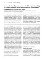
valine. Interestingly, an evolution of the representation of
scissile bonds emerged during the selection (Fig. 2); the
highest variation was observed for the Arg–Ser scissile bond
with continuously increasing recovery of 14, 32, and 42%,
respectively, for the fifth, sixth, and eighth rounds of
selection. A slight increase was also observed in the Arg–
Met and Arg–Ala motif, while an important decrease was
observed for the Arg–Val motif through the selection, which
completely disappeared after eight rounds. The positions
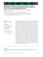
surrounding the scissile bond at the P3, P2, and P¢2subsites
predominantly favoured small or uncharged residues as seen
by the 65, 55, and 70% recovery (Fig. 3). Of these small or
uncharged residues, none in particular was observed more
frequently at these positions. Hydrophobic residues also
appeared in the P3 and P2 subsites in 20% of peptides
whereas no aromatic residues were recovered in the P3 and
P¢2 positions.
CFP fluorescent substrate assay
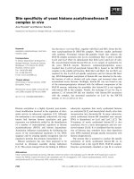
A simple and direct system has been developed to
determine the kinetics of peptide substrate selection from
a phage display library (Fig. 1). All CFP recombinant
proteins can be produced with good yields in bacteria
(1 mg per 50 mL of culture) with 75% being refolded in
stable conformations. To generate the substrate phage, the
CFP–substrate molecule is attached by a His
6
tail to Ni
2+
-
Fig. 1. Schematic outline of the approach used to select substrates for
kallikrein hK2. (1) Phage displaying random peptides fused to a his-
tidine tail (His) are immobilized on an affinity support (Ni
2+
-nitrilo-
triacetic acid sepharose beads). (2) After treatment with kallikrein hK2,
phages expressing sensitive substrates are released from the solid phase,
(3) and are then used to infect F-positive bacteria (4) to be amplified for
a next step of selection (5). Phages from the last round of selection are
cloned by plating onto Petri dishes (6) and DNA of individual phages
are amplified in region encoding for the substrate to determine the
sequences cleaved by the enzyme. (7) Gene encoding the random
substrate was subcloned into an expression vector, in order to be
produced as a fusion protein between the CFP protein and a histidine
tag. (8) The CFP-X5-his protein was fixed to Ni
2+
-nitrilotriacetic acid
magnetic beads and (9) treated by the protease hK2. The released CFP
fluorescent protein was measured with a fluorescence reader (10) which
permitted to determine the percentage of hydrolysis, the specificity
constant and the site of cleavage (11).
Ó FEBS 2002 Substrate specificity of kallikrein hK2 (Eur. J. Biochem. 269) 2749
nitrilotriacetic acid beads; the substrate can then be released
by hydrolysis only. By using two CFP–substrates harbour-
ing a substrate that is either cleavable or resistant to hK2,
we showed that the CFP recombinant protein is cleaved
only in the substrate region and not within the CFP
sequence as no fluorescence was detectable with CFP-
resistant. On the other hand, CFP–PCI was efficiently
cleaved with a first-order curve for the product generation
(data not shown) and the specificity constant k
cat
/K
m
was
20 000
M
)1
Æs
)1
.
Under the same conditions, hK2 cleaved the other 30
peptides constructed as CFP–substrates with catalytic
efficiencies (k
cat
/K
m
) ranging from 1.7 · 10
4
M
)1
Æs
)1
for
LRSRA to 9.9 · 10
1
M
)1
Æs
)1
for peptide ERVSP. Thus,
there is about a 170-fold difference in the efficacy of
cleavage between the different substrates selected by phage
Table 1. Alignment of translated amino acid sequences of random peptide clones selected by substrate phage display with hK2.
Clone
Scissile
bond P5 P4 P3 P2 P1 P¢1 P¢2P¢3P¢4
6.1 RS M T R S N
6.6 K T R S N
6.8 I S P R S
6.11 G V F R S
6.19 G T V R S
5.5 E T K R S
5.2 L G R S L
8.3 R G R S E
6.2 R R S I D
8.11 V L R S P
8.20 L R S R A
8.5 RS GS V
8.9 A R A R S
8.18 RT S D R T A
6.7 K L R T T
8.13 RA R A A M M
5.3 T R A P M
8.17 P G R A P
6.9 V E S R A
6.20 A R A S E
5.19 RV T L Q R V
5.16 R L E R V
5.18 E R V S P
5.12 S S P R V
6.17 RVGP Y
6.4 RM P S A R M
6.14 R G R M A
6.5 T V R M P
8.12 L R M P T
8.14 H R M S S
5.11 RP R P Q E L
6.15 V R P L E
5.7 RL S G R L A
6.12 RF G T L R F
5.1 RN Q W R N S
5.14 RNDKL
6.13 M R N R A
8.19 RD T R D S R
5.4 T G S R D
5.10 RQ I M S R Q
6.3 KG L T T S K
Fig. 2. Frequency of selection of the different scissile bonds.
2750 S. M. Cloutier et al. (Eur. J. Biochem. 269) Ó FEBS 2002
display substrate. The best substrate peptide, giving specif-
icity constant approaching the PCI–peptide and a percent-
age of hydrolysis superior to 90%, contained a serine
residue in P¢1 subsite whereas the less sensitive peptide
contained a valine, an observation that correlates with the
evolution of the number of different scissile bonds during
the selection. The only peptide cleaved at a Lys had a low
specificity constant and gave a percentage of hydrolysis of
only 20% confirming the preference for arginine in the P1
position. No cleavage was observed with the two peptides
that did not contain either arginine or lysine suggesting a
residual background among the selected substrates. All
peptides having a k
cat
/K
m
superior to 5.7 · 10
3
M
)1
Æs
)1
possess two basic amino acids N-terminal to the scissile
bond except for peptide LRSRA where the second basic
residue was found at P¢2(Table2).
Comparison with natural substrate
When compared to previously reported substrates for hK2,
the peptides selected here had scissile bonds containing the
Arg–Ser motif, which is the same bond cleaved in PCI, a
natural inhibitor of hK2 found in seminal plasma, as well as
semenogelin I, antithrombin III, and kininogen. The Arg–
Thr and Arg–Leu motifs are hydrolysed by hK2 in
semenogelins I and II whereas the Arg–Met motif is cleaved
in the plasminogen activator inhibitor-1 and the Arg–Gln
motif is cleaved in IGF-BP-2. Using each of the 44
pentapeptides substrate sequences,
FASTA
and
BLAST
searches were done to look for new potential human protein
substrates of hK2 (Table 3). Among the 11 identical
matches (data not shown), three putative targets were
identified for hK2: ADAM-TS 8 precursor, cadherin-rela-
ted tumour suppressor homologue precursor, and collagen
(IX) chain precursor matching peptides RGRSE, GVFRS
and PGRAP, respectively.
DISCUSSION
A wide variety of critical processes depend on specific
cleavage of targets by different enzymes so an ability to
discriminate among many potential substrates is crucial to
maintaining the fidelity of most biological functions.
However, unnatural cleavage can occur through unpredict-
able reactions between protease and substrate provoking
unexpected biological events such as degradation of extra-
cellular matrix, over-availability of growth factors, or
degradation of tumor suppressor proteins. In the last
5 years, evidence has been mounting that support a role for
hK2 in metastasis and cancer progression by virtue of its
in vitro proteolysis of several biological substrates involved
in cancer biology [8–11]. However, further investigation is
needed to verify this hypothesis. Previously biochemical
characterizations were incomplete due to the limit of
classical iterative methods using already existing or modified
substrates [12,24].
The unbiased approach used in this study clearly defined
the preferential recognition sites for hK2 substrate hydro-
lysis. The phage display substrate technique enables millions
of substrates to be screen simultaneously in a single reaction
[13,25]. Large biological libraries are constructed by
displaying random sequences on the extremities of filamen-
tous phages, then amplified and screened toward a protease
to survey rapidly its specificity.
Most reports using phage display substrate to character-
ize proteases have not reported the extent of diversity of the
library used in the screening, this being a direct product of
the number of different combinations of amino acids
displayed by the phage. Cloning substrates comprising more
than six residues is limited by transformation efficacy, thus
the ability to obtain completely adequate diversity with that
number of amino acids is questionable [26].
Phage display does generate libraries that are many times
more diverse,however, than those using other methods such
as combinatorial chemistry [17] or immobilized positional
peptide libraries [27].
In our experiments, randomised pentapeptides were fused
to a truncated form of g3p to produce a library of 1.8 · 10
8
independent recombinant phages where all possible combi-
nations of sequences even the rarest polypeptides, are
represented. The screening of this library with hK2 showed
that no phage was in duplicate which is in contrast to
selections with other types of phage display libraries
(antibody fragments, ligands, or peptide binders) where
selections often identified only the best clones with highest
reactivity [28,29]. Our results are consistent with other
studies using phage display that reported a broad diversity
but good enrichment in the selection of specific enzyme
substrates [13,15,25].
The determination of the specificity constants of the
substrates showed a positive tendency during the selection.
Most of the better substrates were taken the last rounds.
However, this does not preclude that bad substrates could
be conserved throughout the screening process despite
selection pressure. Therefore, selected substrates need to be
further tested in other configuration than that of fused to a
phage. The CFP system developed in the present work
enabled direct determination of specificity constants and the
site of cleavage of the substrate selected by phage display, an
improvement over the previously described semiquantitative
method [13,25] and chemical synthesis of substrates [15,30].
The effectiveness of our system was validated through a
recombinant CFP carrying a PCI-derived peptide, a
substrate efficiently cleaved by hK2. The k
cat
/K
m
of the
peptide fused to CFP was significantly lower than that
obtained with the same sequence as synthetic fluorogenic
form [12]; this difference could be explained by the
Fig. 3. P3-P¢2 substrate specificity profile of hK2 from selected peptides
tested as CFP fusion protein. Alignment of translated amino-acid
sequences of random peptide clones selected by substrate phage display
with hK2.
Ó FEBS 2002 Substrate specificity of kallikrein hK2 (Eur. J. Biochem. 269) 2751
modification in the Km caused by the peptide being linked
to a fairly large protein (30 kDa). In addition, hydrophobic
fluorophores used to make intramolecularly quenched
fluorogenic substrates are known to modify the affinity of
peptide for the active site of enzyme, increasing the Km [31].
Our results showing that hK2 cleaves quite selectively
after an arginine residue, concurs with previous reports
[12,24]. Nearly one-third of all selected peptides are cleaved
at the Arg–Ser bond despite the large variety of residues
being recovered at the P¢1 position. This result shows that
hK2 can accommodate a broad range of amino acids,
except for basic residues, in the P¢1 position. The strong
preference for small or noncharged residues is also observed
in P3, P2, and P¢2 subsites but no consensus could be
deduced among the amino acids from the selected sequences.
Despite this observation, hK2 seems to be dependent on a
more extended site of binding than R–S bond for an efficient
catalysis as some Arg–Ser peptides possess lower specificity
constants. Nonetheless, the observation that the best three
peptides are cleaved as efficiently as the sequence of PCI–
peptide obtained by the classic iterative methods indicates
the impressive ability of substrate phage technology to
elucidate optimal subsite occupancy for proteases from
large banks of randomly selected candidates.
Interestingly, the Arg–Ser scissile bond found in numer-
ous natural substrates like PCI, semenogelins I and II,
fibronectin and kininogen as well as other preferential
cleavage sites like Arg–Thr or Arg–Met in seminal coagu-
lum proteins and in plasminogen activator inhibitor-1,
respectively; is also preferentially selected by hK2 using
phage display substrates thus confirming the success of
phage display substrate selection.
Finally, a SwissProt database search with selected
sequences identified three potential human protein sub-
strates for hK2. Regions identified in different substrates are
extracellular and thus accessible to proteases. These poten-
tial substrates are not yet well characterized, but are
suspected to be involved in cancer progression. For
example, the desintegrin-like and metalloprotease domain
with thrombospondin type I modules 8 (ADAM-TS8)
Table 2. Comparaison of specificity constant (k
cat
/K
m
) values and the percentage hydrolysis of CFP-X5-his based on selected substrates with hK2.
(Scissile bonds are designated by fl.)
Clone Sequence K
obs
(s
)1
) Hydrolysis (%) k
cat
/K
m
(
M
)1
Æs
)1
)
PCI TFRflSA 3.66 E-04 84.4 19 284
8.20 LRflSRA 3.22 E-04 89.7 16 926
6.2 RRflSID 2.85 E-04 97.9 14 982
8.3 RGRflSE 2.77 E-04 96.2 14 605
6.7 KLRflTT 1.83 E-04 57.1 9646
8.9 ARARflS 1.46 E-04 61.9 7659
6.14 RGRflMA 1.10 E-04 55.8 5765
6.4 PSARflM 1.02 E-04 47.2 5389
8.12 LRflMPT 9.80 E-05 33.1 5158
6.12 GTLRflF 9.54 E-05 43.4 5020
5.5 ETKRflS 8.28 E-05 33.1 4358
6.19 GVFRflS 6.74 E-05 23.2 3545
5.19 TLQRflV 6.53 E-05 24.7 3436
5.3 TRflAPM 6.27 E-05 32.4 3299
8.17 PGRflAP 6.24 E-05 35.1 3282
5.7 SGRflLA 5.78 E-05 30.5 3042
8.11 VLRflSP 5.24 E-05 33.1 2756
8.19 TRDSR 4.84 E-05 30.9 2548
5.10 IMSRflQ 4.77 E-05 27.1 2512
6.5 TVRflMP 4.35 E-05 24.6 2289
6.20 ARflASE 4.10 E-05 23.5 2158
6.6 KTRflSN 3.64 E-05 26.2 1917
6.1 MTRflSN 3.37 E-05 22.9 1772
6.3 LTTSKfl 3.24 E-05 19.6 1705
5.4 TGSRflD 2.69 E-05 16.1 1417
6.15 VRflPLE 2.37 E-05 13.1 1248
5.14 RflNDKL 2.27 E-05 19.1 1196
5.18 ERflVSP 1.89 E-05 11.2 99
MTMQS ND ND ND
QTSLS ND ND ND
Rst AIKFF ND ND ND
Table 3. Identification of potential physiological substrate of hK2 using
the SwissProt data base.
HK2 selected
peptides Sequences Potential protein substrate (residues)
8.3 RGRflSE ADAM-TS 8 precursor (646–50)
6.19 GVFRflS Cadherin-related tumour suppressor
homologue precursor (2473–77)
8.17 PGRflAP Collagen alpha (IX) chain precursor
(753–57)
2752 S. M. Cloutier et al. (Eur. J. Biochem. 269) Ó FEBS 2002
could act as a tumour suppressor through its antiangiogenic
activity [32,33]. Cadherin-related tumour suppressor homo-
logue precursor [34] and collagen alpha (IX) chain precur-
sor, a minor cartilage nonfibrillar collagen associated with
type II collagen fibrils [35], are the two other potential
protein substrates for hK2 that could also have a role in
cancer progression.
In conclusion, we developed an effective phage display
system that enabled rapid and fruitful investigation of hK2
substrate specificity. This powerful technology could
advance the design and the optimization of selective
inhibitors for cancer chemotherapy as well as accelerate
the discovery of new targets. Phage display has already
opened new avenues in kallikrein research that may further
reinforce the role of hK2 in the progression of prostate
cancer.
ACKNOWLEDGEMENTS
This work is supported by a grant from the Ligue Suisse contre le
Cancer. We thank H. Lowman from Genentech Inc. for giving
phagemid and fruitful advices.
REFERENCES
1. Deperthes, D., Chapdelaine, P., Tremblay, R.R., Brunet, C.,
Berton, J., Hebert, J., Lazure, C. & Dube, J.Y. (1995) Isolation of
prostatic kallikrein hK2, also known as hGK-1, in human seminal
plasma. Biochim. Biophys. Acta 1245, 311–316.
2. Tremblay, R.R., Deperthes, D., Teˆ tu,B.&Dube
´
, J.Y. (1997)
Immunohistochemical study suggesting a complementary role of
kallikrein hK2 and hK3 (prostate-specific antigen) in the func-
tional analysis of human prostate tumors. Am.J.Pathol.150,
455–459.
3. Darson, M.F., Pacelli, A., Roche, P., Rittenhouse, H.G., Wolfert,
R.L., Young, C.Y., Klee, G.G., Tindall, D.J. & Bostwick, D.G.
(1997) Human glandular kallikrein 2 (hK2) expression in prostatic
intraepithelial neoplasia and adenocarcinoma: a novel prostate
cancer marker. Urology 49, 857–862.
4. Darson, M.F., Pacelli, A., Roche, P., Rittenhouse, H.G., Wolfert,
R.L., Saeid, M.S., Young, C.Y., Klee, G.G., Tindall, D.J. &
Bostwick, D.G. (1999) Human glandular kallikrein 2 expression in
prostate adenocarcinoma and lymph node metastases. Urology 53,
939–944.
5. Yousef, G.M., Obiezu, C.V., Luo, L.Y., Black, M.H. &
Diamandis, E.P. (1999) Prostase/KLK-L1 is a new member of the
human kallikrein gene family, is expressed in the prostate and
breast tissues, and is hormonally regulated. Cancer Res. 59, 4252–
4256.
6. Diamandis, E.P., Yousef Clements, G.M., Ashworth, J., Yoshida,
L.K., Egelrud, S., Nelson, T., Shiosaka, P.S., Little, S., Lilja, S.,
Stenman, H., Rittenhouse, U.H., & Wain, H.G. (2000) New
nomenclature for the human tissue kallikrein gene family. Clin.
Chem. 46, 1855–1858.
7. Yousef, G.M. & Diamandis, E.P. (2001) The new human tissue
kallikrein gene family: structure, function, and association to
disease. Endocrine Rev. 22, 184–204.
8. Deperthes, D., Frenette, G., Brillard-Bourdet, M., Bourgeois,
L., Gauthier, F., Tremblay, R.R. & Dube, J.Y. (1996) Potential
involvement of kallikrein hK2 in the hydrolysis of the
human seminal vesicle proteins after ejaculation. J. Androl. 17,
659–665.
9. Frenette, G., Tremblay, R.R., Lazure, C. & Dube, J.Y. (1997)
Prostatic kallikrein hK2, but not prostate-specific antigen (hK3),
activates single-chain urokinase-type plasminogen activator. Int. J.
Cancer 71, 897–899.
10. Mikolajczyk, S.D., Millar, L.S., Kumar, A. & Saedi, M.S. (1999)
Prostatic human kallikrein 2 inactivates and complexes with
plasminogen activator inhibitor-1. Int. J. Cancer 81, 438–442.
11. Rehault, S., Monget, P., Mazerbourg, S., Tremblay, R., Gutman,
N., Gauthier, F. & Moreau, T. (2001) Insulin-like growth factor
binding proteins (IGFBPs) as potential physiological substrates
for human kallikreins hK2 and hK3. Eur J. Biochem. 268, 2960–
2968.
12. Bourgeois, L., Brillard-Bourdet, M., Deperthes, D., Juliano,
M.A., Juliano, L., Tremblay, R.R., Dube, J.Y. & Gauthier, F.
(1997) Serpin-derived peptide substrates for investigating the
substrate specificity of human tissue kallikreins hK1 and hK2.
J. Biol. Chem. 272, 29590–29595.
13. Matthews, D.J. & Wells, J.A. (1993) Substrate phage: selection of
protease substrates by monovalent phage display. Science 260,
1113–1117.
14. Matthews,D.J.,Goodman,L.J.,Gorman,C.M.&Wells,J.A.
(1994) A survey of furin substrate specificity using substrate phage
display. Protein Sci. 3, 1197–1205.
15.Coombs,G.S.,Bergstrom,R.C.,Pellequer,J.L.,Baker,S.I.,
Navre, M., Smith, M.M., Tainer, J.A., Madison, E.L. & Corey,
D.R. (1998) Substrate specificity of prostate-specific antigen
(PSA). Chem. Biol. 5, 475–488.
16. Ohkubo, S., Miyadera, K., Sugimoto, Y., Matsuo, K., Wierzba, K.
& Yamada, Y. (1999) Identification of substrate sequences for
membrane type-1 matrix metalloproteinase using bacteriophage
peptide display library. Biochem. Biophys. Res. Commun. 266,
308–313.
17. Harris,J.L.,Backes,B.J.,Leonetti,F.,Mahrus,S.,Ellman,J.A.&
Craik, C.S. (2000) Rapid and general profiling of protease speci-
ficity by using combinatorial fluorogenic substrate libraries. Proc.
Natl Acad. Sci. USA 97, 7754–7759.
18. Frenette, G., Deperthes, D., Tremblay, R.R., Lazure, C. & Dube,
J.Y. (1997) Purification of enzymatically active kallikrein hK2
from human seminal plasma. Biochim. Biophys. Acta 1334, 109–
115.
19. Knight, C.G. (1995) Fluorimetric assays of proteolytic enzymes.
Methods Enzymol. 248, 18–34.
20. Lowman, H.B., Bass, S.H., Simpson, N. & Wells, J.A. (1991)
Selecting high-affinity binding proteins by monovalent phage
display. Biochemistry 12, 10832–10838.
21. Smith, G.P. & Scott, J.K. (1993) Libraries of peptides and proteins
displayed on filamentous phage. Methods Enzymol. 217, 228–257.
22. Laemmli UK (1970) Cleavage of structural proteins during
the assembly of the head of bacteriophage T4. Nature 227, 680–
685.
23. Elmoujahed, A., Gutman, N., Brillard, M. & Gauthier, F. (1990)
Substrate specificity of two kallikrein family gene products
isolated from the rat submaxillary gland. FEBS Lett. 265,
137–140.
24. Mikolajczyk, S.D., Millar, L.S., Kumar, A. & Saedi, M.S. (1998)
Human glandular kallikrein, hK2, shows arginine-restricted spe-
cificity and forms complexes with plasma protease inhibitors.
Prostate 34, 44–50.
25. Smith, M.M., Shi, L. & Navre, M. (1995) Rapid identification of
highly active and selective substrates for stromelysin and
matrilysin using bacteriophage peptide display libraries. J. Biol.
Chem. 270, 6440–6449.
26. Lowman, H.B., Bass, S.H., Simpson, N. & Wells, J.A. (1991)
Selecting high-affinity binding proteins by monovalent phage
display. Biochemistry 30, 10832–10838.
27. Hu, Y., Webb, E., Singh, J., Morgan, B.A., Gainor, J.A., Gordon,
T.D. & Siahaan, T.J. (2001) Rapid determination of substrate
specificity of clostridium histolyticum {phi}-collagenase using an
immobilized peptide library. J. Biol. Chem. 277, 8366–8371.
28. Wrighton, N.C., Farrell, F.X., Chang, R., Kashyap, A.K.,
Barbone, F.P., Mulcahy, L.S., Johnson, D.L., Barrett, R.W.,
Ó FEBS 2002 Substrate specificity of kallikrein hK2 (Eur. J. Biochem. 269) 2753
Jolliffe, L.K. & Dower, W.J. (1996) Small peptides as potent
mimetics of the protein hormone erythropoietin. Science 273,
458–464.
29. Hoogenboom, H.R. & Chames, P. (2000) Natural and designer
binding sites made by phage display technology. Immunol. Today
21, 371–378.
30. Harris, J.L., Peterson, E.P., Hudig, D., Thornberry, N.A. &
Craik, C.S. (1998) Definition and redesign of the extended sub-
strate specificity of granzyme B. J. Biol. Chem. 273, 27364–27373.
31. Charitos, C., Tzougraki, C. & Kokotos, G. (2000) Synthesis and
fluorescence properties of intramolecularly quenched fluorogenic
p-nitroanilides containing coumarin or quinolinone derivatives as
fluorophores. J. Pept. Res. 56, 373–381.
32. Georgiadis, K.E., Hirohata, S., Seldin, M.F. & Apte, S.S. (1999)
ADAM-TS8, a novel metalloprotease of the ADAM-TS family
located on mouse chromosome 9 and human chromosome 11.
Genomics 62, 321–315.
33. Wang, S.S., Virmani, A., Gazdar, A.F., Minna, J.D. & Evans,
G.A. (1999) Refined mapping of two regions of loss of hetero-
zygosity on chromosome band 11q23 in lung cancer. Genes
Chromosomes Cancer 25, 369–387.
34. Dunne, J., Hanby, A.M., Poulsom, R., Jones, T.A., Sheer, D., Da
Chin, W.G.S.M., Zhao, Q., Beverley, P.C. & Owen, M.J. (1995)
Molecular cloning and tissue expression of FAT, the human
homologue of the Drosophila fat gene that is located on chro-
mosome 4q34-q35 and encodes a putative adhesion molecule.
Genomics 30, 207–223.
35.Muragaki,Y.,Kimura,T.,Ninomiya,Y.&Olsen,B.R.
(1990) The complete primary structure of two distinct forms of
human alpha 1 (IX) collagen chains. Eur J. Biochem. 192, 703–708.
2754 S. M. Cloutier et al. (Eur. J. Biochem. 269) Ó FEBS 2002