Randomized trial of surveillance with abbreviated mri in women with a personal history of breast cancer impact on patient anxiety and cancer detection
Bạn đang xem bản rút gọn của tài liệu. Xem và tải ngay bản đầy đủ của tài liệu tại đây (794.78 KB, 7 trang )
(2022) 22:774
Fonseca et al. BMC Cancer
/>
RESEARCH ARTICLE
Open Access
Randomized trial of surveillance
with abbreviated MRI in women with a personal
history of breast cancer– impact on patient
anxiety and cancer detection
Marina Mohallem Fonseca1, Tasneen Alhassan2, Yashmin Nisha3, Diana Koszycki4,5, Betty Anne Schwarz6,
Roanne Segal7, Angel Arnaout8, Tim Ramsay9, Jacqueline Lau10 and Jean M. Seely11*
Abstract
Background: Abbreviated breast MRI (A-MRI) substantially reduces the image acquisition and reading times and has
been reported to have similar diagnostic accuracy as a full diagnostic protocol but has not been evaluated prospectively with respect to impact on psychological distress in women with a prior history of breast cancer (PHBC). This
study aimed to determine if surveillance mammography (MG) plus A-MRI reduced psychological distress and if A-MRI
improved cancer detection rates (CDR) as compared to MG alone.
Methods: This prospective controlled trial of parallel design was performed at a tertiary cancer center on asymptomatic women with PHBC who were randomized into two groups: routine surveillance with MG or intervention of MG
plus A-MRI in a 1:1 ratio. Primary outcome was anxiety measured by four validated questionnaires at three different
time-points during the study. Other parameters including CDR and positive predictive value for biopsy (PPV3) were
compared between imaging modalities of MG and A-MRI. Tissue diagnoses or 1 year of follow-up were used to
establish the reference standard. Linear mixed models were used to analyze anxiety measures and Fisher’s exact test
to compare imaging outcomes.
Results: One hundred ninety-eight patients were allocated to either MG alone (94) or MG plus A-MRI (104). No
significant group difference emerged for improvement in trait anxiety, worry and perceived health status (all Timeby-surveillance group interaction ps > .05). There was some advantage of A-MRI in reducing state anxiety at Time
2 (p < .05). Anxiety scores in all questionnaires were similarly elevated in both groups (50.99 ± 4.6 with MG alone vs
51.73 ± 2.56 with MG plus A-MRI, p > 0.05) and did not change over time. A-MRI detected 5 invasive cancers and 1
ductal carcinoma in situ (DCIS), and MG detected 1 DCIS. A-MRI had higher incremental CDR (48/1000(5/104) vs MG
5/1000(1/198, p = 0.01)) and higher biopsy rates (19.2% (20/104) vs MG 2.1% (2/94), p < 0.00001) with no difference in
PPV3 (A-MRI 28.6% (6/21) vs MG 16.7% (1/6, p > .05).
Author Note This study has been presented previously at the European
Congress of Radiology, in Clinical Trials of Radiology, July 2020.
*Correspondence:
11
Departments of Radiology and Surgery, Department of Medical Imaging,
The Ottawa Hospital, Ottawa Hospital Research Institute, University of Ottawa,
General Campus, 501 Smyth Rd, Ottawa, ON K1H 8L6, Canada
Full list of author information is available at the end of the article
© The Author(s) 2022. Open Access This article is licensed under a Creative Commons Attribution 4.0 International License, which
permits use, sharing, adaptation, distribution and reproduction in any medium or format, as long as you give appropriate credit to the
original author(s) and the source, provide a link to the Creative Commons licence, and indicate if changes were made. The images or
other third party material in this article are included in the article’s Creative Commons licence, unless indicated otherwise in a credit line
to the material. If material is not included in the article’s Creative Commons licence and your intended use is not permitted by statutory
regulation or exceeds the permitted use, you will need to obtain permission directly from the copyright holder. To view a copy of this
licence, visit http://creativecommons.org/licenses/by/4.0/. The Creative Commons Public Domain Dedication waiver (http://creativeco
mmons.org/publicdomain/zero/1.0/) applies to the data made available in this article, unless otherwise stated in a credit line to the data.
Fonseca et al. BMC Cancer
(2022) 22:774
Page 2 of 14
Conclusion: There was no significant impact of A-MRI to patient anxiety or perceived health status. Compared to MG
alone, A-MRI had significantly higher incremental cancer detection in PHBC. Despite a higher rate of biopsies, A-MRI
had no demonstrable impact on anxiety, worry, and perceived health status.
Trial registration: ClinicalTrials.gov (NCT02244593). Prospectively registered on Sept. 14, 2014.
Keywords: Breast cancer, MRI, Anxiety, Abbreviated, Mammography
Background
Women with a prior personal history of breast cancer (PHBC) often have a high level of anxiety related to
breast cancer surveillance [1]. Their actual recurrence
rates are estimated in the order of 1% per year [2, 3], and
depend on tumor size, histology and nodal status at diagnosis, with 5-year risks of recurrence of 7% for stage I,
11% for Stage II and 13% for stage III, and distant recurrences of 10- 41% at 20 years after completion of adjuvant
chemotherapy [4, 5]. Early detection decreases mortality for women with breast cancer [6–8]. In women with
PHBC, the survival benefit is improved if new or recurrent breast cancer is found on surveillance mammography (MG) instead of physical examination [9]. However,
MG has been shown to be less sensitive in women with
PHBC, with sensitivity of 65.4% compared with 76.5% in
women with no PHBC [10]. Breast MRI is the most sensitive test for detecting breast cancer [11]. Breast MRI
is currently recommended for women with PHBC and
dense tissue or those diagnosed by age 50, as per American College of Radiology (ACR) guidelines [12]. Several
other national guidelines do not recommend surveillance imaging with breast MRI after a personal history of
breast cancer unless someone has a hereditary mutation
or mammographically occult malignancy and of itself,
breast tissue density is not an indication for surveillance
breast MRI. Compliance with MRI screening has been
shown to be low, on the order of 25%, due both to lack of
availability and high costs associated with lengthy acquisition times [13, 14]. Abbreviated breast MRI (A-MRI),
which substantially reduces the image acquisition and
reading times, has been reported to have similar diagnostic accuracy as a full diagnostic protocol [13, 15–19].
Currently, A-MRI has not been adopted as the standard for screening for breast cancer and more studies are
required to evaluate outcomes.
Prior studies demonstrated that supplementary MRI
surveillance in women at high risk of breast cancer does
not impact anxiety, cancer-specific distress or healthrelated quality of life [1, 20]. This is the first study to our
knowledge to evaluate the psychological effect of adding abbreviated MRI to MG surveillance in women with
PHBC.
The primary purpose of the current study was to
determine if the intervention of adding A-MRI to MG
surveillance was more effective than MG alone in reducing patient anxiety and, secondarily, if A-MRI improved
cancer detection in women with PHBC. Our hypothesis
was that the MRI group would be superior to mammography alone group to reduce patient anxiety.
Methods
Study subjects
This prospective randomized controlled trial of parallel
design was performed at a large tertiary care academic
medical center and was approved by the hospital’s institutional review board. Our study adheres to the CONSORT guidelines. Patients at a single tertiary care cancer
center were approached by their treating oncologists or
surgeons during routine clinical appointments if they
met the eligibility criteria and their scheduled appointment time allowed. The patients’ oncologists or surgeons
obtained written informed consent. Eight oncologists
and three breast surgeons recruited patients between
2/1/2015 and 4/30/2019. Patients were followed for a
minimum of 12 months.
The eligibility criteria included: (a) female patients
18 years or older; (b) PHBC (including DCIS and invasive
ductal or lobular carcinoma); (c) prior unilateral mastectomy or breast conservation surgery; (d) treatment
for breast cancer completed; and (e) no symptoms of
breast cancer. Patients were excluded if they were considered high-risk (lifetime risk ≥ 25%) [21], were unable to
undergo an MRI due to either physical or mental issues
(i.e.: severe claustrophobia, allergy to gadolinium, severe
renal failure), had bilateral mastectomies, were pregnant
or breastfeeding, or had undergone a breast MRI within
the last 6 months. Regular surveillance imaging consisted
of annual surveillance MG, irrespective of breast tissue
density. All patients had undergone prior mammographic
imaging, and some (< 50%) had undergone prior breast
MRI imaging.
Eligible patients were randomized in a 1:1 allocation
ratio to one of the two arms of the study: 1) surveillance with MG or 2) MG plus A-MRI, with use of permuted blocks of variable length (2, 4, and 6) to ensure
that recruiting physicians remained unaware of the randomization. Researchers or study participants were not
blinded to their allocation. Patients could only participate
once in the study.
Fonseca et al. BMC Cancer
(2022) 22:774
Imaging technique and interpretation
All mammographic examinations were performed using
a full-field digital technique (Hologic, Bedford, MA,
USA) in accordance with national guidelines. Standard
two-dimensional craniocaudal (CC) and mediolateral
oblique (MLO) views were obtained.
All abbreviated dynamic contrast material-enhanced
breast MRIs were performed with one 3 T system (Magnetom TrioTim Syngo, Siemens). The standardized protocol consisted of 8-channel breast coil (Sentinelle Medical
Inc.), T1 localizer, T1 dynamic contrast-enhanced fatsuppressed with one precontrast and one 2 min postcontrast (3D transverse, phase encoding direction right to
left, phase resolution of 60%, phase partial Fourier 6/8, no
interpolation, FA 10 degrees, TR 4.07 ms and TE 1.96 ms,
no IR, NEX 1, Voxel size: 1 × 1x1 mm, acceleration factor 4, no interpolation, base resolution 448,1:01 min,
slice thickness 1 mm). Post-processing axial subtracted
sequences and axial and sagittal maximum intensity
projection were generated of the subtracted images. No
T2-weighted sequences were obtained. For all examinations, gadolinium contrast material (Gadovist) was power
injected (0.1 mmol/kg at 2 mL/s) followed by a 20 mL
saline flush. The entire protocol took 3 min.
Surveillance MG and A-MRI were reviewed by one
of two breast radiologists independently (the first with
8 years of experience reading mammography and breast
MRI and the second reader with 20 years reading mammography and breast MRI) using ACR Breast ImagingReporting Data System (BI-RADS) lexicon[22]. For
patients in the A-MRI group, MG and A-MRI studies
were performed on the same day according to the protocol. Radiologists were not blinded but reported each
modality separately according to the imaging modality
findings, with the mammograms interpreted first. Based
on the imaging findings, additional mammographic
images, including diagnostic tomosynthesis, or targeted
ultrasound were requested at the discretion of the interpreting radiologist. Findings and management were communicated to the patient by telephone by the reporting
radiologist. Subsequent imaging was performed on separate visits, within 3 weeks of the MG or A-MRI. Histologic samples for pathologic diagnosis were obtained
under ultrasound (14G, 5–6 cores), stereotactic (10G,
6–12 cores) or MRI (10G, 6–12 cores) guidance.
Anxiety measures
Patients in both groups were asked to fill out four validated self-report questionnaires that measure anxiety
level and overall health [23–26]) (see supplemental materials). The primary outcome was the State-Trait Anxiety
Inventory (STAI) [23]. This STAI consists of two separate
Page 3 of 14
20-item scales that assess state anxiety (S-Anxiety) (i.e.,
how the person feels at this moment) and trait anxiety
(T-Anxiety) (i.e., how the personal generally feels). The
items are rated on a 1 to 4 scale with total scores ranging from 20–80. Cut-off scores of ≥ 32.2 and ≥ 31.8 indicate elevated levels of state and trait anxiety, respectively.
Both STAI scales have solid psychometric properties
and are sensitive to assessment of longitudinal change.
There are no validated cutoff scores for the STAI scales
in women with PHBC, however a cutoff score of 41 on
the trait form of the STAI and 44 on the state form of
the STAI have been used in previous research to identify clinical levels of anxiety in women with breast cancer [27, 28]. Other psychological measures included the
Penn State Worry Questionnaire (PSWQ) [24], Breast
Cancer Worry Scale (BCWS) [25], and the Health Status Questionnaire 12 (HSQ-12) [26]. The PSWQ [24] is
a 16-item self-report questionnaire which measures frequency and intensity of worry symptoms. Items are rated
on a 5-point scale, with total scores ranging from 16–80.
A score between 16–39 indicates low worry, 40–59 moderate worry and 60–80 high worry. The BCWS [25] is a
3-item scale which measures frequency of breast cancer
worry and the impact of worrying on mood and ability
to perform daily activities. Higher scores indicate greater
cancer worry. The HSQ-12 [26] assesses the impact of
health on social, emotional and physical functioning over
the past four weeks. Depending on the item, questions
are rated of a 3-point, 5-point and 6-point scale. Items
were recoded using the method described by Barry et al.
[26]. Total HSQ scores range from 0 to 800, with higher
scores indicating better health status. The questionnaires
were completed upon enrolment during consultation
at time 1 (T1) when the patients were due for their surveillance test(s) to measure baseline levels of anxiety, at
time 2 (T2) that occurred after the patient received of
their surveillance MG and/or MRI test results, and then
6 months later at time 3 (T3), to determine if there was
a sustained effect observed from the type of surveillance test. T3 questionnaires were mailed to patients and
returned to the study coordination center.
Data collection and statistical analysis
Medical records were reviewed to determine patient
age, family history of breast and/or ovarian cancer in
a first-degree relative, surgery modality, initial breast
tumor stage (TNM), histology, hormone receptor status, months since diagnosis of breast cancer and breast
density. Results were compared between the two groups.
For malignant or atypical/high-risk lesions, surgical
pathologic results were reviewed when available. Imaging and clinical follow-up were determined by review
of the hospital picture archiving system (PACS) and
Fonseca et al. BMC Cancer
(2022) 22:774
medical records as well as the digital imaging repository which includes all clinics and hospitals that serve
the region’s population of 1.2 million. The emigration
rate in the region is < 0.5% per year [29]. Imaging followup for all patients with benign imaging or pathology was
documented with the date of the most recent negative
mammogram.
The anxiety measures were analyzed using SPSS Statistics version 25. Analysis was based on intent-to-treat
(ITT) principles. Data were analyzed using linear mixed
models, with surveillance groups (MG only versus MG
plus A-MRI), time of assessment (T1, T2, T3), and Intervention by Time interaction as fixed factors. Models were
estimated using Restricted Maximum Likelihood (REML)
with an unstructured covariance structure to account for
correlations among repeated measures over time. A significant Time by surveillance group interaction would
suggest that changes in measures over time were different
between the surveillance method; significant interactions
were further analyzed with pairwise least square mean
comparisons. Data from missing questionnaires were
not imputed because our analytical strategy using REML
allowed the estimation of reliable parameters without
the need for imputation of the data under an assumption
of missing at random (MAR) [30]. Descriptive statistics
were calculated using a spreadsheet software program
(Excel, Version 2013, Microsoft). Screening outcomes
were compared between groups using Fisher’s exact test.
Sample size calculation was based on primary outcome
the STAI. There is no generally accepted minimal clinically important difference in the STAI subscales and a
4-point difference was selected to be a minimal clinically
important difference. This was based on previous study
by Millar et al. [28] which used a 4-point difference in
the STAI and on consensus with the research team and
the experience of the psychologist researcher. In order to
have 80% power to detect a 4-point difference between
the groups at any of the three time points, we planned
134 patients per group. Recruitment stopped early due to
differences in cancer detection rates (CDR). Results were
considered significant if p < 0.05.
Imaging modalities (MG, A-MRI), and BI-RADS final
assessment categories for each modality were noted.
Imaging findings and outcomes were documented for all
BI-RADS 3, 4 and 5 lesions, including suspicious extramammary findings. Results were compared between MG
and A-MRI. A screening examination was considered as
positive when additional diagnostic imaging was recommended prior to the next routine screening examination
and included BI-RADS 0, 3, 4 and 5, defined as abnormal interpretations. True positive findings were defined
as a cancer diagnosis within 12 months of a positive
screening examination. Imaging studies were considered
Page 4 of 14
false negatives if there was a tissue diagnosis of cancer
within 12 months of a negative study, or in the surveillance groups if there was a tissue diagnosis of cancer in
the follow-up period. The following performance metrics
were calculated for each modality: CDR, abnormal interpretation rate (AIR), biopsy rate, positive predictive value
for biopsy recommendations (PPV2 = biopsies recommended/cancers diagnosed), positive predictive value for
biopsies performed (PPV3 = biopsies performed/cancers
diagnosed), sensitivity and specificity.
Results
A total of 202 of 1000 patients fulfilled the eligibility criteria (Fig. 1) between 2/1/2015 and 4/30/2019. At enrollment, 94 were randomized to surveillance with MG alone
and 108 to MG plus A-MRI. Of these, four patients from
MG plus A-MRI group withdrew from the study a few
days before undergoing the imaging for different reasons: two patients were discovered to have breast cancer
metastases on separate imaging done prior to undergoing the surveillance imaging, one patient developed sepsis before the imaging was performed and her doctor
decided to postpone contrast injection and one patient
opted to withdraw from the study before undergoing the
imaging. Accordingly, the study population consisted of
198 patients: 47.5% (94/198) randomized to regular surveillance with MG and 52.5% (104/198) to surveillance
with MG plus A-MRI. All patients completed the imaging to which they were randomized and there were no
patient crossovers from the MG only group to A-MRI.
Among the 104 patients who had MG plus A-MRI,
82.7% (86/104) had both imaging exams the same day
and 17.3% (18/104) on different days (average 33.2 days
(range: 1–147)) due to various scheduling conflicts.
Baseline demographic and clinical characteristics
Patients’ demographic and clinical characteristics are
presented in Table 1. No important differences in age,
family history of breast and/or ovarian cancer, surgery
modality, months since diagnosis, breast density, initial
tumor histology, stage, or hormone receptor status were
noted between the two groups, although a nonsignificant
higher number of patient with triple negative cancers was
observed in the group that received A-MRI.
Results regarding anxiety
The observed means (± standard deviations) for the selfreport questionnaires and least square mean difference
between the surveillance groups at T2 and T3 are displayed in Table 2. 197 participants completed questionnaires at T1 (Baseline), 92 patients in MG only and 105
in MG plus A-MRI groups. At Time 2, data were available
for 143 participants; 60 in MG and 83 in MG plus A-MRI
Fonseca et al. BMC Cancer
(2022) 22:774
Page 5 of 14
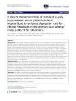
Fig. 1 CONSORT Flow diagram of trial
groups. At T3 data were available for 102 participants;
38 women in MG and 64 in MG plus A-MRI groups. The
surveillance groups did not differ significantly on any of
the baseline measures. Linear mixed models revealed
that our primary outcome STAI-Trait Anxiety did not
change over time (Time main effect p = 0.51) and did not
differ between the groups (Time x Surveillance Group
interaction p = 0.20). However, there was a significant
Time main effect (p < 0.001) and Time x Surveillance
Group interaction (p = 0.022) for the STAI-State Anxiety. Post-hoc tests revealed that for both groups, state
anxiety decreased significantly between T1 and T2 (estimated mean change = -6.80 [95% CI, -8.58 to -5.02] for
MG only (p < 0.001) and -8.17 [95% CI, -9.70 to -6.34] for
MG plus A-MR (p < 0.001), and increased significantly
from T2 to T3 (estimated mean change 5.86 [95% CI, 3.97
to 7.76] for MG only (p < 0.001) and 8.12 [95% CI, 7.23 to
10.40] for MG plus A-MRI (p < 0.001). Between groups
comparisons indicated that state anxiety at T2 was significantly lower in the MG plus A-MRI group (p = 0.03),
but less than a 4-point difference. Levels of worry did not
significantly change over time (Time main effect p = 0.14
and p = 0.73 for the PSWQ and BCWQ, respectively)
and did not differ between the groups (Time x Surveillance Group interaction p = 0.57 and p = 0.48 for the
PSWQ and BCWQ, respectively). There was a significant
Time main effect for self-report health status (p < 0.05),
but no significant Time x Surveillance Group interaction
(p = 0.13). Overall, HSQ-12 scores decreased from T1 to
T3 (estimated mean change -0.27.91 [95% CI -47.60 to
-8.22], p < 01).
Using the cutoff score of 41 on the trait form of the
STAI, the percentage of women with anxiety in the clinical range was 100% at T1 and T2 and T3 for both the MG
Fonseca et al. BMC Cancer
(2022) 22:774
Page 6 of 14
Table 1 Patient demographics according to group (MG versus A-MRI + MG)
Group 1-MG (n = 94)
%
Group 2-A-MRI + MG
(n = 104)
%
P valueb
Age (years)
Mean
59.0
58.2
Median
58.5
58
Range
35–80
38–83
0.44
Family history breast and/ or ovarian cancer
Yes
26
28%
32
31%
0.63
No
68
72%
71
68%
0.53
Unknown
0
0%
1
1%
0.34
Lumpectomy
52
55%
69
66%
0.11
Mastectomy
42
45%
35a
34%
0.11
< 24
42
45%
44
42%
0.73
24
≤ x < 60
37
39%
39
38%
0.79
60
≤ x ≤ 120
13
14%
14
13%
0.94
> 120
2
2%
6
6%
0.19
Unknown
0
0%
1
1%
0.34
ACR A
4
4%
7
7%
0.45
ACR B
45
48%
46
44%
0.61
ACR C
44
47%
45
43%
0.50
ACR D
1
1%
6
6%
0.73
Ductal carcinoma in situ
2
2%
7
7%
0.12
Invasive ductal carcinoma
84
89%
89
86%
0.31
Invasive lobular carcinoma
6
6%
8
8%
0.74
Mucinous carcinoma
1
1%
0
0%
NA
Unknown
1
1%
0
0%
NA
T1
51
54%
44
42%
0.19
T2
27
29%
38
37%
0.14
T3
5
5%
8
8%
0.43
T4
8
9%
5
5%
0.35
NA
1
1%
2
2%
0.59
N0,NX
51
54%
54
52%
0.92
N1
31
33%
33
32%
0.92
M0
94
100%
104
100%
NA
ER positive Her2 negative
69
73%
66
63%
0.33
Her2 positive
12
13%
19
18%
0.21
Triple negative
6
6%
12
12%
0.16
Unknown
4
4%
0
0%
NA
Surgery modality
Months since diagnosis
Breast density
Tumor histology
Tumor stage of initial invasive cancer
Hormone receptor status invasive cancer
a
two patients had mastectomy and contralateral lumpectomy
b
using Fisher’s exact test for comparison between the two groups
and the MG + A-MRI groups. Using a cutoff score of 44
on the state form of the STAI, the majority of women in
the MG and MG + A-MRI groups had scores in the clinical range at T1 (100% and 99%) and T3 (95.9% and 100%).
At T2 however, more (57.1% (32/56)) women in the MG
group than those in MG + A-MRI group (32.9% (27/82))
had scores in the clinical range, with the difference
between groups statistically significant (p < 0.01). Within
Fonseca et al. BMC Cancer
(2022) 22:774
Page 7 of 14
Table 2 Effect of Surveillance Method on Self-Report Measures
Observed means ± standard deviations
Outcome
Time 1
Time 2
Time 3
Estimated mean difference
between groups at Time 2 (95%
CI)
Estimated mean difference
between groups at Time 3
(95% CI)
-0.37 (-1.23 to 0.49)
-0.80 (-1.71 to 0.10)
-2.16 (-4.20 to 0.13)*
- 0.79 (-0.48 to 2.07)
0.83 (-3.4 to 5.1)
2.43 (-1.98 to 6.84)
-0.40 (-1.11 to 0.30)
-0.27 (-0.99 to 0.45)
-6.12 (-48.48 to 36.34)
25.41 (-23.32 to 74.13)
STAI-Trait
MG alone
51.50 ± 2.6
51.55 ± 2.8
51.79 ± 2.5
MG
+ A- MRI
51.61 ± 2.5
51.20 ± 2.2
50.94 ± 2.5
MG alone
51.85 ± 2.8
45.14 ± 5.7
50.99 ± 4.6
MG
+ A-MRI
51.07 ± 2.9
42.88 ± 6.3
51.73 ± 2.6
MG alone
40.23 ± 13.7
39.52 ± 13.4
38.98 ± 14.2
MG
+ A-MRI
42.20 ± 13.4
41.08 ± 14.5
44.41 ± 14.2
MG alone
6.87 ± 2.4
6.93 ± 2.4
6.44 ± 2.1
MG
+ A-MRI
6.73 ± 2.3
6.55 ± 2.3
6.39 ± 2.1
STAI-State
PSWQ
BCWS
HSQ-12
MG alone
617.52 ± 143.7
603.57 ± 122.5
590.54 ± 156.3
MG
+ A-MRI
597.94 ± 145.3
596.73 ± 141.5
611.44 ± 123.5
Note: Analysis is based on the intent-to-treat sample. Questionnaires were completed upon enrolment during consultation (T1; n = 92 MG alone and n = 103
MG + A-MRI), upon receipt of the MG and/or MRI results (T2; n = 61 MG alone and n = 84 MG + A-MRI), and 6- months later (T3; n = 49 MG alone n = 73 MG + A-MRI)
For all questionnaires, higher scores represent higher anxiety or worry
STAI State Trait Anxiety Trait (i.e., how the personal generally feels) and State (i.e., how the person feels at this moment), PSWQ Penn State Worry Questionnaire, BCWQ
Breast Cancer Worry Scale, HSQ Health Status Questionnaire
Range of scores: For the trait-STAI scores range from 20–80 and the state-STAI scores range from 20–80, with cut-off scores of ≥ 31.8 and ≥ 32.2 indicating elevated
levels of trait and state anxiety, respectively. The PSWQ scores range from 16–80 (higher scores indicating higher worry), and the BCWS summed scores range from 3
(low worry) to 12 (high worry). The scores for the HSQ-12 range from 0 to 800, with higher scores indicating better perceived health status
*
p < .05 MG alone vs MG + A- MRI
the MRI group, the 23 recalled patients had significantly
higher PSWQ scores at T2 when compared with the 80
patients who were not recalled (mean score: 47.16 ± 14.7
vs 39.33 ± 14.08, p < 0.05, Cohen’s d = 0.55), with a similar trend found for State STAI scores (45.22 ± 7.1 vs
42.23 ± 5.95, p =
0.075, Cohen’s d
= 0.48). However,
the recalled women had scores that returned to baseline by T3 with no lasting effect on PSWQ (mean score:
48.66 ± 16.29 vs 43.30 ± 13.51, p > 0.05) or State STAI
(52.20 ± 2.32 vs 51.58 ± 2.63, p > 0.05).
Outcomes according to surveillance groups
There was 1 cancer (DCIS) detected in the MG group
during the study, and 3 cancers (2 invasive and 1 DCIS)
were diagnosed at follow-up. In the MG plus A-MRI
surveillance group, 5 breast cancers (1 DCIS and 4 invasive cancers) and 1 breast cancer metastasis to the lung
were detected, with no cancers diagnosed at follow-up.
The outcomes for each group are provided in Table 3.
Although there were significantly more recalls and biopsies performed in the MG plus A-MRI than the MG
group (recalls of 27/104 (26%) vs 4/94 (4.26%) (p < 0.05)
and biopsies of 20/104 (19.2%) vs 2/94 (2.13%)(p = 0.001)
respectively), with lower specificity (77.8% MG + A-MRI
vs 96.7% MG (p = 0.0001), the sensitivity for the MG plus
A-MRI group was higher 5/5(100%) than the MG group
1/4 (25%)(p = 0.048) and there was a higher CDR in the
MG plus A-MRI group (5/104 (48.1/1000)) than the MG
group (1/94 (10.6/1000) (p = 0.1294)).
Findings according to imaging modality
Outcomes according to imaging modality are presented
in Table 4. Among the 302 imaging examinations performed (198 MG and 104 A-MRI), 9 MG and 29 A-MRI
were interpreted as abnormal (17%) (Fig. 2).
Mammography
There were 198 mammographic examinations performed:
94 MG alone and 104 MG with A-MRI; 95.5% (189/198)
were negative or benign (BI-RADS 1 and 2), 4.5% (9/198)
were recalled (BI-RADS 0) and 3.0% (6/198) presented
findings suspicious for malignancy (BI-RADS 4) and
underwent biopsy. One cancer was detected (Table 5)
and no high-risk lesions were identified.