Deletion of trib3 disrupts the tumor progression induced by integrin αvβ3 in lung cancer
Bạn đang xem bản rút gọn của tài liệu. Xem và tải ngay bản đầy đủ của tài liệu tại đây (3.73 MB, 7 trang )
(2022) 22:459
Zhou et al. BMC Cancer
/>
Open Access
RESEARCH
Deletion of TRIB3 disrupts the tumor
progression induced by integrin αvβ3 in lung
cancer
Wen Zhou1,2, Junjun Ma2, Lifeng Meng2, Dabei Liu2 and Jun Chen1*
Abstract
Background: Integrin αvβ3 has been proposed as crucial determinant for tumor sustained progression and a
molecular marker for the estimation of tumor angiogenesis. Our study suggested that integrin αvβ3 could efficiently
promote lung cancer cell proliferation and stem-like phenotypes in a tribbles homolog 3 (TRIB3) dependent manner.
Result: Integrin αvβ3 could mediate the activation of FAK/AKT pro-survival signaling pathway. Meanwhile, activated
TRIB3 interacted with AKT to upregulated FOXO1 and SOX2 expression, resulting in sustained tumor progression in
lung cancer. Our further analysis revealed that TRIB3 was significantly upregulated in lung tumor tissues and correlated with the poor outcome in clinical patients, indicating the potential role of TRIB3 in diagnostic and prognostic
estimation for patients with lung cancer.
Conclusion: Our study showed here for the first time that integrin αvβ3 promote lung cancer development by
activating the FAK/AKT/SOX2 axis in a TRIB3 dependent signaling pathway, and interrupting TRIB3/AKT interaction
significantly improved the outcome of chemotherapy in tumor-bearing mice, representing a promising therapeutic
strategy in lung cancer.
Keywords: Integrin αvβ3, TRIB3, FAK/AKT, Lung cancer
Introduction
Lung cancer is the most common malignant carcinoma
with a leading cause of cancer associated death worldwide. Despite advance in expounding mechanism of
lung carcinogenesis and new surgical/chemotherapeutic protocols, the medium survival time of lung cancer
patients remains less than 5 years [1, 2]. Herein, there is
an urgent demand to explore the underlying mechanism
of lung cancer progression and novel strategies for tumor
therapy.
Integrins are dimeric adhesion receptors that is associated with a series of intracellular signals [3]. Interaction
*Correspondence:
1
Department of Lung Cancer Surgery, Tianjin Medical University General
Hospital, No.154 Anshan Road, Heping District, Tianjin City 300052, China
Full list of author information is available at the end of the article
between integrins and extracellular matrix could regulate diverse cellular functions, which is strictly correlated
with tumor growth and distant metastasis [4]. The alterations in integrin expression level have been extensively
reported and are recognized as crucial determinant for
neoplastic progression. Compelling reports suggested
that the expression of integrin αvβ3 has been detected
in various tumor tissues, which strongly suggested the
potential role integrin αvβ3 in tumor progression [5].
Indeed, increasing evidence demonstrated that integrin
αvβ3 correlated with diverse tumor progression. And
inhibition of integrin αvβ3 signaling could strengthen
antiangiogenic and antitumor effects of radiotherapy in
several tumor types [6, 7]. Also, integrin αvβ3 is capable
of facilitating PI3K/AKT signaling pathway activation to
promote non-small cell lung cancer cells A549 proliferation [8]. However, the underlying mechanism of integrin
© The Author(s) 2022. Open Access This article is licensed under a Creative Commons Attribution 4.0 International License, which
permits use, sharing, adaptation, distribution and reproduction in any medium or format, as long as you give appropriate credit to the
original author(s) and the source, provide a link to the Creative Commons licence, and indicate if changes were made. The images or
other third party material in this article are included in the article’s Creative Commons licence, unless indicated otherwise in a credit line
to the material. If material is not included in the article’s Creative Commons licence and your intended use is not permitted by statutory
regulation or exceeds the permitted use, you will need to obtain permission directly from the copyright holder. To view a copy of this
licence, visit http://creativecommons.org/licenses/by/4.0/. The Creative Commons Public Domain Dedication waiver (http://creativeco
mmons.org/publicdomain/zero/1.0/) applies to the data made available in this article, unless otherwise stated in a credit line to the data.
Zhou et al. BMC Cancer
(2022) 22:459
αvβ3 induced tumor progression remained poorly understood and the failure of integrin αvβ3 inhibitors for
lung cancer treatment in clinical trials indicated the
complex mechanism of integrin αvβ3 associated tumor
progression.
The pseudokinases TRIBs are functional regulators of
cells proliferation and differentiation. TRIBs have been
recognized as a stressor in response to cues from tumor
microenvironment [9, 10]. Increasing evidence suggested that the expression of TRIBs correlated with cisplatin resistance in lung cancer stem cells [11]. Among
TRIB family, TRIB3 have been demonstrated to promote
inflammation and cancer development by interacting
with intracellular signaling molecules and proteins. And
the expression of TRIB3 is strictly correlated with the
progression of several tumor types, including breast cancer, colorectal cancer and glioma [12, 13]. Given the crucial role of TRIB3 in a variety of pro-tumor signals, we
wondered whether TRIB3 contributed to the pathogenesis of lung cancer and correlated with the prognosis of
patients.
In this study, we aimed to further explore the underlying mechanism of integrin αvβ3 induced lung cancer progression. Our findings suggested that integrin αvβ3 could
facilitate the FAK/AKT signaling pathway activation in a
TRIB3 dependent manner. Interrupting the interaction
between TRIB3 and AKT contributed to suppression of
lung cancer progression induced by integrin αvβ3. Our
study further expounded the underlying mechanism of
integrin αvβ3-induced lung cancer progression, which
descripting novel indicator for tumor progression, and
provided innovative target for lung cancer therapy.
Page 2 of 10
from Solarbio (Beijing, China). Cisplatin (Cis) and paclitaxel (PTX) were purchased from Sigma (NJ, USA).
Cell proliferation analysis
Cell proliferation was determined using the CCK8
kit (Biyuntian, Beijing, China). Briefly, 1 × 10³ treated
A549 or PC-9 cells were seeded into 96-well culture
plates. 20 μl of CCK-8 solution was added into the 96
wells in determined time points. After 37 °C incubation
of 2 h, absorbance was measured at 450 nm on a microplate reader (Bio-Rad, MA, USA). Each experiment was
performed for independent three times.
Colony formation
Colony formation assay was conducted to evaluate the
tumorigenic potential of cancer cells. Briefly, A549
or PC-9 cells (200 cells per well) were seeded into the
6-well plates and cultured at 37 °C for 14 days. After
that, the colonies were fixed by 4% paraformaldehyde
and stained by crystal violet. Colonies were pictured and
counted. Each experiment was repeated independently in
triplicate.
Transwell analysis
Transwell analysis was conducted to evaluate cell migration of cancer cells. A549 or PC-9 cells (1 × 10⁵ cells)
were seeded in the upper transwell chamber (8 μm,
Corning, CA, USA). The bottom chamber was filled with
0.5 ml medium containing 20% FBS. After 24 h, cells were
fixed with 4% paraformaldehyde, and then stained with
0.05% crystal violet. The cells numbers were count. Each
experiment was repeated independently in triplicate.
Materials and methods
Cell lines and reagents
Human lung cancer cells A549 (established in 1972 by
D.J. Giard, et al., through an explant culture of adenocarcinomic lung tissue of a 58-year-old Caucasian male,
belonging to hypotriploid alveolar basal epithelial cells)
and PC-9 (a human non-small cell lung cancer (adenocarcinoma) with EGFR mutation) were purchased from
Cell Bank of Chinese Academy of Sciences (Shanghai, China). All cell lines were cultured in RPMI-1640
(Gibico, MA, USA) supplemented with 10% fetal bovine
serum (Gibco, MA, USA). Integrin αvβ3 positive/negative cells were isolated using fluorescence-activated cell
sorting. Tumor cells were labeled with 5 μl anti-integrin
αvβ3 antibody (ab190147, Abcam, Cambridge, UK) per
106 cells. Integrin αvβ3 positive/negative populations
were sorted using a FACSAria machine (BD, CA, USA).
FAK inhibitor Y15 and AKT inhibitor 3CAI were purchased from MCM (NJ, USA). Pep2-Ae was purchased
Western blotting
Western blotting was performed to examine the protein
level of targeted signaling molecule. The protein lysates
from A549 and PC-9 cells were separated by SDS-PAGE
and then transferred to polyvinylidene fluoride (PVDF)
membranes (Millipore, MA, USA). The membrane was
incubated with the primary antibodies against to antip-FAK (ab81298, 1:1000, Abcam, Cambridge, UK),
anti-t-FAK (ab40794, 1:1000, Abcam, Cambridge, UK),
anti-p-AKT (ab38449, 1:1000, Abcam, Cambridge, UK),
anti-t-AKT (ab8805, 1:1000, Abcam, Cambridge, UK),
anti-FOXO1 (ab179450, 1:1000, Abcam, Cambridge,
UK), anti-SOX2 (ab92494, 1:1000, Abcam, Cambridge,
UK), anti-TRIB3 (ab75846, 1:1000, Abcam, Cambridge,
UK) and anti-β-actin (ab8226, 1:1000, Abcam, Cambridge, UK), followed by incubation with an HRP-conjugated secondary antibody (1:1000, Abcam, Cambridge,
UK).
Zhou et al. BMC Cancer
(2022) 22:459
Page 3 of 10
Co‑immunoprecipitation (co‑IP)
Statistical analysis
Sorted tumor cells were lysed with coimmunoprecipitation buffer (25 mM Tri-cl (pH 7.4), 150 mM NaCl, 0.5%
NP-40, 2.5
mM MgCl, 0.5
mM EDTA, 5% Glycerol).
Samples were then incubated with IP antibodies overnight at 4 °C. After that, samples were incubated with
Protein A/G Plus-Agarose (Thermo, MA, USA) for 2 h at
4 °C. AKT-TRIB3 interaction complexes were separated
from the beads by boiling and subjected to SDS-PAGE,
detected using immunoblotting.
The TCGA data were downloaded from http://ualcan.path.uab.edu/index.html and https://www.cbiop
ortal.org/. Each experiment was performed for at least
three independent times. Results were presented as the
mean ± SEM and the statistical significance was analyzed using GraphPad 6.0 software (La Jolla, CA, USA).
Statistical significance between groups was calculated by
Student’s t test for two groups or by one-way ANOVA
for more than two groups. The survival rates were determined by Kaplan–Meier survival analysis, *p < 0.05;
**p < 0.01; ns, no significant difference.
Cytotoxicity analysis
The cytotoxicity of A549 or PC-9 cells to chemotherapy
or inhibitor was analyzed using the FITC-Annexin V/
PE-PI apoptosis detection kit (BD, NJ, USA). Briefly,
agents treated tumor cells were resuspended and stained
with FITC-Annexin V and PE-PI staining solution for
15 min. Then cells apoptosis was detected by flow cytometry on a C6 flow cytometer (BD, NJ, USA). Each experiment was repeated for three independent times.
RNA interference
For small interfering RNA (siRNA) inhibition of TRIB3,
human TRIB3 siRNA (5′-GCGGUUGGAGUUGGAUGA
CAACUUA-3′ and 5′-GCGUGAUCUCAAGCUGUG
UCGCUUU-3′) were obtained from Qingke Co (Beijing,
China). A549 and PC-9 cells were transfected with siRNA
at a concentration of 20 μmol/ ml using lipofectamine
RNAiMAX (Thermo, MA, USA). The TRIB3 silence
efficiency was determined using quantified polymerase
chain reaction (qPCR) or western blotting.
Animal protocols
Female NOD-SCID mice (6 ~ 8 weeks) were purchased
from Huafukang (Beijing, China). All mice were housed
in a specific pathogen-free facility. All animal experiments were performed according to the guidelines
approved by the Institute Ethics Committee of Tianjin Medical University General Hospital. To explore the
anticancer effects of chemotherapy combining molecule
inhibitor, subcutaneous lung cancer model was established. 106 A549 cells (50 μl PBS) were subcutaneously
injected into NOD-SCID mice. After two weeks, mice
were treated with PBS, PTX (5 mg/kg), Cis (5 mg/kg)
and Pep2-Ae (10 mg/kg) by tail vein injection every two
days. The tumor volumes were of mice were recorded
every day (n = 6). Survival was recorded on a daily basis
(n = 6). The calculation formula of tumor volume: tumor
volume = length × width 2/2. For tumorigenesis analysis,
105 A549 cells (50 μl PBS) or PC-3 cells (50 μl PBS) were
subcutaneously injected into NOD-SCID mice. After
30 days, the tumor-bearing mice were counted. Each
experiment was repeated independently in triplicate.
Results
Integrin αvβ3 promoted tumor progression of NSCLC
in vitro
As reported in previous studies, cancer cells with aberrant integrin expression exhibited enhanced stem-like
phenotypes and migratory properties [14]. Our aim was
to elucidate the potential role of integrin αvβ3 in driving
NSCLC progression. To do this, we used fluorescenceactivated cell sorting to isolated integrin αvβ3 positive
cells from NSCLC cell lines A549 and PC-9. The cell proliferation and colony formation were examined ex vivo,
and enhanced capability of proliferation (Fig. 1A) or colony formation (Fig. 1B) was found in αvβ3 positive cells
compared to unsorted or αvβ3 negative cells. In consistent, αvβ3 positive A549 cells also revealed strengthened
tumor growth (Fig. 1C) and tumorigenesis (Fig. 1D) in
immunodeficient mice, indicating that integrin αvβ3
promoted cells proliferation and stem-like phenotypes
in NSCLC cells. Tumor cells with stem-like phenotypes
frequently showed migratory and invasive properties.
Herein, to assess the influence of integrin αvβ3 in cell
migration, transwell analysis were conducted in the A549
and PC-9 cells. As a result, integrin αvβ3 significantly
promoted A549 and PC-9 cells migration (Fig. 1E). We
next explored the role of integrin αvβ3 in NSCLC patient
prognosis. However, no significantly difference of integrin αvβ3 expression was observed in the tumor tissues
or para-carcinoma tissues (Fig. 1F). And patients with
low αvβ3 expression possessed no advantages in overall
survival compared with the high αvβ3 expression group
(Fig. 1G). Those results suggested that integrin αvβ3 promoted tumor progression of NSCLC in vitro, whereas no
positive correlation between integrin αvβ3 and NSCLC
patients prognosis was found.
Integrin αvβ3 mediated FAK/AKT signals to promote
NSCLC progression
Given that integrin αvβ3 have no influence on
NSCLC prognosis, whereas exhibiting pro-tumor
effects in vitro, we sought to explore the underlying
Zhou et al. BMC Cancer
(2022) 22:459
Page 4 of 10
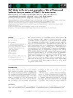
Fig. 1 Integrin αvβ3 promoted NSCLC progression in vitro. A, relative cell proliferation of A549/PC-9, integrin αvβ3 negative A549/PC-9 and integrin
αvβ3 positive A549/PC-9 cells. B, the colony formation rates of A549/PC-9, integrin αvβ3 negative A549/PC-9 and integrin αvβ3 positive A549/PC-9
cells. C, the tumor volumes of A549, integrin αvβ3 negative A549 and integrin αvβ3 positive A549 bearing mice. D, the tumorigenesis percentages
of A549, integrin αvβ3 negative A549 and integrin αvβ3 positive A549 cells in immunodeficient mice. E, relative migrating cell numbers of A549/
PC-9, integrin αvβ3 negative A549/PC-9 and integrin αvβ3 positive A549/PC-9 cells. The scale bar is 15 μm. F, the expression of integrin αvβ3 in
para-carcinoma tissues (n = 59) and tumor tissues (n = 515) from NSCLC patients. G, the overall survival of NSCLC patients divided into high integrin
αvβ3 expression group (n = 127) and low integrin αvβ3 expression (n = 375). n.s means no significant difference, * means p < 0.05, ** means p < 0.01
mechanism of tumor progression induced by integrin
αvβ3. Activation of integrins downstream signaling
pathways, such as AKT signaling pathway for promoting cell survival, was dependent on the phosphorylation
of FAK [15]. Here, the expression of FAK and AKT was
examined in A549 and PC-3 cells. Both phosphorylated FAK and phosphorylated AKT were upregulated
in αvβ3 positive A549/PC-9 cells (Fig. 2A). We next
further suppressed the FAK/AKT signals in NSCLC
cells by using FAK inhibitor Y15 and AKT inhibitor
3CAI to treat integrin αvβ3 positive A549/PC-9 cells.
Intriguingly, blockade of FAK/AKT signals efficiently
suppressed the cells proliferation (Fig. 2B) and colony
formation (Fig. 2C) induced by integrin αvβ3. Meanwhile, integrin αvβ3 positive A549/PC-9 cells revealed
weakened capability of migration after Y15 and 3CAI
treatment (Fig. 2D), suggesting that integrin αvβ3 promoted NSCLC cells proliferation through FAK/AKT
signaling pathway. Subsequently, we continued to evaluate the influence of FAK/AKT on prognosis of NSCLC
patients. However, no direct correlation was observed
between FAK/AKT expression and overall survival
in NSCLC patients (Fig. 2E and F). Those results suggested that additional signaling molecular might participate in the FAK/AKT associated NSCLC progression.
Zhou et al. BMC Cancer
(2022) 22:459
Page 5 of 10
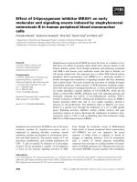
Fig. 2 Integrin αvβ3 mediated FAK/AKT signals to promote NSCLC progression. A, western blotting of phosphorylated FAK, total FAK,
phosphorylated AKT and total AKT in integrin αvβ3 negative A549/PC-9 and integrin αvβ3 positive A549/PC-9 cells. B, relative proliferation of αvβ3
positive A549/PC-9 cells treated with PBS, Y15 (20 nM) and 3CAI (10 nM). C, the colony formation rates of αvβ3 positive A549/PC-9 cells treated
with PBS, Y15 (20 nM) and 3CAI (10 nM). D, relative migrating cell numbers of αvβ3 positive A549/PC-9 cells treated with PBS, Y15 (20 nM) and 3CAI
(10 nM). E, the overall survival of NSCLC patients divided into high FAK expression group (n = 127) and low FAK expression (n = 375). F, the overall
survival of NSCLC patients divided into high AKT expression group (n = 127) and low AKT expression (n = 375). n.s means no significant difference, *
means p < 0.05, ** means p < 0.01
Integrin αvβ3 induced tumor progression was TRIB3
dependent
Compelling findings provided evidence that TRIB3
linked stress signals are capable of promoting tumor
initiation and progression by supporting cancer
stemness, which is coordinated with elevated FOXO1
and AKT1 axis [16]. Herein, we analyzed TRIB3 expression and overall survival in NSCLC patients from
TCGA database. Intriguingly, elevated expression of
TRIB3 was observed in tumor tissues in comparison
with the para-carcinoma tissues (Fig. 3A). And a high
correlation was found in TRIB expression and overall
survival in NSCLC patients (Fig. 3B), indicating that
TRIB3 served as the major regulator in NSCLC progression. Thus, we silenced TRIB3 in A549 and PC-9
cells (Fig. 3C), and further sorted integrin αvβ3 positive NSCLC cells to examine the cell proliferation.
Notably, suppression of TRIB3 retarded the proliferative effects induced by integrin αvβ3 in A549/PC-9
cells (Fig. 3D), whereas no significant difference was
found in integrin αvβ3 negative NSCLC cells (Fig. 3E).
Also, the strengthened capability of colony formation
(Fig. 3F) and cell migration (Fig. 3G) was aborted when
silencing TRIB3 in αvβ3 positive A549 and PC-9 cells.
Collectively, those results suggested that integrin αvβ3
induced tumor progression was TRIB3 dependent.
TRIB3 interacted with AKT1 to up‑regulate FOXO1
and SOX2 expression
Previous documents have proved that TRIB3 could interact with AKT1 to abrogate FOXO1 degradation, resulting
in the up-regulating of FOXO1 and downstream transcription factor SOX2 activation [12]. Firstly, we examined the expression FOXO1 and SOX2 in A549 and PC-3
cells. Notably, enhanced expression of FOXO1 and SOX2
was found in integrin αvβ3 positive A549 and PC-9, and
suppression of AKT or TRIB3 retarded up-regulation
of FOXO1/SOX2 (Fig. 4A). Next, we wondered whether
TRIB3 mediated FOXO1 upregulation by interacting
with AKT1. The endogenous AKT1 was co-immunoprecipitated with TRIB3. And obvious interaction between
AKT1 and TRIB3 was found in integrin αvβ3 positive
A549 and PC-9 cells (Fig. 4B). To further confirm the
role of TRIB3-AKT1 axis in NSCLC progression, we
used Pep2–Ae to interrupt the contact between TRIB3
and AKT1 and examined the cell proliferation of integrin
αvβ3 positive cells. Intriguingly, Pep2–Ae treatment significantly suppressed the cell proliferation (Fig. 4C) and
Zhou et al. BMC Cancer
(2022) 22:459
Page 6 of 10
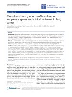
Fig. 3 Integrin αvβ3 induced tumor progression was TRIB3 dependent. A, the expression of TRIB3 in para-carcinoma tissues (n = 59) and tumor
tissues (n = 515) from NSCLC patients. B, the overall survival of NSCLC patients divided into high TRIB expression group (n = 127) and low TRIB
expression (n = 375). C, western blotting of TRIB3 in vector control, TRIB3 silenced #1 and TRIB3 silenced A549/PC-9. D, relative cell proliferation of
vector control, TRIB3 silenced #1 and TRIB3 silenced A549/PC-9 (integrin αvβ3 positive). E, relative cell proliferation of vector control, TRIB3 silenced
#1 and TRIB3 silenced A549/PC-9 (integrin αvβ3 negative). F, the colony formation rates of vector control, TRIB3 silenced #1 and TRIB3 silenced
A549/PC-9 (integrin αvβ3 positive). G, relative migrating cell numbers of vector control, TRIB3 silenced #1 and TRIB3 silenced A549/PC-9 (integrin
αvβ3 positive). * means p < 0.05, ** means p < 0.01
colony formation (Fig. 4D) of integrin αvβ3 positive A549
and PC-9 cells. Meanwhile, interruption of TRIB3-AKT1
axis retarded the cell migration in NSCLC cells (Fig. 4E).
Together, those results suggested that TRIB3 contacted
with AKT1 and upregulated FOXO1 expression, resulting in the SOX2 activation and NSCLC progression.
Disruption of the TRIB3‑AKT interaction suppressed NSCLC
progression
Given the importance of TRIB3-AKT axis in NSCLC
development, we examined whether disruption of TRIB3AKT interaction could improve the anticancer effects in
NSCLC. Firstly, we combined Pep2–Ae with chemotherapeutic PTX/Cis to treat A549 and PC-9. Intriguingly,
Pep2-Ae treatment revealed no influence on the integrin αvβ3 negative cells (Fig. 5A and B), however, significantly strengthened the cytotoxicity of PTX (Fig. 5C) and
Cis (Fig. 5D) to integrin αvβ3 positive A549 and PC-9.
Those results implicated that blockade of TRIB3-AKT
interaction efficiently suppressed the tumor progression
induced by integrin αvβ3 and improved the outcome
of chemotherapy in NSCLC. Next, tumors arising from
the subcutaneous implication of A549 cells in mice was
established for anticancer effects analysis in vivo. Using
Zhou et al. BMC Cancer
(2022) 22:459
Page 7 of 10
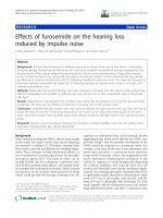
Fig. 4 TRIB3 interacted with AKT1 to up-regulate FOXO1 and SOX2 expression. A, western blotting of FOXO1 and SOX2 in integrin αvβ3 negative
A549/PC-9, integrin αvβ3 positive A549/PC-9 treated with PBS, 3CAI (10 nM) and TRIB3 knockdown. B, co-immunoprecipitation of AKT and TRIB3
in integrin αvβ3 negative A549/PC-9, integrin αvβ3 positive A549/PC-9 cells. C, relative cell proliferation of integrin αvβ3 positive A549/PC-9 cells
treated with PBS or Pep2–Ae (20 μg/ml). D, colony formation rates of integrin αvβ3 positive A549/PC-9 cells treated with PBS or Pep2–Ae (20 μg/
ml). E, relative migrating cell numbers of integrin αvβ3 positive A549/PC-9 cells treated with PBS or Pep2–Ae (20 μg/ml). * means p < 0.05, ** means
p < 0.01
this model, subcutaneous A549 tumors were established
in mice and, then treated with chemotherapeutic Cis,
PTX and Pep2–Ae. Notably, Pep2–Ae treatment resulted
in the suppression of tumor growth, and strengthened
the anticancer effects of Cis (Fig. 5E and F) and PTX
(Fig. 5G and H) in A549 bearing mice. On the basis of our
results, we suggested that suppression of TRIB3-AKT
axis could efficiently impair tumor growth and improve
the outcome of chemotherapy in NSCLC.
Discussion
Previous in vitro and in vivo studies have suggested the
association between integrin αvβ3 and tumor progression [6, 17]. Our observation that integrin αvβ3 could
efficiently promote lung cancer cells progression in vitro,
which is in consistent with previous reports. However,
the TCGA database analysis implicated that no significant difference was found between the patients with
high/low expression of integrin αvβ3. Here, we reported
in the present study that integrin αvβ3 facilitated the lung
cancer proliferation and invasion through FAK/AKT
signaling pathway, which was dependent on the collaboration with TRIB3. Depletion of TRIB3 resulted in the
inactivation of AKT signals, the downstream pro-survival
signals of integrin αvβ3 (Fig. 6). To our knowledge, these
data firstly provided evidence that tumor integrin αvβ3
contributed to the tumor progression through an TRIB3
dependent manner.
Increasing evidence suggested that differential expression cancer stem cell markers, such as ALDH1, CD133
and CD44, was tightly correlated with the pathological
subtypes and tumor development of lung cancer [18, 19].
Importantly, there is recent evidence that integrins also
potentiate cancer stem cell function, especially in lung
cancer. In agreement with previous reports that integrin
αvβ3 correlated with the metastasis of lung cancer cells
[20], we demonstrated that integrin αvβ3 efficiently promoted the cells invasion in lung cancer. Furthermore, we
provided evidence that integrin αvβ3 promoted the stemlike phenotypes and proliferation of A549 and PC-9.
Accordingly, compelling findings suggested that integrin
αvβ3 served as novel cancer stem cells marker, that contributed the stemness up-regulation in melanoma and
breast cancer [21, 22].
In mechanism, C Chetty and his colleagues suggested
that MMP-2 could up-regulate VEGF expression via