Báo cáo " Flavonoid glucosides from the leaves of Croton tonkinensis Gagnep., Euphorbiaceae " docx
Bạn đang xem bản rút gọn của tài liệu. Xem và tải ngay bản đầy đủ của tài liệu tại đây (153.26 KB, 4 trang )
125
Journal of Chemistry, Vol. 42 (1), P. 125 - 128, 2004
FLAVONOID GLUCOSIDES FROM THE LEAVES OF CROTON
TONKINENSIS GAGNEP., EUPHORBIACEAE
Received 14-7-2003
Phan Minh Giang
1
, Jung Joon Lee
2
, Phan Tong Son
1
1
Faculty of Chemistry, College of Natural Science, Vietnam National University
2
Anticancer Agent Research Laboratory, Korea Research Institute of Bioscience
and Biotechnology, Daejeon, Korea
Summary
Two flavone C-glucosides vitexin (1) and isovitexin (2) were isolated as the major
constituents with the total content of 59.5% of the methanol extract of the Croton
tonkinensis leaves along with an minor acylated flavonol O-glucoside kaempferol 3-O-
-
D-(6’’-O-coumaroyl)glucopyranoside (tiliroside, 3) (0.9%). The structures of 1, 2 and 3
were determined on the basis of ESIMS, 1D and 2D NMR spectroscopic data. Although the
flavonoid glucosides found exclusively in the ethyl acetate and n-butanol soluble fractions
are reported for antioxidative and antiinflammatory activities their contribution to the
medicinal properties of C. tonkinensis is demonstrated to be less than the lipid soluble ent-
kaurene diterpenoid constituents of this plant.
In our past studies on the medicinal plant
Croton tonkinensis Gagnep. (Euphorbiaceae)
[1], the phytosterols, the long chain alkyl
alcohols and the ent-kaurene-type diterpenoids
[2] were isolated from the non-polar parts
(n-hexane and CH
2
Cl
2
soluble fractions) of
the methanol extract of the dried leaves of
C. tonkinensis [3]. The investigation of the
methanol extract by reserved-phase high perfor-
mance liquid chromatography (RP HPLC) coupled
with a photodiode array (PDA) detector [4]
revealed the presence of two major classes
of components: ent-kaurene diterpenoids and
flavonoid gluco-sides. Of the flavonoids two
were quantified as the major (total 59.5%) and
one as the minor constituents (0.9%) of the
methanol extract which were found to be
localized in the polar ethyl acetate and n-butanol
soluble fractions. When the ethyl acetate soluble
fraction was dissolved in a minimum amount of
cool methanol a yellow solid was precipitated.
1
H NMR examination of this solid in DMSO-d
6
revealed the presence of a mixture of two major
compounds 1 and 2 (2/1 in ratio). A small amount
(15 mg) of mixture was subjected to preparative
RP HPLC [4] (mobile phase MeOH-H
2
O 1:1) to
afford pure 1 and 2 which were identified as the
flavone C-glucosides vitexin (1) [5, 6] (R
t
10.2 min)
and isovitexin (2) [7, 8] (R
t
10.8 min) on the basis
of the comparison of their spectroscopic data
with the reported values.
Silica gel column chromatographic fractiona-
tion of the ethyl acetate soluble fraction eluting
with gradient: 100% CHCl
3
CHCl
3
-MeOH 2 : 1
100% MeOH, followed by purification by
preparative RP HPLC [4] (mobile phase MeOH-
H
2
O 3 : 2) afforded 3 as a yellow amorphous
powder, mp 250 - 252
o
C. The compound eluted at
R
t
15.4 [5] and displayed on-line UV maxima at
199.2, 218 (shoulder), 260, 310.7 nm indicative
126
for a flavonol. The molecular formula C
3
0
H
2
6
O
1
3
was deduced from the quasimolecular ions 593
[M-H]
+
(ESIMS negative-ion mode) and 617
[M+Na]
+
(ESIMS positive-ion mode) showing
18 degrees of unsaturation of 3. The proton
signals at
H
6.29 and 6.08 (both 1H, d, J = 1.5
Hz), 7.9 and 6.82 (both 2H, d, J = 8.7 Hz)
supported by the correlated carbon signals
derived from the HSQC spectrum and the
13
C
singlet signal at 177 in the
13
C decoupled
spectrum are indicative of the presence of a
5,7,4’-threesubstituted flavonol nucleus. The
anomeric proton signal at
H
5.4 with
configu-
ration (1H, d, J = 7.5 Hz),
C
106.7 (d) and two
proton signals at 4.05 (1H, dd, J = 11.7, 8.3 Hz)
and 4.3 (1H, d br, J = 11.7 Hz) (2H-6”) are
attributable to a glucose moiety in the structure
of 3, the proton and carbon signals in the sugar
sequence were assigned on the basis of COSY
and HSQC spectra. Consistent with the degree
of unsaturation and the
1
H and
13
C NMR spectra,
the last structural fragment of 3 was ascribed to
a trans-p-coumaroyl moiety:
C
166.2 (s, C=O),
H
7.34 (1H), 6.12 (1H) (both d, J = 16 Hz,
trans-disubstituted double bond), 7.38 and 6.74
(both 2H, d, J = 8.7 Hz, p-disubstituted aromatic
ring). This was confirmed by the cross peaks
observed in HMBC spectrum between H-2’’’ (
H
6.12) and H-3’’’ (
H
7.34) and the carbonyl
carbon C-1’’’ (
C
166.2), between H-3’’’ and C-
5’’’ and C-9’’’ (
C
130.2). Further, the HMBC
correlation between the anomeric proton at
H
5.4 and C-3 at
C
133 placed the sugar moiety at
3-O position, and the connection of the
coumaroyl moiety to C-6” of glucose was judged
from the HMBC correlation from the two proton
signals at
H
4.05 (strong) and 4.3 (weak) to the
carbonyl carbon signal at
C
166.2. Thus, the
spectroscopic data were conclusive for the
structure of 3 as kaempferol 3-O-
-D-(6’’-O-
coumaroyl)glucopyranoside (tiliroside) [9]. For
the unambiguous assignments of all
1
H and
1
3
C
signals the correlations in COSY, HMQC and
HMBC spectra were employed.
Figure 1: Chemical structures of vitexin (1), isovitexin (2)
Figure 2: Chemical structure and important H C correlations in HMBC spectrum of tiliroside (3)
OHO
OH O
OH
O
HO
OH
HO
OH
OHO
OH O
OH
O
HO
HO
HO
OH
1
2
9'''
8'''
7'''
6'''
5'''
4'''
3'''
2'''
1'''
6"5"
4"
3"
2"
1"
6'
5'
4'
3'
2'
1'
10
9
8
7
6
5
4
3
2
O
O
OH
O
OH
OH
HO
O
OH
HO
OH
O
O
127
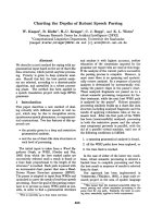
Figure 3: HPLC chromatogram (flavonoid part) of the methanol extract of C. tonkinensis
Tiliroside (kaempferol 3-O-
-D-(6’’-O-
coumaroyl)glucopyranoside) (3). Yellow amor-
phous powder, mp 250 - 252
o
C. UV max: 199.2,
218 (sh), 260, 310.7 nm; ESIMS 593 [M-H]
+
,
617 [M+Na]
+
;
1
H-NMR (300 MHz, DMSO-d
6
):
12.5 (1H, s, 5-OH), 7.9 (1H, d, J = 8.7 Hz, H-2’,
H-6’), 7.38 (1H, d, J = 8.7 Hz, H-5’’’, H-9’’’),
7.34 (1H, d, J = 16 Hz, H-3’’’), 6.82 (1H, d, J =
8.7 Hz, H-3’, H-5’), 6.74 (1H, d, J = 8.7 Hz, H-
6’’’, H-8’’’), 6.29 (1H, d, J = 1.5 Hz, H-8), 6.12
(1H, d, J = 16 Hz, H-2’’’), 6.08 (1H, d, J = 1.5 Hz,
H-6), 5.4 (1H, d, J = 7.5 Hz), 4.3 (1H, d br, J =
11.7 Hz), 4.05 (1H, dd, J = 11.7 Hz, 8.3 Hz),
3-3.8 (4H, m);
13
C-NMR (300 MHz, DMSO-d
6
):
177 (s, C-4), 166.2 (s, C-1’’’), 164.7 (s, C-7),
164.3 (s, C-10), 161 (s, C-5), 159.9 (s, C-7’’’),
159.9 (s, C-4’), 156.3 (s, C-9), 156.2 (s, C-2),
144.7 (d, C-3’’’), 133 (s, C-3), 130.8 (d, C-2’,
C-6’), 130.2 (d, C-5’’’, C-9’’’), 124.9 (s, C-4’’’),
120.7 (s, C-1’), 115.8 (d, C-6’’’, C-8’’’), 115.1
(d, C-3’, C-5’), 113.6 (d, C-2’’’), 106.7 (d, C-1”),
99 (d, C-6), 94 (d, C-8), 76 (d, C-3”), 74 (2d, C-2”,
C-5”), 69.9 (d, C-4”), 63 (t, C-6”).
Taking into account the high contents of the
polar vitexin (27.2%), isovitexin (32.2%) and
tiliroside (0.9%) in the whole leave methanol
extract (Figure 3) the correlation between the
biological activites of the flavonoid glucosides
and medicinal properties of C. tonkinensis [1]
should be put under discussion. While isovitexin
and vitexin displayed moderate antioxidative
activity [10, 11] tiliroside was reported to possess
a potent anticomplementary activity [12] and to
inhibit induced histamine released in rat mast
cells which could be considered as evidence for
the antiinflammatory effect of tiliroside [13]. In
consideration of the association of tumor
promotion with oxidative and inflammatory
tissue damage [14], it would be worthwhile to
determine the possible chemopreventive effects
on carcinogenesis. However, it is noticeable that
the glycosilation of flavonoids often results in
1937
1800
1600
1400
1200
1000
800
600
400
200
-
52
Flavonoid glucosides
Croton tonkinensis
ANAL #5 CT-Me01 UV VIS
8.68 10.00 11.00 12.00 13.00 14.00 15.00 16.00 17.00 18.00
128
the reduction of biological activities regardless
of the types of aglycones and types of linkage
(C- or O-glycosides) possibly due to their
hydrophilicity and consequent diminished ability
to penetrate cell membrane and steric hindrance
caused by their bulky glycosyl residue [15] as
demonstrated with vitexin [16] and isovitexin
[10]. Finally, it is important to underline that in
our bioassays involving the inhibition of the
transcription factor NF-B and iNOS-dependent
NO production the flavonoid glucosides were
non-active. Consistently, in our antiplasmodial
tests against the Plasmodium falciparum strains
the lipid soluble components showed much
more pronounced inhibitory activity than the
water soluble components of C. tonkinensis [17].
Acknowledgements: This work was partly
supported by International Foundation for
Science (Stockholm, Sweden, Grant No. F/2841-2
to Phan Minh Giang). The working facilities
provided by Anticancer Agent Research Labora-
tory, Korea Research Institute of Bioscience and
Biotechnology, Korea, to Phan Minh Giang are
gratefully acknowledged.
References
1. Vo Van Chi. Dictionary of Vietnamese Medi-
cinal Plants. Publishing House Medi-cine
(Ho Chi Minh City) (1997).
2. (a) Phan Tong Son, Van Ngoc Huong, Phan
Minh Giang, C. Taylor Walter. Vietnam J.
Chem., Vol. 37, No. 1-2 (1999). (b) Phan
Tong Son, Phan Minh Giang, C. Taylor
Walter. Austr. J. Chem., Vol. 53, P. 1003 -
1005 (2000). (c) Phan Minh Giang, Jung
Joon Lee, Phan Tong Son. Vietnam J.
Chem., Vol. 41, No. 1 (2003).
3. The air-dried leaves were collected in the
suburbs of Hanoi, and identified by a botanist
Prof. Vu Van Chuyen (Hanoi College of
Pharmacy, Hanoi in 2002.
4. RP HPLC: Dionex HPLC system with a P580
pump, an ASI-100 automated sample injector
and a PDA-100 photodiode array detector.
Analytical condition: YMC ODS-H80 column
(150 × 4.6 mm I.D., S-4 µm), sample injection
size 10 µl, mobile phase gradient 20 - 100%
MeOH in HPLC grade H
2
O, run time 25 min,
flow rate 1 ml/min. Preparative condition:
YMC ODS-H80 column (150 mm × 20 mm
I.D., S-4 µm), flow rate 6 ml/min).
5. G. F. Pauli and P. Junior. Phytochemistry,
Vol. 38, P. 1245 - 1250 (1995).
6. P. -C. Zhang and S. -X. Xu. J. Asian Nat.
Prod. Research, Vol. 5, P. 131 - 136 (2003).
7. M. Haribal and J. A. A. Renwick. Phytoche-
mistry, Vol. 47, P. 1237 - 1240 (1998).
8. G. T. Maatooq, S. H. El-Sharkawy, M. S.
Afifi, and J. P. N. Rosazza. Phytochemistry,
Vol. 44, P. 187 - 190 (1997).
9. N. Backhouse, C. Delporte, R. Negrete, S. A.
San Feliciano, and J. L. Lopez-Perez.
Phytother. Res., Vol. 16, 562 - 566 (2002).
10. C. -M. Lin, C. -T. Chen, H. -H. Lee, and J. -K.
Lin. Planta Med., Vol. 68, P. 365 - 367
(2002).
11. R. Aquino, S. Morelli, M. R. Lauro, S. Abdo,
A. Saija, and A. Tomaino. J. Nat. Prod., Vol.
64, P. 1019 - 1023 (2001).
12. K. Y. Jung, S. R. Oh, S. H. Park, I. S. Lee,
K. S. Ahn, J. J. Lee, and H. K. Lee. Biol.
Pharm. Bull., Vol. 21, P. 1077 - 1078 (1998).
13. T. Tsuruga, Y. Ebizuka, J. Nakajima et al Chem.
Pharm. Bull., Vol. 39, P. 3265 - 3271 (1991).
14. Y. -J. Surh. Food and Chemical Toxicology,
Vol. 40, P. 1091 - 1097 (2002).
15. H. K. Kim, B. S. Cheon, Y. H. Kim, S. Y.
Kim, and H. P. Kim. Biochem. Pharmacol,
Vol. 58, P. 759 - 765 (1999).
16. A. Basile, S. Giordano, J. A. Lopez-Saez, and
R. C. Cobianchi. Phytochemistry, Vol. 52, P.
1479 - 1482 (1999).
17. Phan Tong Son, Le Huyen Tram, Phan
Minh Giang. Vietnam J. Chem., Vol. 40, P.
53 - 57 (2003).