Vaccination-Of-Rabbits-With-Immunodominant-Antigens-From-Sarcoptes-Scabiei-Induced-High-Levels-Of-Humoral-Responses-And-Pro-Inflammatory-Cytokines-But-Confers-Limited-Protection.pdf
Bạn đang xem bản rút gọn của tài liệu. Xem và tải ngay bản đầy đủ của tài liệu tại đây (2.07 MB, 13 trang )
Casais et al. Parasites & Vectors (2016) 9:435
DOI 10.1186/s13071-016-1717-9
RESEARCH
Open Access
Vaccination of rabbits with
immunodominant antigens from Sarcoptes
scabiei induced high levels of humoral
responses and pro-inflammatory cytokines
but confers limited protection
Rosa Casais1, Victor Granda2, Ana Balseiro1, Ana del Cerro1, Kevin P. Dalton3, Roxana González1, Pablo Bravo3,4,
J. M. Prieto1 and Maria Montoya5,6*
Abstract
Background: Vaccination is an attractive ecological alternative to the use of acaricides for parasite control.
However, effective anti-parasite vaccines against sarcoptic mange have not yet been developed. The purpose of
this study was first to identify Sarcoptes scabiei immunodominant antigens and second to evaluate them as vaccine
candidates in a rabbit/S. scabiei var. cuniculi model.
Methods: The S. scabiei Ssλ15 immunodominant antigen was selected by immunoscreening of a S. scabiei var.
hominis cDNA. The full-length cDNA was sequenced and cloned into the pGEX vector and the recombinant protein
expressed in BL21 (DE3) cells and purified. A vaccination trial was performed consisting of a test group (n = 8)
immunised with recAgs (a mix of two recombinant antigens, Ssλ15 and the previously described Ssλ20ΔB3) and
a control group (n = 8) immunised with PBS. All analyses were performed with R Statistical Environment with
α set at 0.050.
Results: The full-length open reading frame of the 1,821 nt cloned cDNA encodes a 64 kDa polypeptide, the
sequence of which had 96 % identity with a hypothetical protein of S. scabiei. Ssλ15 was localised by
immunostaining of skin sections in the tegument surrounding the mouthparts and the coxa in the legs of mites.
Rabbit immunisation with recAgs induced high levels of specific IgG (P < 0.010) and increased levels of total IgEs.
However, no significant clinical protection against S. scabiei challenge was detected. Unexpectedly, the group
immunised with the recAgs mix had significantly higher lesion scores (P = 0.050) although lower mean mite
densities than those observed in the control group. These results might indicate that the lesions in the recAgs
group were due not only to the mites density but also to an exacerbated immunological response after challenge,
which is in agreement with the specific high levels of pro-inflammatory cytokines (IL-1 and TNFα) detected after
challenge in this group.
(Continued on next page)
* Correspondence: ;
5
Centre de Recerca en Sanitat Animal (CReSA), UAB-IRTA, Campus de la
Universitat Autònoma de Barcelona, Bellaterra Cerdanyola del Vallès, Spain
6
The Pirbright Institute, Ash Road, Pirbright, Woking, Surrey, UK
Full list of author information is available at the end of the article
© 2016 The Author(s). Open Access This article is distributed under the terms of the Creative Commons Attribution 4.0
International License ( which permits unrestricted use, distribution, and
reproduction in any medium, provided you give appropriate credit to the original author(s) and the source, provide a link to
the Creative Commons license, and indicate if changes were made. The Creative Commons Public Domain Dedication waiver
( applies to the data made available in this article, unless otherwise stated.
Casais et al. Parasites & Vectors (2016) 9:435
Page 2 of 13
(Continued from previous page)
Conclusions: The selected antigens delivered as recombinant proteins had no clinical protective efficacy against S.
scabiei infestation although immunisation reduced mite density. However, these results pave the way for future
studies on alternative production systems, adjuvants, delivery methods and combinations of antigens in order to
manage stimulation of clinical protective immune responses.
Keywords: Sarcoptic mange, Sarcoptes scabiei, Immunodominant antigens, Vaccine candidates, Clinical protection
Background
Sarcoptic mange (De Geer) (scabies) is a highly contagious parasitic infestation of global distribution, caused
by the burrowing mite S. scabiei that infests humans and
a wide range of domestic and wild mammals [1–3],
causing important economic losses.
Acaricides are used to control sarcoptic mange [4–6]
but they are expensive and can be highly toxic to the environment, foodstuff and animal handlers. Furthermore,
systematic use of acaricides causes development of
strong acaricide resistance in scabies mites in humans
[7], rabbits [8] and dogs [9]. In this sense, and given that
previous studies have shown some degree of protective
immune responses developed after S. scabiei infestation
[10–15], vaccination seems to be a good ecological alternative to the use of acaricides for parasite control. The
necessity of an effective vaccine to control and treat this
skin disease has been mentioned previously for humans
(reviewed in [16]) and animal species [17]. However, effective anti-parasite vaccines against sarcoptic mange
have not yet been developed. This is due to multiple factors such as, the complexity of interactions between the
parasite and the host’s immune system, the fact that we
have yet to understand protective mechanisms employed
by the host and the large number of parasite encoded proteins, which makes it very difficult to find proteins that
have the capacity to confer protective immunity [17].
The generation of comprehensive expressed sequence
tag libraries has enabled the initial characterisation of
molecules of interest for diagnostics, vaccines and drug
resistance development studies [18]. In this sense,
different recombinant antigens have been identified and
assayed as vaccine candidates which have not conferred
complete protection. Vaccination with a mixture of two
fused recombinant antigen portions [Ssag1 (homologous
to the house dust mite Euroglyphus maynei allergen M177, an apolipoprotein from hemolymph) and Ssag2] in
a rabbit/S. scabiei var. canis model did not produce
reduction in the numbers of mites although rabbits did
not exhibit the typical crust characteristics [19].
Sarcoptes scabiei glutathione S-transferase, a target for
vaccine development in several parasitic diseases, did
not induce specific antibodies in mange-infested dogs
and pigs [20]. Likewise, vaccination of rabbits with
tropomyosin allergen of S. scabiei, a protein with proven
immunogenic properties, did not efficiently control sarcoptic mange although the lesion areas were smaller at
the end of the experiment [21].
The purpose of this study was to identify immunodominant antigens in a S. scabiei var. hominis cDNA library
and to evaluate their potential as vaccine candidates in a
rabbit/S. scabiei var. cuniculi model.
Methods
Ethical considerations
Experimental procedures were approved by the SERIDA
Animal Ethics Committee and authorised by the Regional Consejería de Agroganadería y Recursos Autoctonos del Principado de Asturias, Spain. Experiments were
conducted in accordance with the Spanish and European
current legal requirements and guidelines regarding
experimentation and Animal Welfare.
Selection and cloning of recombinant S. scabiei antigens
The recombinant DNA techniques and bacteria strains
used in this study have been previously described [22].
The S. scabiei amplified cDNA library Yv4 used in this
study was kindly provided by Dr. David J. Kemp (Malaria
and Scabies Laboratory, Queensland Institute of Medical
Research, Brisbane, Australia) and contained an average
insert size of 1.6 kb cDNAs [23] flanked by EcoRI and
XhoI restriction sites in the vector λZAP express (Stratagene, La Jolla, USA). Two cDNA clones Ssλ20 and
Ssλ15 were selected by immunoscreening of the Yv4
library using a 1:20 dilution of a serum taken from a
naturally infested chamois and a 1:50 dilution of a serum
taken from an experimentally infested rabbit, respectively, following a previously described procedure [24].
The immunocomplexes were detected using a 1:1,500
dilution of protein G or protein A peroxidaseconjugated (Sigma, St. Louis, MO, USA) and 4-chloro-1naphthol as chromogenic substrate (Sigma, St. Louis,
MO, USA). Positive plaques were re-screened at a lower
density until pure plaque populations were obtained.
This process was followed by in vivo excision of pBKCMV phagemids from the λZAP express vectors following the manufacturer’s instructions.
Cloning, expression and purification of recombinant S.
scabiei antigen Ssλ20ΔB3, the Ssλ20 derivative used in
this study, was previously described in [22]. In order to
Casais et al. Parasites & Vectors (2016) 9:435
produce the selected S. scabiei Ssλ15 specific antigen as a
fusion protein with Glutathione S-transferase (GST), the
positive clone was excised as a phagemid (pBK-CMVSsλ15) which was double-digested with the restriction
endonucleases EcoRI and XhoI. The restriction fragment
corresponding to the S. scabiei cDNA, was gel-purified and
ligated into pGEX-4 T3 digested with the same enzymes
(pGEX-4T3-Ssλ15). Sequence analysis of plasmid DNA
(pBK-CMV-Ssλ15 and pGEX-4 T3-Ssλ15) was done using
the T7 and T3 promoter primers and internal primers
Ss5forward (5′-GAG GAA TCG GAT ATG ATT CG-3′),
representing nucleotides 617–636 of the cDNA) and Ss6reverse (5′-GAC ATA TTT AGA CAT ATG GC-3′), representing nucleotides 1,161–1,142 of the cDNA) to complete
the entire cDNA sequence. Sequencing reactions were
done using BigDye® Terminator v3.1 kit and analysed
on an ABI PRISM 3100 Genetic Analyser. The nucleotide and deduced amino acid sequences were analysed
with Vector NTI (Invitrogen, Carlsbad, California,
USA). For sequence similarity the Blast program at the
NCBI web server was used. The prediction of transmembrane helices in the deduced polypeptide was carried out
by TMHMM Server v. 2.0 ( The presence of a signal peptide was investigated using SignalP-4.1 at the Centre of Biological
Sequence Analysis [25] ( />SignalP-4.1). The hydropathicity plot was calculated according to Kyte & Doolittle [25] using nine residues as
window size.
Purification of recombinant proteins
The recombinant proteins were produced in transformed BL21 E. coli cultures induced with 100 μM
isopropyl-β-D thiogalactopyranoside (IPTG) for 4 h and
purified by affinity chromatography using a GlutathioneSepharose 4B column (Amersham Biosciences, Barcelona, Spain) according to the manufacturer’s instructions. The sarcoptes-derived polypeptide Ssλ20ΔB3 was
excised from the GST by thrombin cleavage, while the
GST-Ssλ15 was either excised from the GST for preparation of specific antisera or directly eluted from the
column as a fusion protein with GST with 50 mM TrisHCl, 10 mM reduced glutathione, pH 8.0 for its use in
the vaccination trial. Proteins were analysed by SDSPAGE and quantified by the Bradford method [26] using
bovine serum albumin as the standard.
Mass spectrometry analysis of protein spots
The gel bands of interest were manually excised from gels.
Proteins selected for analysis were in-gel reduced, alkylated and digested with trypsin according to [27]. Briefly,
the samples were reduced with 10 mM dithioerytritol in
25 mM ammonium bicarbonate for 30 min at 56 °C and
subsequently alkylated with 55 mM iodoacetamide in
Page 3 of 13
25 mM ammonium bicarbonate for 15 min in the dark. Finally, samples were digested with 12.5 ng/μl sequencing
grade trypsin (Roche Molecular Biochemicals, Basel,
Switzerland) in 25 mM ammonium bicarbonate (pH 8.5)
overnight at 37 °C. After digestion, the supernatant was
collected and 1 μl was spotted onto a MALDI target plate
and allowed to air-dry at room temperature. Then, 0.6 μl
of a 3 mg/ml of α-cyano-4-hydroxy-cinnamic acid matrix
(Sigma, St. Louis, MO, USA) in 50 % acetonitrile was
added to the dried peptide digest spots and allowed to airdry again at room temperature. MALDI-TOF MS analyses
were performed in a 4800 Plus Proteomics Analyzer
MALDI-TOF/TOF mass spectrometer (Applied Biosystems, MDS Sciex, Toronto, Canada) at the Genomics and
Proteomics Center, Complutense University of Madrid.
The MALDI-TOF/TOF operated in positive reflector
mode with an accelerating voltage of 20,000 V. All mass
spectra were calibrated internally using peptides from the
auto digestion of trypsin. For protein identification
UniProt-SwissProt Database (date 14th June 2009; 545,388
sequences; 193,948,795 residues) without taxonomy restriction and a home-made data base with the sequence S.
scabiei Ss15-2-A protein (1 sequence; 566 residues) were
searched using MASCOT v 2.3 (www.matrixscience.com)
through the Global Protein Server v 3.6 from ABSCIEX.
Search parameters were: carbamidomethyl-Cystein as
fixed modification and oxidised Methionine as variable
modification, peptide mass tolerance 50 ppm and 1
missed trypsin cleavage site allowed. In all proteins identified, the probability scores were greater than the score
fixed by mascot as significant with a P-value < 0.050.
Western blotting
After SDS-polyacrylamide (10 %) gel electrophoresis, the
proteins were transferred onto Immobilon-P transfer
membranes (Millipore, Billerica, MA, USA) using a Mini
Protean II (Bio-Rad, Hercules, CA, USA) electroblotting apparatus at 100 V for 1 h in 25 mM Tris/
192 mM glycine buffer, pH 8.3 containing 20 % methanol. After a blocking step specific antigens were revealed
with serum from a mange-infested rabbit serum diluted
1:200, a mange-infested chamois diluted 1:100, an antiGST monoclonal antibody diluted 1:5,000 (SIGMA,
Madrid, Spain), and a mix of a rabbit pre-immune serum
(1:100) and a serum from a mange-free chamois (1:200)
followed by the addition of the appropriate speciesspecific peroxidase-conjugated secondary antibody. The
immunocomplexes were revealed using 4-chloro-1-naftol as substrate.
Preparation of antisera
Passive elution of the thrombin excised Ssλ15 70 kDa
protein band from polyacrylamide gel pieces was done
following the protocol of Thermo Scientific and the
Casais et al. Parasites & Vectors (2016) 9:435
efficiency of the process checked analysing the eluted
protein on a 10 % SDS-PAGE gel. One New Zealand
White rabbit was immunised with the purified protein
(70 kDa Ssλ15 gel-eluted polypeptide) for antibody production. The immunisation protocol consisted of 5
intramuscular injections at days 1, 14, 28, 42 and 56,
using 200 μg of the purified protein emulsified with incomplete Freund’s adjuvant for the first boost and without adjuvant for the other 4 injections. The serum from
the bleed on day 63 and the pre-immune serum from
the same rabbit were used for immunolocalisation
studies.
Immunolocalisation of the Ssλ15 encoded antigen
Skin samples from a S. scabiei infested chamois were
fixed in 10 % neutral formalin and embedded in paraffin
using standard procedures. Rabbit skin samples were not
used to avoid background reactions as a consequence of
using the rabbit antisera for detection. For the immunohistochemical study, 4 μm sections were immunostained
using the peroxidase-antiperoxidase (PAP) method [28].
Briefly, the sections were cut, deparaffinised, rehydrated
and rinsed with tap water. Afterwards, samples were
treated to inactivate the endogenous peroxidase by incubation with methanol containing 3 % H2O2 for 10 min,
washed with water for 10 min and then treated to prevent unspecific binding with a 15 min incubation with
10 % normal swine serum (DAKO, Glostrup, Denmark),
3 % BSA in TBS (5 mM Tris/HCl pH 7.6, 136 mM
NaCl). The tissue sections were incubated overnight at
4 °C with a rabbit polyclonal antiserum to the 70 kDa
Ssλ15 derived polypeptide diluted 1:700 in TBS and then
washed three times with TBS. Then, samples were incubated with swine anti-rabbit serum (DAKO) diluted 1:50
in TBS for 30 min at room temperature and washed
three times with TBS followed by incubation with a
rabbit PAP (soluble complexes of rabbit antibody to
horseradish peroxidase-antihorseradish peroxidase) diluted 1:320 in TBS for 30 min at room temperature. Finally, the sections were incubated with the substrate
3,3′-diaminobenzidine tetrahydrochloride (DAB, Sigma,
St. Louis, MO, USA) for 10 min and washed with TBS
and water. After staining for 45 s with haematoxylin the
preparations were dehydrated, pasted with DPX mountant for histology (Fluka, Sigma, St. Louis, MO, USA)
and observed using a light microscope Olympus BH-2
and photographed using a digital camera Olympus DP12. Pre-immune antisera of the rabbits used to produce
the antisera were used as negative controls.
Vaccination trial and mite challenge
Sixteen, 3 month old scabies-free New Zealand White
female rabbits of 2.6–3 kg were housed individually and
kept under observation during an acclimatisation period
Page 4 of 13
of two weeks. Animals were randomly allocated into two
groups (8 rabbits per group): group 1 (recAgs group)
was immunised with a mix of Ssλ20ΔB3 and GST-Ssλ15
S. scabiei recombinant antigens (referred to as recAgs
mix) and Quil A adjuvant (Accurate Chemical and Scientific corp., Westbury, USA), and group 2 (control
group) with PBS pH 7.5 and Quil A. Each animal was
injected subcutaneously in two sites in the back (0.5 ml
per site), group 1 with 200 μg of recombinant proteins
(100 μg of each) and 100 μg of Quil A, and group 2 with
PBS and Quil A (100 μg). The immunisation schedule
consisted of four immunisations, at one-week intervals
for the first three vaccinations and at three-week interval
for the last injection. One week after the last immunisation, all animals were challenged with crusts harbouring
approximately 2,500 mixed life-cycle stage mites (3 g)
taken from previously infested rabbits, and infestations
were allowed to progress for 7 weeks. Mites were inoculated by means of a dressing on the left shaved hind limb
(foot area) for 48 h [15].
The S. scabiei strain used in this experiment derived
from clinically affected wild European rabbits [29] and
was maintained in New Zealand White rabbits as the
source of mites for the challenge of immunised animals.
The inoculum for the challenge was obtained from three
affected donor rabbits, which were euthanatised on the
day of mite collection. Areas of affected skin were cut
into small pieces for the inoculum. For the estimation of
the number of mites ten pieces of lesioned skin were
weighed, placed in Petri dishes on parafilm and incubated for 24 h at 37 °C to encourage mites to migrate
out of the crust of skin. After incubation, the number of
mites per gram was counted under a stereoscope and
the mean value used as an estimation.
Antibody levels
In order to assess the humoral immune response (IgG
and IgE levels) blood samples were collected from the
marginal ear vein prior to vaccination, 6 days after the
second dose, 13 days after the third dose, 8 days after
the fourth dose (just before challenge) and once per
week after infestation. Serum samples were obtained
from blood and stored at -20 °C until use.
Circulating rabbit serum antibody levels were analysed
with an in house recombinant enzyme-linked immunosorbent assay (ELISA) based on the recombinant antigen
Ssλ20ΔB3 [30]. Negative and positive controls were included in all plates for normalisation and consisted of a
pre-immune serum and a serum collected from a mangy
rabbit, respectively. The raw data of the measured OD
were normalised as recommended by [31] expressing
them as a percentage of the positive control in a ratio
correcting for the measured OD of the negative control
Casais et al. Parasites & Vectors (2016) 9:435
according to the formula: Relative OD = ODsample_serum ODnegative_control/ODpositive_control - OD negative_control.
The cut-off value (0.03 relative OD) was defined as the
mean of the relative OD450nm from 21 scabies-free animals plus three times the standard deviation [32, 33].
Due to the lack of an effective secondary anti-rabbit
IgE antibody total levels of IgEs were measured. For the
quantitative determination of total rabbit IgE concentrations in serum we used the Rabbit immunoglobulin E
(IgE) ELISA Kit (CUSABIO BIOTECH co. distributed by
bioNova científica, S.L., Madrid, Spain). Due to economic reasons the analysis was carried out only at four
selected time points (before vaccination, prior to challenge, and 1 and 7 weeks post-challenge).
Cytokine levels
Commercial ELISAs were performed to evaluate serum
titres of three cytokines (IL-1, IL-6 and TNF-α) before
vaccination, prior to challenge and at weeks 1, 2, 3 and 6
post-challenge. The cytokine ELISAs were performed according to the manufacturer’s instructions (ELISA kit,
Uscn Life Science Inc., distributed by bioNova científica,
S.L., Madrid, Spain).
Page 5 of 13
KOH solution containing 1 % Tween 80, incubated for
18 ± 2 h at 45 °C, the material was agitated for 2–3 min
with a vortex and centrifuged at 500 g for 15 min. Then,
the supernatant was decanted to 1 cm above the pellet
(about 2.5 ml were removed), which was suspended in
the residual liquid, then 2 ml 70 % ethanol were added
to rinse the walls of the tube to get a final volume of
4 ml. Twenty replicates of 25 μl digested suspension (a total
of 500 μl) per rabbit were observed under the stereomicroscope, the number of mites counted and the total number
of mites in the 4 ml was estimated.
Statistical analysis
All analyses were performed with R Statistical Environment [35], with confidence intervals stated at 95 %
(α = 0.050). Differences in mite density were established
by Kruskal-Wallis Rank Sum test, as density did not follow
normal distribution. Analyses of variance for repeated
measures for each dependent variable (IgG levels, IgE
levels, cytokines levels, lesion scores and weight) were performed by means of the ez package [36]. Data were analysed, using immunisation group and time as fixed factors
and the rabbit as a random factor to account for repeated
measures variability.
Lesion score
The foot area was chosen for mite inoculation in the
challenge as mange lesions in naturally infested rabbits
have most frequently been initially observed in the
limbs. Afterwards skin lesions caused by mite challenge
were assessed for their extension at weekly intervals
from weeks 1 to 7 post-infestation. The lesion areas were
photographed and measured using a flexible ruler. Lesions were graded as follows: score 0 was assigned if no
limb lesions were observed, score 1 when lesions were
first observed on the limbs (lesions ≤ 7.75 cm2), score 2
when lesions were between 7.75–15.5 cm2, score 3 when
lesions ranged from 15.5–31 cm2 and score 4 when lesions were > 31 cm2, following established procedures
previously published [15]. The effect of infestation on
body condition was also assessed from the changes in
the body weight, which was recorded once per week
from the beginning of the experiment.
Mite burden
Seven weeks after challenge rabbits were euthanised by
intravenous injection of 0.3 ml per kg of body weight of
T-61 Euthanasia Solution® (Intervet) (Embutramide
200 mg; Mebezonium Iodide 50 mg; Tetracaine Hydrochloride 5 mg/ml). Afterwards a fragment of skin was
taken from the lesioned area of the left hind limb and
stored at -20 °C. For mite counts we followed the protocol described by [34], which allows approximately 88 %
recovery of S. scabiei mites. Briefly, 2 cm2 of skin were
cut from the skin fragment, suspended in 4 ml of 10 %
Results
Identification and sequence analysis of S. scabiei
immunodominant antigens
In order to identify S. scabiei antigens to use individually
or as a mixture as candidates for vaccine development
2 × 10⁶ PFU from the S. scabiei var. hominis amplified library Yv4 were screened. Two cDNA clones, Ssλ20 and
Ssλ15, showing the strongest reactions with sera taken
from a naturally infested chamois and an experimentally
infested rabbit, respectively, were selected as the most
immunodominant.
Selection, cloning, expression, purification and immunolocalisation of recombinant S. scabiei antigen Ssλ20ΔB3, a
truncated derivative of Ssλ20 used in this study, was previously described [22]. The Ssλ20ΔB3 cDNA encodes a
28.9 kDa polypeptide of unknown function and contains
one out of the 21 amino acid tandem repeats encoded by
the original Ssλ20 cDNA clone [22]. Ssλ20ΔB3 was detected in Western and dot-blot by sera from an infested
chamois [22].
The second immunodominant antigen, Ssλ15, is described here for the first time. The nucleotide sequence
of Ssλ15 cDNA clone (Fig. 1a) was 1,821 nt long and the
sequence was submitted to the GenBank database on
29th December 2015 (GenBank: KU359774).
Sequence analysis revealed that the Ssλ15 cDNA contained an open reading frame (ORF) starting at cDNA
5′-end and ending at a TAA codon located at nt 1,712–
1,714. This cDNA has a 3′-untraslated region of 78 nt
Casais et al. Parasites & Vectors (2016) 9:435
Page 6 of 13
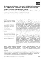
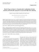
Fig. 1 a Nucleotide sequence of S. scabiei Ssλ15 cDNA (GenBank:
KU359774) and amino acid sequence of the predicted translation
product. The numbers on the right vertical axis refer to the amino
acid position in the Ssλ15 cDNA. The poly (A) tail is shown in
boldface. Internal primers Ss5forward (nucleotides 617–636 of the
cDNA) and Ss6reverse (nucleotides 1161–1142 of the cDNA) used to
complete Ssλ15cDNA nucleotide sequence are boldface and
underlined. b Hydropathicity profile of Ssλ15 polypeptide
preceding the poly (A) tail. This ORF had coding capacity for a 558 amino acid polypeptide with a predicted
molecular weight of 64 kDa, and pI 7.36. The most
abundant amino acids are Glu (12.2 %), Lys (11.1 %) and
Ile (9.9 %). The hydropathicity profile of the deduced
protein is shown in Fig. 1b. No polyadenylation signal
was detected. No signal peptides or transmembrane domains were evident in the polypeptide. No significant
matches for the cDNA nucleotide sequence of Ssλ15
were found in the GenBank non-redundant database
(BLASTn search 8th April 2016). However, the deduced
amino acid sequence of Ssλ15 antigen had a 96 % of
identity (BLASTp search 8th April 2016) with the hypothetical protein QR98_0083330 of S. scabiei (GenBank:
KPM09788.1) [37], whose function has been provisionally noted as a DNA translocase FTSK.
Production and characterisation of recombinant Ssλ15
In order to facilitate the purification of the selected S.
scabiei Ssλ15 specific antigen, it was produced as a fusion protein with GST, by inserting the EcoRI and XhoI
digested cDNA into pGEX-4 T3 expression vector. The
GST-Ssλ15 fusion protein was expressed in transformed
BL21 E. coli cultures and purified by affinity chromatography from a cell-free extract (Fig. 2a, Lane 1). SDSPAGE analysis showed that the molecular weight of the
expressed protein was about 98 kDa (Fig. 2a, Lane 2),
differing slightly from the estimated theoretical weight
of 90 kDa consisting of the predicted 64 kDa Ssλ15 protein and GST (26 kDa). Digestion of the fusion protein
with thrombin liberated three proteins of approximately
70 kDa, 60 kDa and 29 kDa from the affinity column
(Fig. 2a, Lane 3) while GST stayed bound (Fig. 2a, Lane
4). No other thrombin recognition sites (LVPRGS) apart
from the one present in the vector were identified in the
polypeptide amino acid sequence. Mass spectrometry
analysis of the three protein spots (70, 60 and 29 kDa)
revealed that the 60 and 29 kDa bands were fragments
of the Ssλ15 70 kDa moiety (see Additional file 1).
The purified fusion protein GST-Ssλ15 was detected in
Western blot by antiserum from an experimentally
infested rabbit (Fig. 2b), a commercial monoclonal antibody against GST (Fig. 2c), and a naturally infested
chamois (Fig. 2e), while no reaction was observed when
using a mix of sera of a non-infested rabbit and a non-
Casais et al. Parasites & Vectors (2016) 9:435
Page 7 of 13
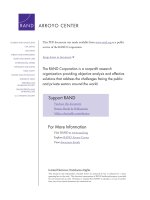
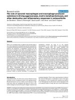
Fig. 2 Analysis of Ssλ15 expression and purification. a Coomassie Blue stained 10 % SDS-PAGE gel. Lane 1, cell-free extract from pGEXSsλ15 transformed E. coli BL21 cells loaded to the affinity column; Lane 2, GST- Ssλ15 fusion protein bound to the column; Lane 3, purified Ssλ15 sarcoptes
moiety after cleavage with thrombin; Lane 4, GST bound to the column after cleavage; M, low molecular weight protein markers (Amersham Biosciences,
Barcelona, Spain). The sample in Lane 3, separated by black lines, was resolved in a different gel from samples M, 1, 2 and 4. b-e Western
blot analysis of GST-Ssλ15 protein using a serum sample from a rabbit experimentally-infested with S. scabiei (b), an anti-GST monoclonal
antibody (c), a mix of sera from a non-infested rabbit and a non-infested chamois (d) and serum from a mange-infested chamois (e). Samples for Western
blot analysis shown in b, c and d were resolved on the same gel, in a different gel from sample e, which was resolved in an independent gel
infested chamois (Fig. 2d). This result confirmed not
only the nature of the fusion protein but also that Ssλ15
was the target of an immune response in mange-infested
animals.
The gel-excised, passive eluted and purified 70 kDa polypeptide was used for the preparation of a specific rabbit
antiserum for further polypeptide immunolocalisation in
the mite’s body. As shown in Fig. 3a and b, Ssλ15 immunolabeling was located in the tegument around the mouthparts and around the coxa of the legs. For the Ssλ20ΔB3
antigen, which forms part of the recAgs vaccine preparation, specific immunostaining is also shown in Fig. 3c.
Ssλ20ΔB3 was located in the integument and the spaces
surrounding the parasite’s vital organs [22]. No staining
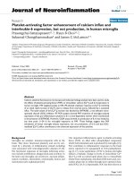
Fig. 3 Immunolocalisation of S. scabiei Ssλ20ΔB3 and Ssλ15 antigens in skin sections derived from a mange-infested chamois. a, b Peroxidase
anti-peroxidase immunostaining after reaction with a rabbit polyclonal anti-Ssλ15 serum, c a rabbit polyclonal anti-Ssλ20ΔB3 serum, d a rabbit
pre-immune serum. Ssλ15 immunolabeling is located in the tegument around the mouthparts and around the coxa of the legs (a and b), and
Ssλ20ΔB3 was located in the integument and the spaces surrounding the parasite’s vital organs (c). No staining is observed in mites and host
tissues when using pre-immune sera, confirming that the detected immunolabeling is specific (d). Abbreviations: A, anterior end of mite; P, posterior
end of mite; Sp, spicules; M, mouthparts; L, legs; SB, stomach blocks; IT, the integument of the epidermis; Sy, synganglion; Mu, striated
muscle. Scale-bars: a, b, 50 μm; c, d, 20 μm
Casais et al. Parasites & Vectors (2016) 9:435
was observed in host tissues, or when using pre-immune
sera, confirming that the detected immunolabeling was
specific (Fig. 3d).
Vaccination trials
In order to determine whether a protective immune response could be elicited using the new recAgs mix a vaccination trial was performed. As described, two groups
of eight rabbits were immunised four times with either
the recAgs mix or PBS with Quil A adjuvant at one week
intervals for the first three immunisations and at three
week interval for the last immunisation. One week after
the last immunisation, all animals were challenged with
crusts containing 2,500 mites taken from previously
infested rabbits.
Antibody responses
Circulating rabbit serum antibody levels (specific IgG
and total IgE levels) were analysed by ELISA. Vaccination induced a significant increase in the levels of total
IgE, these were quantified before vaccination, prior to
challenge, and 1 and 7 weeks post-challenge in the
control and vaccinated groups (Fig. 4a). Immunisation of
rabbits induced an increase in total IgE levels, which
was stronger in the recAgs group. However, S. scabei
challenge induced a rapid and strong increase in the IgE
levels in the control group which was not observed in
the recAgs immunised group. One week after challenge
a slight decrease of the IgE antibody levels was observed
in both groups. No statistically significant differences
were observed between either groups (F(1,19) = 0.406,
P = 0.538). Immunisation resulted in specific IgG
antibody responses in all animals immunised with the
a
Page 8 of 13
recAgs mix, although levels varied between individuals
(Fig. 4b). In the control group, where rabbits received
PBS and adjuvant only, IgG levels remain at preimmunisation levels until challenge when they develop a
specific IgG response. In the recAgs group there was a
strong IgG response after immunisation, which
peaked at week 6 and decreased immediately after
challenge with a slight increased at week 4 postchallenge observed. The IgG levels in the recAgs
immunised group were significantly higher (F(1,14) =
52.31, P < 0.0001) than in the control group.
Cytokines in sera
Systemic immune responses in control and recAgs
immunised groups were measured in serum by means of
an anti-inflammatory cytokine related with lesions or tissue damage (IL-6) and two pro-inflammatory cytokines
(TNF-α, IL-1) before immunisation, prior to challenge
and 1, 2, 3 and 6 weeks post-challenge (Fig. 5). The
serum kinetics of IL-6 response (Fig. 5a) was similar in
both groups, a slight decrease in IL-6 levels was observed after immunisation and a strong increase after
challenge which peaked at week 9 (corresponding to
week 3 post-challenge). Regarding IL-1 levels (Fig. 5b),
rabbits immunised with the recAgs mix exhibited lower
levels of this cytokine in serum during immunisation
than their control counterparts, however after challenge
IL-1 levels were very similar in both experimental
groups remaining high in the recAgs immunised group
as compared with rabbits in the control group. Finally,
the TNF-α profile (Fig. 5c) was rather different, TNF-α
levels exhibited a modest increase during the immunisation period (at week 6 TNF-α levels were about 4 times
b
Fig. 4 Variation of circulating serum S. scabiei-specific IgE (a) and total IgG (b) antibody levels in sera of immunised rabbits detected by ELISA
(Casais et al., 2015). Rabbits were immunised four times (1st, 2nd, 3rd and 4th doses); control refers to the group vaccinated with PBS and Quil A
adjuvant and recAgs refers to the group immunised with a mixture of two S. scabiei recombinant antigens (Ssλ20ΔB3 and GST-Ssλ15) and Quil A.
The dashed line represents the cut-off level of the ELISA used to determine the IgG levels (0.03 relative OD450 nm). Data points correspond to the
mean values, and the error bars represent the standard error. The IgG levels in the recAgs immunised group were significantly higher than in the
control (F(1,14) = 52.31, P < 0.0001), while IgE levels were not significantly different between both groups (F(1,10) = 0.406, P = 0.538)
Casais et al. Parasites & Vectors (2016) 9:435
Page 9 of 13
a
than the control group. No significant differences were
observed between groups in the levels of the three
cytokines investigated, however in the case of the IL1 and TNF-α the recAgs immunisation mix used affected significantly the kinetics observed, as the way
the levels of these two cytokines evolve through the
time in each group is significantly different from the way
they evolve in the control group (F(5,60) = 3.434, P =
0.00854 and F(5,60) = 2.662, P = 0.0307, respectively).
Clinical score and mite density
b
Finally, the protective effect of the potential vaccine was
assessed in the rabbit-S. scabiei var. cuniculi model by
measuring the infested areas over the course of infestation (Fig. 6). All rabbits in the vaccinated and control
group developed mange lesions from week 2–3 postchallenge, which progressed slowly and were mainly observed at the site of inoculation and around the nails as
rough alopecic areas and parakeratotic crusts. Lesions
did not spread to other regions of the body. At seven
weeks post-challenge, the severity of lesions varied between animals with the majority of rabbits having lesion
c
Fig. 5 Serum values of IL-6 (a), IL-1 (b) and TNF-α (c) in rabbits
immunised and challenged with S. scabiei. The kinetics of the
cytokines are depicted in separate graphics, where the data points
correspond to the mean values of the group at each time point, and
the error bars represent the standard error. Rabbits were immunised
four times (1st, 2nd, 3rd and 4th doses), control refers to the group
vaccinated with PBS and Quil A adjuvant and recAgs refers to the
group immunised with a mixture of two S. scabiei recombinant
antigens (Ssλ20ΔB3 and GST-Ssλ15) and Quil A
higher in rabbits belonging to the recAgs group as compared with their control counterpart). After challenge a
rapid and strong increase in the TNF-α levels was observed in the recAgs group, which from week 3 postchallenge TNF-α levels were around 23 times higher
Fig. 6 Lesion scores of New Zealand White rabbits immunised and
challenged with S. scabiei. recAgs refers to the group immunised
with a mixture of two S. scabiei recombinant antigens (Ssλ20ΔB3 and
GST-Ssλ15) and Quil A, and Control refers to the group vaccinated
with PBS and Quil A adjuvant. Data points correspond to the mean
values, and the error bars represent the standard error. The lesions
were graded as follows: score 0 was assigned if no limb lesions were
observed, score 1 when lesions were first observed on the limbs
(lesions ≤ 7.75 cm2), score 2 when lesions were between 7.75–15.5 cm2,
score 3 when lesions ranged from 15.5–31 cm2 and score of 4
when lesions were > 31 cm2. The group immunised with the
recAgs mix had significantly higher lesion scores than the control
group (F(1,14) = 4.608, P = 0.0498) from week 4 to week 6 post-challenge
Casais et al. Parasites & Vectors (2016) 9:435
scores above 2. Unexpectedly, the group vaccinated with
the recAgs mix had significantly higher lesion scores
than the control group (F(1,14) = 4.608, P = 0.0498), as
seen in Fig. 6, from week 4 to week 6 post-challenge.
The density of mites in the skin of all rabbits at
week 7 post-challenge was also analysed as an indicator of the protective value of the potential vaccine
(Fig. 7). The mean number of mites per cm2 was considerably higher in rabbits belonging to the control
group (570.06 ± 704.50, ranging from 144 to 3,952
mites) than in rabbits belonging to the recAgs vaccinated group (357.00 ± 272.90, ranging from 224 to
864 mites/cm2) with a large variability between individuals within each group. The mean mite densities in the
vaccinated group were lower than those observed in the
control group, however no significant differences were
found between groups (χ2 = 0.044, df = 1, P = 0.8335).
No significant effect (F(1,14) = 0.091, P = 0.768) of the
challenge on the weight of the rabbits was observed in
either group (data not shown).
Discussion
In this study, we describe the molecular characterisation
of S. scabiei Ssλ15 cDNA and the immunolocalisation of
the protein encoded by this cDNA in mite infested skin
sections. In addition, the protective value of the immune
responses developed after immunisation with a new
Fig. 7 Box-and-whisker plot of mite densities. Control refers to the
group vaccinated with PBS and Quil A adjuvant and recAgs refers to
the group immunised with a mixture of two S. scabiei recombinant
antigens (Ssλ20ΔB3 and GST-Ssλ15) and Quil A. Box indicates lower
and upper quartiles and horizontal line denotes the median of mite
density; whiskers indicate the lower and upper extremes; the dot
represents an outlier detected in the recAgs group
Page 10 of 13
mix of GST-Ssλ15 and the previously described
Ssλ20B3 antigen was evaluated in a rabbit/S. scabiei
var. cuniculi model.
Two expressed sequence tags from S. scabiei, Ssλ20B3
and Ssλ15, have now been identified by immunoscreening of a S. scabiei var. hominis library with sera from
mangy animals. The two antigens were selected as candidates to include in a potential vaccine because of their
strong reaction with sera from infested animals. Moreover, several clones selected during screenings were
identified as sequences related to Ssλ15 and Ssλ20, suggesting that these cDNAs encoded immunodominant
antigens in infested animals that are derived from
abundant parasite mRNAs. The E. coli expressed fusion
protein GST-Ssλ20B3 was specifically recognised in
Western blot with sera of an infested chamois [22] and
the GST-Ssλ15 with sera of an infested rabbit and an
infested chamois (Fig. 2), which confirms the nature of
the fusion proteins and indicates that both polypeptides are the target of immune responses in mangeinfested animals.
Attempts to elucidate the function and possible crossreactivity of Ssλ20 and Ssλ15 with antigens from other
related parasites through database searches were performed. They did not show homology to any house dust
mite antigens or allergens such as Dermatophagoides
farina, D. pteronyssinus and E. maynei. Ssλ15 antigen
had a 96 % of identity with a hypothetical protein of S.
scabiei (GenBank: KPM09788.1) [37], whose function
has been provisionally noted as a DNA translocase
FTSK. In addition, no positive reactions of Ssλ20B3 with
sera from pigs immunised with D. pteronyssinus and
Acarus siro or with sera from tick-infested red deer were
detected by ELISA indicating the absence of crossreactions between Ssλ20B3 and antigens of the mentioned parasites [22, 38]. Cross-reactivity of Ssλ15 with
those sera has not been checked because this antigen is
not a good candidate to coat ELISA plates for the diagnosis of sarcoptic mange.
Vaccination is the most desirable prophylactic method
for any infectious disease. One of the vaccination strategies explored for complex parasites is the use of proteins isolated directly from them. This strategy is
advantageous over recombinant proteins in that all
structural and immunogenic characteristics that are
native to the organism are displayed in the vaccine.
However, the availability of native proteins at the
required purity and quantity has been the main limiting factor and therefore, production of recombinant
antigens has been one of the most common choices
to test the protective potential of immunodominant
antigens, as was implemented in this study with
recAgs (Ssλ20B3 and GST-Ssλ15 mix) in the rabbit/
S. scabiei var. cuniculi model.
Casais et al. Parasites & Vectors (2016) 9:435
In our system, Ssλ15 antigen was included in the
immunisation mixture as a fusion protein with GST to
prevent problems derived from thrombin digestion. In
addition, GST has immunomodulatory functions and
seems a promising vaccine candidate in human schistosomiasis and other parasite infections including scabies
[39] so the idea was that the presence of GST could
enhance the potential protective immune response elicit
by the vaccine preparation.
The experimental design used in this study included
two groups, one vaccinated with the recAgs mixture
(Ssλ15, Ssλ20ΔB3) plus Quil A adjuvant, and a control
group with PBS and Quil A as has been performed in
other studies [17, 40]. Nonetheless, this design could
have been improved by including a second control group
inoculated only with PBS [21]. However, it is possible to
conclude that significant differences observed between
the recAgs group and the control group are due to the
recombinant antigens present in the recAgs preparation
and not included in the control group. On the other
hand, variations in production of the total IgE and cytokines (IL-1 and IL-6) are observed upon vaccination
with adjuvant only so it should be take into account that
the effects of the vaccination observed could be due not
only to the recombinant antigens but also to Quil A,
highlighting the importance of the chosen adjuvant.
Our results show that both defined antigens (Ssλ20B3
and GST-Ssλ15) delivered as recombinant proteins
produced in E. coli under our vaccination regime of
immunisation of rabbits elicited high specific IgGs levels
(significantly higher than those observed in the control
group) and increased levels of total IgEs (Fig. 4),
however in spite of these humoral immune responses,
no significant clinical protection against S. scabiei
challenge was detected. In this sense, the immunological
response managed to reduce the number of mites per
cm2 of skin in the recAgs rabbits but the lesion area
score in this group was significantly higher than in the
control group, suggesting that mite population might be
more sensitive as an indicator of protection than severity
of lesions in the vaccination challenge experiment [17].
These results might indicate that the lesions observed in
rabbits in the recAgs group were due not only to the
mites themselves but also to an exacerbated immunological response after challenge, which is in agreement
with the high levels of pro-inflammatory cytokines (IL-1
and TNFα) detected. Examples of a detrimental role of
exacerbated pro-inflammatory cytokines have been reported in the literature, particularly in the case of the so
called “cytokine storm” after influenza infection or even
in vaccination studies in pigs [41, 42].
Both vaccination strategies, the production of recombinant antigens and the use of proteins isolated directly
from the parasite, had previously been explored for S.
Page 11 of 13
scabiei with relative success. Thus, recombinant antigens
such as Ssag1 and Ssag2 [19], S. scabiei glutathione Stransferase [20] and tropomyosin [21] had been assayed
as vaccines and did not confer complete protection
against S. scabiei challenge. Likewise, while vaccination
of goats with soluble or insoluble mite proteins did not
produce protective immunity [40], vaccination with a
fresh extract from S. scabiei conferred partial protection of goats [17], slightly reducing the mite population
(P = 0.015) but not affecting the severity of lesions, as
we have reported in this study in the rabbit model. It
has been proposed that the failure to produce complete
protection against sarcoptic mange after sensitisation
or vaccination may be due to denaturation or degradation
of protective antigens, as well as the low abundance or
low “immunoprotection” of some of them [17], suggesting
that identification and production of proteins having vital
function for the mite survival accessible by the host immune system, which constitutes the most important step
in vaccine development, will be difficult.
It has been also suggested that the lack of immune
protection in goats vaccinated with an extract of S. scabiei soluble proteins could be attributed to the absence
of protective levels of IgE, indicating that IgE antibody
play an important role in immunity to S. scabiei infestation [41]. In this regard, our results showed that
immunisation with the selected antigen preparation
(recAgs) was able to induce high levels of IgG and
increased levels of total IgE, with a reduction in the mite
population after challenge observed. However, immunisation was not able to reduce lesion areas, which might
indicate failure of Ssλ20B3 and GST-Ssλ15 recombinant
antigens to elicit protective levels of specific IgE antibody and/or an effective cell mediated response.
The failure to induce complete immune protection in
the vaccinated rabbits might indicate that the selected antigens lost their potential native structural and immunogenic properties during the production and purification
processes. Immunogenicity is of crucial importance when
evaluating expression systems for production of recombinant vaccine antigens [43, 44]. Therefore, we must consider that improvements in expression (for example using
virus as expression vectors) and purification strategies
may lead to peptides that are structurally more similar to
their native counterparts. Likewise, future modifications
in our vaccine regime (adjuvants and delivery methods)
may direct us towards a more appropriate response.
Taking into account the results of our vaccine analysis,
the use of this vaccine mix combined with topical creams
containing corticoids to control the exacerbated immune
response may allow a better control of the disease and
reduction in the use of acaricides. It is also possible that
the selected antigens are not essential for mite survival
and pathogenesis.
Casais et al. Parasites & Vectors (2016) 9:435
Page 12 of 13
Conclusions
In conclusion, the selected immunodominant antigens
(Ssλ15 and Ssλ20ΔB3) delivered as recombinant proteins per
se are not good vaccine candidates against S. scabiei infestation in rabbits. Immunisation with a mix of the two antigens
induced high levels of humoral responses (IgGs and IgEs),
however, immunisation had no clinical protective efficacy (lesion scores were significantly higher although immunisation
reduced mite density). Further studies on alternative production systems, adjuvants, delivery methods, immunisation
protocols and combinations of antigens will be required to
manage stimulation of clinical protective immune responses.
Author details
1
Servicio Regional de Investigación y Desarrollo Agroalimentario (SERIDA),
Centro de, Biotecnología Animal, La Olla-Deva, 33394 Asturias, Spain.
2
Servicio Regional de Investigación y Desarrollo Agroalimentario (SERIDA),
Finca experimental La Mata, Programa de Investigación Forestal (PIF). Área
de Cultivos Hortofrutícolas y Forestales, La Mata s/n, 33825 Asturias, Spain.
3
Instituto Universitario de Biotecnología de Asturias, Departamento de
Bioquímica y Biología Molecular, Edificio Santiago Gascón, Campus El Cristo,
Universidad de Oviedo, 33006 Oviedo, Spain. 4Clinical Research Centre (CRC),
Barts Health NHS Trust, 2 Newark Street, Abernethy Building, Whitechapel,
London, UK. 5Centre de Recerca en Sanitat Animal (CReSA), UAB-IRTA,
Campus de la Universitat Autònoma de Barcelona, Bellaterra Cerdanyola del
Vallès, Spain. 6The Pirbright Institute, Ash Road, Pirbright, Woking, Surrey, UK.
Additional file
References
1. Zahler M, Essig A, Gothe R, Rinder H. Molecular analyses suggest
monospecificity of the genus Sarcoptes (Acari:Sarcoptidae). Int J Parasitol.
1999;29:759–66.
2. Bornstein S, Mörner T, Samuel B. Sarcoptes scabiei and sarcoptic mange.
In: Samuel WM, Pybus MJ, Kocan AA, editors. Parasitic Diseases of Wild
Mammals. 2nd ed. Ames: Iowa State University Press/Ames; 2001. p. 107–19.
3. Pence DB, Ueckermann E. Sarcoptic mange in wildlife. Rev Sci Tech.
2002;21:385–98.
4. Wagner R, Wendlberger U. Field efficacy of moxidectin in dogs and rabbits
naturally infested with Sarcoptes spp., Demodex spp. and Psoroptes spp.
mites. Vet Parasitol. 2000;93:149–58.
5. Voyvoda H, Ulutas B, Eren H, Karagenc T, Bayramli G. Use of doramectin for
treatment of sarcoptic mange in five Angora rabbits. Vet Dermatol. 2005;16:285–88.
6. Kurtdede A, Karaer Z, Acar A, Guzel M, Cingi CC, Ural K, Ica A. Use of
selamectin for the treatment of psoroptic and sarcoptic mite infestation in
rabbits. Vet Dermatol. 2007;18:18–22.
7. Currie BJ, Harumal P, MicKinnon M, Walton SF. First documentation of
in vivo and in vitro ivermectin resistance in Sarcoptes scabiei. Clin Infect Dis.
2004. doi:10.1086/421776.
8. Pasay C, Walton S, Fischer K, Holt D, McCarthy J. PCR-based assay to survey
for knockdown resistance to pyrethroid acaricides in human scabies mites
(Sarcoptes scabiei var. hominis). Am J Trop Med Hyg. 2006;74:649–57.
9. Terada Y, Murayama N, Idemura H, Morita T, Nagata M. Sarcoptes scabiei var. canis
refractory to ivermectin treatment in two dogs. Vet Dermatol. 2010;21:608–12.
10. Mellanby K. Experiments on Scabies Prophylaxis. Br Med J. 1944;1:689–90.
11. Arlian LG, Morgan MS, Vyszenski-Moher DL, Stemmer BL. Sarcoptes scabiei:
The circulating antibody response and induced immunity to scabies. Exp
Parasitol. 1994;78:37–50.
12. Arlian LG, Morgan MS, Rapp CM, Vyszenski-Moher DL. The development of
protective immunity in canine scabies. Vet Parasitol. 1996;62:133–42.
13. Tarigan S. Dermotopathology of caprine scabies and protective immunity in
sensitised goats against Sarcoptes scabiei infection. Jurnal Ilmu Ternak dan
Veteriner. 2003;7:265–71.
14. Rodriguez-Cadenas F, Carvajal-González MT, Fregeneda-Grandes JM,
Aller-Gancedo JM, Rojo-Vázquez FA. Clinical evaluation and antibody responses
in sheep after primary and secondary experimental challenges with the mange
mite Sarcoptes scabiei var. ovis. Vet Immunol Immunopathol. 2010;133:109–16.
15. Casais R, Dalton KP, Millán J, Balseiro A, Oleaga A, Solano P, Goyache F,
Prieto JM, Parra F. Primary and secondary experimental infestation of rabbits
(Oryctolagus cuniculus) with Sarcoptes scabiei from a wild rabbit: factors
determining resistance to reinfestation. Vet Parasitol. 2014;203:173–83.
16. Liu X, Walton S, Mounsey K. Vaccine against scabies: necessity and
possibility. Parasitology. 2014;141:725–32.
17. Tarigan S. Vaccination of goats with fresh extract from Sarcoptes scabiei
confers partial protective immunity. Jurnal Ilmu Ternak dan Veteriner.
2006;11:144–50.
18. Mounsey KE, McCarthy JS, Walton SF. Scratching the itch: new tools to
advance understanding of scabies. Trends Parasitol. 2013;29:35–42.
19. Harumal P, Morgan M, Walton SF, Holt DC, Rode J, Arlian LG, Currie BJ,
Kemp DJ. Identification of a homologue of a house dust mite allergen in a
cDNA library from Sarcoptes scabiei var hominis and evaluation of its vaccine
potential in a rabbit/S. scabiei var. canis model. Am J Trop Med Hyg.
2003;68:54–60.
Additional file 1: Mass spectrometry results. Results from mass
spectrometry analysis of the 70, 60 and 29 kDa protein spots generated
by digestion of GST-Ssλ15 with thrombin during the purification process
of the Ssλ15 antigen. (PDF 560 kb)
Abbreviations
ELISA, enzyme-linked immunosorbent assay; IPTG, isopropyl-Beta-D-hiogalactopyranoside; GST, gluthathione s-transferase
Acknowledgements
The proteomic analysis was carried out in the Proteomics Facility UCM-PCM,
a member of ProteoRed network. Passive elution of the thrombin excised
Ssλ15 70 kDa protein band and preparation of specific antisera was done
by the “Servicios científico-técnicos” of the University of Oviedo. We thank
Dr. Alberto Espí for his help with the mite’s tissue identification.
Funding
This work was partially funded by grant RTA11-00087-00-00 from the Spanish
Instituto Nacional de Investigación y Tecnología Agraria y Alimentaria (INIA),
Fondo Europeo de Desarrollo Regional (FEDER), AGL2010-22200-C02-01 from
Spanish Ministry (MINECO) and the Biotechnology and Biological Sciences
Research Council (BBSRC) grant BBS/E/I/00002014.
Availability of data and materials
The datasets supporting the conclusions of this article are included within
the article and the additional file. The sequence of Ssλ15 cDNA clone is
submitted to the GenBank database under accession number KU359774.
Authors’ contributions
RC participated in the design of the study, manuscript writing and
performed some of the experiments. VG performed the statistical analysis
and prepared figures; AB performed the immunohistochemical study; KPD
participate in the manuscript writing and discussion; AC, RG and PB
participated in the screening of the library, production and purification of
recombinant antigens; JMP participated in vaccination trials, the collection of
mite samples and data; MM participated in the design of the study and
manuscript writing. All authors read and approved the final manuscript.
Competing interests
The authors declare that they have no competing interests.
Consent for publication
Not applicable.
Ethics approval and consent to participate
Experimental procedures were approved by the SERIDA Animal Ethics
Committee and authorised by the Regional Consejería de Agroganadería
y Recursos Autoctonos del Principado de Asturias, Spain. Experiments were
conducted in accordance with the Spanish and European current legal
requirements and guidelines regarding experimentation and Animal Welfare.
Received: 12 February 2016 Accepted: 22 July 2016
Casais et al. Parasites & Vectors (2016) 9:435
20. Pettersson EU, Ljunggren EL, Morrison DA, Mattsson JG. Functional analysis
and localization of a delta-class glutathione S-transfrease from Sarcoptes
scabiei. Int J Parasitol. 2005;35:39–48.
21. Zhang R, Jise Q, Zheng W, Ren Y, Nong X, Wu X, Gu X, Wang S, Peng X,
Lai S, Yang G. Characterisation and evaluation of a Sarcoptes scabiei allergen
as a candidate vaccine. Parasite Vector. 2012;5:176.
22. Casais R, Prieto M, Balseiro A, Solano P, Parra F, Martín Alonso JM.
Identification and heterologous expression of a Sarcoptes scabiei cDNA
encoding a structural antigen with immunodiagnostic potential. Vet Res.
2007;38:435–50.
23. Fischer K, Holt DC, Harumal P, Currie BJ, Walton SF, Kemp DJ. Generation
and characterization of cDNA clones from Sarcoptes scabiei var. hominis for
an expressed sequence TAG library: identification of homologues of house
dust mite allergens. Am J Trop Med Hyg. 2003;68:54–60.
24. Sambrook J, Fritsch EF, Maniatis T. Molecular Cloning, A laboratory
manual, 2nd ed. Cold Spring Harbor, NY: Cold Spring Harbor Laboratory
Press; 1989. Petersen TN, Brunak S, von Heijne G, Nielsen H. SignalP
4.0: discriminating signal peptides from transmembrane regions.
Nat Methods. 2011;8:785–86.
25. Kyte J, Doolittle RF. A simple method for displaying the hydropathic
character of a protein. J Mol Biol. 1982;157:105–32.
26. Bradford MM. A rapid and sensitive method for the quantitation of
microgram quantities of protein utilizing the principle of protein-dye
binding. Anal Biochem. 1976;72:248–54.
27. Sechi S, Chait BT. Modification of cysteine residues by alkylation. A tool in
peptide mapping and protein identification. Anal Chem. 1998;70:5150–58.
28. Sternberger LA, Hardy PH, Cuculis JJ, Meyer HG. The unlabeled antibody
enzyme method of immunohistochemistry: preparation and properties of
soluble antigen-antibody complex (horseradish peroxidase-antihorseradish
peroxidase) and its use in identification of spirochetes. J Histochem
Cytochem. 1970;18:315–33.
29. Millán J. First description of sarcoptic mange in wild European rabbit
(Oryctolagus cuniculus). Eur J Wildl Res. 2010;56:455–57.
30. Casais R, Millán J, Rosell JM, Dalton KP, Prieto JM. Evaluation of an ELISA
using recombinant Ssλ20ΔB3 antigen for the serological diagnosis of
Sarcoptes scabiei infestation of domestic and wild rabbits. Vet Parasitol.
2015;214:315–21.
31. Sanchez J, Dohoo IR, Markham F, Leslie K, Conboy G. Evaluation of the
repeatability of a crude adult indirect Ostertagia ostertagi ELISA and
methods of expressing results. Vet Parasitol. 2002;109:75–90.
32. Bornstein S, Wallgren P. Serodiagnosis of sarcoptic mange in pigs.
Vet Rec. 1997;141:8–12.
33. Hollanders W, Vercruysse J, Raes S, Bornstein S. Evaluation of an enzymelinked immunosorbent assay (ELISA) for the serological diagnosis of
sarcoptic mange in swine. Vet Parasitol. 1997;69:117–23.
34. Mumcuoglu KY. A technique for quantitative evaluation of ectoparasitic
mites and insects of domestic animals. Esp Appl Acarol. 1990;9:97–101.
35. R Core Team. R: A language and environment for statistical computing. R
Foundation for Statistical Computing, Vienna, Austria. 2015. Accessed 26 Jul 2016.
36. Lawrence MA. ez: Easy analysis and visualization of factorial experiments. R
package version 4.2–2. 2013. />Accessed 26 Jul 2016.
37. Rider SD, Morgan MS, Arlian LG. Draft genome of the scabies mite. Parasite
Vector. 2015. doi:10.1186/s13071-015-1198-2.
38. Casais R, Goyena E, Martínez-Carrasco C, Ruiz de Ybáñez R, Alonson de Vega
F, Ramis G, Prieto JM, Berriatua E. Variable performance of a human derived
Sarcoptes scabiei recombinant antigen ELISA in swine mange diagnosis. Vet
Parasitol. 2013;197:397–403.
39. Ouaissi A, Ouaissi M, Sereno D. Glutathione S-transferases and related
proteins from pathogenic human parasites behave as immunomodulatory
factors. Immunol Lett. 2002;81:159–64.
40. Tarigan S, Huntley JF. Failure to protect goats following vaccination with
soluble proteins of Sarcoptes scabiei: Evidence for a role for IgE antibody in
protection. Vet Parasitol. 2005;133:101–9.
41. Gauger PC, Vincent AL, Loving CL, Henningson JN, Lager KM, Janke BH,
Kehrli Jr ME, Roth JA. Kinetics of lung lesion development and proinflammatory cytokine response in pigs with vaccine-associated enhanced
respiratory disease induced by challenge with pandemic (2009) A/H1N1
influenza virus. Vet Pathol. 2012;49:900–12.
Page 13 of 13
42. Van Reeth K, Nauwynck H, Pensaert M. Bronchoalveolar interferon-alpha,
tumor necrosis factor-alpha, interleukin-1, and inflammation during
acute influenza in pigs: a possible model for humans? J Infect Dis.
1998;177:1076–79.
43. Arnot DE, Cavanagh DR, Remarque EJ, Creasey AM, Sowa MP, Morgan WD,
Holder AA, Longacre S, Thomas AW. Comparative testing of six antigenbased malaria vaccine candidates directed toward merozoite-stage
Plasmodium falciparum. Clin Vaccine Immunol. 2008;15:1345–55.
44. Qian F, Wu Y, Muratova O, Zhou H, Dobrescu G, Duggan P, Lynn L, Song G,
Zhang Y, Reiter K, MacDonald N, Narum DL, Long CA, Miller LH, Saul A,
Mullen GE. Conjugating recombinant proteins to Pseudomonas aeruginosa
ExoProtein A: a strategy for enhancing immunogenicity of malaria vaccine
candidates. Vaccine. 2007;25:3923–33.
Submit your next manuscript to BioMed Central
and we will help you at every step:
• We accept pre-submission inquiries
• Our selector tool helps you to find the most relevant journal
• We provide round the clock customer support
• Convenient online submission
• Thorough peer review
• Inclusion in PubMed and all major indexing services
• Maximum visibility for your research
Submit your manuscript at
www.biomedcentral.com/submit