developmental biology protocols - rocky s. tuan and cecilia w. lo
Bạn đang xem bản rút gọn của tài liệu. Xem và tải ngay bản đầy đủ của tài liệu tại đây (3.54 MB, 481 trang )
Developmental
Biology
Protocols
Volume III
Edited by
Rocky S. Tuan
Cecilia W. Lo
HUMANA PRESS
Methods in Molecular Biology
TM
HUMANA PRESS
Methods in Molecular Biology
TM
Developmental
Biology
Protocols
Volume III
Edited by
Rocky S. Tuan
Cecilia W. Lo
VOLUME 137
Overview 3
3
From:
Methods in Molecular Biology, Vol. 137: Developmental Biology Protocols, Vol. III
Edited by: R. S. Tuan and C. W. Lo © Humana Press Inc., Totowa, NJ
1
Developmental Biology Protocols
Overview III
Rocky S. Tuan and Cecilia W. Lo
1. Introduction
The marriage of cell and molecular biology with embryology has produced remark-
able advances for the field of developmental biology. In this third volume of Develop-
mental Biology Protocols, contemporary, practical methods are first presented for the
analysis and manipulation of developmental gene expression. To illustrate how such
techniques, as well as procedures of experimental embryology including those
described in the first two volumes of the series, may be applied in the study of develop-
ment, a panoramic collection of experimental models of morphogenesis, development,
and cellular differentiation are detailed. Both in vivo and in vitro systems are included.
The volume concludes with various examples of developmental models of diseases
and their molecular basis.
2. Manipulation of Developmental Gene Expression and Function
Drosophila has been and remains one of the most versatile model systems for the
manipulation of developmental gene expression. Chapter 2 focuses on a description of
the experimental approaches currently used in ectopic gene expression in Drosophila
to examine the function of a given gene in the desired tissue. Chapter 3 deals with the
utilization of the highly efficient FLP/FRT yeast site-specific recombination system to
generate somatic and germline clones in Drosophila for phenotypic analysis and screening.
Chapters 3 and 4 address the methods used to alter gene expression as well as gene
function in another experimentally highly accessible system, the developing chick
embryo. Chapter 3 describes the application of antisense oligonucleotides to “knock
down” gene expression in somitic stage chick embryos, whereas Chapter 4 discusses
how functional neutralizing monoclonal antibodies may be used to block the activity of
a specific gene product, N-cadherin, in the developing chick embryonic limb bud.
3. Analysis of Gene Expression
The first step in analyzing the molecular basis of any developmental event is to
characterize and compare gene expression profiles, both spatial and temporal, as a function
4 Tuan and Lo
of development. A comprehensive list is provided in this section. Classic methods such
as Northern blotting is not presented here, because relevant protocols are readily avail-
able in many technical manuals of molecular biology. Quantitative methods include
ribonuclease protection assay (Chapter 6), and polymerase chain reaction (PCR) based
methods (Chapters 7 and 8). In situ hybridization (Chapters 9–15) has gained wide
application in visualizing the spatial aspects of gene expression in the developing
embryo, particularly in mapping the dynamics of tissue morphogenesis. In particular,
the ability to carry out multiple in situ hybridizations (Chapter 14), or sequential in situ
hybridization and immunohistochemistry (Chapters 12 and 15), on a given specimen
should be invaluable for analyzing the potential roles of genes and gene products in
development.
The potential of the green fluorescent protein (GFP) of the jellyfish, Aequoria
victoria, as a vital recombinant tag for genes of interest has produced a great deal of
excitement in developmental biology; Chapter 16 provides a thorough discussion of
the principles and techniques in the application of the GFP. Finally, the basic strategy
in the application of monoclonal antibodies, one of the most powerful technical
advances in modern biomedical research that has enjoyed a distinguished history, in
the study of embryonic development is presented (Chapter 17).
4. Models of Morphogenesis and Development
This section presents a number of developmental model systems under active inves-
tigation to illustrate the multitude of experimental questions currently being addressed
in the field of developmental biology. The inductive events of embryogenesis and
means for their analyses are described in Chapters 18 and 19. Techniques for whole or
partial embryo explant cultures for the somitic stage embryos for the analysis of meso-
dermal and neural crest studies are covered in Chapters 20 and 21. Other models of
morphogenesis include those for angiogenesis (Chapter 22), vasculogenesis (Chapter 23),
and epithelial–mesenchyme interactions (Chapter 24). Specific organogenesis models
are also included—limb bud (Chapter 25) and palate (Chapter 26).
5. In Vitro Models and Analysis of Differentiation and Development
Regulation of cell differentiation is one of most active research areas of develop-
mental biology. With the advent of cell and molecular biology, and the identification of
differentiation-associated genes, cell differentiation is often interpreted in terms of
regulation of gene expression. Both cis and trans modes of gene expression regulation
have been found to operate during cell differentiation, leading to active investigation
on structure/function of gene promoters and transcription factors.
This section is a collection of many in vitro cell differentiation systems currently
under active investigation. Early events in development include fertilization (Chapter 27)
and trophoblastic differentiation (Chapter 28). Bone marrow-derived mesechymal pro-
genitor cells have received a great deal of recent attention as candidate cells for cell-
based tissue engineering. It is generally believed that the differentiation potentials of
these cells represent a partial recapitulation of the characteristics of embryonic meso-
dermal cells. Techniques for their isolation, culture, and characterization are described
in Chapter 29. Another cell type important for studying cell differentiation are germ
cells; methods for their isolation and culture are included in Chapter 30. Prostate cell
Overview 5
differentiation is discussed in Chapter 30. Cell differentiation in connective tissues is
presented in the following chapters: striated muscle differentiation (Chapter 31),
somitic myogenesis (Chapter 32), mesenchymal chondrogenesis (Chapters 33–35), and
bone cell differentiation (Chapter 36).
In addition to specific examples and systems of cellular differentiation, methods for
three crucial aspects of cellular activities are also presented. Cell–cell interaction is
illustrated in Chapter 39, which deals with cadherin-mediated events. Cell–matrix inter-
actions as mediated by hyaluronan binding are discussed in Chapter 40. The dynamic
regulation of cytoskeletal architecture, visualized and analyzed by the microinjection
of fluorescently-labeled α-actinin into living cells, is presented in Chapter 41.
6. Developmental Models of Diseases
The experimental paradigms gained from developmental biology lend readily to the
mechanistic analysis of diseases. Several examples are included here. Pax 3, a member
of the vertebrate Pax gene family containing a DNA-binding domain known as the
paired domain, is important for proper formation of the nervous, cardiovascular, and
muscular systems. The molecular analysis of Pax 3 mutations and how the pathways
affected lead to the pathogenesis of specific dysmorphogenic consequences is the sub-
ject of Chapter 42. Finally, one of the most powerful contributions of molecular devel-
opmental biology to the study of diseases is the application of transgenic methodologies
to create animal models of human diseases. The three examples included here all deal
with various aspects of skeletal defects, including both trunk as well as craniofacial
malformations. The methods involve studies utilizing a structural gene (collagen type X,
Chapter 43), cell specific promoter (α1(II) procollagen gene, Chapter 44), as well as
transcription factors (Msx2, Chapter 44).
Ectopic Expression in
Drosophila
9
9
From:
Methods in Molecular Biology, Vol. 137: Developmental Biology Protocols, Vol. III
Edited by: R. S. Tuan and C. W. Lo © Humana Press Inc., Totowa, NJ
2
Ectopic Expression in
Drosophila
Elizabeth L. Wilder
1. Introduction
Ectopic expression in Drosophila has been used extensively to examine the capa-
bilities of a given gene in virtually any tissue. Three general approaches are described
here, and the choice of which to use is determined by the needs of the particular experi-
ment. Certain aspects of each approach can also be combined, providing powerful tools
for the examination of gene function. Because ectopic expression does not involve a
protocol, but rather generation of certain types of transgenic strains, this chapter focuses
on a description of the approaches and in what circumstances each is likely to be useful.
2. Materials
For each of the methods of ectopic expression described here, the production of
transgenic strains is required. The vectors that are widely used in these experiments are
available (1–3).
3. Methods
3.1. Expression Through Defined Promoters
The simplest means of ectopic expression is through the construction of a promoter-
cDNA fusion in which a gene of interest is driven by a defined promoter or enhancer.
Transgenic strains carrying this construct then ectopically express the gene of interest
in the defined pattern.
One of the most commonly used promoters for this purpose is the heat shock protein
70 (hsp70) promoter (1). This promoter allows ubiquitous expression to be induced in
any tissue of the fly through a simple heat shock at 37°C. The inducible nature of
this approach is a great advantage. However, basal levels of expression can be prob-
lematic, and heat shock itself can induce developmental defects. In addition, short
bursts of ectopic expression ubiquitously is often not ideal. Therefore, sustained
expression in defined domains may be preferred.
To achieve ectopic expression within a defined domain, transcriptional regulatory
regions from characterized genes have been linked to genes of interest (4,5). The advan-
tage of this approach is its simplicity. Its primary limitation is that lethality can result
from the ectopic expression. This makes it impossible to establish stable transgenic
10 Wilder
lines. Enhancers that drive expression during late stages of development or in tissues
that are nonessential have been particularly useful, because lethality owing to ectopic
expression is avoided.
The lethality associated with sustained expression of transgenes during develop-
ment, the effort required to generate transgenic strains in which the transgene is
expressed in multiple patterns, and the lack of defined enhancers driving expression in
certain tissues prompted the development of alternative strategies for ectopic expression.
3.2. The GAL4 System
The identification of the yeast transcriptional activator, GAL4, as a highly active,
specific transcription factor that can activate transcription in Drosophila (6) led to the
development of a system of ectopic expression referred to as the GAL4 system (2).
This two-part system is shown in Fig. 1 and involves a cross between a fly expressing
GAL4 in particular cells and a fly carrying a gene of interest under the transcriptional
control of the GAL4 upstream activating sequence, or UAS. In the progeny of such a
cross, the gene of interest will be expressed in cells where GAL4 is synthesized. Tar-
geted ectopic expression of the gene of interest can therefore be achieved by choosing
among many strains that express GAL4 in defined patterns.
Three vectors are generally useful for investigators using this system (2). pGaTB/N
provides either a BamHI site or a NotI site upstream of GAL4, allowing a defined
promoter to drive GAL4 expression. The second, pGawB, is an enhancer-trapping vec-
tor that directs GAL4 expression from genomic enhancers. Finally, pUAST includes
multiple cloning sites behind five copies of an ideal GAL4 binding sequence. Genes of
interest are easily cloned into this vector for GAL4-mediated expression.
Fig. 1. The GAL4 system of ectopic expression (modified from ref. 2). This system allows
the ectopic expression of any gene of interest (Gene X) in a pattern determined by the expres-
sion of the transcriptional activator, GAL4. Hundreds of lines in which GAL4 is expressed in a
variety of patterns have been generated through enhancer trapping or by linking the GAL4
coding sequence to defined regulatory elements. These are crossed to flies carrying the gene of
interest under the transcriptional control of the GAL4 Upstream Activating Sequence (UAS).
The progeny of this cross express the gene of interest in the pattern of choice.
Ectopic Expression in
Drosophila
11
Hundreds of GAL4 strains have been generated through the process of enhancer
trapping. These strains have been characterized by crossing newly generated lines to a
UAS-LacZ strain and characterizing the expression pattern. Many of these strains are
now available through the Drosophila Stock Center at Bloomington, IN. The expres-
sion patterns that have been detected through these enhancers vary from very broad
expression to highly specific patterns. They, thus, offer the possibility of driving ectopic
expression in virtually any tissue.
In addition to the strains generated through enhancer trapping, many lines have been
generated by fusing the GAL4 coding sequence to defined promoters, such as the hsp70
promoter. The latter offers the advantage mentioned above of inducible expression.
The construction of strains expressing GAL4 in defined domains allows any UAS
transgene to be examined within the particular region of interest.
The GAL4 system has contributed to the utility of the FLP-FRT system of inducing
mutant clones (see Chapter 3)
(7). In this system, mitotic recombination is induced via
flip recombinase (FLP), which is under the control of a heat shock promoter. The result-
ing mutant clones are then generated in all mitotically active cell populations. How-
ever, if FLP is placed under the control of GAL4-UAS, mutant clones are only
generated within the GAL4 expression domain. This allows the investigator to deter-
mine whether a particular gene has an endogenous function within cells defined by
GAL4 expression.
The GAL4 system addresses many of the problems associated with simple transgenes.
First, since the UAS transgenic lines are produced in the absence of GAL4 activity,
ectopic expression of the transgene does not occur. Therefore, lethality associated with
ectopic expression is avoided until the transgenic flies are crossed to a GAL4 express-
ing strain. Second, defined enhancers are not required for expression in a particular set
of cells. Sites of expression are only limited by the number of enhancer trapped strains
available, the number of which is continually growing. Finally, the GAL4 system allows
ectopic expression in any number of patterns and conditions with the construction of
only a single UAS transgene.
This system of ectopic expression is extremely powerful for these reasons, but it
does have limitations. First, for undefined reasons, GAL4 does not seem to function in
the germline (A. Brand, personal communication). For experiments where germline
expression is needed, other methods must be used. A more universal limitation of the
GAL4 system is the fact that it is not inducible. Many enhancers drive expression dur-
ing early phases of development, so that GAL4-mediated ectopic expression of certain
UAS transgenes results in embryonic lethality. For investigators interested in later
aspects of development, this has been a serious limitation of the GAL4 system.
This problem can be partially addressed through modulation of temperature. The
optimal temperature for GAL4 activity appears to be the ambient temperature for yeast,
which is 30°C. By rearing flies at lower temperature, GAL4 activity is reduced (8,9),
and in some instances, early lethality associated with higher levels of ectopic expres-
sion from the UAS transgene is avoided. The flies can be shifted at later stages of
development to increase GAL4-mediated expression.
In at least one instance, an inductive ability has been added to the GAL4 system
through the construction of a UAS transgene carrying a cDNA encoding a temperature
sensitive protein (9). Thus, progeny of the GAL4-UAS cross are maintained at the
12 Wilder
restrictive temperature during embryogenesis and shifted to the permissive tempera-
ture at the relevant stages. This permits ectopic activity to begin at the desired stage.
However, since temperature sensitive lesions have not been defined for most genes, the
inability to control expression temporally remains a problem with the GAL4 system in
analysis of postembryonic development.
3.3. Ectopic Expression in Clones
The temporal control of ectopic expression has been critical for the analysis of gene
activity during imaginal development. An ingenious method of ectopically expressing
genes in any region of the imaginal discs was developed by Struhl and Basler (3) (Fig. 2)
and has come to be called the flip-out system. This method involves the generation of
random clones in which the coding region of a gene of interest comes to lie adjacent to
a ubiquitous promoter. In these clones, the gene is ectopically expressed, whereas in
the surrounding tissue, a gene encoding a visible marker is adjacent to the ubiquitous
promoter, separating it from the gene of interest. This is accomplished through the use
of flip recombinase target (FRT) sites flanking the marker gene. In the presence of the
recombinase, the marker is removed, bringing the promoter and the gene of interest
together. The resulting clone of cells is marked by the absence of the marker, which is
ubiquitously present elsewhere in the fly.
This technique requires the generation of a construct in which the gene of interest is
placed within the context of the promoter-FRT-marker-FRT construct (3,10,11). Two
vectors are available that utilize either the Actin-5C promoter or the β-Tubulin pro-
Fig. 2. The Flip-out system of ectopic expression (see ref. 3). Flip recombinase (FLP) target
sites (FRTs) are arranged as direct repeats flanking a visible marker. The expression of this
marker is under the control of the promoter element. However, in the presence of FLP, recom-
bination between the FRTs is induced, resulting in deletion of the marker gene. The gene of
interest is now juxtaposed to the promoter element, resulting in ectopic expression of the gene
of interest. This is an efficient but stochastic process, resulting in clones of cells that express
the gene. The area over which the clones are induced is defined by the region in which the
promoter is active.
Ectopic Expression in
Drosophila
13
moter. Both of these produce ubiquitous expression, so clones can be generated in any
tissue. Levels of expression produced by the Actin-5C promoter are generally higher
than those produced by the β-Tubulin promoter. A third vector uses the Ultrabithorax
(Ubx) promoter, which produces clones in a more restricted pattern. Transgenic lines
carrying the flip-out construct as well as a FLP transgene under the control of the hsp70
promoter (hs-FLP) must be generated. This is done through standard genetic manipula-
tions using any of a number of hsFLP insertions on various chromosomes.
A variation on this method of ectopic expression involves a combination of the
GAL4 system and the flip-out system (12). The promoter-driving expression of the
FRT cassette, in this instance, is the GAL4 UAS. Clones induced via hs-FLP, there-
fore, fall only within the domain of GAL4 expression. The advantage of this combina-
tion lies in the strength of GAL4 as a transcriptional activator. Clones induced in this
way express very high levels of the gene of interest.
The strengths of the flip-out technique are as follows.
1. The clones are efficiently generated randomly throughout the animal. By analyzing a num-
ber of animals, it is very likely that clones will be found in a region of interest.
2. Ectopic expression is completely inducible. Lethality because of early expression is
avoided.
3. The clones are marked molecularly by the ectopic expression of the gene of interest, and
they are marked in the adult cuticle by the absence of the visible marker.
As with any form of clonal analysis, this technique is limited to mitotically active
cells, because cell division is required to generate a clone. A second limitation is that
randomly generated clones are not reproducible; therefore, clones analyzed in the
imaginal discs cannot be analyzed later in the adult cuticle. This contrasts with GAL4-
driven expression that generates reproducible phenotypes. In this instance, one can
precisely correlate imaginal disc phenotypes with the later phenotypes produced in the
adult. Although these limitations need to be considered, the strengths of the flip-out
system make it a very useful way to analyze gene activity during imaginal development.
4. Notes
The foregoing approaches provide enormous temporal and spatial control over
ectopic expression in Drosophila, allowing investigators to analyze gene activity in
virtually any cell at any stage of development. However, in addition to the caveats
mentioned for each of these methods, a few general concerns should be noted.
1. Positional effects can alter the levels of ectopic expression produced from any transgene.
Thus, a transgene under the control of a given regulatory element may not express at the
same level as a different transgene under the control of the same element. Therefore,
multiple transgenic strains should be generated for any experiment to control for posi-
tional effects.
2. Variability in phenotypes produced by ectopic expression is common. The reason for this
is apparent with the flip-out system, because clones are randomly generated. Variation
can be controlled, however, by inducing the clones within a narrow window of develop-
ment. By collecting embryos over a short period before aging them and inducing the
clones, clone size is kept more constant, as is the timing of ectopic expression relative to
other developmental events. Variation in phenotypes using the GAL4 system is less pro-
nounced, but can still be a problem. This can be minimized by rearing flies at a consistent
temperature and by maintaining cultures in uncrowded conditions.
14 Wilder
References
1. Struhl, G. (1985) Near-reciprocal phenotypes caused by inactivation or indiscriminate
expression of the Drosophila segmentation gene ftz. Nature 318, 677–680.
2. Brand, A. and Perrimon, N. (1992) Targeted gene expression as a means of altering cell
fates and generating dominant phenotypes. Development 118, 401–415.
3. Struhl, G. and Basler, K. (1993) Organizing activity of wingless protein in Drosophila.
Cell 72, 527–540.
4. Zuker, C. S., Mismer, D., Hardy, R., and Rubin, G. M. (1988) Ectopic expression of a
minor Drosophila opsin in the major photoreceptor cell class: distinguishing the role of
primary receptor and cellular context. Cell 53, 475–482.
5. Parkhurst, S. M. and Ish-Horowicz, D. (1991) Mis-regulating segmentation gene expres-
sion in Drosophila. Development 111, 1121–1135.
6. Fischer, J. A., Giniger, E., Maniatis, T., and Ptashne, M. (1988) GAL4 activates tran-
scription in Drosophila. Nature 332, 853–856.
7. Duffy, J. B., Harrison, D. A., and Perrimon, N. (1998) Identifying loci required for folli-
cular patterning using directed mosaics. Development 125, 2263–2271.
8. Staehling-Hampton, K., Jackson, P. D., Clark, M. J., Brand, A. H., and Hoffmann, F. M.
(1994) Specificity of bone morphogenesis protein (BMP) related factors: cell fate and
gene expression changes in Drosophila embryos induced by decapentaplegic but not 60A.
Cell Growth Diff. 5, 585–593.
9. Wilder, E. L. and Perrimon, N. (1995) Dual functions of wingless in the Drosophila leg
imaginal disc. Development 121, 477–488.
10. Diaz-Benjumea, F. J. and Cohen, S. M. (1995) Serrate signals through Notch to establish
a Wingless-dependent organizer at the dorsal/ventral compartment boundary of the Droso-
phila wing. Development 121, 4215–4225.
11. Zecca, M., Basler, K., and Struhl, G. (1995) Sequential organizing activities of engrailed,
hedgehog, and decapentaplegic in the Drosophila wing. Development 121, 2265–2278.
12. Nellen, D., Burke, R., Struhl, G., and Basler, K. (1996) Direct and Long Range action of
a Dpp morphogen Gradient. Cell 85, 357–368.
Gene Function in
Drosophila
15
15
From:
Methods in Molecular Biology, Vol. 137: Developmental Biology Protocols, Vol. III
Edited by: R. S. Tuan and C. W. Lo © Humana Press Inc., Totowa, NJ
3
Clonal Analysis in the Examination
of Gene Function in
Drosophila
Jenny E. Rooke, Nicole A. Theodosiou, and Tian Xu
1. Introduction
Clonal analysis in Drosophila has been successfully used to address numerous bio-
logical questions of fundamental importance, including issues of cell lineage, fate
determination, autonomy of gene action and pattern formation (1,2). Clonal analysis
has been particularly useful for the study of genes that would be lethal in a homo-
zygous mutant state; this approach also makes it possible to recover essential genes in
mosaic screens (3).
Among the methods traditionally used by researchers to generate clones in Droso-
phila, the most frequent technique has been the induction of mitotic recombination
through ionizing radiation such as X-rays (4–6). X-ray irradiation causes chromosomal
breaks that can lead to the exchange of homologous chromosome arms; at mitosis,
daughter cells may inherit a homozygous region distal to the point of recombination
(see Fig. 1). Mitotic recombination events induced by X-rays take place at low
frequencies, a factor that cripples the efficiency of most clonal analyses using this
technique.
Use of the FLP–FRT yeast site-specific recombination system provides an efficient
method for generating clones at high frequencies for phenotypic analysis and screening
(see Fig. 2; [7–9]). Strains have been constructed such that expression of the site-spe-
cific FLP recombinase can be driven by a heat-inducible promoter (see Table 1). Clones
for almost any gene of the Drosophila genome can be produced once the gene of inter-
est has been recombined onto specially engineered FRT-carrying chromosome arms
(see Tables 2 and 3). And a sizable array of markers is available, facilitating the choice
of a genetic marker appropriate for the tissue and developmental stage being studied
(see Tables 4–6).
Protocols for using the FLP/FRT system to generate both somatic and germline
clones are given below. Because some genes are not amenable to FLP/FRT clonal
analysis, equivalent protocols for X-ray-induced clone production are also provided.
Successful clone production for both protocols critically depends upon the timing of
clone induction, as mitotic recombination can be induced only in cells that are actively
dividing. For this reason, a timeline of cell divisions in specific tissues of the developing
16 Rooke, Theodosiou, and Xu
fruit fly (see Fig. 3) is included to aid the researcher in designing successful clonal
analyses.
2. Materials
Information for Drosophila strains is provided in Tables 1–6.
3. Methods
3.1. Induction of Somatic Clones
by (a) FLP/FRT or (b) X-rays (
see
Note 1)
1. Set up crosses of the appropriate genotypes at 25°C (Fig. 2; see Notes 2–4).
2. Collect eggs for 12 h at 25°C.
3. Age eggs for 24 h (large adult clones) to 48 h (smaller, more frequent adult clones) at
25°C (see Note 5).
4a. Heat shock vials for 60 min in a 38°C water bath (see Notes 6 and 7).
4b. Place vials containing larvae close to X-ray source and expose to 1000R dose (see Note 6).
5. Return vials to 25°C for recovery.
Fig. 1. (A) Use of the FLP–FRT system or X-rays to induce mitotic recombination and clone
formation. Mutant clones are identifiable by concomitant loss of a marker gene. (B) Because
X-rays induce recombination at random points along the chromosome, the marker gene must
be located more proximal to the centromere than the mutation under study in X-ray induced
clonal analysis. If the marker is more distal, some random X-ray events will generate marked
wild-type clones (false positives). Because the action of FLP-ase is site-specific, proximity of
the marker relative to the mutation is not important in FLP-FRT analysis.
Gene Function in
Drosophila
17
Fig. 2. An example scheme of crosses for recombining an allele of lats onto an FRT chromo-
some for FLP–FRT analysis.
Table 1
FLP Chromosomes
Chromosomes Strains Footnotes
X y w hsFLP1; Adv/CyO
a–c
y w hsFLP1; TM3, Sb/TM6B, Hu
a–c
y hsFLP1; Bc; kar
2
ry
506 a,g
y w hsFLP122
d
y w hsFLP122; TM3, ry
RK
Sb/TM6B, Hu
d,e
y w hsFLP
12
; Sco/CyO
a,f
y w hsFLP
22
; CxD/TM3, Sb
a,f
w; UAS-FLP
c
yw; Ey-FLP
i
2 y; hsFLP38 Bc/CyO; Ki kar
2
ry
506
Tb
a,g
pr pwn hsFLP38/CyO; Ki kar
2
ry
506 a,g
w; UAS-FLP
c
yw; Ey-FLP
i
3 hsFLP3, MKRS/TM6B
a,b,h
w; UAS-FLP
c
a
Golic and Lindquist, 1989;
b
Xu and Rubin, 1993;
c
Xu, T., et al., unpublished;
d
Struhl and Basler,
1993;
e
Ito, N., et al., unpublished;
f
Chou and Perrimon, 1996;
g
Heitzler, P., unpublished;
h
Jan, Y. N.,
et al., unpublished;
i
Dickson, B., unpublished.
18 Rooke, Theodosiou, and Xu
Table 3
Strains for Recombining Mutation onto FRT Arms
Chromosomes Strains Footnotes
X w P[mini-w
+
hsπF]17B FRT18A
a
y w P[mini-w
+
hsπM]5A, 10D FRT19A
a
f
36a
FRT19A; mwh kar
2
ry
506 a,b
2L w; P[mini-w
+
hsπM]36F FRT40A
a
y; P[y
+
ry
+
]25F ck
CH52
FRT40A/CyO; kar
2
ry
506 a,b
2R w; FRT42D P[mini-w
+
, hsπM]45F
a
y; FRT42D pwn P[y
+
, ry
+
]44B/CyO; kar
2
ry
506 a,b
w; FRT43D P[mini-w
+
, hsπM]45F
a
3L y w; P[mini-w
+
hsπM]75C FRT80B
a
yy; mwh (FRT73D?) FRT80B kar
2
ry
506 a,b
3R w; FRT82B P[mini-w
+
hsπM] 87E
a
a
Xu and Rubin, 1993;
b
Heitzler, P., unpublished.
Table 2
FRT Elements
Frequencies
Chromosomes Insertions Code of recombination Footnotes
X P[mini w
+
; FRT]14A-B FRT
101
High
a,c
P[ry
+
, hs-neo; FRT]11A FRT11A ND
b
P[mini w
+
; FRT]18E-F FRT
9-2
High
a,c
P[ry
+
, hs-neo; FRT]18A FRT18A High
b
P[ry
+
, hs-neo; FRT]19A FRT19A High
b
P[ry
+
, hs-neo; FRT]19F FRT19F Low
b
2L P[ry
+
, hs-neo; FRT]29D FRT29D ND
b
P[ry
+
, hs-neo; FRT]34B FRT34B ND
b
P[ry
+
, hs-neo; FRT]40A FRT40A High
b
2R P[mini w
+
; FRT]42B FRT
2R-G13
High
a,c
P[ry
+
, hs-neo; FRT]42B FRT42B Low
b
P[ry
+
, hs-neo; FRT]42C FRT42C Low
b
P[ry
+
, hs-neo; FRT]42D FRT42D Medium
b
P[ry
+
, hs-neo; FRT]43D FRT43D High
b
P[ry
+
, hs-neo; FRT]50B FRT50B ND
b
3L P[ry
+
, hs-neo; FRT]69A FRT69A ND
b
P[ry
+
, hs-neo; FRT]72D FRT72D High
b
P[mini w
+
; FRT]79D-F FRT
3L-2A
High
a,c
P[ry
+
, hs-neo; FRT]80B FRT80B Medium
b
3R P[ry
+
, hs-neo; FRT]82B FRT82B High
b
P[ry
+
, hs-neo; FRT]89B FRT89B ND
b
P[ry
+
, hs-neo; FRT]93D FRT93D ND
b
ND = Not determined.
a
Golic and Lindquist, 1989;
b
Xu and Rubin, 1993;
c
Chou and Perrimon, 1993 and 1996.
Gene Function in
Drosophila
19
3.2. Induction of Germline Clones by FLP/FRT or X-rays
1. Set up crosses at 25°C such that progeny will be trans-heterozygous for a dominant
female-sterile mutation (such as Ovo
D1
) and the mutant gene or marker of interest (see
Notes 2, 3, and 8).
2. Collect eggs for 24 h at 25°C.
3a. Heat-shock vials for 60 min in a 38°C water bath twice over a period of several days while
progeny are in first and second larval instar stages. Adult virgin females collected from
these crosses may be heat-shocked again before mating to initiate mitotic recombination
in ovariole germline cells.
3b. X-ray twice, once during first and once during second larval instar stage. Place vials con-
taining progeny close to X-ray source and expose to 1000R dose. Adult virgin females
collected from these crosses may be X-rayed again before mating to initiate mitotic
recombination in ovariole germline cells.
4. Allow females to recover at 25°C for a day before mating.
Table 4
Strains for Adult Cuticular Clones
Chromosomes Strains Footnotes
X FRT18A; hsFLP3, MKRS/TM6B
a
FRT19A; hsFLP3, MKRS/TM6B
a,b
y w FRT19A
a,b
w sn
3
FRT19A
a,b
f
36a
FRT19A; mwh kar
2
ry
506 a,d
Dp(3;Y;1)M2 y FRT19A/FM7; emc
FX119
mwh kar
2
ry
506 a,d
Dp(3;Y;1)M2 y M(1)o
Sp
FRT19A/FM7; kar
2
ry
506 a,d
2L y w hsFLP1; P[y
+
ry
+
]25F P[w
+
ry
+
]30C FRT40A
a,b
y; P[y
+
ry
+
]25F ck
CH52
FRT40A/CyO; kar
2
ry
506 a,d
2R y w hsFLP1; FRT42D P[y
+
, ry
+
]44B P[w
+
, ry
+
]47A/CyO
a,b
y; FRT 42D pwn P[y
+
, ry
+
]44B/CyO; kar
2
ry
506 a,d
y w; FRT42D P[mini-w
+
, hsπM]45F M(2)S7/CyO; kar
2
ry
506 a,d
y w hsFLP1; FRT43D P[w
+
, ry
+
]47A
a,b
y w hsFLP1; FRT43D P[y
+
, ry
+
]44B
a,b
3L w hsFLP122; P[w
+
]70C FRT80B
a,c
y w hsFLP122; P[ry
+
y
+
]66E P[w
+
]70C FRT80B
a,c
y; mwh (FRT73D?) FRT80B kar
2
ry
506 a,d
y; trc FRT80B kar
2
ry
506
/TM6C ry
CB
Sb Tb
a,d
y w; jv P[ry
+
y
+
]66E P[mini-w
+
hsπM]75C FRT80B
a,d
kar
2
ry
506
/TM3 ry
RK
Sb
y w; M(3)i
55
P[mini-w
+
hsπM]75C FRT80B
a,d
kar
2
ry
506
/TM3 ry
RK
Sb
3R y w hsFLP1; FRT82B P[w
+
; ry
+
]90E P[y
+
ry
+
]96E
a,b
y w hsFLP1; FRT82B P[mini-w
+
hsπM]87E Sb
63b
P[y
+
ry
+
]96E
a
FRT82B kar
2
ry
506 a,d
pr pwn; FRT82B kar
2
ry
506
bx
34e
Dp(2;3)P32/FRT82B kar
2
ry
506 a,d
a
Xu and Rubin, 1993;
b
Xu, T., et al., unpublished;
c
Ito, N., et al., unpublished;
d
Heitzler, P., unpublished.
Note that most eye clones marked with w
–
will appear as dark or black patches against the background of
a wild-type red eye. Only very large clones or clones located at the edge of the eye will appear white.
20 Rooke, Theodosiou, and Xu
Table 5
Strains for Clones in Developing and Internal Tissues
Chromosomes Strains Footnotes
X w P[mini-w+ hsπM]5A, 10D FRT18A; hsFLP3, MKRS/TM6B
a
w P[mini-w+ hsNM]8A FRT18A
a
w P[mini-w+ hsπF]17B FRT18A
a
y w P[mini-w+ hsπM]5A, 10D FRT19A
a
y w P[mini-w+ hsπM]5A, 10D M(1)o
Sp
FRT19A/FM7
a,c
2L w hsFLP1; P[mini-w
+
hsπM]21C, 36F FRT40A
a
w hsFLP1; P[mini-w
+
hsNM]31E FRT40A
a
2R w hsFLP1; FRT42D P[mini-w
+
, hsπM]45F/CyO
a
y w; FRT42D P[mini-w
+
, hsπM]45F M(2)S7/CyO; kar
2
ry
506 a,c
y w hsFLP1; FRT42D P[mini-w
+
, hsNM]46F
a
w hsFLP1; FRT43D P[mini-w
+
, hsπM]45F,47F
a
y w hsFLP1; FRT43D P[mini-w
+
, hsNM]46F
a
3L y w hs FLP122; P[mini-w
+
hsπM]75C FRT80B
a,b
y w hsFLP1; P[mini-w
+
hsNM]67B (FRT73D?) FRT80B
a
y w; jv P[ry
+
y
+
]66E P[mini-w
+
hsπM]75C FRT80B
a,c
kar
2
ry
506
/TM3 ry
RK
Sb
y w; M(3)i
55
P[mini-w
+
hsπM]75C FRT80B kar
2
ry
506
/TM3 ry
RK
Sb
a,c
3R w hsFLP1; FRT82B P[mini-w
+
hsπM]87E,97E
a
y w hsFLP1; FRT82B P[mini-w
+
hsNM]88C
a
a
Xu and Rubin, 1993;
b
Ito, N., et al., unpublished;
c
Heitzler, P., unpublished.
Detailed protocols for dissection of imaginal disc tissues and staining of the π-myc and N-myc markers
can be found in refs. 3 and 13.
4. Notes
1. The heat shock promoter is apparently not active in early embryo divisions. Workers wish-
ing to produce clones in the embryo may need to use X-ray induction.
2. All crosses and egg collections should be carried out on well-yeasted rich medium such as
the standard molasses-agar substrate.
3. It is important to culture flies at 25°C as heat shocking often kills larvae grown at 18°C.
Table 6
Strains for Generating Germline Clones
Chromosomes Strains Footnotes
X C(1)DX, y f/w ovo
D1
v
24
FRT
101
/Y; hsFLP38
a,b
C(1)DX, y f/ovo
D2
v
24
FRT
9-2
/Y; hsFLP38
a,b
2L P[mini w
+
; ovo
D1
]
2L-13X13
FRT40 A/S Sp Ms(2)M bw
D
/CyO
a,c
2R FRT
2R-G13
P[mini w
+
; ovo
D1
]
2R-32X9
/S Sp Ms(2)M bw
D
/CyO
a,b
3L w; P[mini w
+
; ovo
D1
]
3L-2X48
FRT
3L-2A
/ru h st βTub85D
D a,b
ss e
S
/TM3, Sb
3R w; FRT82B P[mini w
+
; ovo
D1
]
3R-C13a31 n9
/ru h st βTub85D
D a,c
ss e
S
/TM3, Sb
a
Chou and Perrimon, 1993; 1996;
b
Golic and Lindquist, 1989;
c
Xu and Rubin, 1993.
Gene Function in
Drosophila
21
Fig. 3. Timeline of cell divisions in different tissues during Drosophila development. Times
are given as hours after egg deposition (AED), except where noted. All times are for 25°C.
Adapted from text and tables in 14–16. AP, after pupariation.
4. Crowded vials will produce divergent development rates among the progeny and thereby
decrease the efficiency with which clones are produced at the precise desired develop-
mental stage. If an experiment calls for large numbers of progeny, set up additional crosses
in individual vials rather than crowd more females into a vial.
5. The production of clones using mitotic recombination is restricted to cells which are
dividing at the time of heat shock (or X-ray). Thus, it is essential to induce FLP expres-
sion/ expose to X-rays when cells in the tissues of interest are actively dividing. Know the
22 Rooke, Theodosiou, and Xu
developmental profile of the tissue(s) you wish to study (see Fig. 3). For Ey-FLP or GAL4/
UAS-FLP, FLP is expressed and will get large clones.
6. When heat-shocking or X-raying older larvae or adult flies, push the cotton stopper down
into the vial to restrict the animals’ movement to as small a space as possible. Then ensure
that this space is fully submerged (in the case of heat-shock) or placed very near the X-ray
source; this will increase the frequency of clone production.
7. The temperature of the water bath for heat-shocking must be at 38°C. One degree less will
dramatically decrease the clone frequency. On the other hand, temperatures higher than
40°C will kill the animals.
8. Remember that only a fraction of females collected from a germline clone experiment
involving a dominant sterile mutation such as Ovo
D1
will be fertile. It is useful to set up
more than enough crosses to produce an excess of the required virgins, and to then be
fastidious about maintaining a daily heat-shock (or X-ray) regimen and frequent collec-
tion of virgins.
References
1. Postlethwait, J. H. (1976) Clonal analysis of Drosophila cuticular patterns, in The Genet-
ics and Biology of Drosophila, vol. 2c (Ashburner, M. and Wright, T. R. F., eds.), Aca-
demic, New York, pp. 359–441.
2. Ashburner, M. (1989) Drosophila: A Laboratory Handbook. Cold Spring Harbor, New York.
3. Xu, T. and Harrison, S. D. (1994) Mosaic analysis using FLP recombinase. Methods Cell
Biol. 44, 655–681.
4. Patterson, J. T. (1929) The production of mutations in somatic cells of Drosophila
melanogaster by means of X-rays. J. Exp. Zool. 53, 327–372.
5. Friesen, H. (1936) Spermatogoniales crossing-over bei Drosophila. Z. Indukt. Abstam-
mungs. Vererbungsl. 71, 501–526.
6. Lawrence, P. A., Johnston, P., and Morata, G. (1986) Methods of marking cells, in Droso-
phila: A Practical Approach (Roberts, D. B., ed.), IRL, Oxford, UK, pp. 229–242.
7. Golic, K. G. and Lindquist, S. (1989) The FLP recombinase of yeast catalyzes site-spe-
cific recombination in the Drosophila genome. Cell 59, 499–509.
8. Golic, K. G. (1991) Site-specific recombination between homologous chromosomes in
Drosophila. Science 252, 958–961.
9. Xu, T. and Rubin, G. M. (1993) Analysis of genetic mosaics in developing and adult
Drosophila tissues. Development 117, 1223–1237.
10. Struhl, G. and Basler, K. (1993) Organizing activity of wingless protein in Drosophila.
Cell 72, 527–540.
11. Chou, T. B. and Perrimon, N. (1992) Use of a yeast site-specific recombinase to produce
female germline chimeras in Drosophila. Genetics 131, 643–653.
12. Chou, T. B. and Perrimon, N. (1996) The autosomal FLP-DFS technique for generating
germline mosaics in Drosophila melanogaster. Genetics 144, 1673–1679.
13. Theodosiou, N. A. and Xu, T. (1998) Use of the FLP-FRT system to study Drosophila
development. Methods, in press.
14. Roberts, D. B. (1986) Basic Drosophila care and techniques, in Drosophila: A Practical
Approach (Roberts, D. B., ed.), IRL, Oxford, UK, pp. 1–38.
15. Foe, V. E., Odell, G. M., and Edgar, B. A. (1993) Mitosis and morphogenesis in the
Drosophila embryo: Point and counterpoint, in The Development of Drosophila melano-
gaster (Bate, M. and Martinez-Arias, A., eds.), Cold Spring Harbor, New York, pp. 149–300.
16. Cohen, S. M. (1993) Imaginal disc development, in The Development of Drosophila
melanogaster (Bate, M. and Martinez-Arias, A., eds.), Cold Spring Harbor, New York,
pp. 747–842.
Antisense ODNs in Developing Chick Embryos 23
23
From:
Methods in Molecular Biology, Vol. 137: Developmental Biology Protocols, Vol. III
Edited by: R. S. Tuan and C. W. Lo © Humana Press Inc., Totowa, NJ
4
Application of Antisense Oligodeoxynucleotides
in Developing Chick Embryos
Peter G. Alexander, George L. Barnes, and Rocky S. Tuan
1. Introduction
Perturbing the expression level of a specific gene in vivo provides a powerful
approach towards explaining its function during embryonic development. One tech-
nique used for perturbing the level of expression of a specific gene is the application of
gene-specific antisense oligodeoxyribonucleotides (ODNs). ODNs are a quick, afford-
able, and effective means to “knock down” the expression of a gene in order to learn
more of its function in vivo. It is a particularly useful tool to study the function of a
gene in a specific target tissue, as it acts in a spatially (i.e., site of injection) and tempo-
rally (i.e., time of treatment) restricted manner.
Antisense ODNs are short DNA sequences designed to be complementary to unique
regions of target mRNAs (1). Upon entering individual cells, ODNs disrupt the expres-
sion of the target gene product by at least three different mechanisms (2–5). The first,
and probably most prevalent, is by binding the antisense ODN to the complementary
mRNA sequences, forming a DNA/RNA hybrid duplex that is degraded by endog-
enous RNase H activity within the cytoplasm. The second postulated mechanism
involves the entry of the ODN into the nucleus of the cell where it binds to the
complementary genomic sequence, thus disrupting gene transcription. The third path-
way is for the ODN to bind and physically perturb the mRNA in the translation pro-
cess. The specific degradation of gene specific mRNAs is generally considered to be
the most probable mechanism of action for antisense ODN perturbation of gene
expression events.
The primary limitation in the use of ODNs in a given developmental system is the
degree of its use is the accessibility of the system to the investigator. For this reason,
antisense ODN studies are frequently performed on embryonic systems maintained in
vitro or using oviparous animal models such as Xenopus, zebrafish and chicken. In this
chapter, we describe protocols we have developed for the application of antisense
ODNs to developing chick embryos by two routes of administration—topical treat-
ment and microinjection—for the study of somite development.
24 Alexander, Barnes, and Tuan
2. Materials
1. Fertilized white leghorn chicken eggs from a commercial source (e.g., Truslow Farms
Inc., Chestertowne, MD or SPAFAS Inc., Preston, CT).
2. Forced-draft commercial egg incubator (e.g., Model 1202, G.Q.F. Manufacturing Co.,
Savannah, GA or Favorite Incubator Leahy Manufacturing Co. Inc., Higginsville, MO).
3. Warm humidified incubator maintained at 38°C for ex ovo embryo culture (see Note 1).
4. Laminar flow hood or otherwise sterile area for manipulating eggs and embryo cultures
and for performing microinjections.
5. Dissection stereo microscope.
6. Pure cellulose chromatography paper for embryo explant rings (Fisher Scientific, Pitts-
burgh, PA, cat. no. 05-714-40).
7. Two- or three-hole paper hole punch (see Note 2).
8. Glass Petri dishes (Fisher cat. no. 08-747C).
9. Autoclaved Spratt Ringers solution (6). A 1X solution contains 120 mM NaCl, 56 mM KCl,
and 2 mM CaCl
2
in ddH
2
O. We prepare 20X stock solutions.
10. Large weigh boats (Fisher cat. no. 02-202D).
11. Parafilm (Fisher cat. no. 13-347-10).
12. 35 mm Petri dishes (Fisher cat. no. 08-757-11YZ).
13. 100 mm Petri dishes (Fisher cat. no. 08-757-12).
14. 150 mm Petri dishes (Fisher cat. no. 08-757-14).
15. Low-melt agarose (Fisher cat. no. BP1360-100).
16. Sterilized Erlenmeyer flasks.
17. Sterilized 30 mL capped polystyrene centrifuge tubes (Fisher cat. no. 3138-0030).
18. Phenol red (Sigma cat. no. P-2417).
19. Metal insert of a heat block.
20. Sterilized thermometer.
21. 50°C Water bath.
22. Rubbermaid storage container (16 in. × 6 in. × 12 in., optional).
23. Microinjector system such as the Drummond Nanoject Variable automatic injector
(cat. no. 3-000-203-XV, see Note 3) (Drummond, Broomall, PA or via the IVD Suppliers
Directory Inc. on the Internet at www.devicelink.com. Similar products are also provided
by World Precision Instruments, Sarasota FL, or ).
24. A micromanipulator such as the Marzhauser MM33 micromanipulator available through
Drummond (cat. no. 3-000-025, see Note 4).
25. Straight or angled micromanipulator base such as those offered by Drummond (cat. no.
3-000-025-SB, see Note 5).
26. Micropipet puller from suppliers such as Narishige (cat. no. PN-30). Narishige (Tokyo,
Japan) can be contacted at www.narishige.co.jp.
27. 7 in. Glass capillaries (Drummond cat. no. 3-00-203-G/XL).
28. Microsurgical scissors (e.g., ROBOZ Surgical Instruments Co. Inc., Rockville MD, cat. no.
RS-5914SC or World Precision Instruments).
29. Microsurgical tweezers (e.g., ROBOZ, cat. no. RS-5045 or similar instrument from World
Precision Instruments).
30. Nile Blue, vital dye (Sigma, St. Louis, MO, cat. no. N 5383).
3. Methods
3.1. Designing ODNs
The design and quality of ODN synthesis are crucial considerations in the design of
an antisense ODN experiment. In designing our ODNs, we begin with the following
Antisense ODNs in Developing Chick Embryos 25
parameters: an ODN length between 15 and 20 nucleotides (nt) (preferably 18), a
cytosine/guanine (GC) content between 45 and 55% and a T
m
between 50 and 60°C.
Adhering to these parameters will maximize the ODNs’ penetration into target cells
and hybridization to target mRNAs while minimizing their cytotoxicity. There are sev-
eral good reviews addressing basic ODN design in the literature (7–10).
The first and foremost consideration in ODN design is targeting the ODN to a unique
portion of the target mRNA (11,12). This decreases the possibility of nonspecific ODN
cross hybridization and undesirable results (see Note 6). In order to address the issue of
cross hybridization with other mRNA species, we routinely perform BLAST searches
with candidate ODN sequences against the GeneBank prior to synthesis (see Note 7).
We have had the best success with ODNs targeted to sequences within the 5' untranslated
regions of target genes, specifically those that lie adjacent to or overlap the ATG trans-
lational start site (see Notes 8 and 9).
A second consideration is how best to modify the ODN in order to maximize its
effective half-life within the cell while minimizing its toxic side effects (11,13,14).
Although there are several options, we routinely use phosphorothioate modified ODNs
to minimize spontaneous and enzymatic degradation (15–17). Because the phosphoro-
thioates themselves can be toxic (see Note 10), we only have the terminal 2–3 bases
on both ends of the antisense ODN phosphorothioated.
Choosing a reliable facility for synthesis is very important because apart from proper
ODN synthesis and modification, the ODNs must be properly purified and free from
unbound modifiers, organics, and salts. We routinely use the services provided by
either IDT (Coralville, IA, www.idtna.com) or Oligos Etc. Inc. (Wilsonville, OR,
www.Oligosetc.com; see Note 10).
Once choices have been made in terms of the variables discussed above, the proper
controls must also be designed (7,9,10). We routinely use at least two types of basic
controls. The first is a sense strand ODN of the complementary sequence to the
antisense. This ODN is designed to provide a control for DNA-based effects of ODNs,
particularly on entry into the nucleus and binding of genomic sequences. The second
control is a base-matched random sequence ODN that controls primarily for nonspe-
cific ODN effects. A third important control involves the use of fluorescently labeled
ODNs that provide proof of ODN localization with respect to the phenotype produced
by the treatment. Although other detection methods are possible, such as the use of
radiolabeled probes, we use fluorescein isothiocyanate (FITC)-conjugated ODNs
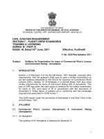
followed by viewing under at 10X under appropriate fluorescence optics (18, Fig. 1).
All control ODNs are phosphorothioated like the antisense ODN in order to control for
the nonspecific effects of applying modified ODNs to a developing embryo.
3.2. Preparing ODN Stock Solutions
We treat lyophilized ODN stocks from the manufacturer as sterile and make concen-
trated stocks in sterile water at 50 mg/mL. ODNs are aliquoted in small volumes and
stored at –20°C to maintain stability (up to a year). This concentration constitutes a
100X solution for topical applications and 10X solutions for injection applications.
Immediately prior to use, the stock is diluted to a working concentration in sterile Spratt
Ringers solution.
26 Alexander, Barnes, and Tuan
3.3. Preparing the Embryo Explant Rings
Make embryo explant rings by cutting 3MM Whatman filter paper (Whatman,
Clifton, NJ) into approximately 3/4 in. squares with a central opening large enough to
surround the chick embryos of desired developmental stage for experimental manipu-
lation. We create the opening by using a two-hole hole punch and punching two par-
tially overlapping holes. Explant rings are sterilized by autoclaving (10 min dry cycle)
in a covered glass Petri dish with cover.
3.4. Preparing Albumen-Agar Embryo Culture Plates
1. Ringer-albumen component: Separate the yolk from the albumen of one fresh, unincubated
egg (12). Add the albumen to 50 mL of sterilized Spratt Ringers solution in sterilized
Erlenmeyer flask. Add 0.5 mg of phenol red (see Note 13). Seal the flask with Parafilm
and shake the contents vigorously for 1 min. Centrifuge the mixture in 30 mL capped
Fig. 1. Assessment of the distribution of ODN administered to chick embryos. To assess the
distribution of fluorescently labeled ODN after several hours in culture, embryos were either
injected with 50 nL of ODN or treated with a direct topical application of 1 µL of the labeled
ODN. The embryos were allowed to develop under normal culture conditions and were observed
immediately after injection (A and B) and after another 6 h (C and D). Arrows indicate the site
of ODN injection. Embryos in (A) and (C) are viewed in Nomarski differential interference
optics and embryos in (B) and (D) are viewed with appropriate fluorescence optics. Immedi-
ately after injection, the fluorescent signal was evident in discrete areas of the somite (B). After
6 h, embryos that had been injected were found to have a localized area of very bright fluores-
cence at the injection site, as well as a diffuse fluorescence over a wider area of the embryo
(D). NT, neural tube; S, somite. Adapted from ref. 18.
Antisense ODNs in Developing Chick Embryos 27
polystyrene centrifuge tubes at 10,000g for 10 min at 4°C. Pour the supernatant into
another sterilized Erlenmeyer flask and place in a preheated 50°C water bath.
2. Ringer-agar component: Autoclave-sterilize (15 min moist cycle) 120 mL of 2% low-melt
agarose dissolved in 1X Spratt Ringers solution. After autoclaving, place the Ringer-agar
mixture in a preheated 50°C water bath. Place a sterilized thermometer into the Ringer-agar.
3. Albumen-agar medium: Once the temperatures of both Ringer-agar and Ringer-albumen
components are equilibrated at 50°C, mix the two together at a ratio of 2 parts Ringer-
albumen to 3 parts Ringer-agar mixtures. Gently mix the two components while maintain-
ing the 50°C temperature in the 50°C water bath. Place 2–3 mL of medium in each 35 mm
Petri dish and allow the mixture to gel at room temperature (see Note 14). Plates can be
stored for up to 1 wk at 4°C in a humidified storage container.
3.5. Pulling the Glass Capillary Microinjection Needles
For the purpose of injecting antisense ODNs into the segmental plate and somites,
we pull needles with tip diameters of approximately 20 µm, the minimal tip diameter
for our model injector system. The strength of the magnet and the intensity of the
heating element of the pipet puller should be adjusted such that the tapered portion is
about 2–3 cm long with the final 0.2–0.5 cm being of almost uniform, minimal diam-
eter (see Note 15). The closed tips of the pulled needles are not cut off until just before
use. Pulled injection needles are kept in a 150-mm Petri dish held in place with
plasticine keeping the tips suspended above the surface. Just before use, a pulled tip is
dipped in 70% ethanol for sterilization. The ethanol will evaporate by the time the
injection needle is mounted on the plunger. Drummond (Broomall, PA) provides an
excellent description of the mounting procedure with their product, which includes a
troubleshooting guide.
3.6. Microinjector Setup
Before the injection procedure is begun, a mock plate is placed on the stage, and the
plane of focus is set upon the surface of the plate. Adjust the height of the manipulator
such that when the infection tip is advanced to three-fourths of its maximal extension
the tip of the injection needle is in the center of the plane of focus just above the embryo
culture albumen-agar plate surface. We set the angle of the injection needle to approxi-
mately 45–60°. Remember to retract the injection needle completely before removing
the plate.
3.7. Harvesting Embryos
Preheat a 500-mL bottle of 1X Spratt Ringer’s solution to 37°C.
Incubate fresh fertilized chicken eggs and harvest appropriately aged chick embryos
are harvested by cracking the egg open and dropping the contents into a sterile 100 mm
Petri dish. We stage embryos according to Hamburger and Hamilton (19) (see Note 16).
An excellent morphological description is given by Bellairs and Osmond (20).
Before the eggs are cracked, rinse and wipe them clean with 70% ethanol to remove
contaminants. We break the eggs by gently cracking the egg about 180° around its
center (see Note 17). Drop the contents of the egg into an open, sterile 100 mm Petri
dish so that the embryo is on top (exposed) surface of the yolk (see Note 18). Lift the
embryo from the yolk by placing an explant ring onto the surface of the yolk sac with
the embryo located in the center of the ring opening. Trim the extra-embryonic mem-
28 Alexander, Barnes, and Tuan
branes along the outer edge of explant ring and lift the embryo from the underlying
yolk. Embryos are rinsed twice by slowly passing them through prewarmed, 38°C
Spratt Ringers solution (see Note 19). The washed embryos are placed onto prewarmed,
38°C albumen-agar plates and quickly transferred to the embryo incubator (see Note 20).
For these procedures, embryos are cultured in an inverted orientation so that the ventral
side of the embryo is exposed (facing up, see Note 21).
3.8. Delivery of ODN by Topical Application and Injection (
see
Note 22)
Prepare and prewarm ODN working solutions. We prepare 5 µg/µL dilutions of ODN
in Spratt Ringer’s solution (see Note 23). Remove harvested embryos from the embryo
incubator in small numbers and examine for condition and health. Discard abnormal or
damaged embryos (see Note 24).
3.8.1. Topical Application
The ODNs are administered with a pipet in a 10-µL drop containing 5 µg/µL of
ODNs suspended in sterile Spratt Ringers solution (see Note 25). After making the
appropriate observations of the pretreated embryo, choose the area to which the ODN
will be applied and position it in the center of the visual field. Under the dissection
scope, bring the pipet tip as close to the embryo as possible. While slowly administer-
ing the drop of ODN, lay the drop on the embryo at close proximity but without touch-
ing to the desired location (see Note 26). Immediately place the treated embryo back in
the incubator for 16–24 h (see Note 27).
3.8.2. Application by Injection (
see
Note 28)
Place selected embryos onto the stage of the dissecting microscope with the
microinjector set up, observed and positioned as described earlier. Advance the tip of
the injection needle slowly until it begins to come into the plane of focus. Again
advance the tip of the injection needle so that it enters the field of view and comes into
focus. Continue to slowly advance the tip towards the embryo using the fine advance-
ment control so that it gently comes in contact with the endoderm of the embryo just to
the side of the desired point of injection (see Note 29).
To inject into the segmental plate, use a swift but controlled 10° turn of the fine
adjustment control to penetrate the endoderm. Slowly retract the needle several
degrees (see Note 30). See that the endoderm is slightly pulled. Upon injection, you
should see a slight movement of the loose segmental plate mesenchymal cells. Sev-
eral microliters of a saturated Nile Blue (a vital dye) may be added to aid in visualiz-
ing the injected material. Retract the needle smoothly and gently out of the embryo
(Fig. 2; see Note 31).
In the case of somitocoel injection, continue to advance the injection needle tip until
it makes contact with the outside of the chosen desired somite. With the fine advance-
ment control, test this contact by nudging the somite gently and seeing that the somite
moves. As described earlier, use a swift but controlled 10° turn of the fine adjustment
control to penetrate the somite and retract. Upon injection, the somite should be seen to
swell (see Note 32). Retract the needle smoothly and gently out of the embryo. Mini-
mal leaking should be seen (Fig. 3).
Antisense ODNs in Developing Chick Embryos 29
3.9. Analysis
Remove treated and control embryos from the incubator and examine under the dis-
secting microscope to determine the condition of the embryo. Only embryos that have
remained in good health are gently lifted from the albumen-agar plates, rinsed twice in
Spratt Ringers solution, and prepared for subsequent analysis.
There are several ways to control the effectiveness of the ODN. Although Northern
blots or reverse transcriptase-polymerase chain reaction (RT-PCR) could be used to
assay for the reduction of a specific message after ODN treatment, we prefer using
whole-mount in situ hybridization (WISH) (Figs. 4 and 5). This procedure provides
Fig. 2. Sequential views of segmental plate injection. Embryos were injected with 50 ng
Paraxis antisense ODNs in 10 nL Ringers solution plus 2% Nile Blue sulfate into a stage 13
chick embryo segmental plate (A–C). In (A) the microinjection needle is positioned inside the
segmental plate ready for injection. After injection and retraction of the needle, the dark,
injected ODN solution remains confined in the segmental plate (B). If the ectoderm on the
underside of the inverted, explanted embryo had been punctured, the dark solution would
bleed into a wider area and would be visible under the neural tube as well as lateral to the seg-
mental plate (C).
Fig. 3. Sequential views of somite injection. Embryos were injected with 50 ng Paraxis
antisense ODNs in 10 nL Ringers solution plus 2% Nile Blue sulfate into stage 13 chick embryo
somites (A–C). In (A) the microinjection needle is positioned inside the somitocoel of the third
epithelialized somite (the 6th somite caudal-rostrally) ready for injection. Part (B) shows the
same somite after injection and retraction of the microinjection needle. The dark ODN suspen-
sion remains contained within the somite. In (C) a different embryo has been injected, this time
into a damaged somite. Evidence of the excessive damage was only apparent after the dark
ODN suspension began to leak from the somite.