evolutionary developmental biology of the cerebral cortex - novartis foundation
Bạn đang xem bản rút gọn của tài liệu. Xem và tải ngay bản đầy đủ của tài liệu tại đây (3.37 MB, 282 trang )
EVOLUTIONARY
DEVELOPMENTAL
BIOLOGY OF THE
CEREBRAL CORTEX
Novartis 228: Evolutionary Developmental Biology ofthe Cerebral Cortex.
Copyright & 2000 JohnWiley & Sons Ltd
Print ISBN 0-471-97978-3 eISBN 0-470-84663-1
The Novartis Foundation is an international scienti¢c and educational
charity (UK Registered Charity No. 313574). Known until September 1997
as the Ciba Foundation, it was established in 1947 by the CIBA company
of Basle, which merged with Sandoz in 1996, to form Novartis. The
Foundation operates independently in London under English trust
law. It was formally opened on 22 June 1949.
The Foundation promotes the study and general knowledge of
science and in particular encourages international co-operation in
scienti¢c research. To this end, it organizes internationally
acclaimed meetings (typically eight symposia and allied open
meetings and 15^20 discussion meetings) and publishes eight books
per year featuring the presented papers and discussions from the symposia.
Although primarily an operational rather than a grant-making foundation,
it awards bursaries to young scientists to attend the symposia and
afterwards work with one of the other participants.
The Foundation's headquarters at 41 Portland Place, London W1N 4BN,
provide library facilities, open to graduates in science and allied disciplines.
Media relations are fostered by regular press conferences and by articles
prepared by the Foundation's Science Writer in Residence. The Foundation
o¡ers accommodation and meeting facilities to visiting scientists and
their societies.
Information on all Foundation activities can be found at
Novartis 228: Evolutionary Developmental Biology ofthe Cerebral Cortex.
Copyright & 2000 JohnWiley & Sons Ltd
Print ISBN0-471-97978-3 eISBN 0-470-84663-1
EVOLUTIONARYEVOLUTIONARY
DEVELOPMENTALDEVELOPMENTAL
BIOLOGY OF THEBIOLOGY OF THE
CEREBRAL CORTEXCEREBRAL CORTEX
2000
JOHN WILEY & SONS, LTD
Chichester ´ New York ´ Weinheim ´ Brisbane ´ Singapore ´ Toronto
Novartis Foundation Symposium 228
Novartis 228: Evolutionary Developmental Biology ofthe Cerebral Cortex.
Copyright & 2000 JohnWiley & Sons Ltd
Print ISBN 0-471-97978-3 eISBN 0-470-84663-1
Copyright & Novartis Foundation 2000
Published in 2000 byJohnWiley & Sons Ltd,
Ba¤ns Lane, Chichester,
West Sussex PO19 1UD, England
National 01243 779777
International (+44) 1243 779777
e-mail (for orders and customer service enquiries):
Visit our Home Page on
or
All Rights Reserved. No part of this book may be reproduced, stored in a retrieval
system, or transmitted, in any form or by any means, electronic, mechanical, photocopying,
recording, scanning or otherwise, except under the terms of the Copyright, Designs and
Patents Act 1988 or under the terms of a licence issued by the Copyright Licensing Agency,
90 Tottenham Court Road, London,W1P 9HE, UK, without the permission in writing
of the publisher.
OtherWiley Editorial O¤ces
John Wiley & Sons, Inc., 605 Third Avenue,
NewYork, NY 10158-0012, USA
WILEY-VCH Verlag GmbH, Pappelallee 3,
D-69469 Weinheim, Germany
Jacaranda Wiley Ltd, 33 Park Road, Milton,
Queensland 4064, Australia
John Wiley & Sons (Asia) Pte Ltd, 2 Clementi Loop #02-01,
Jin Xing Distripark, Singapore 129809
John Wiley & Sons (Canada) Ltd, 22 Worcester Road,
Rexdale, Ontario M9W 1L1, Canada
Novartis Foundation Symposium 228
ix+271 pages, 48 ¢gures, 1 table
Library of Congress Cataloging-in-Publication Data
Evolutionary developmental biology of the cerebral cortex/ [editors, Gregory R. Bock
and Gail Cardew].
p. cm. ^ (Novartis Foundation symposium ; 228)
Symposium on Evolutionary Developmental Biology of the Cerebral Cortex, held at the
Novartis Foundation, London, 20^22 April 1999.
Includes bibliographical references and index.
ISBN 0-471-97978-3 (hbk : alk. paper)
1. Cerebral cortex^Congresses. 2. Brain^Evolution^Congresses. 3. Developmental
neurobiology^Congresses. I. Bock, Gregory. II. Cardew, Gail. III. Novartis Foundation.
IV. Symposium on Evolutionary Developmental Biology of the Cerebral Cortex (1999 :
London, England). V. Series.
QP383.E95 2000
573.8'6^dc21 00-027101
British Library Cataloguing in Publication Data
A catalogue record for this book is available from the British Library
ISBN 0 471 97978 3
Ty p e s e t i n 10
1
Ù
2
on 12
1
Ù
2
pt Garamond by DobbieTypesetting Limited, Tavistock, Devon.
Printed and bound in Great Britain by Biddles Ltd, Guildford and King's Lynn.
This book is printed on acid-free paper responsibly manufactured from sustainable forestry,
in which at least two trees are planted for each one used for paper production.
Novartis 228: Evolutionary Developmental Biology ofthe Cerebral Cortex.
Copyright & 2000 JohnWiley & Sons Ltd
Print ISBN0-471-97978-3 eISBN 0-470-84663-1
Contents
Symposium on Evolutionary developmental biology ofthe cerebral cortex, held atthe Novartis
Foundation, London, 20^22 April1999
This symposium is based on a proposal made by Zolta
¨
nMolna
¨
r
Editors: Gregory R. Bock (organizer) and Gail Cardew
L.Wolpert What is evolutionary developmental biology? 1
Discussion 9
K. Herrup Thoughts on the cerebellum as a model for cerebral cortical development
and evolution 15
Discussion 24
P. Rakic Radial unit hypothesis of neocortical expansion 30
Discussion 42
General discussion I 46
E. Boncinelli, A. Mallamaci and L. Muzio Genetic control of regional identity in
the developing vertebrate forebrain 53
Discussion 61
J. L. R. Rubenstein Intrinsic and extrinsic control of cortical development 67
Discussion 75
A. J. Reiner A hypothesis as to the organization of cerebral cortex in the common
amniote ancestor of modern reptiles and mammals 83
Discussion 102
General discussion II 109
I. Bar and A. M. Go¤net Evolution of cortical lamination: the reelin/Dab1
pathway 114
Discussion 125
v
Novartis 228: Evolutionary Developmental Biology ofthe Cerebral Cortex.
Copyright & 2000 JohnWiley & Sons Ltd
Print ISBN 0-471-97978-3 eISBN 0-470-84663-1
Participants
E. Boncinelli DIBIT, Scienti¢c Institute San Ra¡aele,Via Olgettina 58, Milan,
Italy
F. Bonhoe¡er MPI fÏr Entwicklungsbiologie, Abt. Physikal. Biologie,
Spemannstr. 35/1, 72076 TÏbingen, Germany
V. Broccoli (Bursar) TIGEM Institute Dibit HS Ra¡aele,Via Olgettina 58,
I-20132 Milan, Italy
A. B. Butler Krasnow Institute for Advanced Study and Department of
Psychology, George Mason University, MSN 2A1, Fairfax,VA 22030, USA
S. E. Evans Department of Anatomy and Developmental Biology, University
College London, Gower Street, LondonWC1E 6BT, UK
A. M. Go¤net Neurobiology Unit, University of Namur Medical School,
61 rue de Bruxelles, B5000 Namur, Belgium
K. Herrup Department of Neuroscience and UniversityAlzheimer Research
Center of Cleveland, CaseWestern Reserve University, 10900 Euclid Avenue,
Cleveland, OH 44120, USA
W. Ho d o s Department of Psychology, University of Maryland, College Park,
MD 20742-4411, USA
S. Hunt Department of Anatomy and Developmental Biology, Medawar
Building, University College London, Gower Street, LondonWC1E 6BT, UK
J. H. Kaas Department of Psychology,Vanderbilt University, 301Wilson Hall,
Nashville,TN 37240, USA
H. J. Karten (Chair) Department of Neurosciences, University of California at
SanDiego,LaJolla,CA92093-0608,USA
vii
Novartis 228: Evolutionary Developmental Biology ofthe Cerebral Cortex.
Copyright & 2000 JohnWiley & Sons Ltd
Print ISBN0-471-97978-3 eISBN 0-470-84663-1
Subject index
A
adenylyl cyclase 1 233, 236
adenylate cyclase 1 235
aldolase C 21, 28
amniote
evolution 109^112
relationships 110
see also stem amniotes
amphibian brain 111
Amphioxus 126
amygdala 49, 62, 63, 88, 107, 108
anamniote dorsal thalamus 106
antenna 13
anterior dorsal ventricular ridge (ADVR)
54, 158, 160
see also dorsal ventricular ridge
anterior entopeduncular areas (AEP) 69
anterior neural ridge (ANR) 68, 69
anterior thalamus 13
antidromic activation 238
apical ectodermal ridge (AER) 79
apoptosis 36
apoptotic genes 36^38
archicortex 71
astrocyte 183
auditory system 107
axon guidance molecules 174^175
axonal development 235
B
baboon 8
barrel cortex 229, 235
barrel formation 236
barrelless mice 232^233
basal forebrain cholinergic system 78
basal ganglia 46, 47, 49
basal portion of dorsal ventricular ridge
(BDVR) 54
see also dorsal ventricular ridge
basal striatal domain 54
Bauplan 13, 63
bone morphogenetic proteins (BMPs) 10,
69, 77
brain
building 206^226
maps 192
organization 208, 260
postnatal growth 243
re-wiring 181^182
size 206
brain-derived neurotrophic factor (BDNF)
185
brainstem 182
branchial arches 2^4
cartilage 4
branchial clefts 3
C
cadherins 78
Caenorhabditis elegans 9
Cajal-Retzius cells 18, 19, 22, 56^59, 61, 62,
120, 127, 133, 134, 140, 141
calretinin 56
cAMP 235
canonical circuitry 259
caspase 3 37, 42, 44
caspase 9 37, 38, 42
catecholaminergic amacrine cells 11^12
Cdk5 20, 122^124
cell division 36
cell migration 133
cell populations, evolution 46^52
cell proliferation 173
kinetics 34^36
central nervous system (CNS) 227^228, 232,
263
cerebellar anlage 16
cerebellar ¢eld 15
cerebellar granule cells 17
cerebellum 47, 145
as model for cerebral cortex development
15^29
266
Novartis 228: Evolutionary Developmental Biology ofthe Cerebral Cortex.
Copyright & 2000 JohnWiley & Sons Ltd
Print ISBN0-471-97978-3 eISBN 0-470-84663-1
development 15^18
evolution 20^21
morphogenesis 18^20
cerebral cortex
cerebellum as model 15^29
in common amniote ancestor 83^108
common patterns of organization across
species 212
expansion 44^45
lamination 57, 61, 114^128, 262^263
model of evolution 120^124
neuroepithelium 22
regionalization 173^187
size 34, 39, 42, 206, 235
cerebral peduncle 76
cerebrum 21, 26
chemoattractants 167
chemorepellants 167
chick limb 14
chicken hairy gene 13
claustroamygdaloid complex 49
claustrum 49, 62, 88, 225
CLUSTAL alignments 119
collothalamic pronuclear mass 107
collothalamus 50
common origin hypothesis 89^93, 103
evolutionary transformations 96^98
connectivity 184
patterns 182
regional 154^155
core of dorsal ventricular ridge (CNDVR)
162
see also dorsal ventricular ridge
Cornsweet illusion 245^247
corpus striatum 151
cortical a¡erents 223
cortical cell migration syndromes 19
cortical^cortical aggregates 146
cortical development see cerebral cortex
cortical expansion 39^40
cortical ¢elds 220
development 213^218
cortical gyri 37
cortical interneurons 132^137
cortical migration 55^59
cortical neurons 24
migration 59
cortical patterning 176
cortical plate 39, 40, 58, 128, 130
development 19, 116
neurons 59
radial organization 115^116
cortical sheet 215, 217
corticoclaustral interconnections 62
corticofugal axons 79
corticofugal projections 149, 151
corticogenesis 33, 34, 36
in reptiles 118^119
corticothalamic path¢nding 154
CPP322 44
Craik^O'Brien^Cornsweet e¡ect 245^247
craniofacial defects 237
critical cellular events 31^33
cyclin-dependent kinase-5 20
see also Cdk5
cytoarchitectonic areas 40, 76, 77
cytoarchitectonic maps 82
cytoarchitecture 81
cytochrome oxidase 75, 190
D
Dab1 see reelin/Dab1 pathway
deep cerebellar nuclei (DCN) 16^17, 19
developmental patterns 156^157
developmental plasticity 227^239
developmental potential, cell location for
modulating 175^176
Devonian period 7
Dictyostelium 126
diencephalon 106
digit development 9
DiI (1,1'dioctadecyl-3,3,3',3'-
tetramethylindocarbocyanine
perchlorate) 130, 134^136, 167^169,
238
distalless 63
Dlx-positive cells 51
Dlx1 72
Dlx2 72
DNA 37, 126
dorsal cortex 86, 103, 104, 117, 157
dorsal pronucleus 106
dorsal root ganglia (DRGs) 186
dorsal thalamic neurons 63
dorsal thalamus 166, 238
dorsal ventricular ridge (DVR) 46, 47, 49^
51, 54, 55, 84, 85, 87, 89^93, 99, 103^106,
110, 112, 127, 145, 157, 160, 162, 259,
260
downstream targets 13
SUBJECT INDEX 267
Drosophila 5, 9, 11, 13, 81, 112, 127, 176, 260,
262
E
echidnas 225, 226
electroreceptors 221
embryonic forebrain 152
embryonic pallial organization in reptiles and
mammals 158^160
Emx1 55, 62, 63, 65, 66, 70, 98, 112
Emx2 55^59, 62, 70, 77, 140
En1 16, 18
En2 16, 18
endodermal cells 3
endopyriform nucleus 49
endopyriform region 88
engrailed genes 16, 78
ephrins 5
epidermal growth factor (EGF) 5, 9
receptors 182
epistriatal dorsal ventricular ridge 112
see also dorsal ventricular ridge
eustachian tube 3
eutherian mammals 211^212
evolutionary developmental biology 1^14
external granule cell layer (EGL) 17
extracellular matrix 127
F
fate-mapping 76, 82, 145
Fgf8 15, 18
¢broblast growth factor (FGF) 5, 68, 79
¢eld homology 11^13
¢sh 7, 21, 48
forebrain
development 53^66, 148
genetic control in 53^66
homologous expression patterns 54^55
regional connectivity 154^155
regionalization 68
founder cells 37, 39, 40, 43
fructose-1,6-bisphosphate 21
G
GABA 18, 21, 51, 64, 71^72, 129, 132, 134,
136^137, 140, 141, 143, 184, 232
g-aminobutyric acid see GABA
ganglionic eminence 129^147
see also lateral ganglionic eminence; medial
ganglionic eminence
GAP43 56
Gbx2 71, 75, 77, 78, 145, 168, 170, 176
Gelb e¡ect 254
gene duplication 7
gene expression 65
patterns 260, 262
genetic control in vertebrate forebrain
development 53^66
geniculate nucleus 81
see also lateral geniculate nucleus
Gli3 70
glial cells 59, 184
gliophilic ¢bres 33
globus pallidus 62
granular cells 26
growth di¡erentiation factors 69
growth factor signalling 176^177
growth rates 8
H
handshake hypothesis 149, 223
hawk^goose stimulus 256^257
hedgehog neocortex 92
b-heregulin 177
heterochrony 40
hippocampus 27, 50, 55^56, 78, 86
homeobox genes 13
homology, homologous structures 11^13,
63, 112, 156-157, 215, 260
Hox genes 5^8, 10
HVC 49
hydrocephalus 44
hyperstriatum ventrale 63, 184
hypothalamus 185
I
IGL 18
immunopositive cells 44
incus 65
inferotemporal cortex 102
intermediate zone 25
intralaminar thalamus 95
isocortex 105, 225, 226
L
lamination 47^48, 57, 59, 61, 114^128,
262^263
268 SUBJECT INDEX
LAMP 174^177, 179, 184^186
lateral cortex 104, 105, 157
lateral ganglionic eminence (LGE) 48, 51,
132^134, 137, 139, 141, 167
see also medial ganglionic eminence
lateral geniculate nucleus (LGN) 193
see also geniculate nucleus
lateral limbic cortex 71
lateral pallium 106
see also pallium
Lef1 70
LEF1 transcription factor 69
lemnothalamic nuclei 50
limbic system-associated membrane protein
see LAMP
limbs 7^8
lissencephaly of the Miller^Dieker type 19
lobe-¢nned ¢sh 7
luminance 241, 246, 247
Lxh2 77
M
Mach bands 247^252
malleus 65
marsupials 209^211
Math1 22
Math1-positive cells 17
Mdab1 20
meandertail gene 26
mechanoreceptors 221
medial cortex 117
medial ganglionic eminence (MGE)
133^137, 139, 141, 142, 167, 171
see also lateral gangionic eminence
medial pallium 69
see also pallium
median forebrain bundle 78
midbrain^hindbrain boundary 20, 176
middle temporal visual area (MT) 102^103
migratory cells 125
molecular patterning 174
Monodelphis 158, 167, 224
monotremes 209^211, 225
motor cortex 76
mRNA expression 121, 122
mystacial whisker follicles
cortical representation 228^229
pattern of 231
selective breeding for variations in number
231^232
N
nematode 9, 14
neocortex 10, 11, 70^71, 129, 134, 142,
262
arealization 78, 261
eutherian mammals 212
evolution in mammals 83^88
evolutionary expansion 39^40
size 207, 215
subdivision techniques 206^207
neocortical cells 222
neocortical expansion, radial unit hypothesis
30^45
neocortical lamination 59
neocortical neuronal migration 56
neocortical regionalization 71
neocortical subdivisions 75
neocortical surface 31
neural plate 68, 79
neurogenesis 43, 55, 118
neuromere 11
neuron generation 262
neuronal cell types 129^147
neuronal precursors 36
neurons 24, 32^33, 39, 43, 45, 49^51, 59, 63,
182, 184
cortical and subcortical origins 71^72
neurophilic cells 33
Nkx2.1 69, 70, 79, 176
Nkx2.2 80
NMDA 237
NMDA receptor 229, 236^237
norepinephrine 235
Notch^delta 5
O
ocular dominance columns 192,
221
olfactory bulb 27, 47, 70
olfactory cortex 86, 95
olfactory placode 70
olfactory projections 162
olfactory system 225
ontogeny 1
opossum neocortex 92
optic tectum 194
orbital frontal cortex 225
Otx1 70
Otx2 16, 70
SUBJECT INDEX 269
P
palaeontological consensus 109, 111
pallium 51, 67, 68, 69, 106
stage-wise evolution 95
Pax 10
Pax2 15, 18
Pax6 51, 63, 77, 79, 80, 168, 170
peptides 27
phenotypes 28, 65, 173, 178
phylogeny 1
platypus 210, 220^222, 226
positional information 4^7
postmitotic cells 31, 33, 36
prefrontal cortex 225
prenatal regionalization 70
primitive internal capsule 162
gene expression of stripe of cells 159
transient cells 160^161
progenitor cells 36, 175^177
programmed cell death 36
proliferative zones 43
proto-DVR 96, 98
proto-map 81^82
Purkinje cells 16, 19, 21, 22, 26, 28, 29
pyknotic cells 44
pyramidal cells 27, 28
R
radial columns 34
radial glial cells 31
radial migration 50
radial unit hypothesis of neocortical
expansion 30^45
radiocortical units 44
reeler mutation 58^59, 62
reelin 56, 123
reelin/Dab1 expression 119^120
reelin/Dab1 pathway 114^128
reelin-dependent lamination 126
reticular nucleus 166
retina 11, 47
re-wiring the brain 181^182
rhombencephalic vesicle 45
rhombomeres 1, 2 and 3 78^79
RNA 14, 143
rostral dorsal ventricular ridge 87, 88,
95
see also dorsal ventricular ridge
RTO 107
S
sauropsids 103
schizencephaly 56
second messenger systems 76^77
sensory cortex 76
sensory domain shifts 216
sensory representations 188^205
common features 195^196
congruent borders 194^195
and disruptions in receptor sheet 189^190
and innvervation densities of receptor
sheet 194
and instructions from receptor sheet 196
modular subdivisions 192^194
and order of receptor sheet 189
sensory surfaces, errors in development
190^192
serotonin 75, 78, 235
simultaneous brightness contrast 241^245,
255
somatosensory cortex 71, 75, 76, 194, 224,
229, 231
somatosensory system, reorganization 191
sonic hedgehog (SHH) 5, 10, 69, 70, 79, 176
Sphenodon 89^90, 98, 105, 110, 115, 124
spinal cord 126
stellate cells 25, 145
stem amniote^mammal transition 84, 96
stem amniotes 84
stem cells 29
striatal^cortical aggregates 146
striatal^striatal aggregates 146
striatocortical boundary 79, 155^156
gene expression of stripe of cells 159
striatopallial boundary 79
striatum 166, 183
subcortical dorsal ventricular ridge 86
see also dorsal ventricular ridge
subpallium 65, 66, 68, 105, 262
substantia nigra neurons 182
subventricular zone 24, 25, 31, 48, 49, 182
succinic dehydrogenase (SDH) 91, 190
superior colliculus 126
symmetrical cell divisions 35
T
tangential migration 25
Tbr 112
Tbr1 51, 52, 79, 168, 170
TBR1 transcription factor 72
270 SUBJECT INDEX
tectofugal pathway 102
tectofugal system 184
tecto-thalamocortical pathway 102
telencephalon 84, 86, 87, 104, 105, 262
formation 110
induction 68
patterning 68^70
regionalization 69, 70
temporal neocortex de novo hypothesis
84^86, 88^93, 103
evolutionary transformations 93^95
temporal sulcus 86
tetrapod evolution 109
thalamic a¡erent relationships 216
thalamic axons 75
thalamic input 70^71, 77
thalamic reticular cells 162
thalamocortical a¡erents 213, 224^225
thalamocortical axons 149, 237
thalamocortical circuit formation 148^172
thalamocortical connections 236
thalamocortical development 153
thalamocortical ¢bres 149, 151
thalamocortical path¢nding 154
thalamocortical projections 162
conserved mechanisms 161^164
development in reptiles 157^158
distinct fasciculation patterns 149^154
thalamorecipient sensory areas 92
thymidine autoradiography 139
timing 8, 13, 14, 40
transcription factors 186
transforming growth factor (TGF) 177
transforming growth factor a (TGFa) 185
transforming growth factor b (TGFb)5
transient cells, primitive internal capsule
160^161
transplants 182
TTX 170
TUNEL 42^44
turtle 91, 92
tyrosine hydroxylase 11
U
unc5h3 25
V
ventricular cells 65, 80
ventricular zone 24, 25, 31, 32, 34, 36, 44, 50,
71, 76, 78
progenitors 182
vertebrate forebrain see forebrain
visual cortex 76, 193
visual perception 240^258
simultaneous brightness contrast 241^245
visual system 192
W
whisker follicle
cortical representation 228^229
removal 229^231
Wnt1 15, 16, 18
WNT proteins 69
Wnts 5
Wulst 50
Z
zebra¢sh 8, 10, 262
Zebrin 29
Zebrin II bands 21
Zebrin-negative cell groups 22
Zebrin-positive cell groups 22
SUBJECT INDEX 271
L. A. Krubitzer Center for Neuroscience and Department of Psychology, 1544
Newton Court, University of California, Davis, CA 95616, USA
P. R . Levitt Department of Neurobiology, University of Pittsburgh School of
Medicine, E1440 Biomed ScienceTower, Pittsburgh, PA 15261, USA
A. Lumsden Department of Developmental Neurobiology, King's College
London, Hodgkin House, Guy's Campus, Guy's Hospital, London SE1 9RT,
UK
Z. Molna
¨
r* Institut de Biologie Cellulaire et de Morphologie, Universite¨ de
Lausanne, Rue du Bugnon 9, 1005 Lausanne, Switzerland
D. D. M. O'Leary Laboratory of Molecular Neurobiology,The Salk Institute,
10010 NorthTorrey Pines Road, LaJolla, CA 92037, USA
N. Papalopulu Wellcome/CRC Institute,Tennis Court Road, Cambridge
CB2 1QR, UK
J. G. Parnavelas Department of Anatomy and Developmental Biology,
University College London, Gower Street, LondonWC1E 6BT, UK
J. Pettigrew VisionTouch and Hearing Research Centre, University of
Queensland, Brisbane, QLD 4072, Australia
L. Puelles Dpto. Ciencias Morfologicas, Facultad de Medicina, Universidad de
Murcia, Campus de Espinardo, 30100 Espinardo, Murcia, Espa·a
D. Purves Department of Neurobiology, Box 3209, Duke University Medical
Center, 101-I Bryan Research Building, Durham, NC 27710, USA
P. Rakic Section of Neurobiology,Yale University School of Medicine, New
Haven, CT 06510, USA
A. J. Reiner Department of Anatomy and Neurobiology, College of Medicine,
University of Tennessee, 855 Monroe Avenue, Memphis,TN 38163, USA
viii PARTICIPANTS
*Current address: Department of Human Anatomy and Genetics, University of Oxford, South
Parks Road, Oxford OX1 3QX, UK
J. L. R. Rubenstein Nina Ireland Laboratory of Developmental Neurobiology,
Center for Neurobiology and Psychiatry, Department of Psychiatry and
Programs in Neuroscience, Developmental Biology and Biomedical Sciences,
University of California at San Francisco, 401 Parnassus Avenue, San Francisco,
CA 94143, USA
E.Welker Institut de Biologie Cellulaire et de Morphologie, Universite¨ de
Lausanne, Rue du Bugnon 9, 1005 Lausanne, Switzerland
L.Wolpert Department of Anatomy and Developmental Biology, University
College London, Gower Street, LondonWC1E 6BT, UK
PARTICIPANTS ix
J. G. Parnavelas, S. A. Anderson, A. A. Lavdas, M. Grigoriou,V. Panchis and
J. L. R. Rubenstein The contribution of the ganglionic eminence to the neuronal
cell types of the cerebral cortex 129
Discussion 139
Z. Molna
¨
r Conserved developmental algorithms during thalamocortical circuit
formation in mammals and reptiles 148
Discussion 166
P. Levitt and K. L. Eagleson Regionalization of the cerebral cortex: developmental
mechanisms and models 173
Discussion 181
J.H.Kaas Organizing principles of sensory representations 188
Discussion 198
L. A. Krubitzer How does evolution build a complex brain? 206
Discussion 220
E.Welker Developmental plasticity: to preserve the individual or to create a new
species? 227
Discussion 235
D. Purves, S. M.Williams and R. B. Lotto The relevance of visual perception to
cortical evolution and development 240
Discussion 254
Final discussion 259
Index of contributors 264
Subject index 266
vi CONTENTS
What is evolutionary developmental
biology?
L. Wolpert
Department of Anatomy and Developmental Biology, University College London, London
WC1E 6BT, UK
Abstract. All changes in animal form and function during evolution are due to changes in
their DNA. Such changes determine which proteins are made, and where and when,
during embryonic development. These proteins thus control the behaviour of the cells
of the embryo. In evolution, changes in organs usually involve modi¢cation of the
development of existing structures ö tinkering with what is already there. Good
examples are the evolution of the jaws from the pharyngeal arches of jawless ancestors,
and the incus and stapes of the middle ear from bones originally at the joint between upper
and lower jaws. However, it is possible that new structures could develop, as has been
suggested for the digits of the vertebrate limb, but the developmental mechanisms
would still be similar. It is striking how conserved developmental mechanisms are in
pattern formation, both with respect to the genes involved and the intercellular signals.
For example, many systems use the same positional information but interpret it
di¡erently. One of the ways the developmental programmes have been changed is by
gene duplication, which allows one of the two genes to diverge and take on new
functions ö Hox genes are an example. Another mechanism for change involves the
relative growth rates of parts of a structure.
2000 Evolutionary developmental biology of the cerebral cortex. Wiley, Chichester (Novartis
Foundation Symposium 228) p 1^14
It has been suggested that nothing in biology makes sense unless viewed in the
light of evolution. Certainly it would be di¤cult to make sense of many aspects
of development without an evolutionary perspective. Every structure has two
histories: one that relates to how it developed, i.e. ontogeny; and the other its
evolutionary history, i.e. phylogeny. Ontogeny does not recapitulate phylogeny
as Haeckel once claimed, but embryos often pass through stages that their
evolutionary ancestors passed through. For example, in vertebrate development
despite di¡erent modes of early development, all vertebrate embryos develop to a
similar phylotypic stage after which their development diverges. This shared
phylotypic stage, which is the embryonic stage after neurulation and the
formation of the somites, is probably a stage through which some distant
1
Novartis 228: Evolutionary Developmental Biology ofthe Cerebral Cortex.
Copyright & 2000 JohnWiley & Sons Ltd
Print ISBN0-471-97978-3 eISBN 0-470-84663-1
ancestor of the vertebrates passed. It has persisted ever since, to become a
fundamental characteristic of the development of all vertebrates, whereas the
stages before and after the phylotypic stage have evolved di¡erently in di¡erent
organisms.
Such changes are due to changes in the genes that control development. These
control which proteins are made at the right time and place in the development of
the embryo since it is proteins that determine how cells behave. One of the most
important concepts in evolutionary developmental biology is that any
developmental model for a structure must be able to account for the
development of earlier forms in the ancestors.
Comparisons of embryos of related species has suggested an important
generalization: the more general characteristics of a group of animals, that is
those shared by all members of the group, appear earlier in evolution. In the
vertebrates, a good example of a general characteristic would be the notochord,
which is common to all vertebrates, and is also found in other chordate embryos.
Paired appendages, such as limbs, which develop later, are special characters that
are not found in other chordates, and that di¡er in form among di¡erent
vertebrates. All vertebrate embryos pass through a related phylotypic stage,
which then gives rise to the diverse forms of the di¡erent vertebrate classes.
However, the development of the di¡erent vertebrate classes before the
phylotypic stage is also highly divergent, because of their di¡erent modes of
reproduction; some developmental features that precede the phylotypic stage are
evolutionarily highly advanced, such as the formation of a trophoblast and inner
cell mass by mammals.
Branchial arches
An embryo's development re£ects the evolutionary history of its ancestors.
Structures found at a particular embryonic stage have become modi¢ed during
evolution into di¡erent forms in the di¡erent groups. In vertebrates, one good
example of this is the evolution of the branchial arches and clefts that are present
in all vertebrate embryos, including humans. These are not the relics of the gill
arches and gill slits of an adult ¢sh-like ancestor, but of structures that would
have been present in the embryo of the ¢sh-like ancestor. During evolution, the
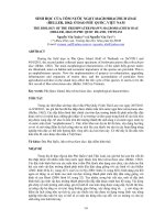
branchial arches have given rise both to the gills of primitive jawless ¢shes and, in a
later modi¢cation, to jaws (Fig. 1). When the ancestor of land vertebrates left the
sea, gills were no longer required but the embryonic structures that gave rise to
them persisted. With time they became modi¢ed, and in mammals, including
humans, they now give rise to di¡erent structures in the face and neck. The cleft
between the ¢rst and second branchial arches provides the opening for the
2 WOLPERT
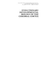
Eustachian tube, and endodermal cells in the clefts give rise to a variety of glands,
such as the thyroid and thymus (Fig. 2).
Evolution rarely generates a completely novel structure out of the blue. New
anatomical features usually arise from modi¢cation of an existing structure. One
EVOLUTIONARY DEVELOPMENTAL BIOLOGY 3
FIG. 1. The ancestral jawless ¢sh had a series of seven gill slits ö branchial clefts ö supported
by cartilaginous or bony arches. Jaws developed from modi¢cation of the ¢rst arch (from
Wolpert et al 1998).
can therefore think of much of evolution as a `tinkering' with existing structures,
which gradually fashions something di¡erent. A nice example of a modi¢cation of
an existing structure is provided by the evolution of the mammalian middle ear.
This is made up of three bones that transmit sound from the eardrum (the tympanic
membrane) to the inner ear. In the reptilian ancestors of mammals, the joint
between the skull and the lower jaw was between the quadrate bone of the skull
and the articular bone of the lower jaw, which were also involved in transmitting
sound. During mammalian evolution, the lower jaw became just one bone, the
dentary, with the articular no longer attached to the lower jaw. By changes in the
development, the articular and the quadrate bones in mammals were modi¢ed into
two bones, the malleus and the incus, whose function was now to transmit sound
from the tympanic membrane to the inner ear. The skull bones of ¢sh remain
unfused and retain the segmental series of the gill arches.
Positional information
One of the ways that the embryo uses to make patterns and organs is based on
positional information, that is the cells acquire a positional value related to
boundary regions and then interpret this according to their genetic constitution
and developmental history. Studies on regeneration of newt limbs and insect
tibia show clearly that even adult cells can retain their positional values and
4 WOLPERT
FIG. 2. Fate of branchial arch cartilage in humans. Cartilage in the branchial arches in the
embryo give rise to elements that include the three auditory ossicles: the malleus and incus
come from the ¢rst arch and the stapes from the second (from Wolpert et al 1998).
generate new ones. One of the ways position can be speci¢ed during development
is by a concentration gradient of a di¡usible morphogen. This has several
important implications for evolution. It means that a major change in
development of the embryo comes from changes in interpretation of positional
information, that is the cells' responses to signals. In fact there are a rather
limited number of signalling molecules in most embryos ö these include the
transforming growth factor
b (TGFb) family, ¢broblast growth factors (FGFs),
sonic hedgehog, Wnts, Notch^delta, the ephrins and epidermal growth factors
(EGFs). Evolution is both conservative and lazy, using the same signals again
and again both within the same embryo and in other distantly related species;
most of the key genes in vertebrate development are similar to those in
Drosophila. Patterning using positional information allows for highly localized
changes in the interpretation of position at particular sites. It is also a feature of
development that the embryo at an early stage is broken up into largely
independent `modules' of a small size which are under separate genetic control.
There is also good evidence that many structures make use of the same positional
information but interpret it di¡erently because of their developmental history. A
classic case is that of the antenna and leg of Drosophila. A single mutation can
convert an antenna into a leg and by making genetic mosaics it was shown that
they use the same positional information but interpret it di¡erently because of
their developmental history ö the antenna is in the anterior region of the body.
Similar considerations apply to the fore- and hindlimbs of vertebrates. These
di¡erences in interpretation involve the Hox genes.
Hox genes are members of the homeobox gene family, which is characterized by
a short 180 base pair motif, the homeobox, which encodes a helix-turn-helix
domain that is involved in transcriptional regulation. Two features characterize
all known Hox genes: the individual genes are organized into one or more gene
clusters or complexes, and the order of expression of individual genes along the
anteroposterior axis is usually the same as their sequential order in the gene
complex.
Hox genes are key genes in the control of development and are expressed
regionally along the anteroposterior axis of the embryo. The apparent
universality of Hox genes, and certain other genes, in animal development has
led to the concept of the zootype. This de¢nes the pattern of expression of these
key genes along the anteroposterior axis of the embryo, which is present in all
animals.
The role of the Hox genes is to specify positional identity in the embryo rather
than the development of any speci¢c structure. These positional values are
interpreted di¡erently in di¡erent embryos to in£uence how the cells in a region
develop into, for example, segments and appendages. The Hox genes exert this
in£uence by their action on the genes controlling the development of these
EVOLUTIONARY DEVELOPMENTAL BIOLOGY 5
structures. Changes in the downstream targets of the Hox genes can thus be a major
source of change in evolution. In addition, changes in the pattern of Hox gene
expression along the body can have important consequences. An example is a
relatively minor modi¢cation of the body plan that has taken place within
vertebrates. One easily distinguishable feature of pattern along the
anteroposterior axis in vertebrates is the number and type of vertebrae in the
main anatomical regions ö cervical (neck), thoracic, lumbar, sacral and caudal.
The number of vertebrae in a particular region varies considerably among the
di¡erent vertebrate classes ö mammals have seven cervical vertebrae, whereas
birds can have between 13 and 15. How does this di¡erence arise? A comparison
between the mouse and the chick shows that the domains of Hox gene expression
have shifted in parallel with the change in number of vertebrae. For example, the
anterior boundary of Hoxc6 expression in the mesoderm in mice and chicks is
always at the boundary of the cervical and thoracic regions. Moreover, the Hoxc6
expression boundary is also at the cervical^thoracic boundary in geese, which have
three more cervical vertebrae than chicks, and in frogs, which only have three or
four cervical vertebrae in all. The changes in the spatial expression of Hoxc6
correlate with the number of cervical vertebrae. Other Hox genes are also
involved in the patterning of the anteroposterior axis, and their boundaries also
shift with a change in anatomy.
Thus a major feature of evolution relates to the downstream targets of the Hox
genes. Unfortunately, these are largely unknown, but they are a major research
area.
There is thus the conservation of some developmental mechanisms at the cellular
and molecular level among distantly related organisms. The widespread use of the
Hox gene complex and of the same few families of protein signalling molecules
provide excellent examples of this. It seems that when a useful developmental
mechanism evolved, it was used again and again. Bird wings and insect wings
have some rather super¢cial similarities and have similar functions, yet are
di¡erent in their structure. The insect wing is a double-layered epithelial
structure, whereas the vertebrate limb develops mainly from a mesenchymal core
surrounded by ectoderm. However, despite these great anatomical di¡erences,
there are striking similarities in the genes and signalling molecules involved in
patterning insect legs, insect wings and vertebrate limbs. All these relationships
suggest that, during evolution, a mechanism for patterning and setting up the
axes of appendages appeared in some common ancestor of insects and
vertebrates. Subsequently, the genes and signals involved acquired di¡erent
downstream targets so that they could interact with di¡erent sets of genes, yet the
same set of signals retain their organizing function in these di¡erent appendages.
The individual genes involved in specifying the limb axes are probably more
ancient than either insect or vertebrate limbs.
6 WOLPERT
Gene duplication
A major general mechanism of evolutionary change has been gene duplication.
Tandem duplication of a gene, which can occur by a variety of mechanisms
during DNA replication, provides the embryo with an additional copy of the
gene. This copy can diverge in its nucleotide sequence and acquire a new
function and regulatory region, so changing its pattern of expression and
downstream targets without depriving the organism of the function of the
original gene. The process of gene duplication has been fundamental in the
evolution of new proteins and new patterns of gene expression; it is clear, for
example, that the di¡erent haemoglobins in humans have arisen as a result of
gene duplication.
One of the clearest examples of the importance of gene duplication in
developmental evolution is provided by the Hox gene complexes. Comparing the
Hox genes of a variety of species, it is possible to reconstruct the way in which they
are likely to have evolved from a simple set of six genes in a common ancestor of all
species. Amphioxus, which is a vertebrate-like chordate, has many features of a
primitive vertebrate: it possesses a dorsal hollow nerve cord, a notochord and
segmental muscles that derive from somites. It has only one Hox gene cluster,
and one can think of this cluster as most closely resembling the common ancestor
of the four vertebrate Hox gene complexes ö Hoxa, Hoxb, Hoxc and Hoxd.Itis
possible that both the vertebrate and Drosophila Hox complexes evolved from a
simpler ancestral complex by gene duplication.
Limbs
The limbs of tetrapod vertebrates are special characters that develop after the
phylotypic stage. Amphibians, reptiles, birds and mammals have limbs, whereas
¢sh have ¢ns. The limbs of the ¢rst land vertebrates evolved from the pelvic and
pectoral ¢ns of their ¢sh-like ancestors. The basic limb pattern is highly conserved
in both the fore- and hindlimbs of all tetrapods, although there are some di¡erences
both between fore- and hindlimbs, and between di¡erent vertebrates.
The fossil record suggests that the transition from ¢ns to limbs occurred in the
Devonian period, between 400 and 360 million years ago. The transition probably
occurred when the ¢sh ancestors of the tetrapod vertebrates living in shallow
waters moved onto the land. The ¢ns of Devonian lobe-¢nned ¢sh the proximal
skeletal elements corresponding to the humerus, radius and ulna of the tetrapod
limb are present in the ancestral ¢sh, but there are no structures corresponding to
digits. How did digits evolve? Some insights have been obtained by examining the
development of ¢ns in a modern ¢sh, the zebra¢sh. The ¢n buds of the zebra¢sh
embryo are initially similar to tetrapod limb buds, but important di¡erences soon
EVOLUTIONARY DEVELOPMENTAL BIOLOGY 7
arise during development. The proximal part of the ¢n bud gives rise to skeletal
elements, which are homologous to the proximal skeletal elements of the
tetrapod limb. There are four main proximal skeletal elements in a zebra¢sh
¢n which arise from the subdivision of a cartilaginous sheet. The essential
di¡erence between ¢n and limb development is in the distal skeletal elements.
In the zebra¢sh ¢n bud, an ectodermal ¢n fold develops at the distal end of the
bud and ¢ne bony ¢n rays are formed within it. These rays have no relation to
anything in the vertebrate limb.
If zebra¢sh ¢n development re£ects that of the primitive ancestor, then tetrapod
digits are novel structures, whose appearance is correlated with a new domain of
Hox gene expression. However, they may have evolved from the distal recruitment
of the same developmental mechanisms and processes that generate the radius and
ulna. There are mechanisms in the limb for generating periodic cartilaginous
structures such as digits. It is likely that such a mechanism was involved in the
evolution of digits by an extension of the region in which the embryonic
cartilaginous elements form, together with the establishment of a new pattern of
Hox gene expression in the more distal region.
Growth and timing
Many of the changes that occur during evolution re£ect changes in the relative
dimensions of parts of the body. Growth can alter the proportions of the
human baby after birth, as the head grows much less than the rest of the
body. The variety of face shapes in the di¡erent breeds of dog, which are all
members of the same species, also provides a good example of the e¡ects of
di¡erential growth after birth. All dogs are born with rounded faces; some
keep this shape but in others the nasal regions and jaws elongate during
growth. The elongated face of the baboon is also the result of growth of this
region after birth.
Because structures can grow at di¡erent rates, the overall shape of an organism
can be changed substantially during evolution by heritable changes in the duration
of growth that lead to an increase in the overall size of the organism. In the horse,
for example, the central digit of the ancestral horse grew faster than the digits on
either side, so that it ended up longer than the lateral digits.
Di¡erences among species in the time at which developmental processes
occur relative to one another can have dramatic e¡ects on structures. For
example, di¡erences in the feet of salamanders re£ects chances in timing of
limb development; in an arboreal species the foot seems to have stopped
growing at an earlier stage than in the terrestrial species. And in legless
lizards and some snakes the absence of limbs is due to development being
blocked at an early stage.
8 WOLPERT
Further reading
Ra¡ RA 1996 The shape of life: genes, development, and the evolution of animal form.
University of Chicago Press, Chicago, IL
Wolpert L, Beddington R, Brockes J, Jessell T, Lawrence P, Meyerowitz E 1998 Principles of
development. Oxford University Press, Oxford
DISCUSSION
Rakic: I enjoyed your presentation, but you didn't mention the importance of
the nematode.
Wolpert: I'll tell you why I didn't mention nematode. In my opinion, studies of
the nematode have not generally helped our understanding of the development of
vertebrates, with the exception of insights into cell death, the netrins and signal
transduction.
Rakic: As I will illustrate in my presentation, this may be quite signi¢cant.
Furthermore, if you assumed that the roles of genes do not change in evolution,
you would not be able to draw any conclusions concerning nematodes and
humans. However, as you have said, genes are conserved, but their roles may be
modi¢ed in di¡erent contexts. An example of this is the sel2 gene, which was
identi¢ed in the nematode and encodes a protein similar to Si28, which has been
implicated in the early onset of Alzheimer's disease (Levitan & Greenwald 1995).
Wolpert: My position on the nematode is that it is peculiar, in the sense that
speci¢cation of cell identity is on a cell-by-cell basis, whereas in Drosophila and in
vertebrates it is on groups of cells. This is why the nematode doesn't tell us a great
deal about vertebrates.
Herrup: I ¢nd it valuable for looking at vertebrates because, as you said, what is
important is not so much the signal itself, but how the cells respond to the signal,
and in Caenorhabditis elegans, you have to work on that problem at the level of the
single cell. Therefore, it's a treat to see one cell doing what an entire cortex full of
neurons are persuaded to do by their genes. However, I do agree that it is not useful
for studying some of the more complex networks in Drosophila, for example.
Levitt: An example of conservation of signalling molecules occurs in the
development of the C. elegans vulva. If the organization of epidermal growth
factor (EGF)-like receptors is alteredöand it is also possible to do this in
vertebratesöthe way the cell interprets the signal is changed, so that the cell
develops into a di¡erent cell type. Maybe intracellular tinkering is what C. elegans
does best.
Herrup: I would like to pursue the topic of digit development. What are the
current theories as to how this occurs, and what can it tell us about how a small
region at the end of a specialized structure can become an apparently novel
morphological structure?
EVOLUTIONARY DEVELOPMENTAL BIOLOGY 9
Wolpert: I wish I knew the answer. During the development of the proximal
elements of the zebra¢sh, a sheet of cartilage breaks up into four elements.
Therefore, the zebra¢sh has a mechanism to make repeated elements.
Presumably, this is primitive and could have been used for making digits.
Timing is an important issue in evolution because changes in timing can produce
dramatic e¡ectsöif development continues for a longer period of time, then it may
give rise to repeated structures at the ends of the digits. Conversely, if limb
development stops early is reduced then this could give rise to loss of digits or
even loss of limbs, as in legless lizards and snakes.
Karten: But there's much more to diversity than this, so the question becomes,
what are the properties that confer these di¡erences? We are ¢nding that many
organisms have common mechanisms, but this doesn't mean they're the same.
Some of the issues concerning derivative gene families and gene duplication are
beginning to give us hints about what underlies diversity and specialization, but
how can we reconcile the constancies in evolution with the divergences that we
observe? And can we specify the mechanisms for this?
Wolpert: The way I think about this is to consider the downstream targets. We
don't understand how an antenna develops di¡erently from a leg, and I can't think
of an example of how downstream targets of a Hox gene control morphology.
Karten: This brings up another critical issue. We talk about high penetrance and
the expression levels of particular genes. For instance, Pax6 is expressed in the eyes
of several animals, and it is also expressed in the olfactory placode. Are we
confounding our search for what genes such as Pax6 are doing by thinking that
just because they are expressed in certain regions it is telling us something
important? How can we use this approach as a strategy?
Rubenstein: There isn't a simple answer. However, some Pax genes are
responsive to sonic hedgehog (Shh) as well as to bone morphogenetic proteins
(BMPS), which tells us something about the position of some of these
transcription factors with regard to patterning centres.
Purves: I'd like to bring the discussion back to the cortex, i.e. whether the cortex
has antecedents or whether it has evolved in some other way. My view of evolution
is that it always proceeds by tinkering, so my question is, what is the alternative to
this tinkering?
Karten: Thirty years ago we viewed the mammalian neocortex as a totally novel
structureöthis was the underlying notion of `neocortex'öand that what existed in
non-mammals was a sort of laminated con¢guration, such as in the olfactory
system. The speci¢c sets of input and output connections involved in
information processing characteristically de¢ned in the studies of the mammalian
cortex within the last 100 years were viewed as properties unique to mammals. It
was argued until about 30 years ago that what we call cortex, in terms of its
structure, constituents wiring and performance, was a novel evolutionary
10 DISCUSSION