cell migration in inflammation and immunity
Bạn đang xem bản rút gọn của tài liệu. Xem và tải ngay bản đầy đủ của tài liệu tại đây (3.89 MB, 269 trang )
Methods in Molecular Biology
TM
Methods in Molecular Biology
TM
Edited by
Daniele D’Ambrosio
Francesco Sinigaglia
Cell Migration
in Inflammation
and Immunity
VOLUME 239
Methods and Protocols
Edited by
Daniele D’Ambrosio
Francesco Sinigaglia
Cell Migration
in Inflammation
and Immunity
Methods and Protocols
Chemotaxis and Vascular or Lymphatic Endothelium 1
1
From: Methods in Molecular Biology, vol. 239: Cell Migration in Inflammation and Immunity
Edited by: D. D'Ambrosio and F. Sinigaglia © Humana Press Inc., Totowa, NJ
1
Chemotaxis and Interaction
with Vascular or Lymphatic Endothelium
Silvano Sozzani, Annunciata Vecchi,
Paola Allavena, and Alberto Mantovani
1. Introduction
Leukocyte recruitment has been recognized as an early event in inflamma-
tory processes since the late 19th century. Accumulation and trafficking of leu-
kocytes in tissues under physiological and pathological conditions are orderly
(typically neutrophils precede mononuclear cells) and selective because, in
certain states, one or more leukocyte subsets are recruited preferentially (e.g.,
eosinophils in allergy). The current paradigm of recruitment is that of a multi-
step process involving the action of chemotactic signals (1,2).
Classical chemoattractants include complement components, formyl peptides,
and leukotriene B4. In addition, various cytokines are able to elicit directional
migration of leukocytes. Whereas molecules, such as monocyte colony-stimu-
lating factor (M-CSF), tumor necrosis factor, and vascular endothelial growth
factor (VEGF), exert chemotactic activity, the main chemotactic cytokines are
a superfamily of molecules known as chemokines (for chemotactic cytokines).
However, the in vivo role of M-CSF and VEGF as chemoattractants is well
established.
Several independent lines of work lead to the identification of chemokines
such as monocyte chemotactic protein-1 (MCP-1) and related molecules. In
the early 1970s it had already been noted that supernatants of activated blood
mononuclear cells contained attractants active on monocytes and neutrophils
(3). Subsequently, a chemotactic factor active on monocytes was identified in
culture supernants of mouse (4) and human (5,6) tumor lines and was called
tumor-derived chemotactic factor (TDCF) human (5–7). At the time, TDCF was
rather unique in that it was active on monocytes but not on neutrophils (6) and
2 Sozzani et al.
had a low molecular weight (5,6). Moreover, correlative evidence suggested its
involvement in the regulation of macrophage infiltration in murine and human
tumors (5,6,8). A molecule with similar cellular specificity and physicochemi-
cal properties was independently identified in the culture supernatant of smooth
muscle cells (SMDCF) (9). The JE gene had been identified as an immediate–
early platelet-derived growth factor (PDGF)-inducible gene in fibroblasts (10,11).
Thus, in the mid-1980s, a gene (JE) was in search of function and a monocyte-
specific attractant was waiting for molecular definition. In 1989, MCP-1 was
successfully purified from supernatants of a human glioma (12), a human mono-
cytic leukemia (13) and a human sarcoma cell line (14–16): sequencing and
molecular cloning revealed its relationship with the long-known JE gene (17–19).
Here, we will focus on selected methods used to investigate chemoattractants
at large, with emphasis on chemokines. In particular, classic protocols used for
studying cell movement including chemotaxis will be presented, along with
methods for transendothelial migration and reverse transmigration. In particu-
lar, sources of vascular endothelium and the generation of lymphatic endothelial
cultures are discussed. In vivo approaches to monitor leukocyte traffic are dis-
cussed elsewhere in this volume; here, we will describe the air-pouch model as
a simple in vivo recruitment system.
2. Materials
2.1. Chemotaxis
1. Micro 48-well Boyden chamber (Neuroprobe).
2. Humidified 5% CO
2
incubator.
3. Peripheral mononuclear cells (PBMCs).
4. 5-µm Polycarbonate filters.
5. Glass slides.
6. RPMI 1640 medium (Biochrom KG) + 0.2% bovine serum albumin (BSA).
7. Chemoattractants.
8. Diff-Quik (Harleco).
2.2. Polarization Assay
1. Purified leukocytes (monocytes, neutrophils, lymphocytes).
2. RPMI 1640 medium (Biochrom KG) + 1% fetal calf serum (FCS) (Hyclone).
3. 10X Concentration chemoattractants.
4. 10% (v/v) Formaldehyde.
5. Humidified 5% CO
2
incubator.
2.3. In Vivo Air-Pouch Model
1. Animals: CD1 mice, either sex.
2. Iota carrageenan, 1% in sterile, apirogen saline.
Chemotaxis and Vascular or Lymphatic Endothelium 3
3. Syringes, 5 mL, with a 25G needle.
4. Plastic Pasteur pipets, 3 mL.
5. Hemocytometer.
6. Test tubes.
7. Centrifuge.
8. Sterile apirogen saline.
2.4. Adhesion to Endothelium
1. Endothelial cells (ECs) obtained with a well-established methodology (20).
2. Tissue culture medium, M199 with 20% FBS +50 µg/mL endothelial cell growth
supplement (ECGS) (Collaborative Research) + 100 µg/mL heparin (Sigma). This
is referred to as a complete medium.
3. Freshly isolated eukocytes.
4. 96-Well plates (Falcon, Becton Dickinson).
5. Cotton fiocs (Johnson and Johnson).
6.
51
Cr (Amersham, 37 MBq, 1 µCi).
7. Humidified 5% CO
2
incubator.
8. Gamma counter windowed for
51
Cr.
9. Phosphate-buffered saline (PBS) (Biochrom KG).
2.5. Transendothelial Migration
1. ECs obtained with a well-established methodology (20).
2. Tissue culture medium, M199 with 20% FBS +50 µg/mL ECGS (Collaborative
Research) + 100 µg/mL heparin (Sigma). This is referred to as complete medium.
3. Freshly isolated leukocytes.
4. Single-well Boyden chambers (Neuroprobe).
5. Nitrocellulose filter (12-mm diameter , 5-µm pore, Sartorius).
6. Polyvinyl-pirrolidonet (PVP)-free polycarbonate filter (12-mm diameter, 5-µm
pore, Sartorius).
7. Fibronectin (Sigma).
8. 24-Well plates (Falcon, Becton Dickinson).
9. Cotton fiocs (Johnson and Johnson).
10.
51
Cr (Amersham, 37 MBq, 1 µCi).
11. Humidified 5% CO
2
incubator.
12. Gamma counter windowed for
51
Cr.
13. PBS (Biochrom KG).
2.6. Reverse Transmigration In Vitro
1. ECs obtained with a well-established methodology (20).
2. Tissue culture medium, M199 with 20% FBS +50 µg/mL ECGS (Collaborative
Research) + 100 µg/mL heparin (Sigma). This is referred to as a complete medium.
3. Freshly isolated leukocytes.
4. Single-well Boyden chambers (Neuroprobe).
4 Sozzani et al.
5. Nitrocellulose filter (12-mm diameter, 5-µm pore, Sartorius).
6. PVP-free polycarbonate filter (12-mm diameter, 5-µm pore, Sartorius).
7. Fibronectin (Sigma).
8. 24-Well plates (Falcon, Becton Dickinson).
9. Cotton fiocs (Johnson and Johnson).
10.
51
Cr (Amersham, 37 MBq, 1 µCi).
11. Humidified 5% CO
2
incubator.
12. Gamma counter windowed for
51
Cr.
13. PBS (Biochrom KG).
14. Stripping buffer: 20 mM NH
4
OH, 0.5% (v/v) Triton X-100.
2.7. Endothelial Cells
2.7.1. Generation of Endothelioma Cell Lines
Note: All culture reagents are from Gibco unless otherwise specified.
1. 15 Days’ gestation fetuses.
2. 0.05% Trypsin + 0.02% EDTA.
3. Dulbecco’s modified Eagle’s medium (DMEM) medium + 20% FCS.
4. 6- and 12-Well tissue culture plates.
5. Retrovirus vector N-TKmT.
6. G418.
7. Ca
2+
- and Mg
2+
-free PBS or saline.
2.7.2. Generation of Lymphatic Endothelial Cell Lines
Note: All culture reagents are from Gibco unless otherwise specified.
1. Incomplete Freund adjuvant (Sigma).
2. 0.05% Trypsin + 0.02% ethylenediamine tetraacetic acid (EDTA).
3. DMEM medium + 10% FCS (HyClone).
4. Nonessential amino acids (NEAA).
5. Na Pyruvate (NaPyr).
6. 6-Well tissue culture plates and T25 flasks (Falcon).
7. Ca
2+
- and Mg
2+
-free PBS or saline.
8. Supernatant of Sarcoma 180.
9. ECGS (Sigma).
10. Heparin (Sigma).
11. Gelatin (Sigma) solution (1% in PBS).
12. Collagenase CLS type I (Worthington Biochem).
3. Methods
3.1. Chemotaxis
Chemotaxis is defined as the directional locomotion of cells sensing a gradi-
ent of the stimulus. Chemotaxis has been extensively studied with leukocytes
Chemotaxis and Vascular or Lymphatic Endothelium 5
that are “professional migrants,” but a variety of cell types, including fibroblasts,
melanoma cells, keratinocytes, and vascular endothelial cells, exhibit directional
locomotion in vitro. Two main techniques have been used to measure migration
in vitro: migration under agarose and chemotaxis across porous membranes.
Although the former approach may more closely resemble the in vivo conditions,
the latter is easier to quantitate and allows analysis of directional versus random
locomotion. We will therefore focus on the description of migration through a
porous membrane using the modified Boyden microchamber method (21,22).
1. Aliquot 25 µL chemoattractant in each lower well. The 25-µL volume may have
some variations (2–3 µL more or less), depending on the microchamber used. It is
worth calibrating the lower wells in advance, so that having seeded the chemo-
attractant, the liquid in the lower well forms a small convex surface that guaran-
tees a perfect adhesion of the filter, avoiding air-bubble formation.
2. Put the filter (25 × 85 mm) on the lower compartment. To avoid confusion in filter
orientation, cut an angle of the filter.
3. Mount the silicon trimming and cover piece. Press the cover piece tightly to avoid
air bubbles.
4. Seed 50 µL cell suspension (1.5 × 10
6
cells) in the upper well by leaning the pipet
tip on the border of the well and quickly ejecting the cell suspension.
5. Incubate the chamber at 37°C in 5% CO
2
for 1.5 h.
6. Unscrew and turn over the chamber. Hold the upper compartment tightly and re-
move the lower compartment, keeping the silicon trimming and the filter adhered
to the upper compartment of the chamber. At this point, the migrated cells are on
the upper surface of the filter.
7. Lift the filter and hold it with a clamp on each end (clamps purchased from the
manufacturer of the chamber—Neuroprobe, Maryland, USA).
8. Wash the opaque side of the filter, where the nonmigrated cells remain, by pass-
ing this side over PBS. Do not immerse the entire filter in PBS or the migrated
cells will be lost.
9. Remove all nonmigrating cells by scraping the opaque surface of the filter against
the special rubber policeman (purchased from the manufacturer).
10. Stain the filter with Diff-Quik.
11. Place the filter on glass slides and cout the migrated cells present on the bright
surface of the filter. Count 5–10 microscopic fields at ×1000 final magnification.
3.2. Polarization Assay
The early phase of leukocyte response to chemotactic factors is character-
ized by shape change. Chemoattract stimulation results in the formation of a
frontal lamellipodia that contains all of the machinery for cell movement and a
rear uropode (23). This front-tail polarization is rapid, being detectable within
minutes, and can easily be observed at the microscope without the need of any
special equipment. Leukocyte polarization is not chemotaxis. However, for
6 Sozzani et al.
the measurement of cell polarization, it is a predictive, and inexpensive, way
to investigate chemotactic factors (23).
1. Prepare cells (10
6
/mL) in RPMI 1640 with 1% FCS and prepare 200 µL samples
(use at least duplicate tubes).
2. Prepare agonists (e.g., fMLP, C5a; Sigma) at a 10X concentration.
3. Prewarm cells at 37°C for 5 min.
4. Add the agonists in a volume of 20 µL.
5. Stimulate the cells at 37°C for 10 min.
6. Stop the stimulation by the addition of an equal volume (200 µL) of ice-cold 10%
(v/v) formaldhyde.
7. The readout of the experiment is the evaluation of the percentage of polarized
cells (head/tail) at the microscope (×400). At least 200 total cells per sample need
to be counteted.
3.3. In Vivo Air-Pouch Model
In vivo leukocyte recruitment can be easily investigated by the use of the
air-pouch model. This technique consists in the creation of a pouch in the back
of the mice. The pouch needs to be prepared some days in advance by the the
injection of sterile air, to allow the internal formation of an epithelial layer (24).
The advantage of this technique is that a chemotactic factor or a pleyotropic
inflammatory agent can be injected locally and cell recruitment can be evalu-
ated by the collection of the local essudate. Cytokines (e.g., chemokines), lipid
mediators, and other components of the inflammatory reaction can also be tested
in the inflammatory essadute. In our experience, this technique works at the
best when pro-inflammatory mediators are inoculated (e.g., endotoxin, inter-
leukin-1, carrageenan). In these conditions, infiltration of mononuclear cells
(monocytes and lymphocytes) as well granulocytes is easily detected. Interleu-
kin (IL)-8 and C5a also represent active in vivo chemotactic signals that pro-
vide clear results. On the contrary, recruitment of monocytes by chemokines
may represent a more difficult task.
1. Inject mice subcutaneously on their back with 5 mL of sterile air (syringes are
prepared under a laminar-flow hood).
2. After 3 d, inject the pouches again with 3 mL of sterile air.
3. On d 6, inject 1 mL of 1% carrageenan into the pouches. Controls are injected with
1 mL of saline.
4. On d 7 (24 h later), sacrifice animals. Incise the skin on the back and gently detach
it to expose the surface of the air sac. Carefully inject the pouch with 1 mL saline;
then, make a small incision in the upper part, recover the liquid with a plastic Pasteur
pipet, and immediately put it in a test tube in ice.
5. Record the total amount of liquid collected. Take an aliquot (usually 100 µL) for
counting the cells; if differential counting is needed, spin the cells in a cytocen-
trifuge and stain with Diff-Quik.
Chemotaxis and Vascular or Lymphatic Endothelium 7
6. If measurement of soluble factors has to be performed, centrifuge the remaining
fluid at 500g for 10 min at 4°C, and collect and store the supernatants at –20°C until
use.
The method described here uses carrageenan as a local stimulus for leuko-
cyte recruitment; other stimuli may be used as well, basically following the same
protocol. It must be noted, however, that water-soluble stimuli (e.g., proteins)
must be prepared in 0.5% sterile (endotoxin-free) carboxymethylcellulose to
avoid the fast absorption of the agent from the local site of injection. The carra-
geenan injection cause a recruitment that lasts a long time, for 7 d, whereas other
stimuli (e.g., IL-1β, recruit cells for shorter times. For IL-1β or IL-8, peak cell
recruitment is observed at 4 h. Conditions here reported refer to CD1 mice. If
other strains of mice are used, experimental conditions need to be validated.
3.4. Adhesion to Endothelium
The emigration of leukocytes from blood to tissues is essential for mediating
immune surveillence and mounting inflammatory responses. The interaction
of leukocytes with endothelial cells (ECs) can be divided into four sequential
steps: tethering, triggering, strong adhesion, and migration. The selectin family
of adhesion molecules mediates tethering; strong adhesion is mediated by the
integrin family, which need to be activated (triggering), and, finally, migration
is induced by local promigratory factors, including some cytokines and chemo-
kines (25,26). We have studied the adhesive properties and transendothelial
migration of leukocytes, but this method may also apply for investigation of
other cell types (e.g., tumor cells). Protocols 5 and 6 describe radioisotopic
assays for monitoring adhesion and transendothelial migration, based on an
assay described in ref. 27. Some leukocytes (e.g., dendritic cells, lymphocytes)
have a peculiar trafficking pattern from tissues into the lumen of blood or lym-
phatic vessels. To mimick this basal-to-apical process of migration in vitro,
we established a transmigration assay, described in ref. 20—the reverse trans-
migration assay.
1. Various leukocyte subsets (neutrophils, monocytes, natural killer [NK] cells, or
lymphocytes) are separated from buffy coats of normal blood donors, as described
(22,28).
2. Resuspend cells at 10
7
cells/mL in RPMI 1640 medium +10% FBS (complete
medium) and label by incubation with 100 µCi
51
Cr for 1 h at 37°C.
3. After labeling, wash extensively and resuspend in complete medium.
4. Culture ECs in 96-well plates (1 × 10
4
/well) in order to reach a confluent mono-
layer in 36–48 h. Stimulate designed wells with IL-1 (10 ng/mL) during the last
18 h of culture.
5. Incubate ECs with 100 µL
51
Cr-labeled cells resuspended at 107 cells/mL and incu-
bate at 37°C for 30 min.
8 Sozzani et al.
6. Carefully remove the supernantant and wash the cells twice to remove nonadherent
cells.
7. Incubate the adherent cells with 100 µL of NaOH 1 M + 1% sodium dodecyl sulfate
(SDS) for 5 min and count radioactivity using a gamma counter. Express cell adhe-
sion as percentage of input cells.
The spontaneous adhesion of resting leukocytes to unstimulated ECs varies
for different subsets. For instance, the adhesion of NK cells is usually 5–15%,
a value intermediate between that of monocytes (20–40%) and the very low value
of T-cells and polymorphonuclear cells (PMNs) (<5%). With monocytes and
NK cells, there is usually a high degree of variability among different donors.
The adhesive capacity of leukocytes to ECs can be modulated by various
signals. When ECs are stimulated with IL-1, leukocyte adhesion increases, as
ECs express new adhesion molecules. The identification of adhesion molecules
involved in the interaction of leukocytes and ECs is performed by the addition
of blocking monoclonal antibodies (mAbs)—most of which are commercially
available—specific for the adhesion structures expressed by leukocytes or ECs.
Studies with specific mAbs have demonstrated that adhesion through resting
ECs is mediated by the LFA-1/ICAM-1,2 pathway, whereas through IL-1, acti-
vated ECs involved both LFA-1/ICAM-1 and VLA-4/VCAM-1 for monocytes
and lymphoid cells, and neutrophils use only the first pathway, being VLA-4
negative.
Cytokines can also affect leukocytes directly. IL-2 and IL-12, for instance,
increases NK cell adhesion to ECs, whereas IL-4 has inhibitory activity.
3.5. Transendothelial Migration
1. Coat PVP-free polycarbonate filters with 1 mL of 10 µg/mL fibronectin in PBS at
room temperature for 2 h) in 24-well plates.
2. Aspirate fibronectin and add 10
5
ECs in 2 mL of M199 complete medium and grow
to confluence (5–6 d).
3. Place 0.2 mL of complete medium in the lower compartment of each Boyden
chamber.
4. Mount the first uncoated filter and on top the second filter coated with ECs.
5. Immediately add 0.15 mL of complete medium. Drying should be avoided.
6. Assemble and screw the upper compartment of the chamber.
7. Label PBMCs (100 µCi
51
Cr at 4°C for 1 h) and seed cells [(3–6) × 10
5
in 0.15 mL
of complete medium] into the upper compartment of the chamber.
8. Incubate the chamber at 37°C for 60 min.
9. Remove the chambers from the incubator.
10. Collect the medium containing nonadherent cells in a 3-mL vial (fraction A).
11. Gently wash the EC monolayers with 0.5 mL warm medium and collect it (frac-
tion B).
Chemotaxis and Vascular or Lymphatic Endothelium 9
12. Scrape (gently) the EC monolayer and adherent leukocytes with cotton fiocs and
transfer to vials (fraction C).
13. Transfer the double filter system to vials together with the medium of the lower
compartment (fraction D).
14. Measure radioactivity in each fraction. Fractions A+B represent nonadherent cells.
Fraction D represent migrated cells. Because migrated cells had first adhered to
ECs, total number of adherent cells is calculated by summing fractions C+D.
The spontaneous transendothelial migration varies for different leukocytes
subsets. Usually, only a proportion (about 30%) of EC-adherent leukocytes effec-
tively transmigrate during the assay. When ECs are activated with IL-1, a greater
number of cells adhere and transmigrate, but the proportion of transmigrated
cells over the input does not dramatically change (usually 30% of EC-adherent
leukocytes). It should be noted that IL-1 does not change the state of confluence
of the monolayer, as determined by staining.
The identification of adhesion molecules involved in the interaction of leuko-
cytes and EC is performed by the addition of blocking mAbs—most of which
are commercially available—specific for the adhesion structures expressed by
leukocytes or ECs. Studies with specific mAbs have demonstrated that, like
adhesion, transmigration through resting ECs is mediated by the LFA-1/ICAM-
1,2 pathway, whereas though IL-1, activated ECs involved both LFA-1/ICAM-1
and VLA-4/VCAM-1 for monocytes and lymphoid cells, and neutrophils use
only the first pathway, being VLA-4 negative. In addition, PECAM (CD31) is
a molecule expressed both by leukocytes and ECs, and plays a major role dur-
ing transmigration (25). Chemoattractants can be seeded in the lower compart-
ment to increase leukocyte transmigration.
3.6. Reverse Transmigration In Vitro
1. Culture EC to confluent monolayers on fibronectin-precoated PVP-free polycar-
bonate filters in 24-well plates as described in Subheading 3.5., item 1.
2. Treat one-half of the filters with 1 mL of stripping buffer for 30 s.
3. Quickly remove the stripping buffer and the digested EC monolayer (ECM) and
wash twice with complete medium. Cover the exposed ECM with 1 mL of com-
plete medium. Drying should be avoided.
4. Place 0.2 mL of complete medium in the lower compartment of each Boyden
chamber. Mount the first filter, coated with ECs, upside down, and on top of the
second filter coated with ECM.
5. Assemble and screw the upper compartment of the chamber.
6. Immediately add 0.15 mL of complete medium. Drying should be avoided.
7.
51
Cr-labeled leukocytes are seeded [(3–6) × 10
5
in 0.15 mL of complete medium]
into the upper compartment of the chamber.
8. Incubate the chamber at 37°C for 60 min.
10 Sozzani et al.
9. Remove the chambers from the incubator.
10. Collect the medium present in the upper chamber (fraction A) and wash the ECM
layer with 0.5 mL warm medium (fraction B).
11. Collect the ECM-adherent cells with cotton fiocs (fraction C), and the transmi-
grated cells in the lower compartment (fraction D).
12. Measure radioactivity in each fraction. Fraction A+fraction B represent the non-
adherent cells. Fraction C + D represent the adherent cells.
13. Fraction D represents the migrated cells. Please note that in the reverse assay, the
transmigrated cells comprise only the radioactivity present in the lower compartment.
3.7. Endothelial Cells
3.7.1. Mouse Vascular Endothelial Cells:
Generation of PMT-Transformed Lines
Normal ECs of human and murine origin are cumbersome to obtain and
culture. EC lines have been generated sporadically, and, in our experience, at
least some of them lack important functions of normal ECs. The polyoma mid-
dle T (PmT) oncogene transforms mouse ECs (29) and can be used to generate
immortalized EC cell lines, possibly representative of microvascular elements.
These lines retain many properties of normal ECs, including production of and
responsiveness to cytokines (30–32).
All procedures must be performed with sterile material in aseptic conditions.
1. Remove fetuses or organs of interest from six to eight fetuses of 15 d gestation.
2. Cut organ or fetus in small pieces and trypsinize (trypsin 0.05% + EDTA 0.02%,
20 min at 37°C).
3. Collect supernatants and add the same volume of DMEM medium with 20% FCS.
4. Centrifuge at 300g for 10 min.
5. Resuspend the pellet in 2–5 mL of DMEM + 10% FCS (complete medium), count,
and bring the suspension to (0.5–1) × 10
6
cells/mL.
6. Distribute 2 mL of cell suspensions to each well of 12-well plates and incubate at
37°C in 5% CO
2
.
7. After 24 h, remove medium and add about 105 neo colony-forming units (CFU)
of the retrovirus vector N-TKmT per well in 1 mL of complete medium. The virus
is produced by the GP+E cell line obtained through the courtesy of Dr. E. Wagner
(Wien, Austria).
8. After 2 h, remove medium and add fresh complete medium.
9. After 72 h, select PmT-infected neomycin-resistant cells with G418, 800 µg/mL.
10. Change the medium twice a week, keeping G418 at 800 µg/mL.
11. Check wells for G418-resistant cells. They usually are observed after 15–20 d.
12. When cells are confluent, wash the wells throroughly two times with PBS Ca
2+
-
and Mg
2+
-free or with saline, add 0.3 mL of trypsin 0.05% + EDTA 0.02% for 2–3
min at 37°C, resuspend the detached cells, and add 1 mL of complete medium.
Chemotaxis and Vascular or Lymphatic Endothelium 11
Transfer all of the suspension to one well of a six-well plate and bring to the final
volume of 3 mL, with G418 at the 800-µg/mL final concentration.
13. Check cells for the growth every day.
14. When confluent, pass the cells 1:3.
At this stage, cells should not be diluted too much, even if they are growing
very well. Confluent monolayers can usually be kept 1–2 d without damage for
the cells. On the other hand, if cells are diluted too much, they stop growing
and can either remain quiescent for some time and eventually grow again, or
die. Maintain selection with G418.
Following this protocol, we have obtained stable cell lines from the heart,
brain, and whole embryo of C57Bl. Cell lines show a cobblestone morphology
at confluency and maintain a monolayer structure without overgrowth. Cells
are positive for CD31/PECAM-1 antigen, show rapid uptake of fluorescinated
acetylated low-density lipoprotein, produce IL-6 constitutively, and are nega-
tive or weakly positive for factor-VIII-related antigen. Transmission electron
microscopy revealed that they were uniformly negative for the presence of
Weibel–Palade bodies.
Transformed cells maintain many characteristics of normal endothelial cells,
such as CD31 expression, modulation of adhesion molecules by cytokines, and
cytokine production (31) . They do not constitutively express ICAM-1, VCAM-1,
E-selectin, and P-selectin; however, they can be induced, with the exception of
ICAM-1, by exposure to tumor necrosis factor-α (TNF-α) and lipopolysaccha-
ride (LPS), but not IL-1.
Endothelioma cells produce IL-6 and CCL2/JE, whose production can be
increased by IL-1 exposure.
Lines originated from embryo tissues infected in vitro with the PmT onco-
gene of the polyoma virus have been growing in this laboratory since the early
1990s. They represent an easy and reliable source of endothelial cells of murine
origin, suitable for studies on EC biology (30,33).These lines do not need exog-
eneous growth factors for proliferation. All lines have been frozen, stored in
liquid nitrogen, and put again in culture without problems. PmT murine EC lines
originated by hemangiomas (34,35) have been used to generate mAbs against
EC-specific antigens expressed constitutively, such as CD31 or induced by cyto-
kines (e.g., VCAM-1 and ELAM-1) (36). EC lines from different organs can
be useful to study the potential diversity of the microvasculature of different
organs.
3.7.2. Lymphatic Endothelial Cells: Isolation and Culture
The lymphatic vessels, by channeling fluid and leukocytes from the periphery
into the lymph nodes, play a central role in the development of the immune
12 Sozzani et al.
response. Despite their importance in homeostasis and diseases, the difficul-
ties in enriching and culturing lymphatic endothelial cells limit studies on their
biology. Recently, isolation, characterization, and short-term culture of human
(37) and mouse (38) lymphatic cells have been reported, but after very few
passages, cells stop growing and die. We report here a metodology to isolate
and stabilize in vitro mouse lymphatic endothelial cells.
All procedures must be performed with sterile material in aseptic conditions.
1. Inject intraperitoneally 0.2 mL of a 1:1 mixture of incomplete Freund adjuvant
and PBS to DBA/2 mice
2. Inject mice for the second time 15 d later, as in step 1.
3. After an additional 15–30 d, recover hyperplastic lymphatic vessels from the liver
and diaphragm, where they appear as small white spots.
4. Treat with collagenase (0.1% in Dulbecco’s phosphate buffer, 3–5 mL), incubate
at 37°C in 5% CO
2
for 45 min and centrifuge at 300g for 10 min.
5. Resuspend the pellet in 2–5 mL of complete medium (DMEM + 10% FCS + NEAA
+ NaPyr + 20% of S180 supernatant + 100 µg/mL ECGS + 100 µg/mL heparin).
6. Check cultures on the microscope. After 7–10 d, clusters of adherent growing cells
with a cobblestonelike morphology can be found.
7. Recover cells with trypsin/EDTA when subconfluent and plate on gelatin-coated
flasks.
8. Check cultures on the microscope. When confluent pass the cells 1/3–1/5.
Following this protocol, we have obtained a cell line that shows a cobble-
stone morphology at confluency and maintains a monolayer structure without
overgrowth. These cells, called MELCs (mouse endothelial lymphatic cells),
were studied for lymphatic endothelial markers and found positive for Flt-4 in
Western and florescein activated cell sorter (FACS) analysis and for podoplanin
and D6 in reverse transcription–polymerase chain reaction; they have been
checked for the expression of endothelial markers and adhesion molecules, rel-
evant for the physiological circulation of leukocytes from tissues to secondary
lyphoid organs through the lymphatics. MELCs were found to express CD34,
ICAM-1, VCAM, and JAM-1, but not CD31, VE-cadherin and E-selectin (TNF-
α induced) and to produce IL-6 and CCL2. Upon stimulation with TNF-α, they
upregulate the expression of adhesion molecules such as ICAM-1 and VCAM
and produce increased amounts of IL-6 and CCL2.
Cells from lymphatic hyperplasia, cultured with appropriate supplements as
reported here, are able to grow in culture for more than 30 passages. They main-
tain the reported markers and response to TNF-α during in vitro culture. MELCs
have also been cloned by limiting dilution and clones mantained the markers
of the original population. Cells have been frozen, stored in liquid nitrogen,
and put again in culture without problems. Hyperplasia of the lymphatic vessel
can be induced by the protocol reported also in other mouse strains (C57Bl/6
Chemotaxis and Vascular or Lymphatic Endothelium 13
and Balb/c). MELCs are now a suitable source of lymphatic endothelial cells
for in vitro studies of their biology.
Acknowledgments
This work was supported by Associazione Italiana per la Ricerca sul Cancro
(AIRC) and Centro di Eccellenza per l’Innovazione Diagnostica e Terapeutica
(IDET).
References
1. Butcher, E. C. (1991) Leukocyte–endothelial cell recognition: three (or more)
steps to specificity and diversity. Cell 67, 1033–1036.
2. Mantovani, A., Bussolino, F., and Dejana, E. (1992) Cytokine regulation of endo-
thelial cell function. FASEB J. 6, 2591–2599.
3. Ward, P. A., Remold, H. G., and David, J. R. (1970) The production by antigen-
stimulated lymphocytes of a lekotactic factor distinct from migration inhibitory
factor. Cell Immunol. 1, 162–174.
4. Meltzer, M. S., Stevenson, M. M., and Leonard, E. J. (1977) Characterization of
macrophage chemotaxis in tumor cell cultures and comparison with lymphocyte-
derived chemotactic factors. Cancer Res. 37, 721–725.
5. Bottazzi, B., Polentarutti, N., Acero, R., et al. (1983) Regulation of the macrophage
content of neoplasms by chemoattractants. Science 220, 210–212.
6. Bottazzi, B., Polentarutti, N., Balsari, A., et al. (1983) Chemotactic activity for
mononuclear phagocytes of culture supernatants from murine and human tumor
cells: evidence for a role in the regulation of the macrophage content of neoplastic
tissues. Int. J. Cancer 31, 55–63.
7. Bottazzi, B., Ghezzi, P., Taraboletti, G., et al. (1985) Tumor-derived chemotactic
factor(s) from human ovarian carcinoma: evidence for a role in the regulation of
macrophage content of neoplastic tissues. Int. J. Cancer 36, 167–173.
8. Mantovani, A., Bottazzi, B., Colotta, F., Sozzani, S., and Ruco, L. (1992) The origin
and function of tumor-associated macrophages. Immunol. Today 13, 265–270.
9. Valente, A. J., Fowler, S. R., Sprague, E. A., Kelley, J. L., Suenram, C. A., and
Schwartz, C. J. (1984) Initial characterization of a paripheral blood mononuclear
cell chemoattractant derived from cultured arterial smooth muscle cells. Am. J.
Pathol. 117, 409–417.
10. Zullo, J. N., Cochran, B. H., Huang, A. S., and Stiles, C. D. (1985) Platelet-derived
growth factor and double-stranded ribonucleic acids stimulate expression of the
same genes in 3T3 cells. Cell 43, 793–800.
11. Rollins, B. J., Morrison, E. D., and Stiles, C. D. (1988) Cloning and expression of
JE, a gene inducible by platelet-derived growth factor and whose product has
cytokine-like properties. Proc. Natl. Acad. Sci. USA 85, 3738–3742.
12. Yoshimura, T., Robinson, E. A., Tanaka, S., Appella, E., Kuratsu, J., and Leonard,
E. J. (1989) Purification and amino acid analysis of two human glioma-derived
monocyte chemoattractants. J. Exp. Med. 169, 1449–1459.
14 Sozzani et al.
13. Matsushima, K., Larsen, C. G., DuBois, G. C., and Oppenheim, J. J. (1989) Purifi-
cation and characterization of a novel monocyte chemotactic and activating factor
produced by a human myelomonocytic cell line. J. Exp. Med. 169, 1485–1490.
14. Zachariae, C. O., Anderson, A. O., Thompson, H. L., et al. (1990) Properties of
monocyte chemotactic and activating factor (MCAF) purified from a human fibro-
sarcoma cell line. J. Exp. Med. 171, 2177–2182.
15. Van Damme, J., Decock, B., Lenaerts, J. P., et al. (1989) Identification by sequence
analysis of chemotactic factors for monocytes produced by normal and transformed
cells stimulated with virus, double-stranded RNA or cytokine. Eur. J. Immunol.
19, 2367–2373.
16. Graves, D. T., Jiang, Y. L., Williamson, M. J., and Valente, A. J. (1989) Identif-
cation of monocyte chemotactic activity produced by malignant cells. Science
245, 1490–1493.
17. Furutani, Y., Nomura, H., Notake, M., et al. (1989) Cloning and sequencing of the
cDNA for human monocyte chemotactic and activating factor (MCAF). Biochem.
Biophys. Res. Commun. 159, 248–255.
18. Yoshimura, T., Yuhki, N., Moore, S. K., Appella, E., Lerman, M. I., and Leonard,
E. J. (1989) Human monocyte chemoattractant proein-1 (MCP-1). Full-length cDNA
cloning, expression in mitogen-stimulation blood mononuclear meukocytes, and
sequence similarity to mouse competence gene JE. FEBS Lett. 244, 487–493.
19. Bottazzi, B., Colotta, F., Sica, A., Nobili, N., and Mantovani, A. (1990) A chemo-
attractant expressed in human sarcoma cells (tumor-derived chemotactic factor,
TDCF) is identical to monocyte chemoattractant protein-1/monocyte chemotactic
and activating factor (MCP-1/MCAF). Int. J. Cancer 45, 795–797.
20. D’Amico, G., Bianchi, G., Bernasconi, S., et al. (1998) Adhesion, transendothelial
migration, and reverse transmigration of in vitro cultured dendritic cells. Blood
92, 207–214.
21. Falk, W., Goodwin, R. H. Jr., and Leonard, E. J. (1980) A 48-well microchemo-
taxis assembly for rapid and accurate measurement of leukocyte migration. J. Immu-
nol. Methods 33,239–245.
22. Sozzani, S., Luini, W., Molino, M., et al. (1991) The signal transduction pathway
involved in the migration induced by a monocyte chemotactic cytokine. J. Immu-
nol. 147, 2215–2221.
23. Wilkinson, P. C. (1998) Assays of leukocyte locomotion and chemotaxis. J. Immu-
nol. Methods 216, 139–153.
24. Romano, M., Sironi, M., Toniatti, C., et al. (1997) Role of IL-6 and its soluble recep-
tor in induction of chemokines and leukocyte recruitment. Immunity 6, 315–325.
25. Springer, T. A. (1994) Traffic signal for lymphocyte recirculation and leukocyte
emigration: the multistep paradigm. Cell 76, 301–314.
26. Adams, D. H. and Shaw, S. (1994) Leucocyte–endothelial interactions and regula-
tion of leucocyte migration. Lancet 343, 831–836.
27. Bianchi, G., Sironi, M., Ghibaudi, E., et al. (1993) Migration of natural killer cells
across endothelial cell monolayers. J. Immunol. 151, 5135–5144.
Chemotaxis and Vascular or Lymphatic Endothelium 15
28. Allavena, P., Paganin, C., Martin Padura, I., et al. (1991) Molecules and structures
involved in the adhesion of natural killer cells to vascular endothelium. J. Exp.
Med. 173, 439–448.
29. Williams, R. L., Courtneidge, S. A., and Wagner, E. F. (1988) Embryonic lethalities
and endothelial tumors in chimeric mice expressing polyoma virus middle T onco-
gene. Cell 52, 121–131.
30. Bussolino, F., De Rossi, M., Sica, A., et al. (1991) Murine endothelioma cell lines
transformed by polyoma middle T oncogene as target for and producers of cyto-
kines. J. Immunol. 147, 2122–2129.
31. Garlanda, C., Parravicini, C., Sironi, M., et al. (1994) Progressive growth in immu-
nodeficient mice and host cell recruitment by mouse endothelial cells transformed
by polyoma middle-sized T antigen: implications for the pathogenesis of oppor-
tunistic vascular tumors. Proc. Natl. Acad. Sci. USA 91, 7291–7295.
32. Taraboletti, G., Belotti, D., Dejana, E., Mantovani, A., and Giavazzi, R. (1993)
Endothelial cell migration and invasiveness are induced by a soluble factor pro-
duced by murine endothelioma cells transformed by polyoma virus middle T onco-
gene. Cancer Res. 53, 3812–3816.
33. Bocchietto, E., Guglielmetti, A., Silvagno, F., et al. (1993) Proliferative and migra-
tory responses of murine microvascular endothelial cells to granulocyte-colony-
stimulating factor. J. Cell Physiol. 155, 89–95.
34. Vecchi, A., Garlanda, C., Lampugnani, M. G., et al. (1994) Monoclonal antibodies
specific for endothelial cells of mouse blood vessels. Their application in the iden-
tification of adult and embryonic endothelium. Eur. J. Cell Biol. 63, 247–254.
35. Piali, L., Albelda, S. M., Baldwin, H. S., Hammel, P., Gisler, R. H., and Imhof, B. A.
(1993) Murine platelet endothelial cell adhesion molecule (PECAM-1)/CD31
modulates beta 2 integrins on lymphokine-activated killer cells. Eur. J. Immunol.
23, 2464–2471.
36. Hahne, M., Jager, U., Isenmann, S., Hallmann, R., and Vestweber, D. (1993) Five
tumor necrosis factor-inducible cell adhesion mechanisms on the surface of mouse
endothelioma cells mediate the binding of leukocytes. J. Cell Biol. 121, 655–664.
37. Kriehuber, E., Breiteneder-Geleff, S., Groeger, M., et al. (2001) Isolation and char-
acterization of dermal lymphatic and blood endothelial cells reveal stable and func-
tionally specialized cell lineages. J. Exp. Med. 194, 797–808.
38. Mancardi, S., Stanta, G., Dusetti, N., et al. (1999) Lymphatic endothelial tumors
induced by intraperitoneal injection of incomplete Freund’s adjuvant. Exp. Cell Res.
246, 368–375.
16 Sozzani et al.
Integrin-Dependent Rapid Adhesion 17
17
From: Methods in Molecular Biology, vol. 239: Cell Migration in Inflammation and Immunity
Edited by: D. D'Ambrosio and F. Sinigaglia © Humana Press Inc., Totowa, NJ
2
Analysis of Integrin-Dependent Rapid
Adhesion Under Laminar-Flow Conditions
Carlo Laudanna
1. Introduction
Adhesion molecules mediate recognition of the blood vessels by circulating
leukocytes and support their selective targeting to different organs (1). In the
vessels, the blood flow imposes peculiar conditions by generating a wall shear
stress that opposes leukocyte stable arrest on the endothelium. As the rapidness
of integrin activation is mandatory to leukocyte adhesion to the blood vessels,
any analysis of adhesion triggering relevant to leukocyte in vivo migration should
be performed under flow conditions. Here, a method is illustrated to quantita-
tively analyze the rapid induction of integrin-dependent lymphocyte adhesion
by chemokines under flow conditions. Glass capillary tubes are cocoated with
purified ligands for selectins and integrins and with chemokines, thus reconstitut-
ing the minimal requirement to support tethering, rolling, and arrest under flow
conditions, with a physiologic wall shear stress of 2 dynes/cm
2
. PNAd, ICAM-1,
and the chemokine CCL21 are used as paradigmatic adhesion molecules and
physiologic proadhesive agonist. Naive lymphocytes isolated from mouse lymph
nodes are used as the cell model. The procedure for quantitative analysis is
discussed.
2. Materials
1. Human tonsils (from the Pediatric Department).
2. Mouse spleens (from Balb/c mice).
3. Tissue potter or blade grinder.
4. CNBr-activated Sepharose™ 4B Fast Flow (Amersham-Pharmacia, code 17-0981-
01) (see Table 1).
18 Laudanna
5. MECA 79, antiperipheral lymph node addressin (PNAd)-associated carbohydrate
epitope mAb (rat IgM; ATCC number: 9479) (2).
6. Y.N.1.7 (rat anti-mouse ICAM-1; from Professor E. C. Butcher, Stanford University).
7. Coupling buffer: 50 mM Tris-HCl, pH 8.3, 150 mM NaCl.
8. Ethanolamine (Sigma).
9. 50 mM Tris-HCl, 0.5 M NaCl, pH 8.0.
10. 50 mM glycine, 0.5 M NaCl, pH 4.0.
11. Phosphate-buffered saline (PBS).
12. Disposable polypropylene minicolumns: filter pore size, 15 µm; length, 11.3 cm;
total volume, 7.5 mL; reservoir, 4.5 mL (Spectra/Chrom
®
, code 104705).
13. Lysis buffer for PNAd and ICAM-1 purification: 0.4% 3-[(cholamido-propyl1)
dimethyl-ammonio]-propanesulfonate (CHAPS) (Boehringer Manneheim), 25 mM
Tris-HCl, pH 8.0, 150 mM NaCl, 1 mM CaCl
2
, 1 mM MgCl
2
, 2 mM NaN
3
; add 1
tablet of protease inhibitor cocktail tablet “Complete, Mini” (Roche Diagnostic
GmbH) to 10 mL of buffer.
14. Wash buffer A: 0.04 % CHAPS, 50 mM Tris-HCl, pH 8.0, 0.5 M NaCl, 1 mM CaCl
2
,
1 mM MgCl
2
, 1 mM NaN
3
.
15. Wash buffer B: 50 mM N-octyl β-D-glucopyranoside (Sigma, cat. no. O-9882),
50 mM Tris-HCl, pH 8.0, 0.5 M NaCl, 1 mM CaCl
2
, 1 mM MgCl
2
, 1 mM NaN
3
.
16. Elution buffer: 50 mM N-octyl β-D-glucopyranoside, 0.2 M acetic acid, 0.5 M NaCl,
2 mM NaN
3
(pH 8.0).
17. 3 M Tris-HCl, pH 8.5.
18. Dot blot standard equipment.
19. Dry milk.
20. Enzymatic-chemiluminescence (ECL) (Pierce).
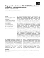
21. Pipets, microcapillary calibrated tubes, 100 µL, color-coded blue (produced by
Drummond Scientific Co., Broomall, PA, USA, cat. no. 2-000-100; purchased from
Sigma, code P-1174) (see Fig. 1).
Table 1
Characteristics of CNBr-Activated Sepharose 4 Fast Flow
Mean particle size 90 µm
Particle size range 45–165 µm
Bead structure Highly crosslinked 4% agarose, spherical
Linear flow
a
150 cm/h at 100 kPa
Coupling capacity 13–26 mg chymotrypsinogen/mL drained gel
Swelling factor 4–5 mL drained gel/g
pH stability 2–11
a
At 25°C in water in an XK 50/60 column, 25-cm bed height. The flow rate
after coupling may differ depending on the ligand.
Integrin-Dependent Rapid Adhesion 19
22. Polypropylene tubes connected to microperfusion needles (“Butterfly”) (G 23,
0.63 × 19 mm; tube internal volume = 0.46 mL).
23. Digital pump with microliter-precise setting capability.
24. 10 mL polypropylene syringe without needle.
25. Adhesion buffer: PBS, pH 7.2, 1 mM CaCl
2
, 1 mM MgCl
2
, 10% heat-inactivated
fetal calf serum (FCS).
26. CCL21 (SLC, 6Ckine, TCA-4, Exodous-2) (from PeproTech).
27. Inverted microscope with ×10 and ×20 phase-contrast objectives, connected to a
high-resolution black-and-white CCD video camera (Sony, Hamamatsu, Ikegami),
monitor (Sony), and analogic S-VHS (400 lines) or digital (540 lines) VCR (Pana-
sonic) with time-stamp capability for image recording and analysis (better if with
SMPTE; see Note 1).
3. Methods
In this section, the described methods (1) purify native PNAd from human
tonsils and native ICAM-1 from mouse spleens, (2) coat the internal surface of
glass capillary tubes with purified adhesion molecules to generate a surface able
to support tethering, rolling, and full arrest under flow, (3) produce and main-
tain a laminar flow to perform under flow adhesion assays, and (4) analyze and
quantify the recorded data. Complex reagents and solutions, described in Sub-
heading 2., are reported as item 1, 2, 3, and so forth.
3.1. Affinity Purification of Adhesion Molecules
3.1.1. Tissue Disruption
Human PNAd supports tethering and rolling of both human and mouse leuko-
cytes. Mouse ICAM-1 mediates stable arrest of either mouse as well as human
leukocytes (3,4). Thus, human PNAd and muse ICAM-1 can be indifferently
used for assays with human or murine leukocytes. Tonsils and spleens are
stored frozen at –80°C. Five to ten human tonsils (corresponding to approx
25–35 g of tissues) or 100–200 mouse spleens (corresponding to about 40–50 g
Fig. 1. Dimensions of the capillary tube and example of coating.
20 Laudanna
of tissue) are disrupted on ice by homogenization with a potter or a blade grinder
and then lysed for 45 min in 30 mL of ice-cold, freshly made lysis buffer, with
occasional stirring. Unlysed nuclei and cell debris are removed by centrifugation
at 4°C for 10 min at 10,000g, followed by centrifugation of the supernatant at
4°C for 45 min at 150,000g. The supernatants are then collected as tonsil or
spleen lysates (see Note 2).
3.1.2. Preparation of the Affinity Column
CNBr-preactivated 4% agarose matrix is used (see Table 1 for the character-
istics of the CNBr-activated beads).
1. Dialyze MECA 79 (for PNAd purification) or Y.N.1.7 (for ICAM-1 purification)
against the coupling buffer.
2. Suspend 1 g of CNBr-activated matrix in 1 mM HCl for 30 min and allow to swell
at room temperature.
3. Wash twice in 100 mL of cold 1 mM HCl.
4. Wash with 50 mL of coupling buffer.
5. Mix the washed gel with 10–15 mg of dialyzed MECA 79 or Y.N.1.7 and incu-
bate overnight at 4°C. (The coupling can also be performed at room temperature
for 3–4 h.) Keep the gel suspended by rotation or slow stirring.
6. Wash the coupled gel in 50 mL of 1 M ethanolamine.
7. Resuspend the coupled gel in 50 mL of 1 M ethanolamine and leave for 4 h at room
temperature to block unused activated sites.
8. Wash the gel four times with alternating 50 mM Tris-HCl, 0.5 M NaCl, pH 8.0,
and 50 mM glycine, 0.5 M NaCl, pH 4.0 buffers.
9. Wash the gel with 50 mL of PBS containing 2 mM NaN
3
.
10. Pack the gel (approx 4 mL of drained gel) into a disposable column with a bottom
filter (see Note 3).
3.1.3. Purification of Adhesion Molecules from Tissue Lysates
The entire procedure is done by gravity and at 4°C.
1. Wash the column with 20 mL of wash buffer A.
2. Wash the column with 5 mL of lysis buffer.
3. Run the tissue lysates twice on the column.
4. Wash the column with 40 mL of wash buffer A to remove the unbound proteins.
5. Wash the column with 10 mL of wash buffer B.
6. Elute bound PNAd or ICAM-1 with 5 mL of elution buffer by collecting 0.5-mL
fractions immediately neutralized with 50 µL of Tris buffer or 5 µL of 10 M NaOH.
The fractions containing purified PNAd or ICAM-1 are then identified by
dot-blot analysis. Usually, fractions 2–4 contain most of the protein. Fractions
are stored at 4°C (see Note 4).
Integrin-Dependent Rapid Adhesion 21
3.2. Cocoating Capillary Tubes
with Adhesion Molecules and Chemokines
Glass capillary tubes are used to establish a biological surface permissive to
lymphocytes tethering, rolling, and arrest under flow. In Fig. 1, the standard
dimensions of the tube are reported and an example of organization of the coated
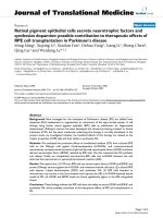
areas is given. Figure 2 depicts the manual procedure for the coating.
1. Identify two distinct sections of the capillary tube (each one 1 cm long) by mark-
ing the external surface of the tube with a pen. The section should be identified in
the middle of the tube.
2. Immediately before use, dilute the human PNAd in 30 µL of PBS; dilution should
be under the critical micelle concentration of N-octyl β-D-glucopyranoside (see
Note 4).
3. Suck diluted PNAd into the tube according to Fig. 2. Completely coat both of the
1-cm-long sections and seal off the ends of the tube to prevent evaporation. Coat-
ing should be done at 4°C for at least 12 h.
4. Wash the tube twice with PBS; to prevent contamination of uncoated areas, wash-
ing has to be done always in the direction of the laminar flow.
5. Immediately before use, dilute mouse ICAM-1 in 15 µL of PBS (see Note 4).
6. Suck diluted ICAM-1 to coat the 1-cm-long downstream section of the tube and
seal off the ends of the tube. Care should be taken to prevent contamination of the
Fig. 2. Schematic drawing of the procedure for capillary coating. Proportions are
not respected.
22 Laudanna
upstream section, which has to be coated only with PNAd. Coating should be
done at 4°C for at least 12 h.
7. Wash the tube twice with PBS.
8. Suck 15 µL of CCL21 (2 µM in PBS) to coat only the 1-cm-long downstream
section of the tube (now cocoated with PNAd and ICAM-1). Coat for 30–60 min
at room temperature.
9. Immediately before the experiment, wash the tube three times with PBS contain-
ing 10% heat-inactivated FCS.
The upstream 1-cm section of tube is now coated only with PNAd, whereas
the downstream 1-cm section is cocoated with PNAd, ICAM-1, and chemokine.
3.3. Under Flow Adhesion Assay
The coating described in Subheading 3.2. supports interaction and stable
arrest of L-selectin, LFA-1, and CCR7-positive cells, such as naïve lymphocytes.
Murine naive lymphocytes (approx 50% B, 50% T) are isolated from mouse
(Balb/c) lymph nodes and resuspended in adhesion buffer (item 25) at 10
6
/mL.
The assay is done at room temperature.
1. Place 10 mL of lymphocyte suspension in a disposable 10-mL polypropylene syringe
connected to a 15-cm-long polypropylene tube (item 22).
2. Connect the syringe to the upstream end of the coated capillary tube; connect the
downstream end of the capillary to a second 15-cm-long polypropylene tube; con-
nect the downstream polypropylene tube to a 15-mL Falcon tube to collect the cells.
3. The entire setting has to be fixed on the inverted microscope to focus from the
bottom the coated sections of the capillary tube; by using ×20 magnification, an
area of 0.2 mm
2
is visualized at video; phase-contrast optics improves the image
quality. The syringe is placed on the digital pump.
4. Set the digital pump to provide a flow rate of 75 mL/h, corresponding to 1250 µL/
min. By applying this flow rate to the capillary tube (with a diameter of 1.025
mm), a wall shear stress (WSS) of 2 dynes/cm
2
is obtained (considering the cell
suspension a Newtonian fluid with a viscosity 0.01 P) (see Note 5).
5. Start the video recording and cell transfusion; 2–3 min are normally necessary to
establish a stable laminar flow and see interacting cells. During the experiment,
care has to be taken to record, as much as possible, separate fields for at least 30 s.
3.4. Analysis and Quantification of Interacting Cells
The described experimental setting reproduces some of the physiological
behaviors of homing lymphocytes, including tethering, rolling, and sticking.
Quantitative differences such as duration of tethering, speed of rolling, and effi-
ciency of stable adhesion can be quantified. To limit the description to chemo-
kine-triggered integrin-dependent rapid adhesion, we will focus only on stable
arrest analysis.
Integrin-Dependent Rapid Adhesion 23
1. Quantitative definition of integrin-dependent stable arrest is a little arbitrary and
has to be defined depending of the experimental setting. For instance, in the pres-
ence of L-selectin-mediated rolling, a stable adhesion of 10 s can be considered
integrin activation; in contrast, in the presence of P-selectin–PSGL-1 mediated inter-
action, rolling is much slower (5) and can be occasionally scored as stable arrest if
a too stringent temporal parameter is adopted. Here, we will consider a stable
arrest of at least 10 s as a manifestation of integrin triggering under flow.
2. The experiments are analyzed by evaluating for at least 30 s as much as possible
of the areas of the coated tube. This will provide much data for statistical analysis.
Start by looking at a noncoated section in order to evaluate eventual background,
nonspecific interactions. Next, evaluate the efficiency of PNAd in supporting tether-
ing and rolling by counting the number of tethering and rolling cells in 30 s.
3. If a cell does not make a complete rotation on itself, score as tethering, otherwise
score as rolling. Normally, an only-tethering cell interacts with the surface for less
than 1 s. Individual cells stable adherent for less than 10 s has to be considered not
arrested but still rolling. In the case of coating with a low number of sites of PNAd,
the number of tethering cells tends to be higher than the number of rolling cells.
Occasional full arrest for 10 or more seconds has to be scored as nonspecific, inte-
grin-mediated adhesion.
4. Move to the section cocoated with PNAd, ICAM-1, and chemokines. Count in each
0.2-mm
2
area the number of total interacting cells, including teething, rolling, and
arrested cells. Analyze frame by frame the fields to precisely evaluate the dura-
tion of full adhesion of single cells. This is important to distinguish between full
adherent cells from only-rolling cells. Various cells adherent in the same field can
be analyzed one by one by going backward and forward with the VCR.
5. The number of interacting cells in noncoated areas (nonspecific interaction) is sub-
tracted form the number of cells interacting on PNAd alone. The number of cells
arrested on PNAd is subtracted from the number of cells arrested on ICAM-1 and
chemokines. The data can be either expressed as total number of only-rolling or
full arrested cells in 0.2 mm
2
in 30 s or as a percentage of arrested cells of the total
interacting cells in 0.2 mm
2
in 30 s (see Note 6.)
4. Notes
1. Identification of single frames by a time code is critical for the accurate measure-
ment of dynamic events. The most accurate time code has been established by the
Society of Motion Pictures and Television Engineers (SMPTE), which identifies
each frame with a unique address in the form hours:minutes:seconds:frames. Nota-
bly, in the PAL system, 1 s of recording contains 25 frames; in the SECAM sys-
tem, 1 s contains 24 frames; in the NTSC system, 1 s contains 30 frames (precisely
29.97). Consequently, the duration of a single frame is 40 ms, for PAL; it is 41.6 ms
for SECAM, and it is 33.33 ms for NTSC. Thus, in the PAL system, a frame iden-
tified as 00:02:25:15 is positioned at 2 min, 25 s, and 15 frames (that is 2' 25" 600
ms) after the beginning of the recording. The time code is essential for quantifica-
24 Laudanna
tion of leukocyte behavior under flow. For example, a cell that started to move after
20 frames was adherent for only 800 ms (in PAL) and, thus, was a rolling cell.
2. It is better to proceed with protein purification immediately after tissue lysate prep-
aration. If necessary, it is possible to store the lysates at –80°C. To prevent protein
aggregation and precipitation (which will reduce the purification efficiency and
clog the affinity column), rapidly freeze the lysates in liquid N
2
before storing them
at –80°C.
3. When choosing the disposable polypropylene minicolumns, pay attention to order
columns with a filter pore size of 15 µm (Spectra/Chrom, code 104705). Note that
the Sepharose 4 Fast Flow particle size range is 45–165 µm (see Table 1). The MECA
79 column appears to be rather unstable and not suitable for more than two puri-
fications, even if care is taken to correctly wash and prevent contamination. The
Y.N.1.7 can be reused several times.
4. It is very important to buffer the fractions immediately after their collection. Never
freeze the fractions. Dilutions must be made immediately before the coating, to
prevent rapid reduction of the titer by adsorption of the protein to the wall of the
tube used for the dilution. Coating can be also done at 37°C for 3–4 h, but more
care has to be taken to prevent evaporation. The specific activity of the ICAM-1
preparation may be determined in static adhesion assay by making several dilutions
of the purified protein to identify the dilution providing the lowest ratio between
background binding in the absence of agonist and binding induced by agonists.
The specific activity of the PNAd preparation has to be evaluated under flow. The
most accurate way to coat the tubes is by calculating the number of molecules
immobilized in 1 µm
2
. This is particularly important with integrin ligands. This
will allow precise evaluation of the role of different modalities of integrin activa-
tion in determine the rapid and stable arrest under flow (4). The number of mol-
ecules immobilized can be determined by using
125
I-mAbs, as reported (6).
5. The wall shear stress (WSS) can be modified to study the efficiency among differ-
ent selectins, integrins, and chemokines in mediating tethering, rolling, and arrest
under flow. This can be combined with changes in the density of immobilized
ligands and agonists to perform a more informative analysis.
The WSS is calculated using the following:
WSS = WSR × P (1)
where WSR is the wall shear rate and P is the viscosity (0.01 for PBS);
The WSR is calculated using:
WSR = 8V
m
/D (2)
where V
m
is the fluid mean velocity and D is the diameter of the tube (1.025 mm in
our case);
The V
m
is extracted using:
Q = V
m
π D
2
/4 × 10
–6
(3)
where Q is the flow rate (75 mL/h in our case) and D is the diameter of the tube
(1.025 mm).