handbook of x ray spectrometry revised and expanded by rene van grieken
Bạn đang xem bản rút gọn của tài liệu. Xem và tải ngay bản đầy đủ của tài liệu tại đây (10.26 MB, 985 trang )
TM
Marcel Dekker, Inc. New York
•
Basel
Handbook of
X-Ray Spectrometry
Second Edition, Revised and Expanded
edited by
René E. Van Grieken
University of Antwerp
Antwerp, Belgium
Andrzej A. Markowicz
Vienna, Austria
Copyright © 2001 by Marcel Dekker, Inc. All Rights Reserved.
Copyright © 2002 Marcel Dekker, Inc.
ISBN: 0-8247-0600-5
First edition was published as Handbook of X-Ray Spectrometry: Methods and Techniques
This book is printed on acid-free paper.
Headquarters
Marcel Dekker, Inc.
270 Madison Avenue, New York, NY 10016
tel: 212-696-9000; fax: 212-685-4540
Eastern Hemisphere Distribution
Marcel Dekker AG
Hutgasse 4, Postfach 812, CH-4001 Basel, Switzerland
tel: 41-61-261-8482; fax: 41-61-261-8896
World Wide Web
http:==www.dekker.com
The publisher offers discounts on this book when ordered in bulk quantities. For more information,
write to Special Sales=Professional Marketing at the headquarters address above.
Copyright # 2002 by Marcel Dekker, Inc. All Rights Reserved.
Neither this book nor any part may be reproduced or transmitted in any form or by any means,
electronic or mechanical, including photocopying, microfilming, and recording, or by any in-
formation storage and retrieval system, without permission in writing from the publisher.
Current printing (last digit):
10987654321
PRINTED IN THE UNITED STATES OF AMERICA
Copyright © 2002 Marcel Dekker, Inc.
Preface to the Second Edition
The positive response to the first edition of Handbook of X-Ray Spectrometry: Methods
and Techniques and its commercial success have shown that in the early 1990s there was
a clear demand for an exhaustive book covering most of the specialized approaches in this
field. Therefore, some five years after the first edition appeared, the idea of publishing
a second edition emerged. In the meantime, remarkable and steady progress has been
made in both instrumental and methodological aspects of x-ray spectrometry. This
progress includes considerable improvements in the design and production technology of
detectors and in capillary optics applied for focusing the primary photon beam. The
advances in instrumentation, spectrum evaluation, and quantification have resulted in
improved analytical performance and in further extensions of the applicability range of x-
ray spectrometry. Consequently, most of the authors who contributed to the first edition
of this book enthusiastically accepted the invitation to update their chapters. The progress
made during the last decade is reflected well in the chapters of the second edition, which
were all considerably revised, updated, and expanded. A completely new chapter on mi-
crobeam x-ray fluorescence analysis has also been included.
Chapter 1 reviews the basic physics behind x-ray emission techniques, and refers to
extensive appendices for all the basic and generally applicable x-ray physics constants.
New analytical expressions have been intro duced for the calculation of fundamental
parameters such as the fluorescence yield, incoherent scattering function, atomic form
factor, and total mass attenuation coefficient.
Chapter 2 outlines established and new instrumentation and discusses the perfor-
mances of wavelength-dispersive x-ray fluorescence (XRF) analysis, whi ch, with probably
15,000 units in operation worldwide today, is still the workhorse of x-ray analysis. Its
applications include process control, materials analysis, metallurgy, mining, and almost
every other major branch of science. The additional material in this edition covers new
sources of excitation and comprehensive comparisons of the technical parameters of newly
produced wavelength-dispersive spectrometers.
Chapter 3 has been completely reconsidered, modified, and rewritten by a new au-
thor. The basic principles, background, and recent advances are described for the tube-
excited energy-dispersive mode, which is invoked so frequent ly in research on environ-
mental and biological samples. This chapter is based on a fresh look and follows
a completely different approach.
Copyright © 2002 Marcel Dekker, Inc.
Chapter 4 reviews in depth the available alternatives for spectrum evaluation and
qualitative analysis. Techniques for deconvolution of spectra have enormously increased
the utility of energy-dispersive x-ray analysis, but deconvolution is still its most critical
step. The second edition includes discussions of partial least-squares regression and
modified Gauss ian shape profiles.
Chapter 5 reviews quantification in XRF analysis of the classical and typical ‘‘in-
finitely thick’’ samples. In addition to being updated, the sections on calibration, quality
control, and mathematical correction methods have been expanded.
Chapter 6, on quantification for ‘‘intermediate-thickness’’ samples, now also in-
cludes the presentation of a modified version of the emission-transmission method and
a discussion of both the accuracy and limitations of such methods .
Chapter 7 is a completely original treatment by a new author of radioisotope-in-
duced and portable XRF. It discusses semiconductor detectors, including the latest types,
analyzes in detail the uncertainty sources, and reviews the recent and increasingly im-
portant applications.
Since the appearance of the first edition, synchrotron-induced x-ray emission ana-
lysis has increased in importance. Chapter 8 was updated and modified by including
a comprehensive review of the major synchrotron facilities.
Although its principles have been known for some time, it is only since the advent of
powerful commercial units and the combination with synchrotron sources that total re-
flection XRF has rapidly grown, mostly now for characterization of surfaces and of liquid
samples. This is the subject of the substantially modified and expanded Chapter 9. The
new authors have taken a radically different approach to the subject .
Polarized-beam XRF and its new commercial instruments are treated in detail in
a substantially revised and expanded Chapter 10.
Capillary optics combined with conventional fine-focus x-ray tubes have enabled the
development of tabletop micro-XRF instruments. The principles of the strongly growing
microbeam XRF and its applications are now covered thoroughly in an additional
chapter, Chapter 11.
Particle-induced x-ray emission analysis has grown recently in its application types
and pa rticularly in its microversion. Chapter 12 discusses the physical backgrounds, in-
strumentation, performance, and applications of this technique. The sections dealing with
the applications were substantially expanded.
Although the practical approaches to electron-induced x-ray emission analysis—
a standard technique with wide applications in all branches of science and technology—
are often quite different from those in other x-ray analysis techniques, a treatment of its
potential for quantitative and spatially resolved analysis is given in Chapter 13. The new
and expanded sections deal with recent absorption correction procedures and with the
quantitative analysis of samples with nonstandard geometries.
Finally, the completely updated and revised Chapter 14 reviews the sample pre-
paration techniques that are invoked most frequently in XRF analysis.
The second edition of this book is again a multiauthored effort. We believe that
having scientists who are actively engaged in a particular technique covering those areas in
which they are particularly qualified outweighs any advantages of uniformity and
homogeneity that characterize a single-authored book. The editors (and one coworker)
again wrote three of the chapters in the new edition. For all the other chapters, we were
fortunate to have the cooperation of truly eminent specialists, some of whom are new
contributors (see Chapters 3, 7, 9, 10 and 11). We wish to thank all the contributors for
their considerable and (in most cases) timely efforts.
Copyright © 2002 Marcel Dekker, Inc.
We hope that novices in x-ray emission analysis will find this revised and expanded
handbook useful and instructive, and that our more experienced colleagues will benefit
from the large amount of readily accessible information available in this compact form,
some of it for the first time. An effort has been made to emphasize the fields and devel-
opments that have come into prominence lately and have not been covered in other
general books on x-ray spectrometry.
We also hope this book will help analytical chemists and other users of x-ray
spectrometry to fully exploit the capabilities of this powerful analytical tool and to further
expand its applications in material and environmental sciences, medicine, toxicology,
forensics, archeometry, and many other fields.
Rene
´
E. Van Grieken
Andrzej A. Markowicz
Copyright © 2002 Marcel Dekker, Inc.
Preface to the First Edition
Scientists in recent years have been somewhat ambivalent regarding the role of x-ray
emission spectrometry in analytical chemistry. Whereas no radically new and stunning
developments have been seen, there has been remarkably steady progress, both instru-
mental and methodological, in the more conventional realms of x-ray fluorescence. For the
more specialized approaches—for example, x-ray emission induced by synchrotron ra-
diation, radioisotopes and polarized x-ray beams, and total-reflection x-ray fluorescence—
and for advanced spectrum analysis methods, exponential growth and=or increasing ac-
ceptance has occurred. Contrary to previous books on x-ray emission analysis, these latter
approaches make up a large portion of the present Handbook of X-Ray Spectrometry.
The major milestone developments that shaped the field of x-ray spectrometry and
now have widespread applications all took place more than twenty years ago. After
wavelength-dispersive x-ray spectrometry had been demonstrated and a high-vacuum
x-ray tube had been introduced by Coolidge in 1913, the prototype of the first modern
commercial x-ray spectrometer with a sealed x-ray tube was built by Friedmann and Birks
in 1948. The first electron microprobe was successfully developed in 1951 by Castaing,
who also outlined the fundamental concepts of quantitative analysis with it. The semi-
conductor or Si(Li) detector, which heralded the advent of energy-dispersive x-ray
fluorescence, was developed around 1965 at Lawrence Berkeley Laboratory. Accelerator-
based particle- induced x-ray emission analysis was developed just before 1970, mostly at
the University of Lund. The various popular matrix correction methods by Lucas-Tooth,
Traill and Lachance, Claisse and Quintin, Tertian, and several others, were all proposed in
the 1960s. One may thus wonder whether the more conventional types of x-ray fluores-
cence analysis have reached a state of saturation and consolidation, typical for a matur e
and routinely applied analysis technique. Reviewing the state of the art and describing
recent progress for wavelength- and energy-dispersive x-ray fluorescence, electron and
heavy charged-particle-induced x-ray emission, quantification, and sample preparation
methods is the purpose of the remaining part of this book.
Chapter 1 reviews the basic physics behind the x-ray emission techniques, and refers
to the appendixes for all the basic and generally applicable x-ray physics constants.
Chapter 2 outlines established and new instrumentation and discusses the performances of
wavelength-dispersive x-ray fluorescence analysis, which, with probably 14,000 units in
operation worldwide today, is still the workhorse of x-ray analysis with applications in
a wide range of disciplines including process control, materials analysis, metallurgy,
Copyright © 2002 Marcel Dekker, Inc.
mining, and almost every other major branch of science. Chapter 3 discusses the basic
principles, background, and recent advances in the tube-e xcited energy-dispersive mode,
which, after hectic growth in the 1970s, has now apparently leveled off to make up ap-
proximately 20% of the x-ray fluorescence market; it is invoked frequently in research on
environmental and biological samples. Chapter 4 reviews in depth the available alter-
natives for spectrum evaluation and qualitative analysis; techniques for deconvolution of
spectra have enormously increased the utility of energy-dispersive x-ray analysis, but de-
convolution is still its most critical step. Chapters 5 and 6 review the quantification pro-
blems in the analysis of samples that are infinitely thick and of intermediate thickness,
respectively. Chapter 7 is a very practical treatment of radioisotope-induced x-ray ana-
lysis, which is now rapidly acquiring wide acceptance for dedicated instruments and field
applications. Chapter 8 reviews synchrotron-in duced x-ray emission analysis, the youngest
branch, with limited accessibility but an exponentially growing literature due to its extreme
sensitivity and microanalysis potential. Although its principles have been known for some
time, it is only since the advent of powerful commercial units that total reflection x-ray
fluorescence has been rapidly introduced, mostly for liquid samples and surface layer
characterization; this is the subject of Chapter 9.
Polarized beam x-ray fluorescence is outlined in Chapter 10. Particle-induced x-ray
emission analysis is available at many accelerator cen ters worldwide; the number of annual
articles on it is growing and it undergoes a revival in its microversion; Chapter 11 treats
the physical backgrounds, instrumentation, performance, and applications of this tech-
nique. Although the practical approaches to electron-induced x-ray emission analysis, now
a standard technique with wide applications in all branches of scienc e and technology, are
often quite different from those in other x-ray analysis techniques, a separate treatment of
its potential for quantitative and spatially resolved analysis is given in Chapter 12. Finally,
Chapter 13 briefly reviews the sample preparation techniques that are invoked most fre-
quently in combination with x-ray fluorescence analysis.
This book is a multi-authored effort. We believe that having scientists who are ac-
tively engaged in a particular technique covering those areas for which they are particu-
larly qualified and presenting their own points of view and general approaches outweighs
any advantages of uniformity and homogeneity that characterize a single-author book.
Three chapters were written by the editors and a coworker. For all the other chapters, we
were fortunate enough to have the cooperation of eminent specialists. The editors wish to
thank all the contributors for their efforts.
We hope that novices in x-ray emission analysis will find this book useful and in-
structive, and that our more experienced colleagues will benefit from the large amount of
readily accessible information available in this compact form, some of it for the first time.
This book is not intended to replace earlier works, some of which were truly excellent, but
to supplement them. Some overlap is inevitable, but an effort has been made to emphasize
the fields and developments that have come into prominence lately and have not been
treated in a handbook before.
Rene
´
E. Van Grieken
Andrzej A. Markowicz
Copyright © 2002 Marcel Dekker, Inc.
Contents
PrefacetotheSecondEdition
PrefacetotheFirstEdition
Contributors
1X-rayPhysics
Andrzej A. Markowicz
I.Introduction
II.History
III.GeneralFeatures
IV.EmissionofContinuousRadiation
V.EmissionofCharacteristicX-rays
VI.InteractionofPhotonswithMatter
VII.IntensityofCharacteristicX-rays
VIII.IUPACNotationforX-raySpectroscopy
Appendixes
I.CriticalAbsorptionWavelengthsandCriticalAbsorptionEnergies
II.CharacteristicX-rayWavelengths(A
˚
)andEnergies(keV)
III.RadiativeTransitionProbabilities
IV. Natural Widths of K and L Levels and K
a
X-rayLines(FWHM),ineV
V.WavelengthsofKSatelliteLines(A
˚
)56
VI.FluorescenceYieldsandCoster–KronigTransitionProbabilities
VII. Coefficients for Calculating the Photoelectric Absorption
CrossSectionst(Barns=Atom)Vialn–lnRepresentation
VIII. Coefficients for Calculating the Incoherent Collision
Cross Sections s
c
(Barns=Atom)Viatheln–lnRepresentation
IX. Coefficients for Calculating the Coherent Scattering
Cross Sections s
R
(Barns=Atom)Viatheln–lnRepresentation
X. Parameters for Calculating the Total Mass Attenuation
CoefficientsintheEnergyRange0.1–1000keV[ViaEq.(78)]
XI.TotalMassAttenuationCoefficientsforLow-EnergyKaLines
XII. Correspondence Between Old Siegbahn and New IUPAC
NotationX-rayDiagramLines
References
Copyright © 2002 Marcel Dekker, Inc.
2Wavelength-DispersiveX-rayFluorescence
Jozef A. Helsen and Andrzej Kuczumow
I.Introduction
II.FundamentalsofWavelengthDispersion
III.LayoutofaSpectrometer
IV.QualitativeandQuantitativeAnalysis
V.ChemicalShiftandSpeciation
VI.Instrumentation
VII.FutureProspects
References
3Energy-DispersiveX-rayFluorescenceAnalysisUsingX-rayTubeExcitation
Andrew T. Ellis
I.Introduction
II.X-rayTubeExcitationSystems
III.SemiconductorDetectors
IV.SemiconductorDetectorElectronics
V.Summary
References
4SpectrumEvaluation
Piet Van Espen
I.Introduction
II.FundamentalAspects
III.SpectrumProcessingMethods
IV.ContinuumEstimationMethods
V.SimpleNetPeakAreaDetermination
VI.Least-SquaresFittingUsingReferenceSpectra
VII.Least-SquaresFittingUsingAnalyticalFunctions
VIII.MethodsBasedontheMonteCarloTechnique
IX.TheLeast-Squares-FittingMethod
X.ComputerImplementationofVariousAlgorithms
References
5QuantificationofInfinitelyThickSpecimensbyXRFAnalysis
Johan L. de Vries and Bruno A. R. Vrebos
I.Introduction
II.CorrelationBetweenCountRateandSpecimenComposition
III.FactorsInfluencingtheAccuracyoftheIntensityMeasurement
IV.CalibrationandStandardSpecimens
V.ConvertingIntensitiestoConcentration
VI.Conclusion
References
Copyright © 2002 Marcel Dekker, Inc.
6QuantificationinXRFAnalysisofIntermediate-ThicknessSamples
Andrzej A. Markowicz and Rene
´
E. Van Grieken
I.Introduction
II.Emission-TransmissionMethod
III.AbsorptionCorrectionMethodsViaScatteredPrimaryRadiation
IV.QuantitationforIntermediate-ThicknessGranularSpecimens
References
7Radioisotope-ExcitedX-rayAnalysis
Stanislaw Piorek
I.Introduction
II.BasicEquations
III.RadioisotopeX-raySourcesandDetectors
IV.X-rayandg-rayTechniques
V.FactorsAffectingtheOverallAccuracyofXRFAnalysis
VI.Applications
VII.FutureofRadioisotope-ExcitedXRFAnalysis
VIII.Conclusions
Appendix: List of Companies that Manufact ure Radioisotope-Based
X-rayAnalyzersandSystems
References
8SynchrotronRadiation-InducedX-rayEmission
Keith W. Jones
I.Introduction
II.PropertiesofSynchrotronRadiation
III.DescriptionofSynchrotronFacilities
IV.ApparatusforX-rayMicroscopy
V.ContinuumandMonochromaticExcitation
VI.Quantitation
VII.SensitivitiesandMinimumDetectionLimits
VIII.Beam-InducedDamage
IX.ApplicationsofSRIXE
X.Tomography
XI.EXAFSandXANES
XII.FutureDirections
References
9TotalReflectionX-rayFluorescence
Peter Kregsamer, Christina Streli, and Peter Wobrauschek
I.Introduction
II.PhysicalPrinciples
III.Instrumentation
IV.ChemicalAnalysis
V.SurfaceAnalysis
VI.ThinFilmsandDepthProfiles
VII.SynchrotronRadiationExcitation
Copyright © 2002 Marcel Dekker, Inc.
VIII.LightElements
IX.RelatedTechniques
References
10PolarizedBeamX-rayFluorescenceAnalysis
Joachim Heckel and Richard W. Ryon
I.Introduction
II.Theory
III.BarklaSystems
IV.BraggSystems
V.Barkla-BraggCombinationSystems
VI.SecondaryTargets
VII.Conclusion
References
11MicrobeamXRF
Anders Rindby and Koen H. A. Janssens
I.IntroductionandHistoricalPerspective
II.TheoreticalBackground
III.InstrumentationforMicrobeamXRF
IV.CollectionandProcessingofm-XRFData
V.Applications
References
12Particle-InducedX-rayEmissionAnalysis
Willy Maenhaut and Klas G. Malmqvist
I.Introduction
II. Interactions of Charged Particles with Matter,
Characteristic X-ray Production, and
ContinuousPhotonBackgroundProduction
III.Instrumentation
IV.Quantitation,DetectionLimits,Accuracy,andPrecision
V. Sample Collection and Sample and Specimen Preparation
forPIXEAnalysis
VI.Applications
VII.ComplementaryIon-Beam-AnalysisTechniques
VIII.Conclusions
References
13Electron-InducedX-rayEmission
John A. Small, Dale E. Newbury, and John T. Armstrong
I.Introduction
II.QuantitativeAnalysis
III.MicroanalysisatLowElectronBeamEnergy
IV.AnalysisofSampleswithNonstandardGeometries
V.SpatiallyResolvedX-rayAnalysis
References
Copyright © 2002 Marcel Dekker, Inc.
14SamplePreparationforX-rayFluorescence
Martina Schmeling and Rene
´
E. Van Grieken
I.Introduction
II.SolidSamples
III.FusedSpecimen
IV.LiquidSpecimen
V.BiologicalSamples
VI.AtmosphericParticles
VII.SampleSupportMaterials
References
Copyright © 2002 Marcel Dekker, Inc.
Contributors
John T. Armstrong, Ph.D. National Institute of Standards and Technology,
Gaithersburg, Maryland
Johan L. de Vries, Ph.D.* Eindhoven, The Netherlands
Andrew T. Ellis, Ph.D. Oxford Instruments Analytical Ltd., High Wycombe,
Buckinghamshire, England
Joachim Heckel, Ph.D. Spectro Analytical Instruments, GmbH & Co. KG, Kleve, Ger-
many
Jozef A. Helsen, Ph.D. Catholic University of Leuven, Leuven, Belgium
Koen H. A. Janssens, Ph.D. University of Antwerp, Antwerp, Belgium
Keith W. Jones, Ph.D. Brookhaven National Laboratory, Upton, New York
Peter Kregsamer, Dr. techn., Dipl. Ing. Atominstitut, Vienna, Austria
Andrzej Kuczumow, Ph.D. Lublin Catholic University, Lublin, Poland
Willy Maenhaut, Ph.D. Ghent University, Ghent, Belgium
Klas G. Malmqvist, Ph.D. Lund University and Lund Institute of Technology, Lund,
Sweden
Andrzej A. Markowicz, Ph.D. Vienna, Austria
Dale E. Newbury, Ph.D. National Institute of Standards and Technology, Gaithersburg,
Maryland
Copyright © 2002 Marcel Dekker, Inc.
Stanislaw Piorek, Ph.D.
{
Niton Corporation, Billerica, Massachusetts
Anders Rindby, Ph.D. Chalmers University of Technology and University of Go
¨
tebo
¨
rg,
Go
¨
tebo
¨
rg, Sweden
Richard W. Ryon, B.A. Lawrence Livermore National Laboratory, Livermore, Califor-
nia
Martina Schmeling, Ph.D. Loyola University Chicago, Chicago, Illinois
John A. Small, Ph.D. National Institute of Standards and Technology, Gaithersburg,
Maryland
Christina Streli, Ph.D. Atominstitut, Vienna, Austria
Piet Van Espen, Ph.D. University of Antwerp, Antwerp, Belgium
Rene
´
E. Van Grieken, Ph.D. University of Antwerp, Antwerp, Belgium
Bruno A. R. Vrebos, Dr. Ir. Philips Analytical, Almelo, The Netherlands
Peter Wobraus chek, Ph.D. Atominstitut, Vienna, Austria
Copyright © 2002 Marcel Dekker, Inc.
1
X-rayPhysics
Andrzej A. Markowicz
Vienna, Austria
I. INTRODUCTION
In this introductory chapter, the basic concepts and processes of x-ray physics that relate
to x-ray spectrometry are presented. Special emphasis is on the emission of the continuum
and characteristic x-rays as well as on the interactions of photons with matter. In the
latter, only major processes of the interactions are covered in detail, and the cross sections
for different types of interactions and the fundamental parameters for other processes
involved in the emission of the characteristic x-rays are given by the analytical expressions
and=or in a tabulated form. Basic equations for the intensity of the cha racteristic x-rays
for the different mo des of x-ray spectrometry are also presented (without derivation).
Detailed expressions relating the emitted intensity of the characteristic x-rays to the
concentration of the element in the specimen are discussed in the subsequent chapters of
this handbook dedicated to specific modes of x-ray spectrometry.
II. HISTORY
X-rays were discovered in 1895 by Wilhelm Conrad Ro
¨
ntgen at the University of
Wu
¨
rzburg, Bavaria. He noticed that some crystals of barium platinocyanide, near a dis-
charge tube completely enclosed in black paper, became luminescent when the discharge
occurred. By examining the shadows cast by the rays. Ro
¨
ntgen traced the origin of the rays
to the walls of the discharge tube. In 1896, Campbell-Swinton introduced a definite target
(platinum) for the cathode rays to hit; this target was called the anticathode.
For his work x-rays, Ro
¨
ntgen received the first Nobel Prize in physics, in 1901. It was
the first of six to be awarded in the field of x-rays by 1927.
The obvious similarities with light led to the crucial tests of established wave optics:
polarization, diffraction, reflection, and refraction. With limited experimental facilities,
Ro
¨
ntgen and his contem poraries could find no evidence of any of these; hence, the des-
ignation ‘‘x’’ (unknown) of the rays, generated by the stoppage at anode targets of the
cathode rays, identified by Thomson in 1897 as elect rons.
The nature of x-rays was the subject of much controversy. In 1906, Barkla found
evidence in scattering experiments that x-rays could be polarized and must therefore by
waves, but W. H. Bragg’s studies of the produced ionization indicated that they were
Copyright © 2002 Marcel Dekker, Inc.
corpuscular. The essential wave nature of x-rays was established in 1912 by Laue,
Friedrich, and Knipping, who showed that x-rays could be diffracted by a crystal (copper
sulfate pentahydrate) that acted as a three-dimensional diffraction grating. W. H. Bragg
and W. L. Bragg (father and son) found the law for the selective reflection of x-rays. In
1908, Barkla and Sadler deduced, by scattering experiments, that x-rays contained com-
ponents characteristic of the material of the target; they called these components K and L
radiations. That these radiations had sharply defined wavelengths was shown by the
diffraction experiments of W. H. Bragg in 1913. These experiments demonstrated clearly
the existence of a line spectrum superimposed upon a continuous (‘‘White’’) spectrum. In
1913, Moseley showed that the wavelengths of the lines were characteristic of the element
of the which the target was made and, further, showed that they had the same sequence as
the atomic numbers, thus enabling atomic numbers to be determined unambiguously for
the first time. The characteristic K absorption was first observed by de Broglie and in-
terpreted by W. L. Bragg and Siegbahn. The effect on x-ray absorption spectra of the
chemical state of the absorber was observed by Bergengren in 1920. The influence of the
chemical state of the emitter on x-ray emission spectra was observed by Lindh and
Lundquist in 1924. The theory of x-ray spectra was worked out by Sommerfeld and
others. In 1919, Stenstro
¨
m found the deviations from Bragg’s law and interpreted them as
the effect of refraction. The anomalous dispersion of x-ray was discovered by Larsson in
1929, and the extended fine structure of x-ray absorption spectra was qualitatively in-
terpreted by Kronig in 1932.
Soon after the first primary spectra excited by electron beams in an x-ray tube were
observed, it was found that secondary fluorescent x-rays were excited in any material ir-
radiated with beams of primary x-rays and that the spectra of these fluorescent x-rays were
identical in wavelengths and relative intensities with those excited when the specimen was
bombarded with electrons. Beginning in 1932, Hevesy, Coster, and others investiga ted in
detail the possibilities of fluorescent x-ray spectroscopy as a means of qualitative and
quantitative elemental analysis.
III. GENERAL FEATURES
X-rays, or Ro
¨
ntgen rays, are electromagnetic radiations having wavelengths roughly
within the range from 0.005 to 10 nm. At the short-wavelength end, they overlap with
g-rays, and at the long-wavelength end, they approach ultraviolet radiation.
The properties of x-rays, some of which are discussed in detail in this chapter, are
summarized as follows:
Invisible
Propagated in straight lines with a velocity of 3610
8
m=s, as is light
Unaffected by electrical and magnetic fields
Differentially absorbed while passing through matter of varying composition,
density, or thickness
Reflected, diffracted, refracted, and polarized
Capable of ionizing gases
Capable of affecting electrical properties of liquids and solids
Capable of blackening a photographic plate
Able to liberate photoelectrons and recoil electrons
Capable of producing biological reactions (e.g., to damage or kill living cells and to
produce genetic mutations)
Copyright © 2002 Marcel Dekker, Inc.
Emitted in a continuous spectrum whose short-wavelength limit is determined only
by the voltage on the tube
Emitted also with a line spectrum characteristic of the chemical elements
Found to have absorption spectra characteristic of the chemical elements
IV. EMISSION OF CONTINUOUS RADIATION
Continuous x-rays are produced when electrons, or other high-energy charged particles,
such as protons or a-particles, lose energy in passing through the Coulomb field of a
nucleus. In this interaction, the radiant energy (photons) lost by the electron is called
bremsstrahlung (from the German bremsen, to brake, and Strahlung, radiation; this term
sometimes designates the interaction itself). The emission of continuous x-rays finds a
simple explanation in terms of classic electromagnetic theory, because, according to this
theory, the acceleration of charged particles should be accompanied by the emission of
radiation. In the case of high-energy electrons striking a target, they must be rapidly
decelerated as they penetrate the material of the target, and such a high negative accel-
eration should produce a pulse of radiation.
The co ntinuous x-ray spectrum generated by electrons in an x-ray tube is char-
acterized by a short-wavelength limit l
min
, corresponding to the maximum energy of the
exciting electrons:
l
min
¼
hc
eV
0
ð1Þ
where h is Planck’s constant, c is the velocity of light, e is the electron charge, and V
0
is the
potential difference applied to the tube. This relation of the short-wavelength limit to the
applied potential is called the Duane–Hunt law.
The probability of radiative energy loss (bremsstrahlung) is roughly proportional to
q
2
Z
2
T=M
2
0
, where q is the particle charge in units of the electron charge e, Z is the atomic
number of the target material, T is the particle kinetic energy, and M
0
is the rest mass of
the particle. Because protons and heavier particles have large masses compared to the
electron mass, they radiate relatively little; for example, the intensity of continuous x-rays
generated by protons is about four orders of magnitude lower than that generated by
electrons.
The ratio of energy lost by bremsstrahlung to that lost by ionization can be
approximated by
m
0
M
0
2
ZT
1600m
0
c
2
ð2Þ
where m
0
the rest mass of the electron.
A. Spectral Distribution
The continuous x-ray spectrum generated by electrons in an x-ray tube (thick target) is
characterized by the following features:
1. Short-wavelength limit, l
min
[Eq. (1)]; below this wavelength, no radiation is
observed.
Copyright © 2002 Marcel Dekker, Inc.
2. Wavelength of maximum intensity l
max
, approximately 1.5 times l
min
; however,
the relationship between l
max
and l
min
depends to some extent on voltage,
voltage waveform, and atomic number.
3. Total intensity nearly proportional to the square of the voltage and the first
power of the atomic number of the target material.
The most complete empirical work on the overall shape of the energy distribution
curve for a thick target has been of Kulenkampff (1922, 1933), who found the following
formula for the energy distribution;
IðvÞdv ¼ iaZv
0
vðÞþbZ
2
dv ð3Þ
where IðnÞdn is the intensity of the continuous x-rays within a frequency range
ðn; n þ dvÞ; i is the electron current striking the target, Z is the atomic number of the
target material, n
0
is the cutoff frequency ð¼c=l
min
Þ above which the intensity is zero, and
a and b are constants independent of atomic number, voltage, and cutoff wavelength. The
second term in Eq. (3) is usually small compared to the first and is often neglected.
The total integrated intensity at all frequencies is
I ¼ iða
0
ZV
2
0
þ b
0
Z
2
V
0
Þð4Þ
in which a
0
¼ aðe
2
=h
2
Þ=2 and b
0
¼ bðe=hÞ. An approximate value for b
0
=a
0
is 16.3 V; thus,
I ¼ a
0
iZV
0
ðV
0
þ 16 :3ZÞð5Þ
The efficiency Eff of conversion of electric power input to x-rays of all frequencies is
given by
Eff ¼
I
V
0
i
¼ a
0
ZðV
0
þ 16 :3ZÞð6Þ
where V
0
is in volts. Experiments give a
0
¼ð1:2 0:1Þ10
9
(Condon, 1958).
The most complete and succ essful efforts to apply quantum theory to explain all
features of the continuous x-ray spectrum are those of Kramers (1923) and Wentzel
(1924). By using the correspondence principle, Kramers found the following formulas for
the energy distribution of the continuous x-rays generated in a thin target:
IðvÞ dv ¼
16p
2
AZ
2
e
5
3
ffiffi
3
p
m
0
V
0
c
3
dv; v < v
0
IðvÞ dv ¼ 0; v > v
0
ð7Þ
where A is the atomic mass of the target material. When the decrease in velocity of the
electrons in a thick target was taken into account by applying the Thomson–Whiddington
law (Dyson, 1973), Kramers found, for a thick target,
IðvÞdv ¼
8pe
2
h
3
ffiffiffi
3
p
lm
0
c
3
Zðv
0
vÞdv ð8Þ
where l is approximately 6. The efficiency of production of the x-rays calculated via
Kramers’ law is given by
Eff ¼ 9:2 10
10
ZV
0
ð9Þ
which is in qualitative agreement with the experiments of Kulenkampff (Stephenson,
1957); for example,
Copyright © 2002 Marcel Dekker, Inc.
Eff¼1510
10
ZV
0
ð10Þ
Itisworthmentioningthattherealcontinuousx-raydistributionisdescribedonlyap-
proximatelybyKramers’equation.Thisisrelated,interalia,tothefactthatthederivation
ignorestheself-absorptionofx-raysandelectronbackscatteringeffects.
Wentzel(1924)usedadifferenttypeofcorrespondenceprinciplethanKramers,and
heexplainedthespatialdistributionasymmetryofthecontinuousx-raysfromthintargets.
Anaccuratedescriptionofcontinuousx-raysiscrucialinallx-rayspectrometry
(XRS).Thespectralintensitydistributionsfromx-raytubesareofgreatimportancefor
applyingfundamentalmathematicalmatrixcorrectionproceduresinquantitativex-ray
fluorescence(XRF)analysis.Asimpleequationfortheaccuratedescriptionoftheactual
continuumdistributionsfromx-raytubeswasproposedbyTertianandBroll(1984).Itis
basedonamodifiedKramers’lawandarefinedx-rayabsorptioncorrection.Also,astrong
needtomodelthespectralBremsstrahlungbackgroundexistsinelectron-probex-ray
microanalysis(EPXMA).First,fittingafunctionthroughthebackgroundportion,on
whichthecharacteristicx-raysaresuperimposedinanEPXMAspectrum,isnoteasy;
severalexperimentalfittingroutinesandmathematicalapproaches,suchastheSimplex
method,havebeenproposedinthiscontext.Second,forbulkmultielementspecimens,the
theoreticalpredictionofthecontinuumBremsstrahlungisnottrivial;indeed,ithasbeen
knownforseveralyearsthatthecommonlyusedKramers’formulawithZdirectlysub-
stitutedbytheaverage
"
Z¼
P
i
W
i
Z
i
(W
i
andZ
i
aretheweightfractionandatomicnumber
oftheithelement,respectively)canleadtosignificanterrors.Inthiscontext,someim-
provementsareofferedbyseveralmodifiedversionsofKramers’formuladevelopedfora
multielementbulkspecimen(Statham,1976;Lifshin,1976;SherryandVanderSande,1977;
SmithandReed,1981).Also,anewexpressionforthecontinuousx-raysemittedbythick
compositespecimenswasproposed(MarkowiczandVanGrieken,1984;Markowiczetal.,
1986);itwasderivedbyintroducingthecompositionaldependenceofthecontinuumx-rays
alreadyintheelementaryequations.Thenewexpressionhasbeencombinedwithknown
equationsfortheself-absorptionofx-rays(WareandReed,1973)andelectronback-
scattering(Statham,1979)toobtainanaccuratedescriptionofthedetectedcontinuum
radiation.Athirdproblemisconnectedwiththedescriptionofthex-raycontinuumgen-
eratedbyelectronsinspecimensofthicknesssmallerthanthecontinuumx-raygeneration
range.ThisproblemarisesintheanalysisofboththinfilmsandparticlesbyEPXMA.
Atheoreticalmodelfortheshapeofthecontinuousx-raysgeneratedinmultielement
specimensoffinitethicknesswasdeveloped(Markowiczetal.,1985);bothcompositionand
thicknessdependencehavebeenconsidered.Furtherrefinementsofthetheoreticalapproach
arehamperedbythelackofknowledgeconcerningtheshapeoftheelectroninteraction
volume,thedistributionoftheelectronwithintheinteractionvolume,andtheanisotropyof
continuousradiationfordifferentx-rayenergiesandfordifferentfilmthickness.
B.SpatialDistributionandPolarization
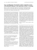
Thespatialdistributionofthecontinuousx-raysemittedbythintargetshasbeenin-
vestigatedbyKulenkampff(1928).Theauthormadeanextensivesurveyoftheintensityat
anglesbetween22
and150
totheelectronbeamintermsofdependenceonwavelength
andvoltage.Thetargetwasa0.6-mm-thickAlfoil.Figure1showsthecontinuousx-ray
intensity observed at different angles for voltages of 37.8, 31.0, 24.0, and 16.4 kV filtered
by 10, 8, 4, and 1.33 mm of Al, respectively (Stephenson, 1957). Curve (a) is repeated as a
dotted line near each of the other curves. The angle of the maximum intensity varied from
Copyright © 2002 Marcel Dekker, Inc.
50
for37.8kVto65
for16.4kV.Figure2illustratestheintensityofthecontinuous
x-raysobservedintheAlfoilfordifferentthicknessesasafunctionoftheanglefora
voltageof30kV(Stephenson,1957).ThetheoreticalcurveisfromthetheoryofScherzer
(1932).Thecontinuousx-rayintensitydropstozeroat180
,andalthoughitisnotzeroat
0
asthetheoryofScherzerpredicts,itcanbeseenfromFigure2thatforathinnerfoil,a
lowerintensityat0
isobtained.Summarizing,itappearsthattheintensityofthecon-
tinuousx-raysemittedbythinfoilshasamaximumatabout55
relativetotheincident
electronbeamandbecomeszeroat180
.
Thecontinuousradiationfromthicktargetsischaracterizedbyamuchsmaller
anisotropythanthatfromthintargets.Thisisbecauseinthicktargetstheelectronsare
rarelystoppedinonecollisionandusuallytheirdirectionshaveconsiderablevariation.
Theuseofelectromagnetictheorypredictsamaximumenergyatrightanglestothein-
cidentelectronbeamatlowvoltages,withthemaximummovingslightlyawayfrom
perpendicularitytowardthedirectionoftheelctronbeamasthevoltageisincreased.In
general,anincreaseintheanisotropyofthecontinuousx-raysfromthicktargetsisob-
servedattheshort-wavelengthlimitandforlow-Ztargets(Dyson,1973).
Figure1Intensityofcontinuousx-raysasafunctionofdirectionfordifferentvoltages.(Curve(a)
is repeated as dotted line.) (From Stephenson, 1957.)
Copyright © 2002 Marcel Dekker, Inc.
Continuous x-ray beams are partially polarized only from extremely thin targets; the
angular region of polarization is sharply peaked about the photon emission angle
y ¼ m
0
c
2
=E
0
, where E
0
is the energy of the primary electron beam. Electron scattering in
the target broadens the peak and shifts the maximum to larger angles. Polarization is
defined by (Kenney, 1966)
Pðy; E
0
; E
n
Þ¼
ds?ðy; E
0
; E
n
Þdskðy; E
0
; E
n
Þ
ds?ðy; E
0
; E
n
Þþdskðy; E
0
; E
n
Þ
ð11Þ
where an electron of energy E
0
radiates a photon of energy E
n
at angle y; ds?ðy; E
0
; E
n
Þ
and dskðy; E
0
; E
n
Þ are the cross sections for generation of the continuous radiation with
the electric vector perpendicular (?) and parallel (k) to the plane defined by the incident
electron and the radiated photon, respectively. Polarization is difficult to observe, and only
thin, low-yield radiators give evidence for this effect. When the electron is relativistic
before and after the radiation, the electrical vector is most probably in the ? direction.
Practical thick-target Bremsstrahlung shows no polarization effects whatever (Dyson,
1973; Stephenson, 1957; Kenney, 1966).
V. EMISSION OF CHARACTERISTIC X-RAYS
The production of characteristic x-ray s involves transitions of the orbital electrons of
atoms in the target material between allowed orbits, or energy states, associated with
ionization of the inner atomic shells. When an electron is ejected from the K shell by
electron bombardment or by the absorption of a photon, the atom becomes ionized and
the ion is left in a high-energy state. The excess energy the ion has over the normal state of
the atom is equal to the energy (the binding energy) required to remove the K electron to a
state of rest outside the atom. If this electron vacancy is filled by an electron coming from
an L level, the transition is accompanied by the emission of an x-ray line known as the Ka
line. This process leaves a vacancy in the L shell. On the other hand, if the atom contains
sufficient electrons, the K shell vacancy might be filled by an electron coming from an M
level that is accompanied by the emission of the Kb line. The L or M state ions that remain
may also give rise to emission if the electron vacancies are filled by electrons falling from
further orbits.
Figure 2 Intensity of continuous x-rays as a function of direction for different thicknesses of the
A1 target together with theoretical prediction. (From Stephenson, 1957.)
Copyright © 2002 Marcel Dekker, Inc.
A. Inner Atomic Shell Ionization
As already mentioned, the emission of characteristic x-ray is preceded by ionization of
inner atomic shells, which can be accomplished either by charged particles (e.g., electrons,
protons, and a-particles) or by photons of sufficient energy. The cross section for ion-
ization of an inner atomic shell of element i by electrons is given by (Bethe, 1930; Green
and Cosslett, 1961; Wernisch, 1985)
Q
i
¼ pe
4
n
s
b
s
ln U
UE
2
c;i
ð12Þ
where U ¼ E=E
c;i
is the overvoltage, defined as the ratio of the instantaneous energy of the
electron at each point of the trajectory to that required to ionize an atom of element i, E
c;i
is the critical excitation energy, and n
s
and b
s
are constants for a particular shell:
s ¼ K: n
s
¼ 2; b
s
¼ 0:35
s ¼ L: n
s
¼ 8; b
s
¼ 0:25
The cross section for ionization Q
i
is a strong function of the overvoltage, which shows a
maximum at U ffi 3–4 (Heinrich, 1981; Goldstein et al., 1981).
The probability (or cross section) of ionization of an inner atomic shell by a charged
particle is given by (Merzbacher and Lewis, 1958)
s
s
¼
8pr
2
0
q
2
f
s
Z
4
Z
s
ð13Þ
where r
0
is the classic radius of the electron equal to 2.818610
15
m, q is the particle
charge, Z is the atomic number of the target material, f
s
is a factor depending on the wave
functions of the electrons for a particular shell, and Z
s
is a function of the energy of the
incident particles.
In the case of electromagne tic radiation (x or g), the ionization of an inner atomic
shell is a result of the photoelectric effect. This effect involves the disappearance of a ra-
diation photon and the photoelectric ejection of one electron from the absorbing atom,
leaving the atom in an excited level. The kinetic energy of the ejected photoelectron is
given by the difference between the photon energy hn and the atomic binding energy of the
electron E
c
(critical excitation energy). Critical absorption wavelengths (Clark, 1963) re-
lated to the critical absorption energies (Burr, 1974) via the equation l(nm) ¼1.24=E(ke V)
are presented in Appendix I. The wavelenghts of K, L, M, and N absorption edges can also
be calculated by using simple empirical equations (Norrish and Tao, 1993).
For energies far from the absorption edge and in the nonrelativistic range, the cross
section t
K
for the ejection of an electron from the K shell is given by (Heitler, 1954)
t
K
¼
32
ffiffiffi
2
p
3
pr
2
0
Z
5
ð137Þ
4
m
0
c
2
hv
7=2
ð14Þ
Equation (14) is not fully adequate in the neighborhood of an absorption edge; in this
case, Eq. (14) should be multiplied by a correction factor f(X ) (Stobbe, 1930):
fðXÞ¼2p
D
hv
1=2
e
4X arccot X
1 e
2pX
ð15Þ
where
Copyright © 2002 Marcel Dekker, Inc.
X ¼
D
hv D
1=2
ð15aÞ
with
D ffi
1
2
ðZ 0:3Þ
2
m
0
c
2
ð137Þ
2
ð15bÞ
When the energy of the incident photon is of the order m
0
c
2
or greater, relativistic
cross sections for the photoelectric effect must be used (Sauter, 1931).
B. Spectral Series i n X-rays
The energy of an emission line can be calculated as the difference between two terms, each
term corresponding to a definite state of the atom. If E
1
and E
2
are the term values re-
presenting the energies of the corresponding levels, the frequency of an x-ray line is given
by the relation
v ¼
E
1
E
2
h
ð16Þ
Using the common notations, one can represent the energies of the levels E by means
of the atomic number and the quantum numbers n, l, s, and j (Sandstro
¨
m, 1957):
E
Rh
¼
ðZ S
n;l
Þ
2
n
2
þ a
2
ðZ d
n;l; j
Þ
2
n
3
1
l þ
1
2
3
4n
!
a
2
ðZ d
n;l; j
Þ
4
n
3
jðj þ1Þlðl þ1Þsðs þ1Þ
2lðl þ
1
2
Þðl þ1Þ
ð17Þ
where S
n;l
and d
n;l; j
are screening constants that must be introduced to correct for the effect
of the electrons on the field in the atom, R is the universal Rydberg constant valid for all
elements with Z > 5 or throughout nearly the whole x-ray region, and a is the fine-
structure constant given by
a ¼
2pe
2
hc
ð17aÞ
The theory of x-ray spectra reveals the existence of a limited number of allowed
transitions; the rest are ‘‘forbidden.’’ The most intense lines create the electric dipole ra-
diation. The transitions are governed by the selection rules for the change of quantum
numbers:
Dl ¼1; Dj ¼ 0 or 1 ð18Þ
The j transition 0 ? 0 is forbidden.
According to Dirac’s theory of radiation (Dirac, 1947), transitions that are forbidden
as dipole radiation can appear as multipole radiation (e.g., as electric quadrupole and
magnetic dipole transitions). The selection rules for the former are
Dl ¼ 0 or 2; Dj ¼ 0; 1; or 2 ð19Þ
The j transitions 0 ? 0,
1
2
?
1
2
, and 0 $ 1 are forbidden.
The selection rules for magnetic dipole transitions are
Copyright © 2002 Marcel Dekker, Inc.
Dl ¼ 0; Dj ¼ 0 or 1 ð20Þ
The j transition 0 ? 0 is forbidden.
The commonly used terminology of energy levels and x-ray lines is shown in Figure 3.
A general expression relating the wavelength of an x-ray characteristic line with the
atomic number of the corresponding element is given by Moseley’s law (Moseley, 1914):
1
l
¼ kðZ sÞ
2
ð21Þ
where k is a constant for a particular spectral series and s is a screening constant for the
repulsion correction due to other electrons in the atom. Moseley’s law plays an important
role in the systematizing of x-ray spectra. Appendix II tabulates the energies and wave-
lengths of the principal x-ray emission lines for the K, L, and M series with their ap-
proximate relative intensities, which can be defined either by means of spectral line peak
intensities or by area below their intensity distribution curve. In practice, the relative
Figure 3 Commonly used terminology of energy levels and x-ray lines. (From Sandstro
¨
m, 1957.)
Copyright © 2002 Marcel Dekker, Inc.