gene therapy for hiv infection
Bạn đang xem bản rút gọn của tài liệu. Xem và tải ngay bản đầy đủ của tài liệu tại đây (268.65 KB, 28 trang )
CHAPTER 11
Gene Therapy for HIV Infection
BRUCE BUNNELL, M.D.
BACKGROUND
In the previous chapters of this text, the technological aspects of gene therapy have
been discussed. The application of these technologies to specific genetic disorders
has also been presented. In this chapter, the application of this technology for
the treatment of an infectious agent will be discussed. Specifically, gene therapy
approaches to limit replication of the human immunodeficiency virus (HIV-1), the
causative agent in acquired immunodeficiency syndrome, will be presented.
INTRODUCTION
Acquired immunodeficiency syndrome (AIDS) is a rapidly expanding global
pandemic. Approximately 15 million people worldwide are infected with HIV-1.
Despite more than a decade of intense research efforts aimed at understanding the
HIV-1 virus and developing an effective therapy for AIDS, HIV-1 infection remains
an incurable and fatal disease. However, significant progress has been made in the
management of HIV-1 replication using traditional drug-based therapies. Most
notable is the advent of the triple-drug regiment, which is composed of three drugs
that inhibit the HIV-1 life cycle at two different stages. A protease inhibitor, which
blocks the normal processing of proteins necessary to generate new HIV-1 parti-
cles, and AZT and 3TC, which are nucleoside analogs that inhibit replication of the
viral genome, are typically the components of the triple-drug cocktail. The high rate
of mutation in the viral genome and the generation of drug-resistant strains of HIV-
1 are the major factors that prevent the development of effective drug-based ther-
apies. The triple-drug regiment has not been sufficiently tested to assess the ability
of the HIV-1 to form drug-resistant mutants. The inability of traditional drug-based
therapies to effectively inhibit the HIV-1 replication has made it necessary to
develop new and innovative therapies for this deadly disease.
As part of the normal virus life cycle, the HIV-1 virus integrates into the host
263
An Introduction to Molecular Medicine and Gene Therapy. Edited by Thomas F. Kresina, PhD
Copyright © 2001 by Wiley-Liss, Inc.
ISBNs: 0-471-39188-3 (Hardback); 0-471-22387-5 (Electronic)
cell’s genome and remains there permanently. Thus AIDS can be considered as an
acquired genetic disorder.As previously discussed, gene therapy holds considerable
potential for the treatment of hereditary and acquired genetic disorders. Human
gene therapy can be defined as the introduction of new genetic material into the
cells of an individual with the intention to produce a therapeutic benefit for
the patient. Therefore, AIDS may be amenable to treatment by gene therapy
approaches to inhibit the replication of HIV-1.
The ultimate goal of gene therapy is to inhibit HIV-1 viral replication and the
resulting AIDS pathogenesis. For gene therapy of HIV infection to be successful, it
will be necessary to introduce genes that are designed to specifically block or inhibit
the gene expression or function of viral gene products such that the replication of
HIV is blocked or limited.This concept was originally denoted as intracellular immu-
nization and is currently being investigated as a therapeutic approach for a wide
variety of infectious agents. In addition to intracellular interventions, gene therapy
may be employed to intervene with the spread of HIV at the extracellular level. Inhi-
bition of viral spread could be accomplished by sustained expression in vivo of a
secreted inhibitory protein or by stimulation of an HIV-specific immune response.
GENETIC ORGANIZATION OF HIV
The HIV-1 virus is a member of the family of viruses denoted as retroviruses. The
retrovirus classification encompasses a heterogeneous group of viruses containing
a single-stranded, positive-sense ribonucleic acid (RNA) genome and the enzyme
reverse transcriptase. Reverse transcriptase functions by copying the viral genomic
RNA into double-stranded deoxyribonucleic and (DNA), which is a critical phase
in the life cycle of retroviruses. Retroviruses have historically been subdivided into
three groups primarily based on the pathologic outcome of infection. The oncovirus
subgroup includes retroviruses that can cause tumor formation in the infected host;
however, this group also includes several apparently benign viruses. Lentiviruses
cause slowly progressing, chronic diseases that most often do not contain a tumor-
forming component. The spumavirus subgroup, although causing marked foamy
cytopathic effect in vitro, have not yet been clearly associated with any disease.
Upon intense investigation into the pathology of HIV infection, it has become clear
that the virus is a member of the lentivirus subgroup. Lentiviruses were initially iso-
lated in the 1960s when it was found that certain slowly evolving, degenerative dis-
eases in sheep were communicable. Interestingly, unlike the oncogenic retroviruses,
the lentiviruses did not form tumors but were cytopathic (caused cells death).
Several members of the lentivirus family have been isolated and described.
Members of the lentivirus family include Visna virus, Simian immunodeficiency
virus, human immunodeficiency virus 1 and 2, caprine arthritis-encephalitis virus,
and equine infectious anemia virus.
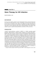
As with all other retroviruses, HIV is an enveloped virus that contains two copies
of single-stranded, positive-sense RNA (Fig. 11.1).The genomic organization of HIV
is shown in Figure 11.2. At the ends of the genome are two identical genetic regions
similar to those found in all retroviruses. The genetic elements are called long
terminal repeats (LTRs). The LTRs contain elements that are responsible for the
proper regulation of gene expression during virus replication such as promoters,
264 GENE THERAPY FOR HIV INFECTION
enhancers, and elements required for efficient messenger RNA (mRNA)
polyadenylation. Between the LTRs are the genes that encode all of the viral pro-
teins. The HIV genome encodes three sets of viral proteins; the structural proteins
(Gag, Pol, and Env), the regulatory proteins (Tat, Rev, and Nef), and the matura-
tion proteins (Vif, Vpu, and Vpr).
As shown in Figure 11.2, the structural proteins can be subdivided into three
groups: core proteins, enzymes, and envelope proteins. These three groups of pro-
teins are encoded by the gag, pol, and env genes, respectively. The gag gene refers
to the group antigen and produces the viral core proteins that have antigens cross-
reacting with other antigens within large retrovirus groups. The Gag proteins are all
produced as a large single polyprotein that is then cleaved into individual proteins
by a virus-encoded protease (p24, p18, and p15). The pol gene products are also
encoded from a single open reading frame as a large polyprotein that is cleaved into
the virus-associated enzymes—protease, reverse transcriptase (RT), ribonuclease,
and integrase. The env gene products are surface glycoproteins that are produced
as a polyprotein (gp160), however, they are cleaved by cellular enzymes to produce
the two HIV surface glycoproteins (gp120 and gp41).
In addition to the structural elements necessary to assemble the virus particle,
the virus genome codes for several nonstructural proteins that play vital roles in the
regulation of the viral life cycle. The nonstructural proteins produced by the HIV
can be divided into two classes, the regulatory proteins and the maturation proteins.
The regulatory proteins include Tat, Rev, and Nef. The Tat protein was the first viral
regulatory protein to be described.The Tat protein, which is encoded by the tat gene,
is a strong transactivator of viral gene expression. In other words, the Tat protein
GENETIC ORGANIZATION OF HIV 265
Gag (p24)
Protease
Ribonuclease
Integrase
Reverse
Transcriptase
Envelope (gp 120/gp41)
RNA Genome
Gag (p17)
Lipid Bilayer
FIGURE 11.1 Structural organization of a mature HIV-1 virion. An HIV virion with struc-
tural and virion accessory proteins identified. HIV particles are approximately 110nm in
diameter. They are composed of a lipid bilayer membrane surrounding a conical nucleocap-
sid. Two copies of single-stranded positive sense RNA are contained with the nucleocapsid.
regulates the function of genes that are not immediately adjacent to its own gene.
The Tat protein binds to the trans-activation response (TAR) element. The TAR
element corresponds to an RNA stem-loop structure present within the untrans-
lated leader sequence of all HIV-1 transcripts, including the RNA genome, and is
required for HIV-1 Tat function. The interaction between Tat and TAR can lead to
266 GENE THERAPY FOR HIV INFECTION
tat
tat
tat
rev
rev
nef
nef
env
env
vpu
vpr
vpr
vpu
pol
pol
gag
gag
vif
vif
TAR RRE
RRE
RRE
HIV-1 Genome Organization
HIV-1 m RNAs
Structural
Proteins
Regulatory
Proteins
Maturation
Proteins
FIGURE 11.2 Genomic organization and mRNA expression pattern of HIV-1.The diagram
depicts the organization of the nine predominant genes of HIV-1. The diagram represents
the major RNAs derived from the HIV-1 genome by alternative splicing of the HIV-1
genome. Three distinct classes of viral proteins are generated from these mRNAs: structural
proteins, regulatory proteins, and maturation proteins. The structural proteins include
the viral envelope protein (gp 120, gp 41) which is encoded by the env gene and the core
proteins (p6, p9, p17, and p24) which are encoded by the gag gene. The pol gene generates
the viral-associated reverse transcriptase, integrase, RNase H, and protease enzyme
activities. The viral-associated regulatory proteins are encoded by the tat, rev, and nef genes,
respectively. The Tat and Rev proteins are powerful regulatory proteins. The Tat protein
interacts with the TAR (tat-responsive) element, which leads to a strong transactivation of
viral gene expression, while the Rev protein interacts with the RRE (rev response element),
which enhances the nuclear export of unspliced and single-spliced viral mRNA. The third
class of viral proteins are the maturation proteins that are encoded by the vif, vpr, and vpu
genes.
a potent transactivation (increasing expression of viral genes by 1000 times their
level of expression in HIV-1 mutants lacking the tat gene) by inducing transcrip-
tional initiation and/or elongation.
A second important regulatory protein is Rev, which produced by the rev gene.
The Rev protein is produced early in the replication phase of HIV and interacts
with a 234-nucleotide region of the env open reading frame in mRNA called the
Rev response element (RRE). The interaction of the Rev protein with the RRE
markedly enhances nuclear export of single-spliced and unspliced viral mRNAs
from the nucleus; these RNAs encode the viral structural proteins. The production
of Rev protein is an absolute requirement for the replication of the HIV virus, since
mutants of the Rev protein are incapable of inducing synthesis of the viral struc-
tural proteins and are, thus, replication defective.
The last member of the regulatory protein family is the Nef protein. The role of
the Nef protein in HIV-1 replication cycle remains unclear. However, the nef gene
product is not required for HIV-1 replication in vitro or SIV in vivo. It is clear that
the nef gene plays a role in the down-regulation of CD4 gene expression in infected
cells. It is also hypothesized that Nef may be involved in the ability of HIV-1 to turn
off its growth and reside dormant in the host cell genome.
In addition to the Gag, Pol, and Env, the late gene products encoded by HIV
include the maturation proteins Vif, Vpu, and Vpr. Both the Vif (virion infectivity
factor) and Vpu (viral protein U) proteins play roles in the maturation and pro-
duction of infectious HIV virion particles. The Vpr (viral protein R) protein has
recently been described as playing an integral role in causing the cell cycle arrest
of HIV-infected cells. Expression of Vpr alone was sufficient to cause arrest of the
cell cycle at the G
2
/M transition phase of the cell cycle. Thus, HIV-infected cells are
unable to progress normally from the G
2
phase of the cell cycle through mitosis to
complete the cell cycle. The cell cycle arrest after infection by HIV causes the
infected cell to remain in an activated state and, thus, may maximize virus produc-
tion from the infected cell.
LIFE CYCLE AND PATHOGENESIS OF HIV-1 INFECTION
As shown in Figure 11.3, the initial stage of infection (the early phase) begins with
the binding of the viral gp120 protein to its cell surface receptor, the CD4 protein.
CD4 is present in high concentration on the surface of peripheral blood lympho-
cytes (PBL) and at lower concentrations on other cells that can be infected by HIV,
including monocytes, macrophages, and dendritic cells. However, CD4 is not the sole
mediator of HIV infection. Previous work in murine cell lines expressing human
CD4 are not infected by HIV, which suggested the existence of a human specific co-
factor. The HIV infection co-factor has recently been identified. This co-factor,
termed fusin (CXCR4), is absolutely required, in addition to CD4, for the entry of
HIV in to human cells. Fusin is an integral membrane glycoprotein and a member
of the chemokine receptor family. Several of these co-factor proteins (CXCR4,
CCR5, and CCR3) have now been identified on various cell types. The binding of
the HIV gp120/gp41 envelope protein induces conformational changes that allow
interaction with the co-receptor and subsequent fusion of the virus with the host
cell plasma membrane. The HIV-1 nucleocapsid is internalized into the cytoplasm
where the viral-genome is uncoated. The RNA genome is reverse transcribed into
LIFE CYCLE AND PATHOGENESIS OF HIV-1 INFECTION 267
a single, negative strand of DNA, by the RT protein encoded by the pol sequences.
The viral-encoded ribonuclease then degrades the viral genomic RNA. The RT
enzyme then encodes the second (positive strand) of DNA, and this double-
stranded viral genome is circularized and transported through the nuclear pore
and into the nucleus of the infected cell. The newly synthesized viral DNA
genome then randomly integrates into the host cell genome by the virally encoded
integrase protein; this integrated form of the virus is denoted as the provirus. The
provirus can replicate immediately or remain latent for extended periods of
time and in so doing is passed along to all progeny cells derived from the original
infected cell.
Although the mechanism of proviral activation is unclear, once the provirus is
activated the intermediate stage of viral infection begins. Activation induces tran-
scription of multiply spliced viral RNAs, which are utilized to produce the Tat and
Rev proteins that act as powerful regulatory proteins during virus replication. As
discussed previously, the Tat protein enhances the transcription elongation of viral
RNA within the nucleus of the infected cell. Whereas, the Rev protein enhances
nuclear export of single-spliced and unspliced viral mRNAs from the nucleus; these
RNAs encode the viral structural proteins.
The late phase of HIV-1 infection begins upon the accumulation of significant
amounts of structural proteins. The late phase consists of assembly of virus parti-
cles containing two copies of the viral RNA genome. The assembled particles are
transported to the cell membrane where the mature virus particles bud off from the
plasma membrane. In theory, the life cycle of the HIV-1 virus can be interrupted by
268 GENE THERAPY FOR HIV INFECTION
INTEGRATION
VIRION
ASSEMBLY
BINDING
BUDDING
REVERSE
TRANSCRIPTION
nuclear
pore
Genomic
RNA
Unintegrated
Genomic DNA
Core
CD4
gp120
Structural
Protein
mRNA's
Gag and Env
Proteins
Mature
HIV Virions
Tat
Rev
Regulatory
mRNA's
Genomic RNA
INTERNALIZATION
FIGURE 11.3 Life cycle and replication of HIV-1.
blocking or inhibiting the function of one or more or the key viral proteins or their
cis-acting regulatory elements.
HIV can kill an infected CD4
+
T lymphocyte in one of two ways.As progeny virus
particles are budded off from the cell membrane, the external envelope protein
gp120 reacts with CD4 molecules found on the surface of the infected cell to disrupt
the integrity of the cell membrane in the areas with high concentrations of CD4.
Disruption of the cell membrane kills the infected cell. Alternatively, an infected
cell may interact with an uninfected cell through the HIV envelope proteins embed-
ded in their cell surface membranes. The interaction is again through the CD4 mol-
ecules found on the surface of the uninfected cell.As the cell fusion occurs, hundreds
of CD4 cells may eventually be involved in the formation of a large syncytium. All
of the cells that fused into the syncytium die, and thus the cytopathic effects of HIV
can extend beyond cells directly infected with the virus. It is predominantly through
these two mechanisms that loss of CD4
+
lymphocytes occurs in HIV-infected
patients. The outcome of HIV infection in monocyte–macrophage lineage cells is
unclear. It appears as though the virus is capable of replication, but it does not
appear to have any obvious cytopathic effects as in T lymphocytes. Similar to
infected T cells, the formation of multinucleated syncytium of macrophage-like cells
is observed in HIV-infected tissues. Macrophages that contain replicating virus may
not be destroyed, but evidence suggests that they become dysfunctional.
GENETIC APPROACHES TO INHIBIT HIV REPLICATION
Approaches to gene therapy for HIV can be divided into three broad categories:
(i) protein approaches such as transdominant negative proteins and single-chain
antibodies, (ii) gene therapies based on nucleic acid moieties, including antisense
DNA/RNA, RNA decoys, and catalytic RNA moieties (ribozymes), and (iii)
immunotherapeutic approaches using genetic vaccines or pathogen-specific lym-
phocytes (Table 11.1). It is further possible that combinations of the aforementioned
approaches may be used simultaneously to inhibit multiple stages of the viral life
cycle or in combination with other approaches, such as hematopoietic stem cell
transplantation or vaccination. The extent to which gene therapy approaches will
be effective against HIV-1 is the direct result of several key factors: (i) selection of
the appropriate target cell in which to deliver the gene therapy, (ii) the efficiency
of the gene delivery system on the target cell, (iii) appropriate expression, regula-
tion, and stability of the anti-HIV gene product(s), and (iv) the strength of the
inhibition of viral replication by the therapeutic entity.
TRANSDOMINANT NEGATIVE PROTEINS
Transdominant negative proteins (TNPs) are mutant versions of regulatory or struc-
tural proteins that display a dominant negative phenotype that can inhibit replica-
tion of HIV. By definition, such mutants not only lack intrinsic wild-type activity but
also inhibit the function of their cognate wild-type protein in trans. Inhibition may
occur because the mutant competes for an essential substrate or co-factor that is
available in limiting amounts, or, for proteins that form multimeric complexes, the
TRANSDOMINANT NEGATIVE PROTEINS 269
mutant may associate with wild-type monomers to form an inactive mixed multi-
mer. A potential drawback in the use of transdominant viral proteins is their possi-
ble immunogenicity when expressed by the transduced cells.The protected cells may
consequently induce an immune response that might result in their own destruc-
tion. This may diminish the efficacy of antiviral gene therapy using transdominant
proteins. HIV-1 regulatory (Tat and Rev) and structural proteins (Env and Gag) are
potential targets for the development of TNPs.
The most thoroughly investigated TNP is a mutant Rev protein denoted RevM10.
The Rev protein is rendered a TNP through a series of mutations introduced into
the rev gene (Fig. 11.4). The RevM10 still retains the ability to multimerize and bind
to the RRE; but as a result of these mutations, the RevM10 protein can no longer
efficiently interact with a cellular co-factor that activates the Rev function. Cell lines
stably expressing RevM10 are protected from HIV-1 infection in long-term cell
culture assays.Transduction of RevM10 into T-cell lines or primary PBL delays virus
replication without any detectable negative effects on the cells. Recently, it has been
demonstrated that RevM10 inhibits HIV-1 replication in chronically infected T cells.
A different TNP Rev protein developed by Morgan et al. (1994) inhibited HIV-1
270 GENE THERAPY FOR HIV INFECTION
TABLE 11.1 Gene Therapy Strategies to Inhibit HIV Replication
Anti-HIV Strategy Potential Mode of Action
Protein-Based Approaches
Transdominant Negative Proteins
Rev Nuclear export of viral mRNA
Tat Viral genome transcription/processing
Gag Viral assembly
Env Viral assembly
Endogenous Proteins
Soluble CD4 Receptor binding/viral assembly
CD4-KDEL Trapping of Env and Rev in ER
E1F-5A
Intrabodies
anti-gp120 Maturation and function of Env protein
Nucleic Acid Approaches
Antisense RNA
Antisense Tat/Rev Translation of Tat and Rev proteins
Antisense Gag Translation of Gag protein
Ribozymes
5¢ leader sequence Translation of viral RNA
Multitarget Translation of viral RNA
Antisnese oligonucleotides
RNA decoys Translation of viral RNA
TAR decoy Viral genome transcription/processing
RRE decoy Nuclear export of viral mRNA
Immunity Augmentation
DNA Vaccines Induction of cellular and humoral response
Env Augments cytotoxic activity to HIV
Virus Specific CTL
replication in T-cell lines and PBL when challenged with both laboratory and clin-
ical HIV-1 isolates.A third type of Rev TNP was generated by deletion of the nucle-
olar localization signal sequence. This sequence functions as a signal to direct the
Rev protein to the nucleolar region of the nucleus of an infected cell. This TNP Rev
is retained in the cytoplasm and prevented the localization of wild-type Rev to the
nucleus by forming inactive oligomers.
The HIV-1 regulatory protein Tat was also utilized to generate TNPs. A TNP Tat
was mutated in its protein binding domain. Upon transduction into T-cell lines, the
TNP Tat inhibited HIV-1 replication for up to 30 days. The mechanism through
which this Tat TNP may function is by sequestration of a cellular factor involved
in Tat-mediated transactivation. Interestingly, in this study a retroviral vector was
developed that was capable of expressing both a Tat and Rev TNP. The multi-TNP
vector was more effective at blocking HIV-1 replication than retroviral vectors
expressing either TNP Tat or Rev alone. This study suggests that the inhibition of
Tat and Rev simultaneously may be a more effective HIV-1 gene therapy. Recently,
a double transdominant Tat/Rev fusion protein (Trev) was designed in an attempt
to inhibit two essential HIV-1 activities simultaneously. Upon transfection or
TRANSDOMINANT NEGATIVE PROTEINS 271
Rev
Rev
TNP
1
2
Inhibition of HIV Replication
Genomic
RNA
Virion
Assembly
Extra-Nuclear
Transport
Structural
Protein
mRNA's
BUDDING
Mature
HIV
virions
FIGURE 11.4 Activity of a transdominant negative Rev protein. (1) The normal function
of the Rev protein is to form multimeric complexes (gray circles) which increase the effi-
ciency of extranuclear transport of genomic viral RNA(s) and (2) the transdominant nega-
tive Rev protein (black circles) forms inactive mixed multimeric complexes with the wild-type
Rev protein (gray circles). These inactive Rev complexes interfere with the normal func-
tioning of the wild-type Rev complexes and inhibit the extra-nuclear transport of unspliced
and singly spliced HIV RNA(s).
transduction of the Trev gene into T cells, they were protected from the cytopathic
effects of HIV-1. Simultaneous inhibition of two HIV-1 functions may have poten-
tial advantages over single-function TNPs.
TNP moieties based on structural proteins have also been investigated for their
anti-HIV-1 functions.The HIV-1 structural proteins (Gag and Env) oligomerize into
multimeric complexes during viral assembly. Multimerization makes them ideal can-
didates for the generation of TNPs. Several Gag TNPs have been investigated and
all are capable of inhibiting HIV-1 replication. The Gag TNPs function by disrupt-
ing distinct stages of the viral life cycle, such as viral assembly, viral budding, uncoat-
ing of the viral genome, or initiation of reverse transcription. Due to inherently low
levels of transcription of gag genes in the absence of the HIV-1 Rev protein, the
application of Gag TNPs has been limited. The low levels of mutant Gag expressed
are insufficient to effectively block HIV-1 replication. Env TNPs have been gen-
erated as well but in initial testing showed only low levels of antiviral activity.
Single-Chain Antibodies (Intrabodies)
One of the more novel classes of antimicrobial gene therapies involves the devel-
opment of intracellularly expressed single-chain antibodies (also called intra-
bodies). The single-chain variable fragment of an antibody is the smallest structural
domain that retains the complete antigen specificity and binding site capabilities
of the parental antibody. Single-chain antibodies are generated by cloning of the
heavy- and light-chain genes from a hybridoma that expresses antibody to a spe-
cific protein target.These genes are used for the intracellular expression of the intra-
body, which consists of an immunoglobulin heavy-chain leader sequence that targets
the intrabody to the endoplasmic reticulum (ER), and rearranged heavy- and light-
chain variable regions that are connected by a flexible interchain linker. Since the
single-chain antibody cannot be secreted, it is efficiently retained within the ER,
probably through its interaction with the ER-specific BiP protein. The BiP protein
binds incompletely folded immunoglobulins and may facilitate the folding and/or
oligomerization of these proteins. Intrabodies can directly bind to and prevent gene
function or may sequester proteins in inappropriate cellular compartments so that
the life cycle of HIV is disrupted.
Expression of an intrabody specific for the CD4 binding region of the HIV-1
gp120 (Env) markedly reduced the HIV-1 replication by trapping the gp160 in the
ER and preventing its maturation by cleavage into the gp120/gp41 proteins (Fig.
11.5). Intrabodies developed to the Rev protein trapped Rev in a cytoplasmic com-
partment and blocked HIV-1 expression by inhibiting the export of HIV-1 RNAs
from the nucleus. Additionally, intrabodies containing an SV40 nuclear localization
signal sequence were developed to Tat. The anti-Tat single-chain antibody blocked
Tat-mediated transactivation of the HIV-1 LTR and rendered T-cell lines resistant
to HIV-1 infection.
Endogenous Cellular Proteins as Anti-HIV Agents
Proteins derived from cellular genes have been identified that exhibit specific gene
inhibitory activity (Fig. 11.5). These activities may act by preventing the binding of
HIV to cells, by binding directly to the regulatory/structural proteins, or indirectly
272 GENE THERAPY FOR HIV INFECTION
by inducing or repressing cellular factors that in turn influence viral gene expres-
sion. One of the most successful in vitro uses of endogenous cellular proteins to
inhibit an infectious agent is the use of a soluble version of the HIV receptor CD4
(sCD4). The T helper cell antigen CD4 functions as the receptor for the HIV
through the physical interaction of the HIV envelope glycoprotein gp120 and the
CD4 protein. Based on these results, investigators have demonstrated that sCD4
protein can effectively bind to and inhibit HIV infection in CD4
+
cells. The effect
of this strategy is to compete for binding of HIV to cellular CD4 with high con-
centrations of sCD4. In order for this strategy to be efficacious, a high level of con-
tinuous expression of sCD4 will be required. Retroviral vectors expressing sCD4
have been shown to protect T-cell lines from HIV infection in vitro. A significant
limitation to this strategy is the ability to achieve sufficiently high levels of sCD4 to
neutralize HIV effectively. The use of sCD4 for the gene therapy of HIV infection
in a clinical setting has been disappointing. The intravenous infusion of recombi-
nant sCD4 protein in HIV-infected patients failed to show efficacy in phase I
clinical trials. In contrast to the laboratory strains of the HIV virus, clinical isolates
TRANSDOMINANT NEGATIVE PROTEINS 273
HIV Binding
gp 120
CD4
sCD4
Nucleus
HIV Production
CD4-Lyso
CD4-KDEL
gp 120
Intrabody
gp 120
Expression
gp 120
Expression
Lysosome
FIGURE 11.5 Cellular protein-based approaches for the inhibition of HIV-1 replication.
The intracellular production of a soluble CD4 protein (sCD4) can prevent both the binding
of infectious HIV particles and the production of new virus particles from an infected cells
by saturating all of the available envelope protein. The attachment of a endoplasmic reticu-
lum (ER) retention signal (KDEL) to the CD4 protein brings about the inhibition of virus
replication by retaining the gp160 envelope complexes with the endoplasmic reticulum. The
incorporation of a lysosomal targeting sequence into the CD4 protein leads to the inhibition
of gp160 expression through targeted degradation in the lysosomes. The expression of single-
chain antibodies (intrabodies) can also lead to the retention of viral proteins in the ER by
specific interaction with the BiP protein.
have shown a significant increase resistance to the neutralizing characteristics of
soluble CD4 protein.
A variation on the CD4-based anti-HIV gene therapy approach is the develop-
ment of a mutated soluble CD4 molecule that contains a specific ER retention signal
(Lys-Asp-Glu-Leu or KDEL). This hybrid molecule blocked secretion of gp120 and
cell surface expression of gp120/41, when expressed intracellularly (Fig. 11.5). The
CD4-KDEL/gp120 complex was retained within the endoplasmic reticulum, thus
preventing maturation of infectious HIV-1 particles. It has also been demonstrated
that the mutations that decrease the affinity of CD4 for gp120 discussed above
have little effect on the ability of CD4-KDEL to retain gp120 in the ER. A similar
approach is to specifically target soluble CD4/gp160 complexes to the lysosomes
through the incorporation of lysosome targeting domains onto the soluble CD4.The
CD4-lysosomal domain/gp160 complexes are degraded in the lysosomes of the cells
and production of mature HIV particles is diminished (Fig. 11.5).
It has recently been demonstrated that the Rev protein interacts specifically with
cellular factors in order to perform its normal function in the infected cell. The
eukaryotic initiation factor 5A (eIF-5A) is a cellular transcription factor that inter-
acts with Rev by binding to the Rev activation domain. The interaction between
mutants of the eIF-5A and Rev can effectively inhibit HIV-1 replication in vitro.
Utilization of the interactions between cellular factors and HIV could provide an
additional approach for the development of HIV genetic therapies. Since these are
endogenous cellular proteins, they are nonantigenic. Therefore, cells engineered
with these cellular inhibitory genes may not be eliminated by the patient’s immune
system. This is an advantage as compared to the use of genes encoding potentially
immunogenic, trans-dominant viral proteins for gene therapy.
A novel strategy exploiting the interaction of CD4/fusin with the gp120/gp41
protein of the HIV virus has been developed. As a consequence of HIV replication,
infected cells express the gp120/gp41 envelope protein on their surface in order for
the assembly of new virus particles to occur. The expression of gp120/gp41 on the
cell surface lead investigators to hypothesize that a virus could be engineered to
contain the HIV receptor and co-receptor in its envelope in place of endogenous
viral envelope proteins. Generation of these hybrid virus particles would specifically
target these hybrid virus particles to infected cells where replication of the virus
would kill the cell. The vesicular stomatitis virus (VSV) has been engineered in this
manner. In the VSV studies, deletion of the VSV gene and substitution with the
genes for CD4 and CXCR4 lead to the formation of recombinant VSV particles
that specifically infected HIV-infected cells. Upon infection, the replication of VSV
was cytopathic in HIV-infected cells.
NUCLEIC-ACID-BASED GENE THERAPY APPROACHES
RNA Decoys
This approach disrupts the normal interaction of the HIV regulatory proteins with
their cis-acting regulatory elements through the overexpression of short RNA mol-
ecules (decoys) that compete with viral RNA elements for binding of proteins that
are required for virus replication (Fig. 11.6). The TAR (transactivation response)
274 GENE THERAPY FOR HIV INFECTION
and RRE (Rev response element) are two such viral regulatory elements found in
HIV and are the binding sites for the transactivating proteins Tat and Rev, respec-
tively. The antiviral activity of the TAR element decoys was examined by retrovi-
ral-mediated gene transfer into T-cell lines in vitro. Overexpression of the TAR
decoys inhibited the transcriptional activation mediated by the Tat protein that, in
turn, markedly reduced HIV replication of laboratory HIV isolates.The TAR decoy
inhibition of virus replication results from the decreased binding of the Tat protein
to the endogenous TAR elements, which in turn inhibits the transcriptional activa-
tion necessary for efficient virus replication. Expression of a tandem repeat TAR
decoy composed of as many as 50 TAR repeats has also been demonstrated to effec-
tively inhibit HIV-1 replication in both T-cell lines and primary lymphocytes.
Furthermore, the multimerized TAR decoy was shown to efficiently inhibit virus
replication in lymphocytes from late-stage AIDS patients.
The enhanced expression of RRE decoys by retroviral vectors resulted in long-
term inhibition of HIV replication in human T-cell lines. RRE decoys may function
by preventing the binding of REV to the normal RRE sequences, which decreases
NUCLEIC-ACID-BASED GENE THERAPY APPROACHES 275
Decoys Decoys
Viral mRNA
Viral mRNA
Viral mRNA
Degradation by
RNase H
Cleavage of
RNA Backbone
Tat
Transactivator
Antisense
Nucleic Acids
Protein
Translation
Tar
Element
Saturation of
Tat Protein
3'
3'
5'
5'
5'
5'
5'
3'
5'
5'
3'
3'
3'
3'
Viral mRNA
FIGURE 11.6 Nucleic-acid-based gene therapies for HIV-1. Anti-HIV genes can be
expressed in the context of antisense nucleic acids, as antisense oligonucleotides, antisense
RNA, or ribozymes. All of these antisense approaches promote the destruction of the target
sequence by RNAses or block the translation of the mRNA. Ribozymes function by specific
interaction with a target sequence within the RNA and functionally inactivate it by cleavage
of the phosphodiester backbone. Overexpression of short RNA molecules (decoys) that cor-
respond to the viral cis-acting elements Transactivation response element (TAR) or the Rev
response element (RRE) inhibit the binding of the viral protein to the cognate sequences
found on the viral mRNAs.
the levels of singly spliced and unspliced HIV-1 mRNAs that are exported from the
nucleus of an infected cell. It is clear that the overexpression of RRE decoys has
strong antiviral activity, but there is some concern as to the long-term effects that
the expression of the RNA decoys will have on the normal function of the cell. In
addition to viral proteins, both TAR and RRE bind cellular co-factors. The overex-
pression of the decoys may have negative effects on cell viability or activity through
the sequestration of proteins required for normal cell function. To limit the inter-
action between the RRE decoy and cellular proteins, a minimal RRE decoy com-
posed of only 13 nucleotides that retained the rev binding domain was tested for
antiviral activity. This minimal RRE decoy was shown to effectively suppress HIV-
1 replication in vitro.
Antisense DNA and RNA
Antisense nucleic acid technology encompasses a broad spectrum of methods all
directed toward the specific silencing of gene expression. The silencing of gene
expression is achieved through the introduction into the cell or tissue of an anti-
sense RNA or single-stranded DNA moiety (oligodeoxynucleotide), which is com-
plementary to a target mRNA (Fig. 11.6). In theory, the antisense nucleic acids
utilize Watson–Crick nucleic acid base pairing to block gene expression in a
sequence-specific fashion. One of the most intensely investigated approaches for
application of antisense RNA is the introduction of DNA oligonucleotides that have
been chemically modified in an attempt to increase their stability (half-life) within
a cell. A variety of synthetic antisense oligonucleotides have been designed that
inhibit the replication of HIV-1. However, their use for the inhibition of HIV-1 has
been extremely limited because uptake of free oligonucleotides from the extracel-
lular environment in vivo is extremely inefficient, and effective oligonucleotide
delivery systems have not yet been devised. Also, the oligonucleotide moieties that
are internalized into the target cells are ultimately degraded by cellular enzymes
such that any inhibitory activity on gene expression is only transient. An additional
problem with the use of DNA oligonucleotides is that the gene inhibition that is
observed is often nonspecific. In other words, the inhibition of expression is most
often not the direct result of the interaction between the oligonucleotide and the
target sequence, but the interaction with RNA in a broad nonspecific manner.
The other approach for antisense nucleic-acid-mediated inhibition of gene
expression is the direct introduction or intracellular production of antisense RNA
in cells or tissues of the organism. The direct introduction of RNA transcripts into
cells can be accomplished through microinjection of an in vitro transcription
product or as a chemically modified oligonucleotide. The direct administration of
antisense RNA transcripts in vivo is not plausible for gene therapy due to the vast
number of cells that need to receive the therapeutic RNA.
An alternative approach to the delivery of antisense RNA for gene therapy is
the use of vector-based systems, which produce the antisense RNA within the cell
or tissue of the organism. Most often recombinant viral vector systems, such as retro-
viruses, are used because they efficiently target large numbers of cells. The use of
retrovirus vector-based systems for the intracellular production of antisense RNA
has an additional advantage. That is, the vector integrates into the host cell genome
and, thus, the antisense effects are more prolonged in comparison to oligonu-
276 GENE THERAPY FOR HIV INFECTION
cleotides.Also, the use of regulatable or inducible promoters would permit the levels
of inhibition to be tightly controlled.
Although the mechanism of antisense-mediated inhibition of gene expression is
not completely understood, it is hypothesized that RNA duplexes (antisense RNA
and target RNA) are degraded by RNase H or by blocking subsequent translation
of the mRNA. The limitations of antisense RNA transcripts are similar to those
observed with oligonucleotides. With higher levels of expression of antisense tran-
scripts the gene inhibition observed is often nonspecific.There is another major lim-
itation to the use of stable expression of antisense sequences as a therapy for HIV-1
infection. Long-term high levels of antisense expression are required in order
to effectively inhibit viral replication. The mechanism through which antisense
moieties inhibit gene expression requires that one antisense molecule efficiently
bind to one target molecule. The stoichiometry of antisense sequences to target
sequences must be at a minimum: 1 to 1 (antisense to target), but ratios of 5 to 1 or
10 to 1 lead to more effective inhibition of gene expression.Thus, the antisense gene
expression must be quantatively higher than the levels of HIV-1 gene expression
for an antisense gene therapy strategy to be effective. Standard gene therapy vectors
containing pol II promoters often do not produce sufficient levels of antisense
sequence to inhibit viral replication. To subvert this problem, vectors containing
alternative promoter systems have been developed. A retroviral vector containing
a pol III promoter has been demonstrated to significantly increase the levels of
expression. The transcription of transfer RNAs (tRNAs) and small nuclear RNAs
(snRNAs) found in eukaryotic cells are controlled by pol lII promoters. Pol III pro-
moters are multipartite (composed of two distinct parts). Interestingly, they are
found internal to the transcriptional start site. This means RNA polymerase III
reaches backwards to initiate transcription of a pol III gene. A pol-III-based retro-
viral vector expressing an antisense to the TAR sequence has successfully inhibited
HIV-1 replication in vitro. Alternatively, coordinating the expression of antisense
RNA with HIV-1 infection would permit the efficient expression of antisense
sequences within cells following infection. This could be accomplished by the devel-
opment of a retroviral vector in which the HIV-1 LTR is used as a promoter. This
vector permits the efficient expression of antisense sequences within cells following
infection of a lymphocyte by HIV-1.
A number of antisense transcripts have been designed to target various regions
of the HIV-1 genome. Stable intracellular expression of antisense HIV-1 transcripts
is currently the most efficient method by which antisense technology can be utilized
to inhibit HIV-1 gene expression. A number of studies have shown only limited
antiviral activity using antisense transcripts to the viral genes tat, rev, vpu, and gag.
An in-depth analysis of potential HIV-1 antisense gene sequences was performed
in which various antisense RNAs targeted to 10 different regions of the HIV-1
genome were compared for their antiviral effects.The antisense gene sequences with
the greatest antiviral activity were those that targeted a 1-kb region within the gag
gene and a sequence specific for a 562-bp genomic fragment encompassing the tat
and rev genes. Further analysis of the antiviral effects of the antisense tat/rev gene
fragment has demonstrated a strong inhibition of HIV-1 replication in a T-cell line
and primary CD4
+
PBL, but loss of the protective effects was observed as the
number of infectious HIV particles used to infect the protected cell population was
increased.
NUCLEIC-ACID-BASED GENE THERAPY APPROACHES 277
Ribozymes (Catalytic Antisense RNA)
Ribozymes are antisense RNA molecules that have catalytic activity. They function
by binding to the target RNA moiety through antisense sequence-specific hybridiza-
tion. Inactivation occurs by cleavage of the phosphodiester backbone at a specific
site (Fig. 11.6). The two most thoroughly studied classes of ribozymes are the
hammerhead and hairpin ribozymes (the names are derived from their theoreti-
cal secondary structures). Hammerhead ribozymes cleave RNA at the nucleotide
sequence U-H (H = A, C, or U) by hydrolysis of a 3¢–5¢ phosphodiester bond.
Hairpin ribozymes utilize the nucleotide sequence C-U-G as their cleavage site. A
distinct advantage of ribozymes over traditional antisense RNA is that they are not
consumed during the target cleavage reaction.Therefore, a single ribozyme can inac-
tivate a large number of target molecules. Additionally, ribozymes can be generated
from very small transcriptional units. Thus, multiple ribozymes targeting different
genomic regions could be incorporated into the same vector. Due to their unique
catalytic properties, ribozymes have the potential to be highly efficient inhibitors of
gene expression, even at low concentrations. Ribozymes also have greater sequence
specificity than antisense RNA because the target must have the correct target
sequence to allow binding. In addition, the cleavage site must be present in the right
position within the antisense fragment. However, the functionality and the extent
of catalytic activity that ribozymes actually have for their RNA targets in vivo is
presently unclear. However, a potent limitation to the use of ribozymes for HIV-1
gene therapy is that they are inherently limited in effectiveness due to the high rate
of mutation associated with HIV-1 replication. Any alteration of the binding or
cleavage sites within the target sequence required by the ribozyme for activity could
render the ribozyme totally inactive.
The first investigation into ribozymes designed to inhibit HIV-1 was performed
by transfecting a hammerhead ribozyme targeted to the viral gag sequence into
human fibroblasts that express CD4 antigen. Upon challenge with HIV-1, the cells
were demonstrated to express reduced levels of full-length gag RNA molecules and
markedly reduced levels of the gag-derived protein p24. Ribozymes developed to
target the 5¢ leader sequence of HIV-1 were shown to significantly inhibit HIV-1
replication in T-cell lines and PBL.These ribozymes inactivate incoming viral RNAs
prior to integration into the genome. Thus, ribozymes targeted to the 5¢ leader
sequence of HIV prevent the establishment of infection. The ability to prevent
infection by HIV-1 in the long term may allow uninfected cells to become perma-
nently resistant to HIV-1 infection. Interestingly, this ribozyme may have the poten-
tial to globally inhibit viral gene expression because the leader sequence is
contained within all of the HIV-1-derived RNAs. Multitarget ribozymes have also
been developed in which a single ribozyme cleaves at multiple highly conserved
targets within the HIV-1 genome. A multitarget ribozyme to conserved regions of
the env sequences has been shown to effectively inhibit replication of several HIV-
1 isolates.
Ribozyme transcription units are small enough that several ribozymes could be
incorporated into a single vector, thus ribozymes targeted to several regions of the
HIV-1 genome can be delivered within the same cell. Improved ribozyme-mediated
inhibition of HIV replication may be achievable by the development of ribozymes
that co-localize with their HIV-1 target RNA to the same subcellular compartment.
278 GENE THERAPY FOR HIV INFECTION
As a test of this strategy, a ribozyme transcript that contained the retroviral pack-
aging signal was demonstrated to efficiently inactivate newly synthesized MoMLV
genomic RNA prior to particle assembly resulting in a marked decrease in the
release of mature particles.
GENETIC APPROACHES TO ENHANCE IMMUNITY IN AIDS STIMULATION
OF AN HIV-SPECIFIC IMMUNE RESPONSE
DNA Vaccines
A novel nucleic-acid-based approach for gene therapy is to attempt to elicit an
immune response to native proteins of the HIV synthesized by the transfer of
plasmid DNA into cells; in other words, DNA-based vaccinations. The preliminary
observations leading to the development of genetic vaccination were made based
on the determination that plasmid DNA encoding marker genes could be expressed
following intramuscular injection in mice. Although the levels of gene transfer were
low, it was determined that the internalized plasmid persisted and was expressed for
the life span of the animal. The generation of an immune response to marker pro-
teins encoded by plasmids was demonstrated by two groups using plasmid DNA
introduced into the skin of mice by a biolistic gene delivery system. The develop-
ment of a protective immune response by immunization with a genetic vaccine was
initially demonstrated in mice that underwent intramuscular injection of naked
plasmid DNA encoding the internal nucleoprotein of the influenza virus.The poten-
tial efficacy of DNA vaccination into postmitotic muscle cells has since been demon-
strated in a variety of murine and animal models infected with bacterial, viral, or
parasitic pathogens. The rationale behind these gene vaccines is to generate both a
specific, cytotoxic T-cell response as well as a humoral response.
There are a number of theoretical advantages of the DNA-based vaccination
technology over traditional vaccine strategies. These include (i) the ease of pro-
duction and preparation of plasmid DNA, (ii) the expression of antigens in their
native form, which leads to the efficient generation of both cytotoxic and helper T
cells, (iii) the long-term immunity elicited suggests the potential to reduce the
number of doses of vaccine required to generate a protective immune response, and
(iv) the cells need not be the target cells that are normally infected by the infec-
tious agent.
The potential disadvantages of DNA vaccination include (i) accidental intro-
duction of the plasmid DNA into other than intended cell types, (ii) generation of
anti-DNA antibodies to the plasmid used for the vaccination, and (iii) random inte-
gration of the injected DNA into the target cells, which may activate an oncogene
or inactivate a tumor suppressor gene by insertional mutagenesis.
This approach is being actively investigated as a technique to optimize HIV-1
vaccination strategies. Introduction of the HIV-1 env gene via injection of naked
plasmid DNA into cells led to the formation of highly specific humoral and
cellular immune responses in mice. In addition, expression plasmids encoding either
the HIV-1 envelope glycoprotein or a defective noninfectious HIV particle were
shown to produce transient antibody to Env and expression of the defective
HIV genome raised persistent cytotoxic T-cell activity to the HIV Gag p24 protein.
GENETIC APPROACHES TO ENHANCE IMMUNITY IN AIDS STIMULATION 279
Recently, results from a study using a DNA-based anti-HIV vaccine approach in
chimpanzees indicated that although the DNA injections yielded a variable immune
response, the vaccinated animals remained infection-free following HIV challenge.
Taken together, these studies elucidate the potential for the formation of
strong HIV-directed immune responses. However, the ability of such an immune
response to persist and protect against polymorphic HIV-1 strains remains to be
demonstrated.
HIV-Specific Cytotoxic T-Lymphocytes
The use of the infected individual’s own cells (CD4
+
and CD8
+
lymphocytes, CD34
+
hematopoietic stem cells, or antigen presenting cells such as macrophages) for the
passive restoration of the immune system function is another technique in the devel-
opment of genetic therapies for HIV infection. Direct immunotherapy approaches
involve the ex vivo expansion of selected T-cell populations, either CD4 or CD8
lymphocytes, followed by reinfusion of the expanded lymphocyte population into
the HIV-1-infected individual. The major area of focus for adoptive cell therapy for
HIV-1 infection has been the use of CD8 cells. Although the importance of MHC
class 1 restricted CD8 CTL in controlling HIV-1 infection is not understood, it is
clear that early in the infection the increase in HIV-specific CD8 cells is correlative
with the resolution of viremia. This data strongly suggests that MHC class 1
restricted CD8 cells play a role in limiting infection during the acute phase (early
stages) of infection. The development of MHC class 1 restricted CD8 specific for
several HIV proteins, including Env, Gag, Pol, Vif, and Nef, has been demonstrated
in HIV-1-infected individuals. HIV-specific cytotoxic T lymphocytes (Tc) are gen-
erated by the ex vivo expansion of pools of CD8
+
T cells in the presence of HIV-1
antigens (gag peptides, env peptides, etc.). Individual clones of antigen-specific CD8
cells are isolated, expanded, and used for autologous reinfusion into the HIV-1-
infected individual. The data to support the use of HIV-1 specific individual Tc
clones to limit HIV-1 infection is based on observations that CD8 T cells can inhibit
replication of HIV-1 in human PBL in vitro.
PRACTICAL ASPECTS OF GENE THERAPY FOR HIV
Cellular Targets for Gene Therapy
The dominant reservoir of HIV-1 infection and replication are the cells of lymphoid
(lymphopcytes) and myeloid (macrophages, monocytes) origin (see also Chapter 6).
In order for HIV-1 gene therapy to be effective, it is vital that cells derived from
these lineages be utilized as recipients for anti-HIV-1 gene therapies.
The pluripotent hematopoietic stem cells (HSCs) generate all cells of lymphoid
and myeloid origin.Therefore, these cells are the ultimate candidates for use in gene
therapy. In theory, permanent protection from HIV-1 infection could be achieved
through the introduction of anti-HIV-1 genes into HSCs because these cells are self-
regenerating and therefore, will indefinitely produce a population of HIV-resistant
cells. However, it is not currently possible to isolate pure HSC populations, but
several enrichment techniques based on selection for CD34
+
cells have been devel-
280 GENE THERAPY FOR HIV INFECTION
oped. CD34
+
-enriched cells can be isolated directly from the bone marrow, from
mobilized peripheral blood cells, or from umbilical cord blood. CD34
+
cells isolated
from all of these various sources have successfully been used for in vitro analysis of
HIV-1 gene therapies. Unfortunately, the levels of gene transfer obtained after these
cells are reintroduced in vivo is very low (1 to 5%). It has been determined that
CD34
+
cells that initially express the introduced gene may silence gene expression
over time. It is believed that the silencing of gene expression may be the result of
methylation of the gene therapy construct. Thus, improvements in the gene trans-
fer technology and enhanced gene expression may eventually make HSCs viable
candidates for use in gene therapy.
Due to the inefficiency of gene transfer into HSCs, investigators have turned to
the predominant host cell of HIV-1, the mature CD4
+
T cell, as an alternative target
for gene therapy. The CD4
+
lymphocytes are desirable due to their ease of isolation
from the peripheral blood and ease of enrichment by depletion of CD8
+
cells. Using
CD4
+
cells, high levels of transduction can be achieved, and the cells can be selected
for during expansion in tissue culture prior to reinfusion into a patient. The ques-
tions surrounding the use of CD4
+
lymphocytes for gene therapy deal with the in
vivo growth potential and length of life span of the cells. Preliminary investigations
using nonhuman primates revealed a small number of transduced autologous T cells
that could be recovered from the peripheral blood of rhesus monkeys 2 years after
a single injection of gene-marked cells. Human studies using gene-marked tumor
infiltrating lymphocytes (TIL) demonstrated that reinfused transduced TIL cells
survived several weeks in vivo. Recent primate studies in which autologous CD4
+
lymphocytes transduced with an anti-HIV-1 retroviral vector indicate that a low
level of transduced lymphocytes survive for several months in the peripheral blood
and lymph nodes of these animals. A gene-marking clinical protocol involving iden-
tical twins suggests that gene-marked lymphocytes survive at low levels for up to
14 weeks in the HIV-1-infected individuals. Results from a adenosine deaminase
(ADA-SCID) clinical human gene therapy trial indicate that the infused lympho-
cytes survive and divide for up to 4 years postinfusion.The transduced lymphocytes
used in the ADA clinical trial may have an increased life span because it is hypoth-
esized that the transduced cells have a survival advantage over nontransduced lym-
phocytes due the presence of the ADA gene product. Taken together, there is
mounting evidence to indicate that long-term persistence of mature lymphocytes
can be achieved in some experimental settings.
Gene Transfer Systems
In order for gene therapy to be effective, it is necessary to efficiently deliver genes
into the target cells. The most efficient gene delivery systems are based on recom-
binant viral vector systems. A variety of gene delivery systems, including viral
vectors based on retrovirus, herpes virus, adenovirus, and adenoassociated virus,
have been developed (see Chapter 4). Several nonviral-based gene transfer tech-
niques are currently being evaluated for their effectiveness for gene delivery. The
viral-derived gene transfer systems have been extensively tested on both HSCs and
PBL. Only the retroviral vectors effectively deliver genes into these target cells,
albeit at low levels in HSCs. Thus, retroviral-mediated gene transfer is currently
the optimal gene transfer system available for use in HIV-1 gene therapy. The
PRACTICAL ASPECTS OF GENE THERAPY FOR HIV 281
limitations of retroviral vectors include the inability to infect nondividing cells due
to the requirement for DNA replication in order to efficiently integrate into the
host cell genome.
Nonviral-mediated gene transfer systems include liposomes, molecular conju-
gates, receptor ligands, direct injection of naked DNA, and particle-mediated gene
transfer (see Chapter 5). These gene transfer methods are effective in situations
where transient expression of the gene product is desired. In general, most of these
systems are not effective on lympho-hematopoietic cells. To this point, investigators
have used the particle bombardment technology to deliver the RevM10 transdom-
inant negative protein to human CD4
+
lymphocytes. The initial levels of gene trans-
fer ranged from between 0.1 and 10%, which are below the levels of gene transfer
that are achievable with retroviral vectors. Also, the gene expression resulting from
particle bombardment was transient and any therapeutic benefit was only transient.
This experiment demonstrates the potential for the use of particle-mediated gene
transfer in HIV-1 gene therapy, but the aforementioned technological problems
must be overcome before the technology is widely used as a gene transfer system.
ANTI-HIV GENE THERAPY CLINICAL TRIALS
Eighteen different anti-HIV gene therapy protocols have been reviewed and
approved by the Recombinant DNA Advisory Committee (RAC) (as of November
1997). The clinical protocols can be divided into three categories: (i) gene marking
studies; (ii) immunotherapy; these gene therapy strategies are aimed at stimulating
an anti-HIV-1 immune response; and (iii) inhibition of virus replication; these
anti-HIV-1 strategies are aimed at the intracellular inhibition of virus replication
(Table 11.2).All of the proposed clinical trials use the technologies discussed earlier,
such as transdominant negative proteins, ribozymes, virus-specific cytotoxic T cells,
antisense nucleic acids, or single-chain antibodies. Examples of a few of the current
HIV-1 gene therapy trials are discussed below.
1. Marking of Sygeneic T cells A gene marker study on the safety and survival
of the adoptive transfer of genetically marked syngeneic lymphocytes in HIV-
discordant identical twins (one HIV-infected twin and an uninfected twin) has been
initiated. The objective of this phase I/II pilot project is to evaluate the distribution
and survival, tolerance, safety, and efficacy of infusions of activated, gene-marked
syngeneic T lymphocytes obtained from HIV-seronegative identical twins on the
functional immune status of HIV-infected twin recipients. This protocol represents
the initial step in a sequence of studies designed to evaluate the potential value of
genetically modified T lymphocytes (CD4
+
and CD8
+
) in an attempt to prevent or
control HIV infection. This study will provide the initial baseline data needed to
prospectively evaluate the fate of activated CD4
+
and CD8
+
cells after reinfusion in
HIV-infected individuals.
2. Marking of Cytotoxic T cells A second gene marking study involves adoptive
immunotherapy using genetically modified HIV-specific CD8
+
T cells for an
HIV-seropositive patient. The objectives of this trial are (i) to evaluate the safety
and toxicity of administering increasing doses of autologous CD8
+
class I
282 GENE THERAPY FOR HIV INFECTION
ANTI-HIV GENE THERAPY CLINICAL TRIALS 283
TABLE 11.2 Clinical Trials for HIV-1
Protocol Description Status Investigator Institute
Gene Marking Protocols
Safety of adoptive transfer of Open Walker National Institutes of
syngeneic gene-modified Health
lymphocytes in HIV-1-
infected identical twins
Safety of adoptive transfer of Open Walker National Institutes of
syngeneic gene-modified Health
cytotoxic T cells in HIV-1
infected identical twins
(phase I/II)
Transduction of CD34
+
cells Open Kohn Children’s Hospital, Los
from the bone marrow of Angeles
HIV-1-infected children:
comparative marking by an
RRE decoy (phase I)
Transduction of CD34
+
Open Kohn Childrens Hospital, Los
autologous peripheral blood Angeles
progentior cells from HIV-1-
infected persons: a study of
comparative marking using a
ribozyme gene and neutral
gene (phase I)
Immunotherapy Protocols
Safety of cellular adoptive Fred Hutchinson
immunotherapy using Open Greenberg Cancer Center
gentically modified CD8
+
HIV-specific T cells
Safety and biologic effects of Closed Viagene, Inc.
murine retroviral vectors
encoding HIV-1 IT(V) in
asymptomatic individuals
(phase I)
Safety and biologic activity Closed University of
of HIV-1 IT(V) in HIV-1- California,
infected individuals (phase San Diego
I/II)
Repeat dose safety and efficacy Open Haubrich Multiinstitute
study of HIV-IT(V) in HIV-
1-infected individuals with
≥100 CD4
+
cells (phase II)
Double-blinded study to Closed Merritt VIRx, Inc. Viagene, Inc.
evaluate the safety and
optimal CTL inducing dose
of HIV-IT(V) in HIV-infected
subjects (phase I/II)
MHC-restricted HIV-specific cytotoxic T-cell clones transduced by retroviral-medi-
ated gene transfer to express a marker/suicide gene, (ii) to determine the survival
of adoptively transfered HIV-specific T-cell clones, and (iii) to evaluate markers of
HIV disease activity in these recipients.
The importance of MHC class-I-restricted CD8
+
CTL in controlling infection has
not been as well documented for HIV as for other viruses for which small animal
284 GENE THERAPY FOR HIV INFECTION
TABLE 11.2 (Continued)
Protocol Description Status Investigator Institute
Safety of cellular adoptive Open Riddell Fred Hutchinson
immunotherapy using Cancer Center
autologous unmodified and
genetically modified CD8
+
HIV-specific T cells in
seropositive individuals
(phase I)
Inhibition of Replication Protocols
Effects of a transdominant Open Nabel University of Michigan
negative from of Rev (phase I)
Safety and effects of a ribozyme Open Wong-Staal University of California,
that cleaves HIV-1 in HIV-1 San Diego
RNA in infected humans
(phase I)
Retroviral mediated gene transfer Open Morgan National Institutes of
to deliver HIV-1 antisense TAR Health
and transdominant Rev protein
gene to syngeneic lymphocytes
in HIV-1-infected identical
twins (phase I)
Intracellular antibodies against Open Marasco Dana Farber Cancer
HIV-1 envelope protein for Institute
AIDS (phase I)
Autologous CD3
+
hematopoietic Open Rosenblatt University of California,
progenitor cells transduced with Los Angeles
an anti-HIV ribozyme (phase I)
Randomized, controlled, study of Open Connick Multiinsitute
the activity and safety of
autologous CD4-Zeta gene-
modified T cells in HIV-infected
patients (phase II)
Safety and in vivo persistence of Open Gilbert Fred Huthchinson
adoptively transferred autologous Cancer Center
CD4
+
T cells genetically modified
to resist HIV replication (phase I)
Intracellular immunization against Open Pomerantz Thomas Jefferson
HIV-1 infection using an University
anti-Rev single-chain variable
fragment (Sfv) (phase I)
models exist but is supported by correlative data from HIV-1-infected patients.
Prior to developing AIDS, HIV-seropositive patients commonly have MHC class-
I-restricted CD8
+
CTL detectable in high frequency in peripheral blood, specific for
numerous HIV proteins, including Env,Gag, Pol,Vif, and Nef.The rationale for using
HIV-specific T-cell clones to limit HIV-1 infection is based on the observations that
CD8
+
T cells inhibit replication of HIV in human lymphocytes in vitro. In addition,
adoptive immunotherapy using in vitro expanded CMV-specific clones has proven
effective for reconstituting CMV-specific T-cell responses following BMT. Hence,
adoptive immunotherapy with in vitro expanded HIV-specific CD8
+
CTL may have
a beneficial antiviral effect.
3. Trans-Dominant Rev Based on the encouraging preclinical data (discussed pre-
viously in this chapter) obtained with the Rev M10 transdominant mutant, a clini-
cal protocol was proposed, whereby CD4
+
T lymphocytes from an HIV-1-infected
individual will be engineered with Rev M10 expression vectors. In this study, the
efficacy of intracellular inhibition of HIV-1 infection by the M10 trans-dominant
mutant Rev protein will be evaluated. The aim of this proposal is to determine
whether expression of M10 can prolong the survival of PBL in AIDS patients, thus
conferring protection against HIV-1 infection. CD4
+
T lymphocytes will be geneti-
cally modified in patients using either particle-mediated gene transfer or retroviral-
mediated gene transfer. In each case, a control vector identical to the Rev M10 but
with a frameshift that inactivates gene expression will be used to transduce a par-
allel population of CD4
+
cells. Retroviral transductions and particle-mediated trans-
fections will be performed after stimulation of CD4
+
-enriched cells with IL-2 and
either anti-CD3 or anti-CD28 antibodies. Activation of endogenous HIV-1 is inhib-
ited by addition of reverse transcriptase inhibitors plus an HIV-specific toxin gene
(CD4-PE40). The engineered and expanded cells will be returned to the patient,
and the survival of the cells in each group compared by limiting dilution PCR.
The effect of Rev M10 on HIV-1 status and immunological parameters will also be
evaluated.
4. Trans-dominant Rev in Combination with Antisense TAR To specifically inhibit
the function of Rev, a novel trans-dominant Rev mutant (RevTD) based on the pre-
viously described Rev M10 mutant was genereated. It was shown that the presence
of just one point mutation in the activator domain was sufficient to confer a domi-
nant negative phenotype. To inhibit Tat function, an antisense strategy was devel-
oped and targeted at the HIV-1 transactivation response (TAR) element. A
retroviral vector was constructed that expresses a chimeric tRNA
i
-Met-antisense
TAR (pol III promoter-antisense TAR) fusion transcript complementary to the
HIV-1 TAR region. Using transient and stable transfection assays, it was shown that
antisense TAR inhibited Tat-mediated transactivation of HIV-1 LTR-containing
expression vectors. The exact mechanism involved in this inhibition is not fully
understood but may involve inhibition of Tat binding on the TAR element or RNase
degradation of the RNA duplex between the antisense TAR and its complemen-
tary target sequence. This RNA duplex may also inhibit ribosome binding and con-
sequently inhibit translation.
An additional clinical protocol for AIDS gene therapy uses retroviral-mediated
gene transfer to deliver antisense TAR and RevTD genes to syngeneic lymphocytes
in identical twins discordant for HIV-1 infection. This phase I pilot study is based
ANTI-HIV GENE THERAPY CLINICAL TRIALS 285
on the preclinical data obtained with the antisense TAR and RevTD retroviral
vectors and on the adoptive transfer of neomycin-marked syngeneic CD4
+
T cell in
HIV-1 discordant identical twins described above. In this clinical trial, the safety,
survival, and potential efficacy of the adoptive transfer of genetically engineered
syngeneic lymphocytes obtained from HIV-seronegative identical twins on the func-
tional immune status of HIV-infected twin recipients will be evaluated.
5. Anti-HIV Ribozyme A clinical protocol for AIDS gene therapy using the HIV-
1 leader-specific hairpin ribozyme has been proposed. In this phase I clinical trial,
the safety and efficacy of ribozyme gene therapy will be evaluated in HIV-1-infected
patients by reinfusing autologous CD4
+
T cells that have been transduced ex vivo
with a retroviral vector that expresses a ribozyme to the HIV-1 leader sequence.
Transduction of HIV-1-infected cells in vitro will require culture conditions that
inhibit the spread of endogenous HIV-1, which can be accomplished through the
addition of the anti-HIV agents nevirapine and CD4-PE40. The in vivo kinetics and
survival of ribozyme-transduced cells will be compared by limiting dilution PCR
with those of a separate aliquot of cells transduced with a control vector that is iden-
tical except for the ribozyme cassette. The level and persistence of ribozyme expres-
sion will also be assessed. The results will determine whether this ribozyme can
protect CD4
+
T cells in patients with HIV-1 infection and will aid the design future
trials of hematopoietic stem cell gene therapy for AIDS.
6. Gene Vaccines Two related clinical protocols have been approved by the
RAC/Food and Drug Administration (FDA) to test the safety and potential efficacy
of genetic vaccination in HIV-1-infected individuals. In one protocol, HIV-1-
infected patients will have their fibroblasts removed for ex vivo transduction with
a potentially immunotherapeutic MoMLV-based retroviral vector encoding the
HIV-1 Env/Rev proteins (designated as HIV-IT). In the other protocol, HIV-IT will
be injected intramuscularly into the HIV-1-infected patient to achieve in situ trans-
ductions. The ex vivo genetic vaccination phase I clincical protocol involves three
successive doses (and a booster set of three successive doses) of HIV-IT-transduced
autologous fibroblasts. These fibroblasts will be obtained from a skin biopsy and
subsequently transduced with the HIV-IT vector, selected, irradiated, quality
control tested, and returned to the donor. The direct in vivo injection protocol is a
phase I placebo-controlled clinical trial involving the administration of the HIV-IT
vector or a diluent control to HIV-infected, seropositive, asymptomatic individuals
not currently receiving antiretroviral treatment. Direct vector treatment consists of
a series of three monthly intramuscular injections (using a two-tier dosing sched-
ule). Treated individuals will be evaluated for acute toxicity and for normal clinical
parameters, CD4 levels, HIV-specific T-cell responses and viral load prior to, during,
and following treatment. Preliminary clinical data suggest that HIV-infected
patients treated with vector-transduced autologous fibroblasts show augmented
HIV-1 IIIB Env specific CD8
+
CTL responses. It is hoped that the retroviral vector-
mediated immunization will result in a balanced in vivo immune attack by HIV-
specific CTL and antibody responses that may eliminate HIV-infected cells and
clear cell-free virus from an HIV-1-infected individual.
7. Intracellular Antibodies As described previously, intracellular antibodies can be
constructed that target a variety of HIV proteins. A protocol proposes to use the
antienvelope (gp120) intracellular antibody sFv105 in an anti-HIV gene therapy
286 GENE THERAPY FOR HIV INFECTION
trial. The choice of the HIV envelope as a target for attack is supported by the
potential detrimental role of gp160 in syncytium formation, single-cell killing, and
potential virus-independent cytopathology. This study plan is to enroll six patients
who will undergo lymphopheresis from which CD4
+
enriched PBMC will be
obtained. Again, as in the other protocol using HIV-infected cells, the anti-HIV
drugs nevarapine and CD4-PE40 will be used to inhibit in vitro HIV expansion.
Two identical aliquots of lymphocytes will then be transduced with either the
sFv105 expressing retroviral vector or with a control Neo gene-containing vector
(LN). Following transduction, it is proposed to enrich for gene-engineered cells by
selection for the Neo gene by growth in G418-containing medium. Large numbers
of transduced and culture expanded cells are proposed to be returned to the patient.
Patients will subsequently be monitored by limiting dilution PCR to quantitative
transduced cells in the circulation, to evaluate in vivo expression of the sFv105
transgene in transduced lymphocytes, and to make preliminary observations on the
effects of gene therapy on HIV viral burden and CD4
+
lymphocyte levels.
CONCLUSIONS
A large variety of anti-HIV-1 gene therapy strategies have been developed that
effectively inhibit HIV-1 in vitro. Significant progress has recently been made in
demonstrating that primary CD4
+
T lymphocytes can be protected from infection
with HIV-1, including primary patient isolates, using gene therapy approaches based
on transdominant mutant HIV-1 proteins, antisense, and ribozymes.The recent data
obtained by vector-mediated immunization are also encouraging since long-term
persistence of CTL and cross-protection against heterologous polymorphic HIV-1
strains has been demonstrated in animal models. Based on these preclinical find-
ings, several anti-HIV gene therapy strategies have received RAC/FDA approval
for testing in HIV-1-infected individuals. It is hoped that these clinical trials will be
able to address the question of whether rendering a cell resistant to HIV-1 infec-
tion by gene therapy will have a therapeutic benefit to the patient.
KEY CONCEPTS
•
Intracellular immunization refers to the introduction of genes that are designed
to specifically block or inhibit the gene expression or function of viral gene
products such that the replication of HIV is blocked or limited.
•
A number of gene therapy strategies have been developed for the inhibition of
HIV replication.These approaches include (i) protein approaches such as trans-
dominant negative proteins and single-chain antibodies and (ii) gene therapies
based on nucleic acid moieties, including antisense DNA/RNA, RNA decoys,
and ribozymes.
•
Several immunotherapeutic gene therapy strategies directed at restoration or
supplementation of the immune response to HIV-1 have also been developed
and include (i) the development of DNA vaccines and (ii) passive restoration
of immune function using pathogen-specific cytotoxic T lymphocytes.
KEY CONCEPTS 287