inorganic biochemistry of iron metabolism
Bạn đang xem bản rút gọn của tài liệu. Xem và tải ngay bản đầy đủ của tài liệu tại đây (7.32 MB, 355 trang )
Inorganic Biochemistry of Iron Metabolism: From Molecular Mechanisms to
Clinical Consequences. 2e
Robert Crichton
Copyright 2001 John Wiley & Sons Ltd
ISBNs: 0-471-49223-X (Hardback); 0-470-84579-1 (Electronic)
Inorganic Biochemistry
of Iron Metabolism
Inorganic Biochemistry
of Iron Metabolism
From Molecular Mechanisms
to Clinical Consequences
Second Edition
Robert Crichton
Universit
´
eCatholiquedeLouvain,Belgium
With the collaboration of
Johan R. Boelaert, Volkmar Braun, Klaus Hantke, Jo J. M. Marx,
Manuela Santos and Roberta Ward
JOHN WILEY & SONS, LTD
Chichester
•
New York
•
Weinheim
•
Brisbane
•
Singapore
•
Toronto
Copyright 2001 John Wiley & Sons, Ltd
Baffins Lane, Chichester,
West Sussex PO19 1UD, England
National 01243 779777
International (+44) 1243 779777
e-mail (for orders and customer service enquiries):
Visit our Home Page on
or
All Rights Reserved. No part of this publication may be reproduced, stored
in a retrieval system, or transmitted, in any form or by any means, electronic,
mechanical, photocopying, recording, scanning or otherwise, except under the terms
of the Copyright, Designs and Patents Act 1988 or under the terms of a licence issued
by the Copyright Licensing Agency, 90 Tottenham Court Road, London W1P 0LP, UK
without the permission of the Publisher and the copyright owner, with the exception
of any material supplied specifically for the purpose of being entered and executed
on a computer system, for the exclusive use by the purchaser of the publication.
Other Wiley Editorial Offices
John Wiley & Sons, Inc., 605 Third Avenue,
New York, NY 10158-0012, USA
Wiley-VCH Verlag GmbH, Pappelallee 3,
D-69469 Weinheim, Germany
John Wiley & Sons Australia Ltd, 33 Park Road, Milton,
Queensland 4064, Australia
John Wiley & Sons (Asia) Pte Ltd, 2 Clementi Loop #02-01,
Jin Xing Distripark, Singapore 129809
John Wiley & Sons (Canada) Ltd, 22 Worcester Road,
Rexdale, Ontario, M9W 1L1, Canada
Library of Congress Cataloguing-in-Publication Data
Crichton, Robert R.
Inorganic biochemistry of iron metabolism : from molecular mechanisms to clinical
consequences / Robert Crichton ; with the collaboration of Johan Boelart [et al.] 2nd ed.
p. cm.
Includes bibliographical references and index.
ISBN 0-471-49223-X (alk. paper)
1. Iron Metabolism. 2. Iron proteins. 3. Iron Metabolism Disorders. I. Boelart,
Johan. II. Title.
QP535.F4 C75 2001
572
.5174 dc21
2001026202
British Library Cataloguing in Publication Data
A catalogue record for this book is available from the British Library.
ISBN 0-471-49223-X
Typeset in 10.5/12.5pt Palatino by Laser Words, Chennai, India
Printed and bound in Great Britain by Antony Rowe, Chippenham, Wiltshire.
This book is printed on acid-free paper responsibly manufactured from sustainable
forestry in which at least two trees are planted for each one used for paper production.
Contents
Preface xv
1 Solution Chemistry of Iron in Biological Media 1
1.1 Aqueous Solution Chemistry of Iron 1
1.2 Oxygen Free Radicals 2
1.3 Iron Hydrolysis – A Ubiquitous Phenomenon 6
1.4 Hydrolysis of Iron(III) in Acid Media – Formation
of Polynuclear Species
6
1.5 Formation of Precipitates 8
1.5.1 Ageing of Amorphous Ferrihydrite to More-crystalline Products 9
1.6 Biomineralization 11
1.6.1 Magnetite Biomineralization by Magnetotactic Bacteria 11
1.7 References 14
2 The Importance o f Iron for Biological Systems 17
2.1 Introduction 17
2.2 Physical Techniques for the Study of Iron in Biological Systems 20
2.3 Haemoproteins 22
2.3.1 Oxygen Carriers 22
2.3.2 Activators of Molecular Oxygen 25
2.3.3 Electron Transport Proteins 31
vi Contents
2.4 Iron–Sulfur Proteins 34
2.5 Other Iron-containing Proteins 37
2.5.1 Mononuclear Non-haem Iron Enzymes 40
Dioxygenases 40
Hydroxylases 41
a-Ketoacid-dependent Enzymes 42
Isopenicillin N Synthase 43
Superoxide Dismutases 43
2.5.2 Dinuclear Non-haem Iron Enzymes 44
(m-Carboxylato)diiron Proteins 45
2.6 References 48
3 Microbial Iron Uptake 49
3.1 Introduction 49
3.2 Siderophores 51
3.2.1 FhuA-mediated Ferrichrome Transport A cross
the Outer Membrane–of E. coli 54
3.2.2 FhuA as an Antibiotic Transporter 58
3.2.3 Transport of Ferrichrome Across the Cytoplasmic Membrane 58
3.2.4 Variety of Fe
3+
Transport Systems in Bacteria 63
3.3 Ferrous Iron Transport Systems 63
3.4 Iron Metabolism 64
3.5 Iron Regulation in Bacteria – the Fur Protein 65
3.5.1 The Fur Regulon 66
3.5.2 Siderophore Biosynthesis and Uptake 66
3.5.3 Iron Metabolism and Oxidative Stress Response 70
3.5.4 Genes Regulated by Fur 71
3.5.5 Virulence-Associated Genes 71
3.5.6 Fur-like Proteins 71
DtxR-like Regulators 72
3.5.7 Regulation by Fe
3+
Siderophores 73
3.5.8 Regulation of Outer Membrane Transporter Synthesis
by Phase Variation 74
3.5.9 Iron-related Bacterial Virulence 75
3.6 Acknowledgements 76
3.7 References 76
Contents vii
4 Iron Uptake by Plants and Yeast 83
4.1 Iron Acquisition by Plants 83
4.1.1 Introduction 83
4.1.2 Iron Acquisition by the Roots of Plants 84
Dicotyledons
and Non-grass Monocotyledons (Strategy I) – Ferrous Iron Transport
84
Graminaceous Plants (Strategy – Ferric Iron Transport 88
Mutants Affected in Iron Transport 90
4.2 Plant Ferritins 91
4.2.1 Developmental Regulation of Ferritin Synthesis 91
4.2.2 Iron-regulated Expression of Ferritin Genes 92
4.3 Iron Acquisition by Yeast 92
4.3.1 Introduction – Pathways for Iron Uptake 93
4.3.2 Cell Surface Reductases 93
4.3.3 Iron Uptake Across the Plasma Membrane 94
Low Affinity Iron-Transport System 94
High Affinity Iron-Transport S ystem 95
SMF Family of Transporters 97
Siderophore-mediated Iron Uptake 97
Recovery of Iron from the Vacuole 98
4.4 Intracellular Iron Metabolism 99
4.4.1 Mitochondrial Iron Transport 100
4.4.2 Iron Storage in S. cerevisiae 101
4.5 Iron Transport in Other Fungi 101
4.6 References 102
5 Cellular Iron Uptake in Mammals 107
5.1 The Transferrins 107
5.1.1 Structure of Transferrins 108
5.1.2 Transferrin Iron Binding and Release 111
5.2 Iron Uptake by Mammalian Cells – Uptake of Transferrin-bound Iron 115
5.2.1 The Transferrin Receptor 115
5.2.2 Transferrin Binding to Its Receptor 118
5.2.3 Transferrin Receptor Binding to Hereditary
Haemochromatosis Protein HFE 120
viii Contents
5.2.4 The Transferrin-to-cell Cycle 121
5.2.5 Receptor-independent Uptake of Transferrin Iron 124
5.3 Iron Uptake by Mammalian Cells – Uptake of
Non-transferrin Bound Iron
124
5.3.1 Non-protein-Bound Iron 125
5.3.2 Ferritin-bound Iron 126
5.3.3 Haemopexin as an I ron Transporter 126
5.4 References 127
6 Intracellular Iron Storage and Biomineralization 133
6.1 Intracellular Iron Storage 133
6.1.1 Ferritin: Distribution and Primary Structure 134
6.1.2 Ferritin: Three-dimensional Structure 138
L-Chain Ferritins 139
H-chain Ferritins 145
Bacterioferritins 146
Ferritin-like Proteins 147
6.1.3 The Mineral Core 149
6.1.4 Iron Deposition in Ferritin 151
Iron Pathways into Ferritin 151
Iron Oxidation at Dinuclear Centres 152
Ferrihydrite Nucleation Sites 154
Crystal Growth 155
6.1.5 Iron Mobilization from Ferritin 156
6.1.6 Haemosiderin 157
6.2 Biomineralization 159
6.3 References 161
7 Intracellular Iron Metabolism and Cellular
Iron Homeostasis
167
7.1 Intracellular Iron Metabolism 167
7.1.1 The Labile Iron Pool 167
7.1.2 Haem Biosynthesis 168
7.1.3 Friedrich’s Ataxia and Mitochondrial Iron Metabolism 171
Contents ix
7.1.4 Synthesis of Non-haem Iron Centres 172
7.1.5 Intracellular Haem Degradation – Haem Oxygenase 174
7.2 Metal Ion Homeostasis 176
7.2.1 Structural Features of IREs 178
7.2.2 Hereditary Hyperferritinaemia–Cataract Syndrome 180
7.2.3 mRNA T ranslation – IRE Translation Regulators 181
7.2.4 mRNA S tability – IRE Turnover Regulators 182
7.2.5 Iron Regulatory Proteins 1 and 2 183
7.3 References 186
8 Iron Absorption in Mammals with Particular
Reference to Man
191
8.1 Iron Metabolism in Man: An Overview 191
8.2 Sources of Dietary Iron in Man and the Importance of
Luminal Factors
192
8.3 Molecular Mechanisms of Mucosal Iron Absorption 194
8.3.1 Iron Uptake at the Apical Pole 196
8.3.2 Iron Transfer Across the Mucosal Cell 197
8.3.3 Release of Iron at the Basolateral Membrane
and Uptake by Apotransferrin 199
8.4 A Model of Iron Uptake and Regulation of Iron Homeostasis by the
Enterocyte
202
8.5 References 204
9 Pathophysiology of Iron Deficiency and Iron
Overload in Man
207
9.1 Introduction: Acquired and Genetic Disorders of Iron Metabolism 207
9.2 Body Iron Regulation 208
9.2.1 Communication Between Iron Donor and Iron Acceptor Cells 208
9.2.2 Maintenance of Iron Balance in Cells 210
x Contents
9.3 Iron Absorption in Disorders of Iron Metabolism 211
9.3.1 Genotype and Phenotype of Animal and Human Iron Disorders 216
9.3.2 Macrophages and Hepatocytes in Disorders of Iron Metabolism 217
9.4 Iron Deficiency 220
9.4.1 Prevalence and Global Distribution of Iron Deficiency 220
9.4.2 Acquired Iron Deficiency 221
9.4.3 Genetic Forms of Iron Deficiency 221
9.4.4 Clinical Stages of Iron Deficiency 222
9.4.5 Symptoms and Signs of Iron Deficiency 223
9.4.6 Treatment of Iron Deficiency 223
9.5 Iron Overload 223
9.5.1 The b2m
−/−
Mouse as a Model for Human Hereditary
Haemochromatosis 223
9.5.2 Adaptive Response of I ron Absorption in Iron-overload Diseases 224
9.5.3 Causes of Iron Overload 225
9.5.4 Heterogeneity of Phenotypes in Hereditary Haemochromatosis 225
9.5.5 Findings in C282Y Heterozygotes 227
9.5.6 Haemochromatosis and Porphyria Cutanea Tarda 227
9.5.7 Treatment of Iron Overload 228
9.6 Conclusion 228
9.7 References 229
10 Iron and Oxidative Stress 235
10.1 Introduction 235
10.2 Iron and Fenton Chemistry 235
10.2.1 Reactive Nitrogen Species 236
10.3 Importance of Cytoprotection 236
Glutathione (GSH) 237
10.3.1 Glutathione Reductase 238
10.3.2 Glutathione Peroxidase 238
10.3.3 Superoxide Dismutase 239
10.3.4 Catalase 239
10.3.5 Pentose Phosphate Pathway, PPP 239
10.3.6 Haem Oxygenase 240
10.4 Importance of Cell Type in Response to Oxidative Stress 240
10.4.1 Cancer Cells 241
10.4.2 Neutrophils and Macrophages 242
Contents xi
10.5 Natural Resistance-associated Macrophage Protein (Nramp1) 244
10.6 Ageing of Cells 244
10.7 Cell Signalling and Iron 244
10.7.1 Oxidative Stress in Bacteria 245
10.7.2 Oxidative Signalling in Yeast 245
10.7.3 Oxidative Stress in Plants 245
10.7.4 Oxidative Stress in Mammalian Cells 246
10.8 Apoptosis 249
10.9 Relationship Between NFKB and NO 249
10.10 How Does NO and H
2
O
2
Affect the Iron
Regulatory Proteins IRP-1 and IRP-2
251
10.11 Diseases in which Increases in Iron may be Associated with
Increased Oxidative Stress in the Cell
252
10.11.1 Iron and Inflammation 252
10.12 Diseases in which Iron Plays an Important Role 252
10.12.1 Genetic Haemochromatosis 252
10.12.2 Secondary Iron Overload 253
Thalassaemia 253
HIV 253
10.13 Neurodegenerative Diseases 253
Parkinson’s Disease 253
Alzheimer’s Disease 254
Friedrich’s Ataxia 255
10.14 References 255
11 Iron and Infection 259
11.1 Introduction 259
11.2 Microbial Strategies to Overcome the Iron-withholding Imposed by
the Host, and its Potential Clinical Consequences
259
11.2.1 Siderophore Production 259
11.2.2 Binding of Diferric-transferrin or -lactoferrin 262
11.2.3 Binding of Haem-containing Compounds 264
xii Contents
11.2.4 Reduction of Fe(III) and Uptake of Fe(II) 266
11.2.5 Multiple Intracellular Microbial Strategies 266
11.2.6 Comment 269
11.3 The Impact of Chronic Inflammation/Infection on Iron Metabolism 269
11.4 The Impact of Iron Excess on Infection 271
11.4.1 Iron Excess Increases the Risk and Aggravates
the Outcome of Many Infections 271
11.4.2 What may be the Mechanisms? 273
11.5 The Role of Iron-related Genes on the Risk and Outcome of Infection 273
11.5.1 The Nramp1 Gene 274
11.5.2 The Haptoglobin Gene 276
11.6 Can Iron Depletion be Applied to Combat Infection? 277
11.6.1 Phlebotomy 277
11.6.2 Iron Chelators 277
11.6.3 Chloroquine 279
11.6.4 Vaccination 280
11.7 References 280
12 Interactions between Iron and other Metals 285
12.1 Introduction 285
12.2 Interactions Between Iron and Essential Metals 285
12.2.1 Mars and Venus – Iron and Copper 286
Introduction 286
Copper Chemistry, Its Interactions with Iron, and Evolution 287
Copper Chaperones 289
Iron and Copper Interactions in Mammals and Man 292
12.2.2 Iron and Zinc 294
Introduction 294
Zinc Chemistry and Biochemistry 295
Iron and Zinc Interactions in Man 296
12.2.3 Iron and Manganese 297
Introduction 297
Manganese Chemistry and Biochemistry 297
Iron–Manganese Interactions in Man 298
12.2.4 Iron and Cobalt 299
Cobalt Chemistry and Biochemistry 299
Iron–Cobalt Interactions in Man 301
Contents xiii
12.3 Iron and Toxic Metals 302
12.3.1 Iron and Aluminium 302
Introduction 302
Aluminium Chemistry and Biochemistry 303
Iron–Aluminium Interactions and Aluminium Toxicity 304
12.3.2 Iron and Lead 306
12.3.3 Cadmium 307
12.4 References 308
13 Concluding Remarks 313
13.1 References 318
Index 321
Preface
Two roads diverged in a yellow wood,
And sorry I could not travel both
And be one traveller, long I stood
And looked down one as far as I could
Then took the other as just as fair,
Oh, I kept the first for another day !
Yet knowing how way leads on to way,
I doubted if I should ever come back.
I shall be telling this with a sigh
Somewhere ages and ages hence:
Two roads diverged in a wood, and I –
I took the one less traveled by,
And that has made all the difference.
Robert Frost ‘The road not taken’ from Mountain Interval (1916)
When one reflects on the choice that one has made in the course of a career (and
I might well have gone down the path of Kierkegardian existentialist philosophy,
and ended up as a Church of Scotland minister), Frost’s reflections seem particularly
appropriate. I came to iron and protein chemistry simultaneously and serendipi-
tously, via cytochrome c in Glasgow, insect haemoglobins in Munich, and ferritins
and transferrins in Glasgow, Berlin and Louvain-la-Neuve. I have remained faithful
to the p roteins involved in iron metabolism for nearly forty years’ and make no
apology for continuing to do so.
Over this time the importance of metals in biology – described as inorganic
biochemistry or bioinorganic chemistry respectively by biochemists and inorganic
chemists, and more recently neutralized by the appellation ‘biological inorganic
chemistry – has been increasingly recognized. The growth of interest in inorganic
biochemistry, as I shall call this area, has been modestly helped on its way by
the author who, together with Cees Veeger from the Agricultural University
in Wageningen, has organized in Louvain-la-Neuve (with the financial support,
initially of the Federation of European B iochemical Societies (FEBS), and subse-
quently the European Union and the European Science Foundation) 15 Advanced
Courses on ‘Chemistry of Metals in Biological Systems’. These with the enthusi-
astic help of a devoted faculty, have trained more than 500 young (and not so
young) biochemists, physicists, inorganic and physical chemists in the spectro-
scopic and biochemical techniques that enable us to investigate the role of metals
in biological systems. A measure of our success was the presence 2 years ago of
35 former students among some 300 participants at the fourth European Biological
Inorganic Chemistry Congress in Seville, many of who gave invited lectures. The
xvi Preface
list of faculty reads like a roll call of Europe’s best inorganic biochemists – Fraser
Armstrong, Lucia Banci, Ernesto Carafoli, Bob Eady, Dave Garner, Fred Hagen,
Peter Kroneck, Claudio Luchinat, Daniel Mansuy, Jan Reedijk, Helmut Sigel, Barry
Smith, Alfred Trautwein, Bob Williams and Antonio Xavier, to mention but a few.
While Robert Frost might have doubted whether he would ever come back, I
have done so, not by the other path but by the same, I trusts with renewed relish
at the prospect of once again confronting the wonderful world of iron metabolism.
At the outset of this next step, along a road already travelled, I would like first of
all to address myself to you, my dear readers, and to thank you for the enormous
encouragement you have given me over the last few years to continue, and develop
the project that I began in 1990. This encouragement has been an important stimulus
in the preparation of a second edition, if only through seeing the well-thumbed
copies of the first edition on your desks and bookshelves. Another factor was
hearing from so many of you about how useful you found the first edition for
giving a panoramic view of iron metabolism from the point of view of a participant’
who was at least prepared to come down on one side of the fence, rather than the
other
†
. I continue to adopt the position that when writing a review (or even more
important, as in this case, an overview), it is important not simply to describe the
current literature, but to take a reasoned position concerning the probability that one
particular viewpoint is correct.
A list of some of the inorganic elements (and a few selected non-metals) that
play an important part in biology with their relative abundance in the earth’s
crust and in seawater, together with examples of specific functions, is presented in
Table 1. The basic principles involved in the bioselection of elements conform to
four fundamental rules (Frausto da Silvo and Williams, 1991; Orchiai, 1986), namely
(i) the abundance of the element, (ii) its e fficacy, (iii) its basic fitness for a given
task, and (iv) the evolutionary pressure. A rapid examination of Table 1 shows
that abundance, for example, is not an adequate requirement for biological fitness
(aluminium is perhaps the best example, and owes its inclusion to the fact that it
has more or less been brought into our present day biological environment by man
himself). Individual elements are particularly fitted for specific functions, often as
a direct consequence of their chemical properties – for instance sodium and potas-
sium, which form complexes of very low stability and are therefore very mobile
in biological media, are ideally suited for use in electrolytic circuits. Yet, if each of
these elements has its own particular specificities with regard to biological function,
the present text will consider one metal only (with the exception of a brief excursion
into its interactions with other metals), the one that I consider to be of capital impor-
tance, namely iron, and that, as a glance at the table will show, has a multiplicity
of functions. The reader will, I trust, forgive this selectivity, for it is with iron that I
have passed the last four decades, and it is the metal with which I am most familiar.
This, fortunately, does not breed contempt on my part, but rather an increasing
wonder at what iron, associated with low molecular weight and protein ligands,
and often with other metal and non-metal cofactors, can do in biological systems.
The importance of well-defined amounts of iron for the survival, replication and
differentiation of the cells of animals, plants and almost all microorganisms (one
†
Unlike the mythical mugwump, reputed to sit on fences with his mug on one side and his wump on the other.
Preface xvii
Table 1 Relative abundance and examples of functions of inorganic elements (and a few selected
non-metals) that play an important role in biology. Adapted extensively from Mason and Moore,
1982.
Crustal Seawater
Metal average (ppm) (mg/l) Examples of specific functions
Sodium 2.8 × 10
4
1.1 × 10
4
Osmotic control, electrolytic equilibria, currents
Magnesium 2.1 × 10
4
1.4 × 10
3
Phosphate metabolism, chlorophyll
Aluminium 8.1 × 10
4
1 × 10
−3
Neurotoxic, solubilized by acid rain
Silicon 2.8 × 10
5
3 Prevents aluminium toxicity
Potassium 2.6 × 10
4
3.9 × 10
−2
Osmotic control, electrolytic equilibria, currents
Calcium 3.6 × 10
4
4.1 × 10
−2
Second messenger, muscle activation, biominerals
Vanadium 135 2 × 10
−3
Nitrogenase, peroxidases
Chromium 100 5 × 10
−4
Glucose metabolism?
Manganese 950 2 × 10
−3
Oxygen production and metabolism, structure
Cobalt 25 4 × 10
−4
B
12
coezymes, alkyl transfer
Nickel 75 7 × 10
−3
Hydrogenases, urease
Copper 55 3 × 10
−3
Electron transfer, oxidases, oxygen transport
Zinc 70 1 × 10
−2
Lewis a cid catalysis, regulation (DNA binding)
Selenium 5 × 10
−2
9 × 10
−9
Glutathione peroxidase
Molybdenum 1.5 1 × 10
−2
Nitrogenase, oxidases, oxo-transfer
Tungsten 1.5 1 × 10
−4
Dehydrogenases
Iron 5 × 10
4
3 × 10
−3
Oxygen transport, storage, activation and
detoxification, electron transfer, nitrogen
fixation, ribose reduction, etc.
notable exception is the Lactobacillus family) is well established (Crichton, 1991).
However, iron in excess is toxic, particularly in man, and iron deficiency is also a
general problem in biology, such that iron homeostasis is a major preoccupation in
biology. Some examples of the multiple roles of iron in biology have been selected to
give the reader a panoramic view of the importance of this element. This panorama
is by no means comprehensive, nor is it intended to be. It should, like the ‘ameuse
geule’ served before the meal in many French restaurants, whet the appetite of the
reader for what is to follow.
While iron is the fourth most abundant element in the earth’s crust, it is only
present in trace concentrations in seawater (Table 1). Indeed, it is arguably the
most important transition metal ion in the oceans on account of its relatively l ow
abundance. Throughout large regions of our oceans, low levels of iron (20–50 pM)
limit primary production of phytoplankton (Martin and Fitzwater, 1989). It has
been suggested that addition of substantial amounts of iron, thereby stimulating
massive algal blooms which fix carbon dioxide by photosynthesis, could mitigate the
greenhouse effect relatively inexpensively, (the ‘iron hypothesis’). Iron enrichment
in the high-nitrate, low-chlorophyll, equatorial Pacific ocean – the experiments
Ironex I (Martin et al., 1994) and Ironex II (Coale et al. – 1996), showed that iron
supply controls phytoplankton photosynthesis (Behrenfeld et al., 1996), r esulting
in enhanced algal stocks and associated macronutrient uptake (Coale et al., 1996).
However, modelling simulations indicate that the equatorial Pacific is unlikely to be
a significant region for iron-mediated car bon sequestration, whereas the southern
ocean is the largest repository of unused macronutrients i n surface waters, and
is thought to have a disproportionate influence on the functioning of the global
xviii Preface
carbon cycle in both the present and the geological past. This has been recently
tested by conducting the in situ mesoscale southern ocean iron release experiment
(SOIREE) over 13 days in February 1999. The result showed that increased iron
supply (up to 3 nM over an area of around 50 km
2
) led to elevated phytoplankton
biomass and rates of photosynthesis in surface waters, causing a large drawdown
of carbon dioxide and macronutrients, mostly due to the proliferation of diatom
stocks (Boyd et al., 2000). However, no significant iron-increased export production
was observed, so it was not possible to test the second tenet of the ‘iron hypothesis’,
namely that fixed carbon is subsequently sequestered in the dee p ocean. Many
oceanic bacteria have been shown to produce siderophores (see next paragraph),
and although little is known about how phytoplankton sequesters iron, it seems
likely that cell-surface reductases are used to obtain iron from chelated complexes
(reviewed in Butler, 1999).
Ironically, the propensity of some bacteriophages and colicines to kill certain
strains of E. coli, depends on the recognition of an outer membrane receptor,
normally reserved for uptake of iron–siderophore complexes – siderophores are
low molecular weight chelators secreted by microorganisms into their extracellular
medium, where they complex ferric iron and transport the ferri-siderophore via
a receptor-mediated mechanism into the microbial cell. The receptors for some of
these siderophores were identified genetically as the receptors for T1 and other
bacteriophages before their real biological role, which is to ensure the essential
iron requirements of the microbial cells was identified (Neilands, 1982). Indeed the
tonB protein (discussed in more detail in Chapter 3) is so named because it was the
second protein component to be identified as essential for infection of E. coli strains
by the T1 bacteriophage. The other, Ton A, originally discovered as the receptor for
the phages TI, T5 and for colicin M, is now known as FhuA, the receptor for ferric
hydroxamate uptake. The bacteriophages and the colicines bind to the siderophore
receptors of the host bacteria, and are used to shuttle the aggressor into the inside
of the bacterial cell as if it were a veritable ferri-siderophore. It is equally ironic
that my first iron publication was with Volkmar Braun (Braun et al. 1968), who
subsequently followed me to Berlin where he set up a research group to work on
the bacteriophage receptors of E. coli,onlytoreturntoironwhenitbecameclear
that they were in fact siderophore receptors. Since his appointment to the chair
of microbiology in T
¨
ubingen, he has been enormously influential in advancing
our understanding of iron uptake by bacteria, E. coli in particular. That is why I
asked him together with his long-standing collaborator Klaus Hantke, to contribute
Chapter 3, although, as with all the contributed chapters, I remain responsible for
their final form (or lack of it, as the case may be !).
And, following thee, in thy ovation splendid,
Thine almoner, the wind, scatters the golden l eaves
Longfellow, ‘Autumn’
As I write this Preface looking out on the superbly changing colours of the ‘Season
of mists and mellow fruitfulness’ (Keats), and trying very hard to avoid what
GeorgeBernardShawcalledthedifferencebetween inspiration and transpiration,
I am reminded that not only are the golden and other colours of the leaves largely
Preface xix
due to iron-containing enzymes, but that as they fall, they also take with them
most of the inorganic matter that constitutes the foliage of the tree. Come the
springtime, all of these elements must be reassimilated from the soil by the roots
and pumped, often many tens of metres, up to the branches where the leaves
with their iron-intensive photosynthetic apparatus will be resynthesized. In the
case of iron, this will involve the uptake of soil iron by one or more of three
basic strategies: (i) protonation, i.e. acidification of the soil in order to displace the
equilibrium in favour of dissociation of the Fe(OH)
3
complexes – Fe(III) solubility
increases one-thousand fold for each unit decrease in pH;(ii) chelation, involving
the release of high-affinity Fe(III) siderophores, both those synthesized and secreted
by soil microorganisms and those produced by a number of plant species, and;
(iii) reduction of Fe(III) by membrane-bound reductases at the plasmalemma of the
plant root cells, resulting in iron being assimilated as the much more water-soluble
ferrous iron.
The advent of monoclonal antibodies led to the hunt for tumour-specific antigens,
particularly those expressed on the outer surface of tumour cells. Among the
many candidates found was a 180-kD transmembrane disulfide-linked dimeric
protein, that turned out to be the receptor for transferrin, the protein that, like
Mercury, the messenger of the Gods, transports iron in the circulation and ensures
the supply of iron to the mammalian cells that require it. Tumour cells (i.e.
cells with an excessively high growth rate) express an extremely high level of
transferrin receptors, and indeed most mammalian cells seem to regulate their iron
requirements by regulating the expression of transferrin receptors. However, it
has also become clear that in many mammalian cells there is reciprocal regulation
of transferrin receptors and the intracellular iron storage protein, ferritin, at the
level of mRNA translation, by a family of RNA-binding proteins known as iron
regulatory factors. When iron is scarce TfR protein synthesis increases several fold
while Ft mRNA is not translated. Inversely, in iron repletion, Ft i s synthesized and
Tfr mRNA is degraded.
One of the most impressive developments in our understanding of iron metabo-
lism since publication of the first edition of this book has been the application
of the revolutionary techniques of molecular biology to identify new genes, their
gene products involved in iron uptake and cellular utilization, and progressively
to approach an understanding of their function. The possible role of some of these
new candidates implied in the uptake of iron across the gastrointestinal tract is
discussed in Chapter 8. This approach, initially pioneered in the model eukaryote
Saccharomyces cerevisiae, has not only led to our recognition of the link between iron
and copper – discussed briefly later – but also to the i dentification of a candidate
gene for genetic haemochromatosis, HFE (Feder et al., 1996). Within a very short
time of it’s discovery, the X-ray structure of HFE has been determined, and it has
been shown to form a complex with the transferrin receptor and to influence binding
of diferric transferrin to its receptor. Haemochromatosis, an autosomal, recessive,
HLA-linked, disease, is one of the most frequent genetic d isorders in man, with an
estimated carrier frequency of 1in 200 in Caucasian populations, and characterized
by a defect in the regulation of iron absorption, resulting in a progressive iron
accumulation in parenchymal t issues. If undiagnosed and untreated, premature
death occurs as a result of liver, pancreatic and cardiac dysfunction. Some 83 % of
xx Preface
patients with GH have the same mutation of Tyr for Cys at position 282 of the HFE
protein. Our colleague Jo Marx from the University of Utrecht, whose laboratory
with Maria de Sousa of the University of Porto, developed the first animal model
of the disease using b2-microglobulin knock-out mice (Santos et al., 1996), has
undertaken (with Manuela Santas) to contribute Chapter 9 which treats both iron
overload and iron deficiency in man. While neither of these conditions is ‘sexy’ in
terms of their treatment – theone requires iron repletion either orally or parenterally,
while the other requires iron depletion by venesection – their high penetration in
the human population (one in three for iron deficiency and one in two-hundred for
genetic haemochromatosis) means that they merit serious clinical attention.
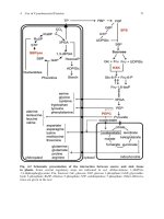
As we shall explain in more detail later, iron is particularly indicated for catalysis
of reactions which necessitate a free radical mechanism. One such reaction, of
primordial importance forDNA replication and cell division, is the transformationof
ribose to deoxyribose, catalysed by ribonucleotide reductases. The best characterized
enzyme, from E. coli, has a binuclear iron centre and a stable tyrosyl free radical
as cofactor. However, as we will see in Chapter 2, iron can also catalyse reactions
with molecular oxygen and its reduced forms superoxide and hydrogen peroxide,
leading ultimately to production of the highly reactive hydroxyl radical. This is the
so-called oxygen paradox, whereby oxygen is absolutely essential for aerobic life,
yet under certain conditions, is also toxic. The capacity of iron to catalyse Fenton
chemistry, with concomitant production of hydroxyl radicals, means that iron is
intimately involved in a catalogue of diseases involving oxidative stress – indeed,
as has been pointed out, in the former Soviet Union, cancer and atherosclerosis
are now referred to as the ‘rusting diseases’ (Parke, 1992). This link between
iron and oxidative damage is dealt with in Chapter 10 by our long-standing (and
long-suffering) collaborator Roberta Ward.
Iron is ne cessary for the multitude of bacteria, fungi and protozoa that causes
infections in vertebrate hosts, and its importance in this respect is underlined
by the observations that in all such infections, iron withholding is a classic host
response – plasma iron is rapidly decreased, whilst conditions of reticuloendothelial
iron overload greatly increases the severity of infections. Very often the strains of
microorganisms that are responsible for the most v irulent infections have a specific,
and often plasmid-borne, iron uptake mechanism. A good example is the ColV
plasmid associated with virulence in E.coli strains (Williams, 1979) that codes for
the aerobactin iron-transport system. Epidemiological evidence supports the role
of aerobactin in bacterial virulence in meningitis, septicaemia, and epidemics of
Salmonella poisoning. Siderophore-mediated iron uptake systems are also known
as virulence factors in fish pathogens (Vibrio anguilarum –anguibactin), and in
Pseudomonas species – the fluorescent pyoverdins and pyochelins (Crosa, 1989),
while exogenous siderophores supplied to hosts infected with Salmonella, Vibrio
and Yersinia can severely enhance their virulence (Weinberg, 1989). Indeed certain
pathogenic bacteria do not use siderophores at all, but use iron sources that
occur in their host – haem, both alone or bound to haemopexin, haemoglobin, both
alone and bound to haptoglobin, while Neisseria and Haemophilis influenzae also use
iron bound to transferrin and lactoferrin ( see Chapter 3). Johan Boelaert, a Brugge-
based clinician who has for many years charted the important relationship of iron
and infection, deals with this in Chapter 11, and his emphasis on the potential
Preface xxi
of iron depletion to combat infection is reflected in his organization of important
international meetings on iron and HIV over the last few years.
Finally, we can cite the powerful fascination for human civilization of Mars, the
Roman god of war and the old chemists name for iron, and Venus, the Roman goddess
of love and beauty, and the alchemists, name for copper (Crichton and Pierre, 2001).
During prebiotic times, water-soluble ferrous iron was present and was the form
used in the first stages of life. The natural abundance and the redox properties of
iron allowed the chemistry that was suited to life. At the same time, copper was
in the water-insoluble Cu(I) form, as highly insoluble sulfides, and was not avail-
able for life. About 10
9
years ago, evolution of dioxygen i nto the earth’s atmosphere
began to develop. Iron was oxidized and transformed into the insoluble Fe(III)state,
while at the same time the oxidation of insoluble Cu(I) led to soluble Cu(II). As iron
biochemistryevolved andchanged, sotoo anew roleof copper evolved with enzymes
of higher redox potentials taking advantage of the oxidizing power of dioxygen.
In Roman mythology, allusions not only to interactions but even to connivances
between Mars and Venus, leading even to seduction, abound (the superb represen-
tations of the love of Mars and Venus in Pompeii, for e xample). As we will see in
our final chapter, t he metabolism of iron and copper are intimately interconnected.
Though little remains from the Iron Age, compared with the to Bronze Age (the alloy
of copper and tin), or gold and silver, iron always has the last word:
‘Gold is for the mistress – silver for the maid –
Copper for the craftsman cunning at his trade.’
‘Good !’ said the Baron, sitting in his hall,
‘But Iron – Cold Iron – is master of them all.’
Rudyard Kipling ‘Cold Iron’ (1902)
These few examples give only the mariner’s view of the iceberg concerning the
fundamental importance of iron in biological systems. There remains a great deal
more that we have not seen in this titillation of the reader’s palate, but we shall try to
make amends in what follows. Of course, rules always must have their exceptions,
and the Lactobacillae quoted in an earlier paragraph are no doubt that. They have
evolved in ecological niches where the use of a metal other than iron for key
metabolic roles was an evolutionary plus. For the vast majority of living organisms,
iron is absolutely necessary for the maintenance, the defence, the differentiation
and last, but by no means least, the growth and cellular division of almost all living
organisms.
That is why I have devoted this book to the inorganic biochemistry of iron.
References
Behrenfeld, M.J., Bale, A.J., Kolber, Z.S., Aiken, J. and Falkowski, P.G. (1996). Nature, 383,
508–10.
Boyd, P.W., Watson, A.J., Law, C.S., Abraham, E.R. et al. (2000). Nature, 407, 695–702.
Braun, V., Crichton, R.R. and Braunitzer, G. (1968). Hoppe Zeyler’s Z. Physiol. Chem., 349,
197–210.
xxii Preface
Butler, A. (1999). Science, 281, 207–10.
Coale, K.H., Johnson, K.S., Fitzwater, S.E., Gordon, R.M. et al. (1996). Nature, 383, 495–501.
Crichton, R.R. (1991). Inorganic Biochemistry of Iron Metabolism, Ellis Horwood, Chichester,
263 pp.
Crichton, R.R. and Pierre, J L. (2001). Biometals, in press.
Crosa, J.H. (1989). Microbiol. Rev., 53, 517–30.
Feder, J.N., Gnirke, A., Thomas, W., Tsuchihashi, Z. et al. (1996). Nat. Genet., 13, 399–408.
Frausto da Silvo, J.J.R. and Williams, R.J.P. (1991). The Biological Chemistry of the Elements,
Clarendon Press, Oxford, 561 pp.
Martin, J.H. and Fitzwater, S.E. (1989). Nature, 331, 341–3.
Martin, J.H., Coale K.H., Johnson, K.S., Fitzwater, S.E. et al. (1994). Nature, 371, 123–9.
Mason, B. and Moore, C.B. (1982). Principles of Geochemistry, Fourth Edition, Wiley, New
York.
Neilands, J.B. (1982). Ann. Rev. Microbiol., 36, 285–309.
Orchiai, E.I. (1986). J. Chem. Educ., 63, 942–4.
Parke, D.V. (1992). The Biochemist, 15, 48.
Santos, M., Schilham, M.W., Rademake rs, L.H.M.P., Marx, J.J.M., de Sousa, M. and Clevers,
H. (1996). J. Exp. Med., 184, 1975–85.
Weinberg, E.D. (1989). Quart. Rev. Biol., 64, 261–90.
Williams, P.H. (1979). Infect. Immunol., 26, 925–32.
Inorganic Biochemistry of Iron Metabolism: From Molecular Mechanisms to
Clinical Consequences. 2e
Robert Crichton
Copyright 2001 John Wiley & Sons Ltd
ISBNs: 0-471-49223-X (Hardback); 0-470-84579-1 (Electronic)
The following are the list of pages where ‘Plate X’ appears.
Chapter2–33(2occurances),42(2occurances).
Chapter 3 – 54, 58, 72.
Chapter 5 – 111 (2 occurances), 112 (2 occurances), 117, 118 (2 occurances), 120.
Chapter 6 – 146, 147, 148 (2 occurances), 149, 154.
Chapter 7 – 170, 185.
Chapter 9 – 217, 220.
Chapter 10 – 250.
Chapter 12 – 290.
Plate 1
Figure 2.7M(a) A front view of the nitrite reductase dimer with the five haems in each monomer
in white, a bound Ca
2+
ion in grey and Lys-133, which coordinates the active site iron of haem 1,
in yellow. (b) The haem arrangement. The overall orientation corresponds to (a), with the active
site located at haem 1. Reprinted with permission from Einsle et al., 1999. Copyright (1999),
Macmillan Magazines Limited.
(a)
(b)
Plate 2
Figure 2.8MThe structure of the dimeric cytochrome bc
1
complex of the respiratory chain. (a) The
cave for chemistry constituted by the hollow between the two monomers (the ‘essential dimer’)
in a cartoon representation. Reprinted with permission from Smith, 1998. Copyright (1998),
American Association for the Advancement of Science. (b) The structure viewed perpendicular to
the twofold axis and parallel to the membrane. All of the eleven subunits are completely traced
and their sequences assigned. The top of the molecule extends 3.8inm into the intermembrane
space, the middle spans the membrane (4.2inm), and the bottom extends some 7.5inm into the
matrix. Reprinted with permission from Iwata et al., 1998. Copyright (1998) American Association
for the Advancement of Science.
?
Heme b
L
Cyt b
Heme b
H
Q
P
Q
N
Heme
c
1
?
Cyt c
1
Subunit 8
Fe
2
S
2
Core 1
Core 2
Subunits 6, 9
Rieske
protein
subunits
7,10, 11
Matrix
Milochondrial
inner
membrane
Inter-
membrane
space
–
+
(a)
(b)
Plate 3
Figure 2.12M(a) Model of the human phenylalanine tetramer, created by combining the structure
of the regulatory/catalytic domain crystal structure and the catalytic/tetramer domain crystal
structure. The monomeric form of the enzyme is presented in the left panel, the tetramer viewed
down the tetramerization domain in the right panel. (b) The metal-binding domains of
phenylalanine hydroxylase and tyrosine hydroxylase are shown on the left and right respectively.
(a) and (b) reprinted with permission from Flatmark and Stevens, 1999. Copyright (1999),
American Chemical Society.
(a)
(b)
Figure 3.3 MComparison of the FepA and FhuA crystal structures. A portion of the ß-barrel (in
violet is removed to show the globular cork domain (in yellow) that inserts from the periplasm
into the channel of the ß-barrel. FhuA is loaded with ferrichrome (iron is shown as a green ball)
(Ferguson et al., 1998; Locher et al., 1998). The FepA crystal structure does not reveal
Fe
3+
–enterobactin, but the FepA structure shown might be partially occupied by enterobactin
(Buchanan et al., 1999).
Figure 3.5MStructures of FhuA ligands as determined by X-ray analysis of co-crystals with FhuA.
Albomycin adopts an extended and a compact conformation in the FhuA crystal, and rifamycin
CGP 4832 binds to the same FhuA site as ferrichrome and albomycin although it assumes a
different conformation.
Plate 4