lung cancer, volume 1
Bạn đang xem bản rút gọn của tài liệu. Xem và tải ngay bản đầy đủ của tài liệu tại đây (11.05 MB, 531 trang )
Humana Press
Humana Press
M E T H O D S I N M O L E C U L A R M E D I C I N E
TM
Lung Cancer
Edited by
Barbara Driscoll
Volume I
Molecular Pathology
Methods and Reviews
Lung Cancer
Edited by
Barbara Driscoll
Volume I
Molecular Pathology
Methods and Reviews
Genetic Alterations 3
3
From:
Methods in Molecular Medicine, vol. 74: Lung Cancer, Vol. 1: Molecular Pathology Methods and Reviews
Edited by: B. Driscoll © Humana Press Inc., Totowa, NJ
1
Characteristic Genetic Alterations in Lung Cancer
Ignacio I. Wistuba and Adi F. Gazdar
1. Introduction
Lung cancer is the most frequent cause of cancer deaths in both men and
women in the U.S. (1). Although tobacco smoking is accepted as the number
one cause of this devastating disease, our understanding of the acquired genetic
changes leading to lung cancer is still rudimentary. Lung cancer is classifi ed
into two major clinic-pathological groups, small cell lung carcinoma (SCLC)
and non-small cell lung carcinoma (NSCLC) (2). Squamous cell carcinoma,
adenocarcinoma, and large cell carcinoma are the major histologic types of
NSCLC. As with other epithelial malignancies, lung cancers are believed to
arise after a series of progressive pathological changes (preneoplastic lesions)
(3). Many of these preneoplastic changes are frequently detected accompany-
ing lung cancers and in the respiratory mucosa of smokers (3). Although
many molecular abnormalities have been described in clinically evident lung
cancers (4), relatively little is known about the molecular events preceding the
development of lung carcinomas and the underlying genetic basis of tobacco-
related lung carcinogenesis.
To investigate the molecular abnormalities involved in the multistep patho-
genesis of lung carcinomas, we have developed a fi ve-step analysis scheme that
included the study of: 1) lung cancer cell lines; 2) microdissected primary lung
tumors of the three major histologic types (SCLC, squamous cell carcinoma,
and adenocarcinoma); and normal and abnormal respiratory epithelium from
3) lung cancer patients; 4) from smoker subjects without lung cancer; and from
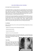
5) never smoker subjects (see Fig. 1). Under this strategy we systematically
search for mutations in tumor cell-lines specimens, and in archival tumor
tissues, preneoplastic lesions, and normal epithelium, using paraffi n-embedded
CH01,1-28,28pgs 07/22/02, 7:28 AM3
4 Wistuba and Gazdar
materials. Recently, we have also analyzed genetic changes present in cytologic
specimens bronchial brushes from smokers (5). In tissues samples, using a
precise microdissection technique, under direct microscopic observation a
variable number of cells from those areas are precisely isolated along with
invasive primary tumor and stromal lymphocytes (as a source of normal
constitutional DNA). Using polymerase chain reaction (PCR)-based techniques,
these different specimens are examined for molecular abnormalities (mainly
gene mutations and allele losses) at chromosomal regions frequently mutated
or deleted in clinically evident lung carcinomas.
The risk population for targeting lung cancer early detection efforts has
been defi ned (current and heavy smokers, and patients who have survived one
cancer of the upper aerodigestive tract). However, conventional morphologic
methods for the identifi cation of premalignant cell populations in the airways
have limitations. This has led to a search for other biological properties
(including genetic changes) of respiratory mucosa that may provide new
methods for assessing the risk of developing invasive lung cancer in smokers,
for early detection, and for monitoring their response to chemopreventive
regimens.
Fig. 1. Schema showing the strategy developed to study the molecular abnormalities
involved in the pathogenesis of lung cancer.
CH01,1-28,28pgs 07/22/02, 7:28 AM4
Genetic Alterations 5
2. Tumor-Cell and Tissue-Specimens Methodologies Used
in the Analysis of Lung Cancer Molecular Abnormalities
To investigate the molecular abnormalities involved in the pathogenesis
of lung cancers, we have utilized a panel of paired lung tumor cell lines and
corresponding normal lymphoblastoid cells (6), as well as microdissection
technique of archival paraffi n-embedded tumor and nonmalignant epithelial
tissues (7–9) (see Fig. 2). Both methodologies have played a pivotal role in the
study of the molecular abnormalities of the pathogenesis of lung cancer.
2.1. Paired Lung Tumor and Normal Lymphoblastoid Cell Lines
Despite the pivotal role played by human lung cancer cell lines in biomedical
research, there is a widespread belief in the scientifi c community that they are
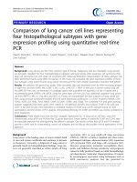
Fig. 2. Comparison of phenotypic properties between primary lung cancer tissues
and their corresponding cancer cell lines. Upper panels, tumor tissue and corresponding
cell line showing squamous cell differentiation with keratinization features. Lower
panel, tumor tissue and corresponding cell line showing adenocarcinoma features with
gland-like structures formation and p53 nuclear immunostaining.
CH01,1-28,28pgs 07/22/02, 7:28 AM5
6 Wistuba and Gazdar
not representative of the tumors from which they were derived. Lung cancer
cell lines have demonstrated advanced molecular changes, including extensive
chromosomal rearrangements, oncogene mutations, and multiple sites of allelic
loss and gene amplifi cation (10,11). Thus, many investigators presume that
loss of phenotypic properties and additional molecular changes develop during
the prolonged time required for cell-culture establishment and subsequent
passage.
To investigate this phenomenon we compared the morphologic, phenotypic,
and genetic changes in lung cancer cell lines and in their corresponding tumor
tissues (12). We compared the properties of a series of 12 human NSCLC
cell lines (cultured for a median period of 39 mo, range 12–69) and their
corresponding archival tumor tissues. Other than differences in the degree of
aneuploidy, the other properties studied demonstrated a remarkable degree
of concordance between lung tumors and their corresponding cancer cell
lines (see Table 1). These features included morphologic characteristics (see
Fig. 2), presence of aneuploidy, immunohistochemical expression profi le
for HER2/neu and p53 proteins, and a similar K-RAS and TP53 gene muta-
tions allelic loss and MA pattern for multiple loci frequently deleted in
lung carcinoma. The concordance between tumors and cell lines for all of
the comparisons was independent of the time on culture, indicating that the
properties of cell lines usually closely resemble those of their parental tumors
for culture periods up to 69 mo.
While p53 immunohistochemical protein expression was detected in all
of the lung tumor cell lines and their corresponding tumor tissues (100%
correlation). TP53 gene mutations in exons 5–8 were detected in 10 (83%) of
12 lung tumor cell lines, and six of those corresponding tumor tissues exhibited
the identical TP53 gene mutation. K-RAS gene mutations at codon 12 were
detected in two adenocarcinomas cell lines (17% of the NSCLCs and 33% of
adenocarcinoma cases), and identical K-RAS mutations were identifi ed in their
corresponding tumors. We also determined chromosomal deletions expressed
by loss of heterozygosity (LOH) at 13 chromosomal regions frequently deleted
in lung cancers. Nearly identical high LOH frequencies at all chromosomal
regions analyzed were detected between tumors and theirs corresponding cell
lines (see Table 1). For all of the individual markers there was an excellent
correlation between tumors and cell lines (mean concordance of 89%). In all
of the 115 (100%) comparisons, when allelic loss of a particular microsatellite
was present in both the tumor and corresponding cell line, the identical parental
allele was lost in both, confi rming that the allelic loss originated in the original
tumor tissue. In addition, tumor cell did not develop greater frequency of
genomic instability phenomenon in culture and they retain some of the unstable
properties of their parental tumors after lengthy culture periods (up to 69 mo).
CH01,1-28,28pgs 07/22/02, 7:28 AM6
Genetic Alterations 7
Our fi ndings also indicated that successfully cultured NSCLCs represent the
general population of tumors and their cell lines are useful models for studying
this important type of lung neoplasm.
2.2. Tissue Microdissection Technique
The molecular examination of pathologically altered cells and tissues at the
DNA, RNA, and protein level has revolutionized research and diagnostics in
tumor pathogenesis. However, the inherent heterogeneity of primary tissues
with an admixture of various reactive cell populations can affect the out-
come and interpretation of molecular studies. Recently, microdissection of
tissue sections and cytological preparations has been used increasingly for the
Table 1
Comparison of Properties Between 12 Lung Cancer Tumor Tissues
and Their Corresponding Cancer Cell Lines
Frequency
Feature Tumor tissue Cell lines
Aneuploidy 100% 100%
Protein immunohistochemical expression
HER2/neu 25% 25%
p53 protein 100% 100%
Chromosomal region with LOH
3p25 38% 38%
3p22–24 55% 55%
3p21 58% 58%
3p14–21 22% 22%
3p14.2 (FHIT gene) 50% 50%
3p12 25% 25%
Any 3p 67% 67%
5q22 (APC-MCC region) 44% 44%
8p23 91% 91%
8p22 91% 91%
8p21 58% 75%
Any 8p 100% 100%
9p21 78% 89%
13q (RB gene) 33% 33%
17p (TP53 gene) 78% 89%
Microsatellite Alteration (MA) 54% 58%
Gene Mutations
TP53 gene (Exons 5–8) 58% 83%
K-RAS gene (Codons 12–13) 17% 17%
CH01,1-28,28pgs 07/22/02, 7:28 AM7
8 Wistuba and Gazdar
isolation of homogeneous, morphologically identifi ed cell populations, thus
overcoming the obstacle of tissue complexity. In conjunction with sensitive
analytical techniques, such as the PCR, microdissection allows precise in
vitro examination of cell populations, such as normal epithelial or dysplastic
cells, which are otherwise inaccessible for conventional molecular studies (see
Fig. 3). However, most of manual microdissection techniques are time-
consuming and require a high degree of manual dexterity, which limits
their practical use. Microdissection under microscopic visualization using
micromanipulator is very precise, but very time-consuming. Laser capture
microdissection (LCM), a novel technique developed at the National Cancer
Institute, is an important advance in terms of speed, ease of use, and versatility
of microdissection (13). LCM is based on the adherence of visually selected
cells to a thermoplastic membrane, which overlies the dehydrated tissue section
and is focally melted by triggering of a low-energy infrared laser pulse. The
melted membrane forms a composite with the selected tissue area, which
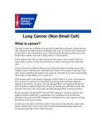
Fig. 3. Representative example of precise tissue microdissection technique of
bronchial epithelium (a and b) and adenocarcinoma of the lung (c and d) (before and
after microdissection). Note that only tumor and epithelial cells were microdissected
without contamination with stromal cells.
CH01,1-28,28pgs 07/22/02, 7:28 AM8
Genetic Alterations 9
can be removed by simple lifting of the membrane. LCM can be applied to a
wide range of cell and tissue preparations including paraffi n wax-embedded
material. The use of immunohistochemical stains allows the selection of cells
according to phenotypic and functional characteristics. Depending on the
starting material, DNA, good-quality mRNA, and proteins can be extracted
successfully from captured tissue fragments, down to the single-cell level. In
combination with techniques like expression library construction, cDNA array
hybridization, and differential display (14–16), the use of this microdissection
technique has allowed to us to analyze minute amount of lung tissues and
perform most of our studies on the genetic changes involved in lung cancer
pathogenesis (7–9,12,17–25).
3. Overview of Molecular Abnormalities in Lung Cancer
Several cytogenetic, allelotyping, and comparative genomic hybridization
(CGH) studies have revealed that multiple genetic changes (estimated to be
between 10 and 20) are found in clinically evident lung cancers, and involve
known and putative recessive oncogenes as well as several dominant oncogenes
(4). The major molecular changes detected in lung cancers are summarized
in Table 1.
3.1. Growth Stimulation and Oncogenes
Many growth factors or regulatory peptides and their receptors are expressed
by cancer cells or adjacent normal cells in the lung, and thus provide a series of
autocrine and paracrine growth stimulatory loops in this neoplasm (26). Several but
not all components of these stimulatory pathways are proto-oncogene products.
3.2. Gastrin-Releasing Peptide (GRP)/Bombesin (BN)
Autocrine Loop
There is good evidence that the GRP/BN and GRP receptor autocrine loop
is involved in the growth of lung cancer, particularly SCLC (26). Immuno-
histochemical studies demonstrate that most SCLCs express the ligand por-
tion of the autocrine loop GRP/BN, whereas NSCLC express GRP/BN less
frequently (27).
3.3. Tyrosine Kinases
Neuregulins and their receptors, the ERBB family of transmembrane recep-
tor tyrosine kinases (ERBB1 and ERBB2), constitute a potential growth
stimulatory loops in lung cancer (27). However, NSCLCs but not SCLCs often
demonstrate abnormalities of ERBB gene family. The KIT proto-oncogene,
which encodes yet another tyrosine kinase receptor, CD117, and its ligand,
stem cell factor (SCF), are co-expressed in many SCLC (28). Other putative
CH01,1-28,28pgs 07/22/02, 7:28 AM9
10 Wistuba and Gazdar
loops involve insulin-like growth factor 1 (IGF-1), insulin-like growth factor
2 (IGF-2), and the type I insulin-like growth factor receptor (IGF-1R), which
are frequently co-expressed in SCLC, as well as platelet-derived growth factor
(PDGF) and its receptor (4).
3.4.
MYC
Family
MYC family genes are frequently altered in SCLC and include MYC, MYCN,
and MYCL, all of which can be involved in SCLC pathogenesis. Of the well-
characterized MYC genes, MYC is most frequently activated in both SCLC and
NSCLC, whereas abnormalities of MYCN and MYCL usually only affect SCLC.
In nearly all cases, only one MYC family member is activated in each individual
tumor. Activation of the MYC genes may occur via gene amplifi cation (20–115
copies per cell) or via transcriptional dysregulation, both of which lead to
protein overexpression (29). Amplifi cation or overexpression of MYC family
members has been reported more frequently in SCLCs (18–31%) than NSCLCs
(8–20%) (27).
3.5.
BCL-2
, BAX, and Apoptosis
There is accumulating evidence that tumor cells acquire the ability to escape
pathways leading cells to undergo programmed cell death (apoptosis) when
exposed to conditions such as growth factor deprivation or DNA damage. Key
members of the normal apoptotic pathways are the BCL-2 proto-oncogene
product and the TP53 TSG product. BCL-2 protects cells from the apoptotic
process and thus probably plays a role in determining the chemotherapy
response of cancer cells. Whereas BCL-2 protein immunohistochemical
expression (and thus upregulation) is present in most SCLCs (75–95%),
BCL-2 immunostaining is far less frequent in NSCLC (10–35%) (4). BAX,
which is a BCL-2 related protein that promotes apoptosis, is a downstream
transcriptional target of p53. BAX and BCL-2 immunostaining are inversely
related in neuroendocrine lung cancers, with most SCLCs having high BCL-2
and low BAX expression (30).
Recent CGH studies have shown that lung cancer cell lines and tumor
tissues demonstrate increased copy number consistent with amplifi cation of
underlying dominant oncogenes at several chromosomal regions, including 1p,
1q, 2p, 3q, 5q, 11q, 16p, 17q, 19q, and Xq (31). Some of these regions, such as
1p32 (L-MYC), 2p25 (N-MYC), and 8q24 (C-MYC) contain known dominant
oncogenes, while in others the genes need to be identifi ed.
3.6. Recessive Oncogenes
The list of recessive oncogenes that are involved in lung cancer is likely to
include as many 10–15 known and putative genes (4). These include changes
CH01,1-28,28pgs 07/22/02, 7:28 AM10
Genetic Alterations 11
in TP53 (17p13), RB (13q14), p16
6ink4
(9p21), and new candidate recessive
oncogenes in the short arms of chromosome 3 (3p) at 3p12 (DUTT1 gene),
3p14.2 (FHIT gene), 3p21 (RASFF1 gene), 3p22-24 (BAP-1 gene), and
3p25 regions (4). Recessive oncogenes are believed to be inactivated via a
two-step process involving both alleles. Knudson has proposed that the fi rst
“hit” frequently is a point mutation, while the second allele is subsequently
inactivated via a chromosomal deletion, translocation, or other event such as
methylation (32).
3.6.1. TP53 Gene
Loss of p53 function allows inappropriate survival of genetically damaged
cells, setting the stage for the accumulation of multiple mutations and the
subsequent evolution of a cancer cell (33). Missense TP53 mutations prolong
the protein’s half-life, leading to accumulation of high levels of mutant p53
protein readily detected by immunohistochemistry. Multiple studies have
shown abnormal p53 protein expression by immunohistochemistry in 40–70%
of lung cancer (4). TP53 abnormalities play a critical role in lung cancer
pathogenesis (4). Chromosome 17p13 sequences, the site of the TP53 locus,
are frequently hemizygously lost in SCLC (90%) and NSCLC (65%) (9),
and mutational inactivation of the remaining allele occurs in 50–75% of
these neoplasms (34). TP53 mutations in lung tumors correlate with cigarette
smoking and are mostly the G-T transversions expected of tobacco-smoke car-
cinogens (33). Furthermore, in lung cancers a relationship has been described
between mutational hot-spots at the TP53 gene and adduct hot-spots caused by
benzo[α]pyrene metabolites of cigarette smoke (35).
3.6.2. The p16-Cyclin D1-CDK4-Retinoblastoma Pathway
In SCLC, this pathway is usually disrupted by retinoblastoma gene (RB)
inactivation, while cyclin D1, CDK4, and p16 abnormalities are rare in SCLC
but common (particularly p16) in NSCLC (4). The major growth-suppressing
function of RB protein is to block G1-S progression. Inactivation of both RB
alleles at chromosomal region 13q14 is common in SCLC (36), with protein
abnormalities reported at frequencies of over 90% (37). There is frequent loss
of one the RB 13q14 alleles. Functional loss of the remaining RB allele can
include deletion, nonsense mutations, or splicing abnormalities, leading to a
truncated RB protein encoded by the remaining allele.
3.6.3. PTEN/MMAC1
A new TSG, PTEN (Phosphatase and Tensin homolog deleted on chromo-
some 10), also called MMAC1 (Mutated in Multiple Advanced Cancers),
has been identifi ed and localized to chromosome region 10q23.3 (38). Allelo-
CH01,1-28,28pgs 07/22/02, 7:28 AM11
12 Wistuba and Gazdar
typing analysis utilizing microsatellite markers in close proximity to the
PTEN/MMAC1 gene have demonstrated high incidence of LOH in lung
cancers, especially SCLCs (91% LOH in SCLC, and 41% in NSCLC) (39).
Homozygous deletions interrupting the PTEN/MMAC1 gene have been detected
in 8% of SCLC cell lines examined and in a few uncultured primary SCLCs
(40). However PTEN/MMAC1 mutations were detected in only 11% of lung
cancers, including both SCLC and NSCLC tumors (40).
3.6.4. Other Candidate TSGs
TSG101 is a recently discovered candidate TSG that maps to 11p15 (41). It
has been reported that the mutant TSG101 transcript was expressed simultane-
ously with wild-type TSG101 transcript in almost all SCLC cell lines. In
contrast, normal lung tissue, as well primary NSCLC specimens, express only
a wild-type transcript. DMBT1 is a candidate TSG located at 10q25.3-26.1
(42). Recent data demonstrate that DMBT1 gene expression is frequently lost
in both SCLC and NSCLC (43), suggesting that inactivation of DMBT1 may
play an important role in lung tumorigenesis.
3.6.5. Candidate TSGs at Chromosome Region 3p
The very frequent loss of alleles on chromosome 3p in both SCLC (>90%)
and NSCLC (>80%) (24) provides strong evidence for the existence of one or
more TSGs on this chromosomal arm. Several distinct 3p regions have been
identifi ed by high-density allelotyping including 3p25-26, 3p24, 3p21.3-22
(several sites), 3p14.2 (FHIT), and 3p12 (U2020 deletion site) suggesting that
there are several different TSGs located on 3p. The 3p21.3 region has been
extensively examined for putative TSGs, although the identity of such gene(s)
remains elusive (44). Currently, two distinct 3p21.3 regions are under study
because of the existence of multiple homozygous deletion in lung cancer cell
lines. Although several genes identifi ed so far in both homozygous deletions
none of them have been shown to have frequent mutations in lung cancer (44).
Recently, one of the splicing isoforms of the RASSF1 gene (RASSF1A) located
in the 370 kb deletion region has been shown to undergo tumor promoter
hypermethylation as a mechanism of inactivation (>90% of SCLCs and 60%
of NSCLC) (45). This gene when re-expressed in lung cancer cells suppresses
the malignant phenotype (45,46).
The FHIT gene maps to 3p14.2 and encompasses approx 1 Mb of genomic
DNA, which includes the human common fragile site (FRA3B). FHIT is a
candidate TSG for lung cancer on the basis of frequent 3p14.2 allele loss in
lung cancer (100% of SCLCs and 88% of NSCLCs) and homozygous deletion
in several lung cancer cell lines (47,48). Lung cancer cells (40–80%) express
CH01,1-28,28pgs 07/22/02, 7:28 AM12
Genetic Alterations 13
abnormal mRNA transcripts of FHIT but nearly always also express very
low levels of wild-type FHIT transcripts (47,48). However, unlike classic
TSG inactivation, FHIT point mutations are rare (47,48), and a few abnormal
transcripts can be found in normal lung tissue. Of importance, most lung
cancers, expressed undetectable or very low levels of FHIT mRNA, and
exhibited loss of Fhit protein expression by immunohistochemistry. Recently,
it has been demonstrated that hypermethylation of the promoter region of the
FHIT gene is a frequent event in lung cancer cell lines (SCLC 64% and NSCLC
64%) and noncultured NSCLC primary tumors (37%) (5).
There are other candidate TSGs of lung cancer on chromosome 3p. The Von
Hippel-Lindau (VHL) TSG at 3p25 and the BRCA1-associated protein, BAP-1,
at 3p21. However, these genes have been infrequently mutated in lung cancer,
including SCLC (4). Recently, a new candidate TSG, DUTT1, has been cloned
residing in the U2020 3p12 deletion region and crossing a small (>100 KB) lung
cancer homozygous deletion at 3p12 (49). However, DUTT1 tumor-suppressing
activity and protein expression patterns in tumors are unknown.
3.6.6. Other Candidate Lung Cancer Tumor Suppressor Genes Loci
Besides the candidate and known TSGs mentioned earlier, cytogenetic
and allelotyping studies have shown allelic loss of many other chromosomal
regions, in both SCLC and NSCLC, suggesting the involvement of other
tumor-suppressor genes in its pathogenesis. The chromosomal regions include
1q, 2q, 5q, 6p, 6q, 8p, 8q, 10q, 11p, 14q, 17q, 18q, and 22q (11,39). These
novel sites will direct the search for new candidate TSGs. Future investigations
with an even higher resolution of microsatellite markers will be crucial to
narrow down the sites of frequent allelic loss. In addition, the presence of
homozygously deleted chromosomal regions 2q33, 5p13-q14, 8, and X/Y
in lung cancer provide further evidence that these regions harbor as yet
unidentifi ed TSGs (4).
3.7. Genetic Instability in Lung Cancer
In addition to the specifi c genetic changes discussed earlier, other evidence
indicates that genomic instability occurs in lung cancer. This evidence includes
changes in the number of short-tandem DNA repeats (also known as micro-
satellite markers), frequently present in a wide variety of cancer types,
including SCLC. Microsatellite instability, was initially reported in hereditary
nonpolyposis colorectal cancer, resulting from inherited defects in DNA
mismatch-repair enzymes, which induce large-scale genetic instability with
the formation of a ladder-like pattern replacing the normal allele pattern.
This type has not been seen in lung cancer. Another form of microsatellite
CH01,1-28,28pgs 07/22/02, 7:28 AM13
14 Wistuba and Gazdar
change, where only a single band of altered size is found, has been described
in many forms of sporadic cancers, including SCLC and NSCLC, referred to
as microsatellite alteration (MA). While the relationship of MAs to the DNA
repair mechanism has not been established, the former probably represents
evidence of some form of genomic instability (50). Multiple studies have
reported MAs in lung cancers. Overall, 35% (range 0–76%) of SCLCs and 22%
(range 2–49%) of NSCLCs have shown some evidence of MA at individual loci
(4). Although the mechanisms are unknown, a signifi cantly higher frequency
of MAs have been detected in lung tumors arising in HIV-positive individuals
(20) and in patients with secondary lung tumors after treatment for Hodgkin’s
disease (25), compared to lung cancers in the general population.
3.8. Aberrant Methylation in Lung Cancer
TSGs need to inactivate both alleles to exert their tumor-promoting effects.
One method of gene silencing is via the epigenetic phenomenon of aberrant
methylation of gene promoters. DNA methylation only occurs at CpG sites
(known as “CpG islands”). In the human genome CpG sites are usually
concentrated in the promoter regions of about half of all human genes, and
normally these islands are completely unmethylated (51). During carcinogen-
esis, the promoter regions of several genes are methylated, resulting in gene
silencing. It is estimated that the average number of CpG islands methylated in
individual human tumors may be as high as 600 (range 0–4500) (52).
As predicted, several genes are methylated in lung cancers, and the list is
increasing rapidly. Esteller et al. (53), detected promoter hypermethylation of
at least one of four genes examined (p16
6ink4
, DAP kinase, GSTP1, and MGMT)
in 15 of 22 (68%) NSCLC tumors but not in any paired normal lung tissue.
Interestingly, 11 of 15 (73%) matched serum samples obtained from primary
tumors with aberrant methylation also had abnormal methylated DNA. None of
the sera from patients with tumors not demonstrating methylation was positive.
These fi ndings suggest that detection of aberrant promoter hypermethylation
of cancer-related genes in serum may be useful for cancer diagnosis or the
detection of recurrence. Recently, Tang et al. (54) reported that patients with
pathologic stage I NSCLC whose tumors exhibited DAP kinase gene promoter
hypermethylation (44%) had a statistically signifi cantly poorer probability of
overall 5-yr survival after surgery than those without such hypermethylation.
These fi nding suggest that abnormal promoter-gene methylation may be useful
as prognostic marker in lung cancer patients.
In lung cancer, regional hypermethylation has been found at chromosome 3p,
but the precise gene target(s) until recently have been uncertain. Recently, three
reports have shown in lung cancer frequent methylation of promoter sequences
of three genes located at chromosome 3p regions, which are frequently deleted
CH01,1-28,28pgs 07/22/02, 7:28 AM14
Genetic Alterations 15
in this neoplasm (46,55). Dammann et al. (46) and Burbee et al. (45) described
a human RAS effector homologue (RASSF1) gene located in a small 120-kb
region of minimal homozygous deletion in 3p21.3, with frequent (>90%
of SCLC and ~40% of NSCLCs) methylation of its CpG-island promoter
sequence, which correlates with loss of gene expression. Virmani et al. (55)
reported a high frequency of methylation of the RARβ gene in lung cancers,
particularly SCLC (72%) compared to NSCLC (41%). In addition, high
frequencies of methylation of lung cancers has been recently detected in FHIT
gene (3p14.2), in both SCLC and NSCLC cell lines (64%) and NSCLC primary
tumors (37%) (5).
Recently, it has been shown a relatively high frequency of other genes
methylation (TIMP-3 26%, p16
ink4
25%, MGMT 21%, DAPK 19%, ECAD 18%,
p14
ARF
8%, and GSTP1 7%) in a panel of 107 NSCLC primary tumors (56).
Frequent abnormal methylaton of the CDH13 (H-Cadherin) gene (16q24.2-3)
has been also demonstrated in lung cancer, particularly in NSCLC cell lines
(50%) and primary tumors (43%) (57). The number of genes showing a high
incidence of abnormal methylation in lung cancer is rapidly increasing. All
these recent fi ndings suggest that aberrant methylation of genes is a frequent
abnormality in lung cancers and may have applications for risk assessment,
diagnosis, and for development of novel therapeutic approaches.
4. Tumor Type-Specifi c Genetic Changes in Lung Cancers
Studies of large numbers of lung cancers have demonstrated different
patterns of involvement between the two major groups of lung carcinomas
(SCLC and NSCLC) (39) and between the three major histologic types of
lung carcinomas (SCLC, squamous cell carcinomas, and adenocarcinomas)
(9,22,24,58). The major differences found between SCLC and NSCLC are
summarized in Table 1. Our results (9,22,24,39,58) of allelotyping lung cancer
cell lines and microdissected invasive primary tumors indicate that SCLC
demonstrate more frequent losses at 4p, 4q, 5q21 (APC-MCC region), 10q,
and 13q14 (RB), while losses at 9p21 and 8p21-23 are more frequent in
NSCLCs. Recently, Girard et al. (11) performed a high-resolution genome-wide
allelotyping analysis of a similar panel of lung cancer (SCLC and NSCLC)
and detected 22 different “hot spots” for LOH, 13 with a preference for SCLC,
7 for NSCLC, and 2 affecting both. This provides clear evidence on a genome-
wide scale that SCLC and NSCLC different signifi cantly in the TSGs that
are inactivated during their pathogenesis. Similarly, the recent fi ndings on the
methylation pattern of a number of genes in lung cancer indicate that there are
differences in between SCLCs and NSCLCs (55).
In addition, we have found different patterns of allelic loss involving the
two major types of NSCLC (squamous cell and adenocarcinoma), with higher
CH01,1-28,28pgs 07/22/02, 7:28 AM15
16 Wistuba and Gazdar
incidences of deletions at 17p13 (TP53), 13q14 (RB), 9p21 (p16
6ink4
), 8p21-23,
and several 3p regions in squamous cell carcinomas. These results suggest
that more genetic changes accumulate during tumorigenesis in squamous cell
carcinomas than in adenocarcinomas. Several of those studies have identifi ed
different allele loss patterns between SCLC and NSCLC.
5. Preneoplasia and the Development of Lung Cancer
Lung cancers are believed to arise after a series of progressive pathological
changes (preneoplastic or precursor lesions) in the respiratory mucosa. While
the sequential preneoplastic changes have been defi ned for centrally arising
squamous carcinomas, they have been poorly documented for large-cell
carcinomas, adenocarcinomas, and SCLCs (3) (see Table 2). Mucosal changes
in the large airways that may precede or accompany invasive squamous
cell carcinoma include hyperplasia (basal cell hyperplasia and goblet cell
hyperplasia), squamous metaplasia, squamous dysplasia, and carcinoma in
situ (3). While hyperplasia and squamous metaplasia are considered reactive
and reversible changes, dysplasia and carcinoma in situ are the changes
most frequently associated with the development of squamous cell lung
carcinomas. Adenocarcinomas may be accompanied by changes including
atypical adenomatous hyperplasia (AAH) (3) in peripheral airway cells,
although the malignant potential of these lesions has not been demonstrated.
For SCLC, no specific preneoplastic changes have been described in the
respiratory epithelium.
Currently available information suggests that lung preneoplastic lesions
frequently are extensive and multifocal throughout the lung, indicating a fi eld
effect (“fi eld cancerization”) by which much of the respiratory epithelium has
been mutagenized, presumably from exposure to carcinogens.
6. Genetic Abnormalities in the Sequential Development
of Lung Cancer
Although our knowledge of the molecular events in invasive lung cancer
is relatively extensive, until recently we knew little about the sequence of
events in preneoplastic lesions. A few studies have provided suggestions that
molecular lesions can be identifi ed at early stages of the pathogenesis of
lung cancer. Myc upregulation, cyclin D1 expression, p53 immunostaining,
and DNA aneuploidy have been detected in dysplastic epithelium adjacent to
invasive lung carcinomas (59–61). K-RAS mutations have been also detected
in atypical adenomatous hyperplasia (62), which may be a potential precursor
lesion of adenocarcinoma. TP53 gene abnormalities (including mutations,
deletions, and overexpression) have been demonstrated in nonmalignant
epithelium of lung specimens resected for lung cancer (63). They also occur in
CH01,1-28,28pgs 07/22/02, 7:28 AM16
Genetic Alterations 17
Table 2
Major Differences in the Pathogenesis of SCLC and NSCLC
SCLC NSCLC
Frequency 20%–25% 80%–85%
Neuroendocrine cells Yes No
Putative autocrine loop GRP/GRP receptor HGM/MET
SCF/KIT NDF/ERBB
RAS mutations <1% 15%–20%
MYC amplifi cation 18%–31% 18%–20%
BCL-2 IHC 75%–95% 10%–35%
TP53 abnormalities
LOH 90% 65%
Mutation 75% ~50%
p53 IHQ 40%–70% 40%–60%
RB abnormalities
LOH 67% 31%
rb abnormalities (IHC) 90% 15%–30%
p16
6ink4
abnormalities
LOH 53% 66%
Mutation <1% 10%–40%
p16 IHC 0%–10% 30%–70%
PTEN/MMAC1 loci LOH 1191% 141%
TSG101 abnormal transcripts ~100% 110%
DMBT1 abnormal expression 1100% 143%
3p LOH various regions 1>90% >80%
4p LOH various regions 1150% ~20%
4q LOH various regions 1180% 130%
8p21-23 LOH 80%–90% 80%–100%
Other specifi c LOH regions 1q23, 9q22-32, 10p15, 13q34 13q11, Xq22.1
Microsatellite alterations 35% 22%
Promoter hypermethylation
RASSF1 gene >90% ~40%
RARβ gene 172% 141%
Other genes Not studied 10–40% various genes
*
Preneoplastic changes
Histopathology Unknown Relatively known
LOH multiple loci 90% 31%
MA frequency 68% 11%
GRP, Gastrin-releasing peptide; HFG, hepatocyte growth factor; MET, MET proto-oncogene;
SCF, stem cell factor; KIT, KIT proto-oncogene; NDF, neu differentiation factor; ERBB,
neuregulin receptor; LOH, loss of heterozygosity; IHC, immunohistochemistry; BCL-2, BCL-2
anti-apoptotic proto-oncogene.
*
p16, death-associated protein (DAP) kinase, glutathione S
transferase P1 (GSTP1), and O6-methylguanine-DNA methyltransferase (MGMT).
CH01,1-28,28pgs 07/22/02, 7:28 AM17
18 Wistuba and Gazdar
the histologically normal and abnormal epithelium of smokers (7,64). Recently,
Franklin et al. described an identical TP53 gene mutation widely dispersed in
normal and preneoplastic epithelium of a smoker without lung cancer (65).
Our recent studies allowed us to identify some of the genetic changes involved
in the pathogenesis of lung cancer (summarized in Table 2). Because the
preneoplastic changes have been well established only for squamous cell
carcinoma of the lung, most of our fi ndings are referred to this histologic
type of lung cancer.
6.1. Mutations Follow a Sequence
Our data have demonstrated that in lung cancer the developmental sequence
of molecular changes is not random, with LOH at one or more 3p regions
(especially telomeric regions 3p21, 3p22-24, and 3p25) and 9p21, and to
a lesser extent at 8p21-23, 13q14 (RB), and 17p13 (TP53), being detected
frequently very early in pathogenesis (histologically normal epithelium)
(8,22,24) (see Fig. 4). In contrast, LOH at 5q21 (APC-MCC region) and
K-RAS mutations were only detected at the carcinoma in situ stage, and TP53
mutations appear at variable times. Detailed examination of all our material
suggests that the order of events (allelic losses) is usually either 3p→9p→8p
or 3p→8p→9p deletions followed by TP53 deletions. In early lesions (normal
epithelium-metaplasia), the 3p losses are small and multifocal, commencing
at the central (3p21) and telomeric end of the chromosomal arm (24). In later
lesions (carcinoma in situ and invasive cancers), all or almost the entire chromo-
some is lost. Similar fi ndings were detected on chromosome 8p analysis (22).
6.2. Accumulation of Genetic Changes in the Development
of Lung Cancer
The development of epithelial cancers requires multiple mutations stepwise
accumulation of which may represent a mutator phenotype. Thus, it is possi-
ble that those preneoplastic lesions that have accumulated multiple mutations
are at higher risk for progression to invasive cancer. Of interest, using a panel
of microsatellite markers targeting chromosomal regions frequently deleted in
invasive lung carcinomas, we have detected similar incidences of LOH between
histologically normal epithelium and slightly abnormal epithelial changes
(hyperplasia and squamous metaplasia) accompanying lung carcinomas (8).
These fi ndings may indicate the latter foci may represent reactive foci, and are
not at higher risk for progression to invasive carcinomas. However, high-grade
dysplasias and carcinoma in situ accompanying invasive squamous cell lung
carcinomas demonstrated a signifi cant increase of total number of allelic
losses (8), suggesting that the accumulation of mutations correlates with the
CH01,1-28,28pgs 07/22/02, 7:29 AM18
Genetic Alterations 19
Fig. 4. Sequential histologic and molecular changes during the multistage pathogenesis of lung cancer.
Adapted from ref. 71.
CH01,1-28,28pgs 07/22/02, 7:29 AM19
20 Wistuba and Gazdar
morphologic changes and may lead to development of invasive carcinomas
(sequential theory of lung cancer development).
In our recent study (9), comparing MA frequency in lung cancer types
and their accompanying bronchial epithelia, SCLCs (50%) demonstrated a
signifi cantly higher incidence of MAs than NSCLC tumor types (24–32%),
suggesting that more widespread and more extensive genetic damage is
present in bronchial epithelium in patients with SCLC. The fi nding of some
specimens of normal or mildly abnormal epithelia accompanying SCLCs have
demonstrated a very high incidence of genetic changes (9), suggests that
SCLC may arise directly from histologically normal or from mildly abnormal
epithelium, without passing through the entire histologic sequence (parallel
theory of cancer development).
6.3. Allele-Specifi c Mutations
We have noted that the specifi c parental allelic lost in chromosomal deletions
present in preneoplastic lesions and their accompanying cancers are similar
(8,18,19). We have referred to this phenomenon as allele-specifi c mutations
(ASM). We have detected ASMs in preneoplastic lesions located in all regions
of the respiratory epithelium and in a wide spectrum of preneoplastic lesions,
including hyperplasia, squamous metaplasia, dysplasia, and carcinoma in
situ (8,18,19). Of great interest, we have detected ASMs in smoking-related
damaged epithelium, even in biopsy samples obtained from different lungs (7).
Although the mechanism by which this phenomenon occurs is unknown, ASM
is likely to be a phenomenon of major biological signifi cance.
6.4. Aberrant Methylation in the Pathogenesis of Lung Cancer
The fi nding of p16
6ink4
methylation in the early stages of progression of
squamous cell lung carcinoma of the lung support the critical role for this
molecular change (66). p16
6ink4
methylation has been detected in 75% of
carcinoma in situ adjacent to squamous cell carcinomas and the frequency of
this event increased during disease progression from basal cell hyperplasia
(17%) to squamous metaplasia (24%) to carcinoma in situ lesions (50%).
Recently, aberrant methylation of the p16
6ink4
and/or O
6
-methyl-guanine-DNA
methyltransferase promoters have been detected in DNA from sputum in
100% of patients with squamous cell lung carcinoma up to 3 yr before clinical
diagnosis (67). Preliminary results on methylation analysis of several genes
(RARβ H-cadherin, APC, p16
6ink4
, and RASFF1) indicate that abnormal gene
methylation is a relatively frequent (at least one gene, 35%) in oropharyngeal
and bronchial epithelial cells in heavy smokers with evidence of sputum atypia
(Zöxhbauer-Muller et al., in preparation). Although more studies need to be
performed in lung cancer preneoplastic lesions, the recent fi ndings suggest
CH01,1-28,28pgs 07/22/02, 7:29 AM20
Genetic Alterations 21
Table 3
Summary of the Histopathological and Molecular Abnormalities in the Major Three Types of Lung Cancer
Squamous cell
Abnormality SCLC carcinoma Adenocarcinoma
Histopathology
Precursor lesion Unknown Normal epithelium Known Squamous Probable Adenomatous
and hyperplasia? dysplasia and CIS atypical hyperplasia (AAH)?
Theory of development Parallel Sequential Probably Sequential
Molecular
Gene Abnormalities MYC overexpression TP53 LOH and mutation K-RAS mutation
TP53 LOH and mutation
LOH High Intermediate Low
Frequency 10% 54% 90%
Chromosomal regions 9p21, 17p/TP531 8p21-23, 9p21, 17p/TP53 5q21, 8p21–23, 9p21, 17p/TP53
Genetic instability High Intermediate Low
Frequency 13% 10% 68%
CH01,1-28,28pgs 07/22/02, 7:29 AM21
22 Wistuba and Gazdar
that aberrant gene methylation can be an early event in lung cancer and may
constitute in this neoplasm new marker for risk assessment, early detection,
and monitoring of chemoprevention trials.
7. Smoking-Damaged Bronchial Epithelium
It has been established that advanced lung preneoplastic changes occur far
more frequently in smokers than in nonsmokers and increase in frequency
with amount of smoking, adjusted by age. Although morphologic recovery
occurs after smoking cessation, elevated lung cancer risk persists. Changes in
bronchial epithelium, including metaplasia and dysplasia, have been utilized
as surrogate end points for chemoprevention studies. Risk factors that identify
normal and premalignant bronchial tissue at risk for malignant progression
need to be better defi ned. However, only scant information is available about
molecular changes in the respiratory epithelium of smokers without cancer.
Two independent studies showed that the genetic changes (LOH and
MA) found in invasive cancers and preneoplasia can also be identifi ed in
morphologically normal-appearing bronchial epithelium from current or former
smokers and may persist for many years after smoking cessation (7,64). In
general, such genetic changes are not found in the bronchial epithelium from
true, lifetime, never-smokers. In our study (7) 86% of the individuals who
smoked demonstrated LOH in one or more biopsies and 24% showed LOH
in all biopsies. Somewhat surprising, about half of the histologically normal
epithelium showed LOH; however, the frequency of LOH and the severity of
histologic changes did not correspond until the carcinoma in situ stage. As it
has been observed in epithelial foci accompanying invasive lung carcinoma
(8), allelic losses on chromosome 3p and 9p were more frequent than deletions
in chromosomes 5q21, 17p13 (TP53 gene), and 13q14 (RB gene). All these
fi ndings suggest the hypothesis that identifying biopsies with extensive or
certain patterns of allelic loss may provide new methods for assessing the risk
in smokers of developing invasive lung cancer and for monitoring response
to chemoprevention.
8. Molecular Markers for Early Detection of Lung Cancer
Mutant K-RAS and TP53 genes have been detected in the sputum some
months prior to diagnosis of cancer (68) and K-RAS mutations have been
detected in bronchoalveolar lavage fl uids from patients with adenocarcinoma
(56%), but not in patients with squamous cell carcinoma or with other diagnosis
(69). Recently, Ahrendt et al. (70) have reported that molecular assays could
identify cancer cells in bronchoalveolar lavage fl uid from patients with early-
stage lung cancers. Using PCR-based assays for K-RAS and TP53 gene muta-
CH01,1-28,28pgs 07/22/02, 7:29 AM22
Genetic Alterations 23
tions, CpG-island methylation of the p16
6ink4
gene and for microsatellite
instability, they were able to detect identical molecular abnormalities in the
bronchoalveolar fl uid and corresponding tumors in 23 of 43 (53%) of the cases.
These fi ndings suggest that molecular strategies may detect the presence of
neoplastic cells in the central and peripheral airways in patients with early-
stage lung carcinomas.
As we stated earlier, several genes are methylated in lung cancers, and
the list is increasing rapidly. Aberrant methylation commences during the
multistage pathogenesis, in bronchial epithelium with mildly abnormal changes
(hyperplasia/squamous metaplasia) (66). Because methylated DNA sequences
can be found even when they represent a small fraction within total normal
DNA, they are very attractive candidates for early molecular detection tools
and for following chemoprevention studies. The potential of using assays for
aberrant p16
6ink4
methylation to identify disease and/or risk was validated
by detection of this change in sputum from a small series of patients with
cancer and smoker individual without lung cancer (66). Recently, abnormal
methylation of the FHIT gene has been shown in bronchial brushes from heavy
smokers (17%) subjects (5). Thus, aberrant methylation may be useful for early
detection, risk assessment, and for monitoring the effi cacy of chemoprevention
trials.
9. Summary
Our understanding of the molecular pathology of lung cancer is advancing
rapidly with several specifi c genes and chromosomal regions being identifi ed.
Lung cancer appears to require many mutations in both dominant and recessive
oncogenes before they become invasive. Several genetic and epigenetic changes
are common to all lung cancer histologic types, while others appear to be
tumor-type specifi c. The identifi cation of those specifi c genes undergoing such
mutations and the sequence of cumulative changes that lead the neoplastic
changes for each lung tumor histologic type remain to be fully elucidated.
Recent fi ndings in normal and preneoplastic bronchial epithelium from lung
cancer patients and smoker subjects suggest that genetic changes may provide
in this neoplasm new methods for early diagnosis, risk assessment, and for
monitoring response to chemoprevention.
References
1. Greenlee, R. T., Hill-Harmon, M. B., Murray, T., and Thun, M. (2001) Cancer
statistics, 2001. CA Cancer J. Clin. 51, 15–36.
2. Colby, T. V., Koss, M. N., and Travis, W. D. (1995) Tumors of the lower respiratory
tract, 3rd. series, Fascicle 13, Armed Forces Institute of Pathology, Washington,
DC, pp. 1–554.
CH01,1-28,28pgs 07/22/02, 7:29 AM23
24 Wistuba and Gazdar
3. Colby, T. V., Wistuba, I. I, and Gazdar, A. (1998) Precursors to pulmonary
neoplasia. Adv. Anat. Pathol. 5, 205–215.
4. Sekido, Y., Fong, K. M., and Minna, J. D. (1998) Progress in understanding the
molecular pathogenesis of human lung cancer. Biochim. Biophys. Acta. 1378,
F21–F59.
5. Zochbauer-Muller, S., Fong, K. M., Maitra, A., Lam, S., Geradts, J., Ashfaq, R.,
et al. 5′ CpG island methylation of the FHIT gene is correlated with loss of gene
expression in lung and breast cancer. Cancer Res. In press.
6. Gazdar, A. F. and Minna, J. D. (1996) NCI series of cell lines: an historical
perspective. J. Cell Biochem. Suppl. 24, 1–11.
7. Wistuba, I. I., Lam, S., Behrens, C., Virmani, A. K., Fong, K. M., LeRiche, J., et
al. (1997) Molecular damage in the bronchial epithelium of current and former
smokers. J. Natl. Cancer Inst. 89, 1366–1373.
8. Wistuba, I. I., Behrens, C., Milchgrub, S., Bryant, D., Hung, J., Minna, J. D., and
Gazdar, A. F. (1999) Sequential molecular abnormalities are involved in the mul-
tistage development of squamous cell lung carcinoma. Oncogene 18, 643–650.
9. Wistuba, I. I., Berry, J., Behrens, C., Maitra, A., Shivapurkar, N., Milchgrub, S.,
et al. (2000) Molecular changes in the bronchial epithelium of patients with small
cell lung cancer. Clin. Cancer Res. 6, 2604–2610.
10. Phelps, R. M., Johnson, B. E., Ihde, D. C., Gazdar, A. F., Linnoila, R. I., Matthews,
M. J., et al. (1996) NCI-Navy Medical Oncology Branch cell line data base.
J. Cell. Biochem. (Suppl. 24), 32–91.
11. Girard, L., Zöchbauer-Müller, S., Virmani, A. K., Gazdar, A. F., and Minna, J. D.
(2000) Genome-wide allelotyping of lung cancer identifi es new regions of allelic
loss, differences between small cell and non-small cell lung cancer, and loci
clustering. Cancer Res. 60, 4894–4906.
12. Wistuba, II, Bryant, D., Behrens, C., Milchgrub, S., Virmani, A. K., Ashfaq, R.,
et al. (1999) Comparison of features of human lung cancer cell lines and their
corresponding tumors. Clin. Cancer Res. 5, 991–1000.
13. Emmert-Buck, M. R., Bonner, R. F., Smith, P. D., Chuaqui, R. F., Zhuang, Z.,
Goldstein, S. R., et al. (1996) Laser capture microdissection. Science 274,
998–1001.
14. Maitra, A., Wistuba, II, Virmani, A. K., Sakaguchi, M., Park, I., Stucky, A.,
et al. (1999) Enrichment of epithelial cells for molecular studies. Nat. Med. 5,
459–463.
15. Fend, F., Emmert-Buck, M. R., Chuaqui, R., Cole, K., Lee, J., Liotta, L. A., and
Raffeld, M. (1999) Immuno-LCM: laser capture microdissection of immunostained
frozen sections for mRNA analysis. Am. J. Pathol. 154, 61–66.
16. Simone, N. L., Remaley, A. T., Charboneau, L., Petricoin, E. F., Glickman, J. W.,
Emmert-Buck, M. R., et al. (2000) Sensitive immunoassay of tissue cell proteins
procured by laser capture microdissection. Am. J. Pathol. 156, 445–452.
17. Sugio, K., Kishimoto, Y., Virmani, A., Hung, J. Y., and Gazdar, A. F. (1994) K-ras
mutations are a relatively late event in the pathogenesis of lung carcinomas.
Cancer Res. 54, 5811–5815.
CH01,1-28,28pgs 07/22/02, 7:29 AM24
Genetic Alterations 25
18. Hung, J., Kishimoto, Y., Sugio, K., Virmani, A., McIntire, D. D., Minna, J. D., and
Gazdar, A. F. (1995) Allele-specifi c chromosome 3p deletions occur at an early
stage in the pathogenesis of lung carcinoma. JAMA 273, 558–563.
19. Kishimoto, Y., Sugio, K., Mitsudomi, T., Oyama, T., Virmani, A., McIntire, D. D., and
Gazdar, A. F. (1995) Allele specifi c loss of chromosome 9p in preneoplastic lesions
accompanying non-small cell lung cancers. J. Natl. Cancer Inst. 87, 1224–1229.
20. Wistuba, II, Behrens, C., Milchgrub, S., Virmani, A. K., Jagirdar, J., Thomas, B.,
et al. (1998) Comparison of molecular changes in lung cancers in HIV-positive
and HIV- indeterminate subjects. JAMA 279, 1554–1559.
21. Onuki, N., Wistuba, II, Travis, W. D., Virmani, A. K., Yashima, K., Brambilla,
E., et al. (1999) Genetic changes in the spectrum of neuroendocrine lung tumors.
Cancer 85, 600–607.
22. Wistuba, II, Behrens, C., Virmani, A. K., Milchgrub, S., Syed, S., Lam, S., et al.
(1999) Allelic losses at chromosome 8p21-23 are early and frequent events in the
pathogenesis of lung cancer. Cancer Res. 59, 1973–1979.
23. Park, I. W., Wistuba, II, Maitra, A., Milchgrub, S., Virmani, A. K., Minna, J. D.,
and Gazdar, A. F. (1999) Multiple clonal abnormalities in the bronchial epithelium
of patients with lung cancer. J. Natl. Cancer Inst. 91, 1863–1868.
24. Wistuba, II, Behrens, C., Virmani, A. K., Mele, G., Milchgrub, S., Girard, L.,
et al. (2000) High resolution chromosome 3p allelotyping of human lung cancer
and preneoplastic/preinvasive bronchial epithelium reveals multiple, discontinuous
sites of 3p allele loss and three regions of frequent breakpoints. Cancer Res.
60, 1949–1960.
25. Behrens, C., Travis, L. B., Wistuba, II, Davis, S., Maitra, A., Clarke, E. A., et al.
(2000) Molecular changes in second primary lung and breast cancers after therapy
for Hodgkin’s disease. Cancer Epidemiol. Biomarkers Prev. 9, 1027–1035.
26. Viallet, J. and Sausville, E. A. (1996) Involvement of signal transduction pathways
in lung cancer biology. J. Cell Biochem. (Suppl. 24), 228–236.
27. Richardson, G. E. and Johnson, B. E. (1993) The biology of lung cancer. Semin.
Oncol. 20, 105–127.
28. Krystal, G. W., Hines, S. J., and Organ, C. P. (1996) Autocrine growth of small
cell lung cancer mediated by coexpression of c-kit and stem cell factor. Cancer
Res. 56, 370–376.
29. Krystal, G., Birrer, M., Way, J., Nau, M., Sausville, E., Thompson, C., et al. (1988)
Multiple mechanisms for transcriptional regulation of the myc gene family in
small-cell lung cancer. Mol. Cell. Biol. 8, 3373–3381.
30. Brambilla, E., Negoescu, A., Gazzeri, S., Lantuejoul, S., Moro, D., Brambilla,
C., and Coll, J. L. (1996) Apoptosis-related factors p53, Bcl2, and Bax in
neuroendocrine lung tumors. Am. J. Pathol. 149, 1941–1952.
31. Levin, N. A., Brzoska, P. M., Warnock, M. L., Gray, J. W., and Christman, M. F.
(1995) Identifi cation of novel regions of altered DNA copy number in small cell
lung tumors. Genes Chromosomes Cancer 13, 175–185.
32. Knudson, A. G. (1989) Hereditary cancers: clues to mechanisms of carcinogenesis.
Br. J. Cancer 59, 661–666.
CH01,1-28,28pgs 07/22/02, 7:29 AM25
26 Wistuba and Gazdar
33. Harris, C. C. (1996) p53 Tumor suppressor gene: from the basic research laboratory
to the clinic—an abridged historical perspective. Carcinogenesis 17, 1187–1198.
34. Takahashi, T., Nau, M. M., Chiba, I., Birrer, M. J., Rosenberg, R. K., Vinocour,
M., et al. (1989) p53: A frequent target for genetic abnormalities in lung cancer.
Science 246, 491–494.
35. Denissenko, M. F., Pao, A., Tang, M S., and Pfeifer, G. P. (1996) Preferential
formation of benz[a]pyrene adducts in lung cancer mutational hotspots in p53.
Science 274, 430–433.
36. Harbour, J. W., Sali, S. L., Whang-Peng, J., Gazdar, A. F., Minna, J. D., and Kaye,
F. J. (1988) Abnormalities in structure and expression of the human retinoblastoma
gene in SCLC. Science 241, 353–357.
37. Cagle, P. T., el-Naggar, A. K., Xu, H. J., Hu, S. X., and Benedict, W. F. (1997)
Differential retinoblastoma protein expression in neuroendocrine tumors of the
lung. Potential diagnostic implications. Am. J. Pathol. 150, 393–400.
38. Li, J., Yen, C., Liaw, D., Podyspanina, K., Bose, S., Wang, S. I., et al. (1997)
PTEN, a putative protein tyrosine phosphatase gene mutated in human brain,
breast and prostate cancer. Science 275, 1943–1947.
39. Virmani, A. K., Fong, K. M., Kodagoda, D., McIntire, D., Hung, J., Tonk, V.,
et al. (1998) Allelotyping demonstrates common and distinct patterns of chro-
mosomal loss in human lung cancer types. Genes Chromosomes Cancer 21,
308–319.
40. Forgacs, E., Biesterveld, E. J., Sekido, Y., Fong, K., Muneer, S., Wistuba, II, et al.
(1998) Mutation analysis of the PTEN/MMAC1 gene in lung cancer. Oncogene
17, 1557–1565.
41. Ponting, C. P., Cai, Y. D., and Bork, P. (1997) The breast cancer gene product
TSG101: a regulator of ubiquitination? J. Mol. Med. 75, 467–469.
42. Mollenhauer, J., Wiemann, S., Scheurlen, W., Korn, B., Hayashi, Y., Wilgenbus,
K. K., et al. (1997) DMBT1, a new member of the SRCR superfamily, on chromo-
some 10q25.3-26.1 is deleted in malignant brain tumours. Nat Genet. 17, 32–39.
43. Wu, W., Kemp, B. L., Proctor, M. L., Gazdar, A. F., Minna, J. D., Hong, W. K.,
and Mao, L. (1999) Expression of DMBT1, a candidate tumor suppressor gene, is
frequently lost in lung cancer. Cancer Res. 59, 1846–1851.
44. Lerman, M. I. and Minna, J. D. (2000) The 630-kb lung cancer homozygous
deletion region on human chromosome 3p21.3: identifi cation and evaluation of
the resident candidate tumor suppressor genes. The International Lung Cancer
Chromosome 3p21.3 Tumor Suppressor Gene Consortium. Cancer Res. 60,
6116–6133.
45. Burbee, D., Forgacs, E., Zöchbauer-Müller, S., Shivakuma, L., Fong, K., Gao, B.,
et al. RASFF1A in the 3p21.3 homozygous deletion region: epigenetic inactivation
in lung an breast cancer and suppression of the malignant phenotype. J. Natl.
Cancer Inst. In press.
46. Dammann, R., Li, C., Yoon, J. H., Chin, P. L., Bates, S., and Pfeifer, G. P. (2000)
Epigenetic inactivation of a RAS association domain family protein from the lung
tumour suppressor locus 3p21.3. Nat. Genet. 25, 315–319.
CH01,1-28,28pgs 07/22/02, 7:29 AM26