molecular diagnosis of genetic diseases
Bạn đang xem bản rút gọn của tài liệu. Xem và tải ngay bản đầy đủ của tài liệu tại đây (26.37 MB, 352 trang )
1
An Overview of Clinical Molecular Genetics
Rob Elles
1. Introduction
Clinical molecular genetics has only recently become recognizable as a
diagnostic discipline m its own right-gradually becoming distinct from its
academic- and research-based origins. This chapter seeks to give some shape
and context to the contrtbutions that follow and add to previously published
ideas of how diagnostic laboratories are structured and evolving (2,2). The
chapter largely draws on the UK experience of the field and does not claim to
be authoritative on developments in North America, Europe, Australasia, or
other parts of the world.
2. Clinical Molecular Genetics and Other Diagnostic Disciplines
Diagnosis of genetic disease usually involves a consideration of the mher-
ited nature of the condition and therefore often involves a family study. This
imposes unique disciplmes and requirements on the molecular diagnostic labo-
ratory which distinguishes it from other categories of clinical laboratory. The
family is the unit of study m contrast to the individual. This will remain true
even when mutation screening takes over from linkage analysis.
Furthermore, inheritance across generations and horizontally in the extended
kindred gives the information generated by the genetic laboratory a lasting
relevance. It places on the laboratory a responsibility for long-term and careful
storage and retrieval of clinical information, For instance this requirement may
be met by a report format suitable for long-term access deposited in the indi-
vidual or the family file held by the genetic counseling service.
Similarly, key samples must be reliably stored and readily retrievable. Such
long-term sample storage provides a challenge in terms of space, safety, and
reliability, and data storage and retrieval (see Section 7.4.).
From Methods in Molecular Medrcrne Molecular Dlagnosls of Genebc Diseases
Edited by R Elles Humana Press Inc , Totowa, NJ
I
2 El/es
As well as this long-term cycle of storage and testing, a molecular genetics
laboratory also requires the flexibility to respond to urgent clinical needs. These
include prenatal diagnosis (PND) and carrier detection tests during pregnancy.
In the neonatal period, cystic fibrosis (CF) mutation screening is an example of
a test which may be urgently required in order to influence management of the
child’s condition. In addition some presymptomatic programs (e.g., Huntmg-
ton’s disease [HD]), which are set in a rigorous counseling protocol, require a
rapid results service (see Section 3.5.).
Both of these disciplines of urgent and long-term testing require clear lines of
commumcation with clinicians and the clinical genetics infrastructure. For PND,
one key individual who can coordinate the patient and the family doctor, obstetric,
genetic counseling, and laboratory services is important for their smooth pro-
vision A second example is the existence of a reliable mechanism for the clmi-
cal service and the laboratory to coordinate and prioritize testing within a family
and ensure the availability of key samples required in a linkage or carrier detection
study. This may be achieved by a regular meeting between individual counselors/
clinicians and the laboratory scientist responsible for a particular diagnostic area.
The establishment of voluntary family registers m the United Kingdom has
provided a structure which lends itself to the long-term continuity of contact
required for effective counseling and carrier and presymptomatic testing within
the extended family (see Chapter 11). A geographical area-based structure for
genetic services serving populations of 14 million prevents duplicated provi-
sion of services and gives an effective catchment size for genetic diseases all of
which are relatively rare.
However, diagnostic testing at a distance is quite possible as long as the
requirements and limitations of testing are appreciated. The referring clmi-
clans must understand that there may be a requirement for a correct diagnosis
in an index case, for key specimens, the need to establish informativeness, the
error rates inherent in the test, and the lag time in some procedures (mutation
screening for example). The laboratory must be aware of the degree of urgency
in a particular case and be realistic about quoting turnaround times for the test.
The widespread implications of genetic testing also impose a requirement
for a reference point to the social and ethical considerations connected with the
generation of this type of data. Practically this means a close working relation-
ship between the laboratory and the clinic-usually clinical geneticists and
nonclinical counselors.
3. Categories of Test
Clinical molecular genetics testing falls into five main categories. The mix
of cases within these categories will to some extent define the resources
required in the laboratory and the characteristics of the laboratory.
Overview of Molecular Genetics 3
3. I. Differential Diagnostic Testing
This category includes differential diagnosis for the X-linked muscular dys-
trophies and for some of the neurological disorders where neurological symp-
toms exist for example to differentiate HD from other rare conditions, to
confirm or exclude Fragile X (FraX) disease as a cause of mental retardation,
and to clarify a diagnosis or suspicion of CF or Angelman/Prader Willi syn-
drome. A feature of these molecular tests is that they are often highly specific
but not highly sensitive. For example failure to detect a deletion in Duchenne
or Becker muscular dystrophy (DMD/BMD) does not exclude the diagnosis
because a high proportion of these remaining cases may be the result of a point
mutation.
3.2. Carrier Detection Within Families
These tests are relevant for instance where an index case exists for congeni-
tal adrenal hyperplasia owing to 2 1 -hydroxylase deficiency and carrier detec-
tion is required for a sibling or close blood relative. Molecular genetic testing
is a powerful tool for this kind of diagnosis and may be the only method suit-
able for deriving carrrer information, Linkage-based carrier testing in DMD
may involve introducing risks derived from biochemical and pedigree data and
the complex calculations require skills in using and interpreting the computer-
based statistical packages available for this type of analysis (see Chapter 8).
3.3. Carrier Detection Within Populations
Molecular testing for autosomal recessive diseases may not be the most effi-
cient way of carrier testing in populations-for hemoglobinopathies for
instance. However in some cases like CF, it is the only method available and may
be sufficiently efricient to be effective (see Chapter 5 for methods). Molecular
genetics laboratories set up to handle this type of program must be capable of
handling relatively large numbers of cases and have the sample processing,
testing, and reporting systems appropriate for the task. The limitations on this
kind of program are based on social acceptability, the existence of an adequate
counseling service, and cost effectiveness in detecting heterozygotes couples.
3.4. PND
A demand for PND from parents is usually apparent for severe childhood
onset diseases where there is a poor prognosis and no effective treatment. The
demand on the molecular genetics laboratory IS to cope with an urgent test in
pregnancy in a situation where the test may be complex. The answer is to have
a close collaboration with the clinicians ideally to gather the required speci-
mens from the index case and from family members prior to the requirement
4 El/es
for PND. The laboratory then has the opportunity to ascertain in advance the
tests required (i.e., to make the family informative for a linkage-based test or to
define the genetic mutations involved). The prenatal test can then proceed m a
more controlled fashion with a faster and more predictable turnaround time.
3.5.
Presympfomafic Diagnosis
Presymptomatic diagnosis for adult onset disorders also requires a close
liaison between the laboratory and the referring clinicians. Counselmg proto-
cols may place the test in the urgent category once a decision to proceed has
been taken by the patient. An example of this is HD. It is felt to be of para-
mount importance to minimize the period of anxiety prior to receiving the
result. The laboratory must be m a position to meet these demands (3). Other
tests may require extensive effort before a test can be offered to the family, for
mstance m familial adenomatous polyposis coli or familial breast-ovarian cancer,
the work involved in finding the mutation is a considerable undertaking.
4. Introducing New Genetic Tests
The human genome project is generating a huge amount of data and charac-
terizmg genes capable of producing human disease at an impressive rate. This
presents an enormous challenge to the molecular diagnostic laboratory in terms
of the possrble choice of diagnostic areas to resource and develop. However a
number of constraints and consrderations impose themselves in these choices.
4.1. Disease Frequency and Patient Demand for Testing
The first diagnostic tests to be developed naturally tended toward those dis-
eases that are most frequent, for instance the hemoglobmopathies-DMD and
CF. There is, however, a relationship between the demand for testing and the
perceived individual burden of a disease. This may depend on whether it is
treatable or not, causes mental or physical handicap, its age of onset, average
impairment of function, and loss of life years and life quality. Hemophilia A,
although as prevalent as DMD, does not present a large demand for molecular
carrier detection or prenatal diagnosis at least to UK laboratories. Families
may consider that the problem of HIV contamination of factor VIII has been
controlled and the disease is treatable and does not warrant PND.
4.2. Resource/Benefit Trade Off
Given current technologies, the choice of a diagnostic area may be dictated
by the available resources m the laboratory. For instance, hydrocephalus is
perceived to be a serious condition with a considerable patient demand for
carrier testing and PND. However, the offer of a service is tempered by the low
detection rate of mutations m the L 1 CAM gene owing to possible genetic het-
Overview of Molecular Genetics 5
Table 1
Comparison of Mutation Detection Services for CF and Hydrocephalus
Gene
screened
Number
of exonic
fragments
to screen
Mutations
detected by SSCP/
heteroduplex
analysis (%)
cost/
Estimated mutation
turnaround Estimated found
ttme (wk) cost (LJS$)
ww
CFTR 20 9ga
32 1100 1122
LICAM 27 lgb 32 1475 8194
5creenmg of 20 exonlc fragments detects approx 98% of mutations in UK populations
bDetectlon rate m the cases referred (S. Ramsden, personal commumcatlon)
erogeneity, phenocopies,
and the laborious nature of screens given current strat-
egies. These tests may involve a single-stranded conformatlonal polymorphism
(SSCP)/heteroduplex analysis or denaturing gradient gel electrophoresis
(DGGE) prescreen followed by sequencing and development of a mutation-
specific assay. Laboratories may attempt to alter the resource/benefit ratio by
selecting the diagnostic criteria acceptable for a referral to be accepted. In the
case of hydrocephalus, perhaps referrals by limiting to clear X-linked familial
cases. In contrast, rare mutation screening for CF provides a high detection
rate (>95%) and the demand for testing is high. Typical referrals are the result
of equivocal diagnosis of CF or for carrier screening where only one mutation
segregating in a family is recognized. The cost per mutation detected is much
less for CF than for LlCAM (Table l), although the cost of detection should be
divided by the average number of persons who will take up and benefit from
the test. Without doubt the resource/benefit equation will alter rapidly as
new technologies to find unknown and uncommon mutations in genes come
on-stream in the future.
4.3. Technical Difficulty
Other criteria which may be considered are the degree of technical difficulty
involved in an analysis and the current level of sophistication of the laboratory.
For example, strategies of analysis involving RNA as the analytical material
may not be tenable. In the same way, linkage-based risk analysis using com-
puter programs may not be an expertise available in the laboratory.
4.4. Clinical Limitations
Other problems may be exterior to the laboratory. For instance, it may be
difficult to set up a linkage-based service for a familial cancer like neuro-
fibromatosis type 2 (bilateral meningioma) where early death may mean that
families are frequently fragmented and the key samples are simply unavail-
6 El/es
able. Similarly, if the clinical infrastructure to collect key specimens and clini-
cal diagnostic and pedigree information is not available, then providing a ser-
vice IS difficult. Thus, the choice of a laboratory service may be closely tied to
local clinical expertise, Interests, and resources.
4.5. Rare Disorders Versus Population Screening
Climcal molecular genetics laboratories began by being mostly concerned
with diagnosis of relatively rare disorders m an index case and in carrier testing
within the immediate family-persons at high prior risk of carrying and per-
haps expressing the disease gene in question. The possibility now exists for
genetic diagnosis among the general population at relatively low prior risk of
carrier status in relevant recessrves and of genetic susceptibility to common
diseases. Chapters 5, 16, and 19 discuss techniques relevant to populatton-based
screens in CF and cardiovascular disease. These programs have not yet taken
hold on a large scale. However if they do, they will signal a profound shift m
the scale and organization of the clinical molecular genetics laboratories that
undertake them and indeed of the services required to counsel those screened.
Laboratory and clinical genetic services are faced with the choice of entermg
these areas which will greatly change the nature and emphasis of their work.
5. Services for Rarer Disorders
Limited demand because of the rarity of a disorder limits efficiency by slow-
ing the development of expertise and by not allowing batch efficiencies in a
reasonable turnaround time. One answer to this problem is to widen the
catchment population for a service speciality. In the United Kingdom, most
laboratories serving a National Health Service (NHS) Region of 14 million
people provide core services for CF, DMD, FraX, and HD, but only one or two
laboratories specialize m rarer disorders such as mitochondrial myopathies or
a- 1 antitrypsin deficiency. These more specialized services may develop m the
public sector by the adoption of formal or informal arrangements between cen-
ters to promote sample flows.
6. Relationship Between Research and Diagnostic Service
Molecular diagnostics has a short transfer time from the research laboratory
to the service laboratory (largely because new diagnoses are usually new appli-
cations of a generic DNA-based technology). This transfer time may mvolve a
validation period of only a few weeks from the publication of a characterized
gene to the new diagnostic test-the trinucleottde repeat expansion mutation
in HD is a case in point. It is not surprising that there is often a close relation-
ship between university academic research teams and diagnostic facilities. In
many examples research groups take on the initial cohort of diagnostic cases.
Overview of Molecular Genetics 7
These studies form an integral part of the search for or characterization of a
gene, the spectrum of pathological mutations within it, and the range of
expressed phenotypes. However, for a variety of reasons, such as the ending of
research potential, increasing demand, changmg interests, or medico-legal con-
siderations, research laboratories invariably and quite properly wish to pass on
diagnostic work to diagnostic facilities. Physical and organizational links
between the research and diagnostic laboratories are then of enormous benefit
in facilitating this transfer of technology and application. Similarly, the diag-
nostic service may be of benefit to the research effort in providing mfrastruc-
ture facilities, a continuity of expertise in the technology, a resource for
laboratory quality, and access to a DNA sample bank and its associated clinical
information.
The initial application of a new diagnosis is usually itself of research inter-
est and it is in this level of development that the diagnostic laboratory is most
active. In the public sector the controllers or purchasers of health care may be
quite rigorous in their approach to this kind of research. They may require or
commission it as an evaluation to determine whether outcomes in terms of the
costs and benefits to the persons tested are suffictently great to allow addi-
tional resources for a new service development (46).
7. Space Requirements
for the Clinical Molecular Genetics Laboratory
The technological base of clinical molecular genetics has yet to stabilize
making it difficult to make statements on specialized facilities that will be
required in the future. However, the current situation can be outlined together
with an idea on whether the requirements will diminish or grow.
7.1. Specialized Facilities for Specimen Handling
Handling facilities are required to receive and process specimens (mostly
blood, but also prenatal samples, solid tissues, and mouthwashes). The space
must take account of the biohazard associated with these specimens. This haz-
ard is generally a population frequency risk of HIV and hepatitis B, unless
certain high-risk groups are being routinely dealt with.
Specimen preparation requires centrifugation facilities and may involve han-
dling hazardous chemicals (phenol and chloroform) depending on the chemis-
try chosen. Parts of the process may be dealt with by automated equipment.
The clinical and data processing involved in sample handling must not be
overlooked and access is required to the laboratory database via a computer
terminal, and sufficient space must be provided for a clean and dry area within
the sample preparation room separated from the actual sample handling facil-
ity for efficient clerical procedures to be carried out.
8 El/es
A laboratory serving a population of 4 million people may expect to receive
60-70 samples/wk, but this obviously will depend heavily on the clinical mfra-
structure available, the mix of disease categories offered as a laboratory ser-
vice, and whether a population screening program is being offered. The ideal is
for a separate room to be provided for sample handling to give a physical sepa-
ration of the biological and chemical hazards involved from other laboratory
actrvrties, to provide a clear barrier to contamination by polymerase chain
reaction (PCR) products, and to provide an efficient environment for the cleri-
cal procedures required.
7.2. General Operations
Adequate space is required for general operations including PCR, poly-
acrylamide and agarose gel electrophoresis, restriction enzyme digestion, cen-
trifugation, Southern blotting, silver staining, and chemiluminescent imaging
techniques. Specialized areas required for these activities include containment
for chemical hazards and a clean area for setting up PCRs.
7.3. Radioisotopes
Although the trend has been away from radioisotope techniques in recent
years, the use of 32P and 33P and 35S is still required for Southern blotting,
certain fragment sizing techniques, and the Protein Truncation Test. These
techniques are still standard for instance in sizing FraX and myotonic dystro-
phy alleles and m sequencmg. The ideal is a separate room for radiorsotope
handling requiring fume extract, sealed floors, nonabsorbent working surfaces,
and so on to meet national and local isotope handling regulations.
7.4. Storage
The accumulation of an archived bank of DNA specimens is an inevitable
consequence of setting up a clinical molecular genetics service and thought
needs to be given to suitable storage facilities. DNA is inherently stable and
very low temperatures are not required. However, a storage temperature of -20°C
or below is recommended. A DNA bank of 25,000 specimens stored in 2-nL
cryotubes racked m vertical towers in a chest freezer occupies approx 0.5 m3 of
freezer space. This space should be doubled if a pohcy of splitting samples for
safety from tire, security, or other incident is adopted. The duplicate bank
should be in a separate part of the building for extra protection against the
possibility of serious mishap (7). A bank serving 4 million people can be expected
to grow at a rate of up to 2500-3500 samples/yr (5000-7000 including dupli-
cates). Account must be taken of the heat generated from freezers in planning
storage space.
Overview of Molecular Genetics
9
7.5. Imaging
Radioisotope imaging requires specialized instrumentation or standard
autoradiography. Autoradiography requires access to a -70°C freezer and
facilities for developing standard X-ray films. In addition, ethidium bromide
stained gels must be visualized and recorded under UV illumination. These
operations require constant access to a darkroom which is standard to a
molecular genetics laboratory.
7.6. lnsfrumentation
Recently, more automated instrumentation has become important in
molecular genetics. Fluorescent labeling techniques coupled with automated
detection allow analysis of sequencing gels and fragment analysis for
microsatellites, SSCP, and similar techniques. Space needs to be allowed for
this type of instrumentation and associated computer and printmg equipment.
7.7. Microbiology
PCR has largely taken over from the use of recombinant DNA probes in
clinical molecular genetics. However, facilities to propagate plasmid or cosmid
DNA in bacteria are required for some techniques including analysis of FraX
disease, myotonic dystrophy, and Angelmann/Prader Willi syndromes and for
fluorescent
in situ
hybridization studies. The alternative may be to purchase
these materials commercially. These facilities may be available m association
with academic research programs involved in cloning and screening for DNA
sequences from libraries. Otherwise these facilities will have to be provided.
The space will need to account for national and local regulations covering the
handling of genetically manipulated organisms. Generally these operations
require precautions appropriate to the lowest level of containment consistent
with good microbiological practice and will not require negative pressure
rooms, extraordinary equipment, or room fixtures. Nevertheless the ideal situ-
ation is a separate laboratory devoted to microbiological work.
7.8. Other Space Requirements
The clinical molecular genetics laboratory also requires access to adequate
office, information, and communication facilities and preparation, autoclave,
and storage areas.
8. Equipment and Choices of Technology
The technology in molecular genetics is shifting, but a number of key tech-
nologies will be important in the next 5 yr and these may be borne in mind in
the choices of capital equipment purchased and in setting up techniques. The
technologies which are likely to become more important are:
10 Elles
1. Rapid fluorescent sequencmg and fragment analysis;
2. Nonradioactive hybridization techniques imaging systems;
3. Kit-based diagnostic systems,
4. Automated sample handling devices;
5. Information technologies access to the Internet;
6 Laboratory databases and reporting systems
9. Staffing of the Clinical Molecular Genetics Laboratory
The staffing of molecular diagnostic laboratories has reflected the research
origins of the discipline. In many cases those first employed in diagnostics, at
least m the United Kingdom, came from a research background and in the years
following, graduate scientists have largely been employed. It is still true that
the nature of the work is relatively nonroutine and automation and kit-based
technologtes have yet to make a major impact on molecular genetic testing.
Because of this, a number of characteristics are required of the core staff in a
laboratory: an abihty to innovate and troubleshoot, a deep understanding of the
technology, result interpretation, data and risk analysis, and the relationship
between the laboratory and climcal genetics. These criteria dictate that the time
of relatively skilled and motivated graduates IS available to the laboratory either
directly running the diagnostic service or overseeing its activities. Academic
scientists may be able to give this input at least at the beginning of the service.
9.1. Growth in Staffing in the United Kingdom
The last 8-l 0 yr have seen a steady growth in public sector (NHS) laborato-
ries in the United Kingdom. Table 2 indicates this growth and illustrates that most
of this expansion has been by employing graduate scientists. The other grades
of staff commonly found in this kind of laboratory are technical support work-
ers and short-term funded workers on academic research assistant scales or the
same type of NHS scientific scale as the graduate scientists.
9.2. Training
In the Umted Kingdom since 1990, 2-yr postgraduate training programs
accredited and controlled by the UK Clinical Molecular Genetics Society
(CMGS), have become available. This training is workplace based and relies
on the achievement of competences. It should give the trainee a wide experi-
ence of the main diagnostic areas and techniques but also includes a theoretical
program and a research project. This is one route to the main career grade for
diagnostic scientists. Specialist career grade training qualifications by exami-
nation are available to allow molecular geneticists to achieve Membership of
the Royal College of Pathologists (MRCPath). Postqualification Continued
Professional Development by attendance at accredited meetings or participa-
Overvjew of Molecular Genetics
II
Table 2
Growth in UK Staffing from 1988-19948
Category of staff
1986 1994
Qualified graduate scientists 15 107
Trainee graduate scientists
-
14
Technical/support workers 5
27
Academic-related staff/short-term 18 38
funded graduate staff
%ource UK Climcal Molecular Genetm Society (CMGS) survey
Change (%)
+613
-
+440
+111
tion in approved relevant activities has become a recent requirement for the
laboratory scientist. In North America, the American Board of Medical Genet-
ics and the Canadian College of Medical Genetics accredit training programs
for clinical molecular geneticists (8).
9.3. Individual Skills
One characteristic of molecular diagnostics in recent years has been a con-
stant change within the technology (Southern blotting to PCR) and in the
method of diagnosis (linkage to direct mutation analysis). This shifting ground
has dictated that staff retain a contact with the research base and develop an
individual expertise in a diagnostic area. For the diagnostic laboratory this may
have the strength of allowing up-to-the minute research developments to be
quickly brought into service and for building quality into tests. The weakness
is that this expertise may be embodied in one person who may move on and
damage the overall capability of what remain relatively small laboratories in
most cases. This problem of overspecialization can be overcome by deliber-
ately spreading responsibilities as laboratories expand. It also will diminish as
techniques become more standardized, automated, and kit-based, and some
work in the laboratory becomes relatively deskilled from graduate scientist to
technician level.
10. Audit
As part of the evaluation of the effectiveness of molecular genetic diagno-
sis, it has become necessary to standardize the collection of workload and
activity data. In the United Kingdom, audit data is collected by the CMGS. The
three main categories of data are samples entering the laboratory for testing or
archiving, tests indicated as genotypes, and output as reports. The definition of
samples is self-explanatory but the working definition of genotypes and reports
is more problematic and worth outlining.
12
Elles
A genotype is the sequence, variant, or mutation data generated by one PCR
reaction or Southern blot track. In many cases the definition is straightforward,
but in some cases is somewhat complicated. A multiplex of nine exons ampli-
fied from the dystrophin gene would count as one genotype. An Amplification
Refractory Mutation System test may involve two PCR reactions but counts as one
genotype for audit purposes as both reactions are required to produce a result.
A report is defined as the answer to one clinical question m one individual.
A family-specific report for DMD may include the characterization of the
dystrophm mutation in the index case and say two carrier tests on female rela-
tives. Thts would count as three reports for audit. When a couple is tested for
informativeness in advance of a pregnancy this counts as one report because in
this context the results on one individual are meaningless without those of their
partner. The value of standardized audit data 1s in allowing the laboratory to
track trends in workload, provide accurate costs, and make internal and exter-
nal comparisons (Tables 3 and 4).
10.1. UK Trends
Although the number of UK laboratories submitting audit returns had stabi-
lized by 1990, the audit figures demonstrate an impressive increase in activity
over the next 3-yr period. Samples processed doubled and reports issued rose
by approaching 200%. The fact that genotypes only rose by a factor of 45%
reflects the move away from linkage-based tests, the increased emphasis on
PCR technology, a reduced failure rate, and an increase in low prior risk-popu-
lation-based tests (CF and FraX). Over a similar period, the number of services
available increased by 65%, but most of these comprise relatively rare disorders.
11. Quality Issues-External Quality Assessment
An emphasis m diagnostics is a systematic attempt to assess and control the
quality of tests. To this end, a number of single disease external quality assess-
ment (EQA) exercises have been undertaken (9). In addition North America,
Australasia, the United Kingdom, and parts of Europe are some way into set-
tmg up standing multidisease EQA systems involving testing reference speci-
mens and some form of mterpretatton of the results or of theoretical results
(see Chapter 20).
1 I. 1. Internal Quality Assurance
Internal quality assurance includes all the controls and checks that a labora-
tory builds into its procedures to prevent sample mix-up and to ensure a consis-
tent and adequate quality of testing. Some examples of these measures are given
in Chapter 20.
Overview of Molecular Genetics
13
Table 3
UK Activity Statistics from 1990-l 9948
Activity indicator
Samples processed
Genotypes
Reports issued
Number of laboratories
submitting audit returns
%ource CMGS surveys
1990
1993-1994
Change, %
19,446 42,505 +118
101,379 146,562
+45
8551 24,618 t-188
25 27 +8
Table 4
UK Service Categories Offered from 1988-I 9938
1988 1991 1993
Number of services offered
%ource CMGS surveys
32
49
81
11.2. Laboratory Accreditation
In the United States, several individual states, most notably New York, have
developed accreditation systems for diagnostic molecular genetics laborato-
ries. The accreditation requires that the materials used (probes or PCR marker
systems) meet certain standards (e.g., having well-established recombination
frequencies between the marker and the disease in question). It also requires
the staff to be qualified specialists.
In the United Kingdom, an independent company set up by the Royal
College of Pathologists-College of Pathologists Accreditation trains inspec-
tors and has the power to accredit facilities. Inspection Includes examination of
the effectiveness of the management structure, the equipment and facilities
available in the laboratory, and safety and maintenance standards. Also the
quality and consistency of documentation relating to the tracking of specimens
through the laboratory, staff facilities, and training are examined. Although
accreditation is only in the earliest stages of development in the United King-
dom, the pressure to become accredited will increase from the public sector
health service purchasing organizations which fund genetic services.
12. Role of the Professional Bodies
In the United Kingdom, a number of professional bodies have had an inter-
est in the development of clinical molecular genetics over the last 10 yr. Of
note are the Clinical Genetics Society, the Association of Clmical Cytogeneti-
14 El/es
cists, and the Royal College of Pathologists. The American College of Medical
Genetics and the American College of Pathologists have broadly similar roles
in the United States. The UK professional body with the most direct interest in
the field is the CMGS. Since 1987, the CMGS has organized laboratory-based
scientists mostly working in NHS diagnostic laboratories. The society promotes
the discipline through training, audit, quality assessment schemes, best prac-
tice guidelines, and scientific meetings.
13. Conclusions
Clinical molecular genetics will continue to grow as the benefits of testing
become apparent, as the number of possible tests increases, and as they become
available to new populations. The technology will change and become more
kit-based and automated. However, for some time the discipline will retain and
enjoy its close links with the research community as the human genome project
reaches its successive goals.
Whatever scientific and technical developments bring, scientists working in
this field will continue to be anxious that the testing they carry out should be
provided in an adequate counseling framework and after an informed debate
on the social and ethical impact of the mtroduction of genetic testing. They
also will be concerned to retain the confidence of the public in genetic testing
by promotmg an improving standard of quality in all the centers involved.
Acknowledgments
My thanks to Andrew Read, Simon Ramsden, and Andrew Wallace for dis-
cussion during the preparation of this chapter.
References
1. Harris, R., Elles, R., Craufurd, D., Dodge, A., Ivinson, A., Hodgkinson, IS., et al.
(1989) Molecular genetics in the Natronal Health Service in Britain. J.
Med. Genet.
26,219-225.
2.
Rona, R. J., Swan, A. V., Beech, R., Wilson, 0. M., Kavanagh, F. B., Brown, C.,
et al. (1992) DNA probe technology: implications for service planning in Britain.
Clm. Genet. 42, 186-195.
3. Tyler, A., Ball, D., and Craufurd, D., on behalf of the United Kingdom Hunt-
ington’s Disease Prediction Consortium (1992) Presymptomatic testing for
Huntington’s disease in the United Kingdom.
Br. Med. J. 304, 1593-1596.
4. MacDonald, F., Morton, D. G., Rindl, P. M., Haydon, J., Cullen, R., Gibson, J., et
al. (1992) Predictive diagnosis of familial adenomatous polyposis with linked
DNA markers: populatron based study. Br Med J 304,86!I-872.
5. Elles, R. G., Hodgkinson, K. A., Mallick, N. P., O’Donoghue, D. J., Read, A. P.,
Rimmer, S., Watters, E. A., and Harris, R. (1994) Diagnosis of adult polycystic
kidney disease by genetic markers and ultrasonographic imaging in a voluntary
family
register.
J A4ed Genet 31, 115-120.
Overview of Molecular Genetics 15
6. Read, A. P., Kerzm-Storrar, L., Mountford, R. C., Elles, R. G., and Hans, R.
(1986) A regrster based system for gene tracking in Duchenne muscular dystro-
phy J. Med. Genet. 23,581-586.
7 Yates, J., Malcolm, S., and Read, A. P. (1989) Guidelines for DNA bankings report
of a working party of the Clinical Genetics Society J Med. Genet 26,245-250
8. Andrews, L. B., Fullarton, J. E., Holtzman, N A., and Motulsky, A. G. (eds.)
(1994) Assessing Genetics Risks-Implications for Health and SocluE Policy,
National Academy Press, Washington, DC, pp. 202-233.
9. Cuppens, H. and Cassimans, J. J. (1995) A Quality Control Study of CFTR
Mutation Screening in 40 different European Laboratones. Eur. J Hum. Genet.,
3,235-245
PCR Techniques for Deletion, Linkage,
and Mutation Analysis in DuchennelBecker
Muscular Dystrophy
Roger Mountford
1. Introduction
Duchenne muscular dystrophy (DMD) and Becker muscular dystrophy
(BMD) are allelic disorders caused by mutations in the dystrophin gene. The
molecular genetic analysis of these disorders is among the most difficult
encountered in a routine diagnostic laboratory. The analysis is made difficult
by the size and structure of the gene, which is 2.4 Mb in size, and comprises 79
exons encoding a 14-kb mRNA transcript (1,2). The exons are all small (~200
bp), whereas the introns vary from 109 bp to >200 kb. The interpretation of
results is hampered further by the incidence of new mutation (approximately
one-third of DMD cases), the greater than normal level of recombination across
the gene (approx 10% [3,4]), and finally the occurrence of a significant level
of germline mosaicism (5,6).
1. I. Strategy
It is difficult to define a set procedure for the analysis of all DMD/BMD
cases, since the exact tests performed will depend on the pedigree structure and
the availability of key samples. However, the following set of guidelines will
cover most cases seen in a diagnostic laboratory.
1. I. 1. Mutation Detection
Approximately two-thirds of boys with DMD and a similar proportion of
affected males with BMD have a deletion of one or more exons of the
dystrophin gene (73). The deletions vary in size and location, but are clustered
in two “hot spots,” the major site encompassing exons 45-52, and a minor
From: Methods in Molecular Medlclne* Molecular Diagnosis of Genetrc Diseases
E&ted by I? Elles Humana Press Inc , Totowa, NJ
17
18 Mountford
region including exons 3-19. Deletions are detected using a multiplex poly-
merase chain reaction (PCR) method (9), in which 18 exons are analyzed in
two separate PCR reactions. These exons were chosen to include the two dele-
tion “hot spots,” and this system is estimated to identify approx 98% of all
deletions. Further exons can be studied to increase the sensitivity of the test or
to define the extent of deletions identified by the initial screen, However, full
characterization of a deletion may require analysis with cDNA probes.
A further 5-10% (7) of affected males have a duplication of one or more
exons, and the remainder are assumed to have point mutations. The duplica-
tions have traditionally been detected using dosage estimation of cDNA-probed
Southern blots. Autoradiograph signals from blots have proven very difficult
to quantify, and many laboratories do not screen routinely for duplications.
Alternatively, duplications can be detected using pulsed-field gel electrophore-
sis (PFGE) (see Chapter 17) or by RNA analysis, but these methods are labor-
intensive, technically demanding procedures that are used in very few routine
laboratories. However, the advent of automated fluorescent dosage analysis
will make duplication screening a reality for more laboratories in the future.
Point mutation screening is very difficult given the size of the gene. Muta-
tions can be identified systemattcally in patients using reverse transcriptase-
polymerase chain reaction (RT-PCR) analysis of illegitimate transcripts of the
gene in peripheral lymphocytes followed by the use of the protein truncation
test (IO,ll) (see Chapter 4). However, this system is only used in a research
context, and has not been transferred to a routine diagnostic setting. Some point
mutations may be identified by single-stranded conformational polymorphism
(SSCP)/beteroduplex analysis on the 18 exons used for the multiplex deletion
screening assay. This system requires no extra resources m the laboratory in
terms of primers. However, there is no evidence for clustermg of such
mutations (12,13), and therefore, this approach has a limited detection rate.
1.2. Direct Carrier Defection
1.2.1. Deletion Detection
If a deletion is detected m a family, then carrier detection can be performed
using one of a number of direct tests. The simplest method is to analyze the
family with one or more polymorphisms from withm the deleted region (14). If
a woman is heterozygous for the appropriate marker, then she cannot be a car-
rier (excluding germline mosaicism-+ee Section 1.2.3.). If a woman is a car-
rier, this can manifest itself as a failure to inherit a maternal allele for the
appropriate marker, although this is dependent on the right combination of
alleles being present in the woman’s parents. This approach is quick and effec-
tive, but is limited because there are no markers available for all the deleted
regions (see Table l), and those that are used may not always be informative.
Table 1
Sequences of Primers for Multiplex Deletion Screena
Exon
Product,
bp
Forward primer, 5’-3’ Reverse prtmer, 5’-3’
5’ Reaction
1
19
3
8
13
6
G
4
3’ Reaction
48
44
51
43
45
50
53
47
42
60
52
535
GAA GAT CTA GAC AGT GGA TAC ATA ACA
TX TCC GAA GGT AAT TGC CTC CCA GAT CTG
AAT GCA TG AGT CC
459
TTC TAC CAC ATC CCA TTT TCT TCC A
GAT GGC AAA AGT GTT GAG AAA AAG TC
410 TCATCCATCATCTTCGGCAGATTAA CAG GCG GTA GAG TAT GCC AAA TGA AAA TCA
360 GTC CTT TAC ACA CTT TAC CTG TTG AG GGCCTCATTCTCATGTTCTAATTAG
238
AAT AGG AGT ACC TGA GAT GTA GCA GAA AT
CTG ACC TTA AGT TGT TCT TCC AAA GCA G
202 CCA CAT GTA GGT CAA AAA TGT AAT GAA GTCTCAGTAATCTTCTTACCTATGACTATGG
196
-M-G TCG GTC TCC TGC TGG TCA GTG CAA AGC CCT CAC TCA AAC ATG AAG C
506 TTG AAT ACA TTG GTT AAA TCC CAA CAT G CCT GAA TAA AGT CTT CCT TAC CAC AC
426
GTTGTGTGTACATCGTAGGTGTGTA
TCC ATC ACC CTT CAG AAC CTG ATC T
388
GAA ATT GGC TCT TTA GCT TGT GII”T TC
GGA GAG TAA AGT GAT TGG TGG AAA ATC
357 GAA CAT GTC AAA GTC ACT GGA CTT CAT GG ATA TAT GTG TT’A CCT ACC CTT GTC GGT CC
307
CTTTCTTTGCCAGTACAACTGCATGTG CAT TCC TAT TAG ATC TGT CGC CCT AC
271
CAC CAA ATG GAT TAA GAT GTT CAT GAA T TCT CTC TCA CCC AGT CAT CAC TTC ATA G
212 TTG AAA GAA TTC AGA ATC AGT GGG ATG CTT GGT TTC TGT GAT TTT CTT TTG GAT TG
181 CGT TGT TGC ATT TGT CTG TlT CAG TTA C GTC TAA CCT TTA TCC ACT GGA GAT TTG
155 CACACTGTCCGTGAAGAAACGATGATG TTA GCA CAG AGG TCA GGA GCA T-I-G AG
139 AGG AGA AAT TGC GCC TCT GAA AGA GAA CG CTG CAG AAG CTT CCA TCT GGT GTT CAG G
113 AAT GCA GGA TTT GGA ACA GAG GCG TCC TTC GAT CCG TAA TGA TTG TTC TAG CCT C
“Adapted from ref 9.
20
Mountford
An alternative direct carrier detection method is to use fluorescent in situ
hybridization (FISH) of standard metaphase chromosome spreads with cosmid
probes spectfic for given dystrophin exons (25). If a carrier has a deletion that
includes the relevant cosmid, then she will show a signal on only one of her X
chromosomes, whereas a noncarrier will have a signal on both. A number of
cells (minimum 10) are analyzed to rule out false-negative results owing to
hybridization failure. This direct technique has advantages over the use of poly-
morphic markers m that a result is more certain. However, cosmids are not
currently available for all the deleted exons, and the size of the deletion is
crmcal. If the deletion does not encompass the whole of the region comple-
mentary to the cloned DNA in the cosmtd, then the labeled cosmid will hybrid-
ize to the deleted chromosome and the test becomes invalid. Therefore, if the
cosmid includes an exon at either end of the deletion, then an affected boy or
an obligate carrier should be tested to validate the test in each specific family.
This method will usually be performed in, or in conjunction with, a cytogenet-
its laboratory.
Other direct tests include the use of PFGE (16) or RT-PCR analysis of
ectoplc dystrophin transcripts (l&11). PFGE is a very effective method of
detecting deletion carriers and, in addition, has the ability to detect duplica-
tions. However it requires a positive commitment to the technology. This
method is considered in more detail m Chapter 17. Analysis of ectopic
dystrophin RNA transcripts from peripheral lymphocytes is a potentially use-
ful method of carrier detection, but is technically difficult. The effect of X chromo-
some mactivation on such low levels of transcript is not understood, and therefore,
it is not possible to say a woman is not a carrier with complete certainty.
A new method of deletion detection is the use of automated fluorescent DNA
analysis to measure dosage on PCR products using the exons of the multiplex
deletion screen (I 7,18). This involves the use of modified fluorescent primers
or the incorporation of a fluorescent-labeled nucleotide in the multiplex PCR
assay. The number of cycles of amphfication is kept below 24 to ensure the
reaction is still in the logarithmic phase. The levels of fluorescence m each
exon can then be analyzed and compared with each other either visually using
peak heights or statistically using peak areas. The ratio of a deleted exon to
nondeleted exons in a carrier would be approximately half that in a noncarrier.
This method is new, but the technique has proven to be accurate and is being
introduced mto routine service laboratories.
1.2.2. Point Mutation Detection
If a point mutation has been detected in a family, then carrier detection
should be carried out using an appropriately designed assay. If the mutation
alters a restriction enzyme site, then a simple assay based on the enzyme should
PCR Techniques for DMDBMD 21
be used. If no restriction site is involved, a modified oligonucleotide primer
can be designed to create a novel restriction site involving either the normal or
mutant sequence, or alternatively, primers may be designed for an amplitica-
tion refractory mutation system (ARMS)-based assay (see Chapter 5). If these
methods are not possible, then an assay using allele-spectfic oligonucleotides
(ASOs) specific for the normal or mutant sequence can be used, or finally direct
sequencing of potential carriers can be performed.
1.2.3. Germline Mosaicism
Interpretation of all direct carrier tests is complicated by the presence of
germline mosaicism. It has been demonstrated that where the mother of an
affected male has been shown not to be a carrter by any one of the direct detec-
tion methods available using somatic material, she still has a 5% chance of
having another affected child ($6). Therefore, the mother of an affected male
can never be told she is definitely not a carrier.
If a woman is definitely a carrier and her affected son(s) has inherited the
grand-paternal haplotype for some/all markers across the gene, then there is a
chance that the grandfather could have been a germinal mosaic carrier. This has
implications for any maternal aunts of affected males. Cases of grand-paternal
mosaicism have been demonstrated, but there are no figures available for
its frequency.
1.3. Indirect Carrier Detection
If no mutation is detectable m a family or a direct test is uninformative, then
carrier detection and prenatal diagnosis can be carried out indirectly using
linked markers. There are over 20 intragenic polymorphisms described in the
dystrophin gene (Table 2). These range from restriction fragment length poly-
morphisms (RFLPs) with two alleles to highly polymorphic microsatellite
markers. They can be used to track the disease through a family, but interpreta-
tion of the results is complicated by the high level of intragenic recombination
and by the high frequency of new mutations. There are two recombination “hot
spots” located in introns 3 and 44 of the dystrophin gene.
Ideally, when carrying out linkage analysis, markers from the 5’ and 3’ ends
of the gene plus a marker between introns 3 and 44 should be used to reduce
the possibility of double recombinants going undetected. However, not all
families are informative with this combination of markers.
The results of linked marker analysis can be combined with details of the
pedigree and information on serum creatinine kinase levels to produce relative
carrier risks. Such risks are often calculated using the MLINK option of the
LINKAGE computer program (19) (see Chapter 8).
Table 2
Sequences of Primers for Dystrophin-Specific Markers
Marker Forward primer, S-3’ Reverse pruner, 5’-3’
DYSI
DYSII
PERT 84-l/Mae111
NM7173
PERT 87-1lBstNI
pERT87- 1 SIBamHI
pERT87-8/TaqI
pERT87- 15lXmnI
pERTW- 15
TuqI
Cala/PstI
Cf23amqI
Exon 43 TA
STR44
Exon 45-SSCP
STR45
Exon 4804seI
DXS997
STR49
STR50
Exon 53-SSCP
DMDI
566
STR62163
STRHI
MPlP
3/DYS
ACT GTA AAT GAA ATT GTT TTC TAA GTG CC
TGA GTA CTT GCA CAC AAA GC
CAG GGA TGC AAA GGA ACT GGG
ATC CCA TCC TGT TCT ATT TT
CTA TCA TGC CTT TGA CAT TCC AG
TCC AGT AAC GGA AAG TGC
GTC AGT TGG TCA GTA AAA GCC
GAC TGG AGC AAG GGT CGC C
GAC TTT CGA TGT TGA GAT TAC TTT CCC
GAA TGG CCT GCC CTT GGG GAT TCA G
ATT CAG CAG GGG GTG AAT CTG A
GAA CAT GTC AAA GTC ACT GGA CTT CAT GG
TCC AAC ATT GGA AAT CAC ATT TCA A
CTT TCT TTG CCA GTA CAA CTG CAT GTG
GAG GCT ATA ATT CTT TAA CTT TGG C
AAG CTT GAA GAC CTT GAA GAG C
TGG CTT TAT TTT AAG AGG AC
CGT TTA CCA GCT CAA AAT CTC AAC
AAG GGT TCC TCC AGT AAC AGA TTT GG
TTG AAA GAA TTC AGA ATC AGT GGG ATG
TGT CTG TCT TCA G’M’ ATA TG
GCA GCT ATA TGT TTC CCA AGA TTG A
TTC TTC GTC GAT ACC CCC ATT CCA
ACGACAAGAGTGAGACTCTG
ATC AGA GTG AGT AAT CGG TTG G
GAA AGA TTG TAA ACT AAA GTG TGC
GTT AAC AAA ATG TCC TTC AGT TCT ATC C
TAG TGT TTT CCT AAG GGG TT
CAG TTT GTT TAA CAG TCA CTC
ACT GGC ATG CAT TAT TTT GT
CTC AAT AAG AGT TGG ATT CAT TC
ATA ATT CTG AAT AGT CAC AAA AAG
CCAATTAAAACCACAGCAG
ACA ATT TCC CTT TCA TTC CAG
AAG CTT GAG ATG CTC TCA CCT TTT CC
AGT GTT AAG TTC TTT GAG TTC TGT CTC AAG
GTT GTA AGT TGT CTC CTC TTT GC
ATA TAT GTG TTA CCT ACC CTT GTC GGT CC
TCA TCA CAA ATA GAT GTT TCA CAG
CAT TCC TAT TAG ATC TGT CGC CCT AC
CTCTTTCCCTCTTTATTCATGTTAC
CCT GAA TAA AGT CTT CCT TAC CAC AC
GTT TTC AGT TTC CTG GGT
CAT ATG ATA CGA TTC GTG TTT TGC
TAT GCT ACA TAG TAT GTC CTC AGA C
CTT GGT TTC TGT GAT TTT CTT TTG GAT TG
ATA ACT TAC CCA AGT CAT GT
GAG GTT CTT TGG AGG AAT AC
CTC TTT GAG TTT GAA GTT ACC TGA
ATA TAT CAA ATA TAG TCA CTT AGG
ATC TAG CAG CAG GAA GCT GAA TG
GGATGCAAAACAATGCGCTGCCTC
PCR Techniques for DMDIBMD
23
2. Materials
Analytical-grade reagents should be used at all stages, unless otherwise indicated.
2.1. Multiplex Deletion Screening
1. 10X PCR buffer: 670 mMTris-HCl, pH 8.3, 166 mMammonium sulfate, 500 mM
KCl, 37 amagnesium chloride, and 0.85 mg/mL bovine serum albumin (BSA).
Filter sterilize and store as 1-mL aliquots at -20°C.
2 Deoxynucleoside triphosphates (dNTPs): Dissolve 10 mg of individual nucle-
otides (Sigma, St. Louis, MO) in sterile dHzO to a concentratron of 20 m&f, and
mix together to form an equimolar mix of all four dNTPs. Store 400~pL aliquots
at -2OOC. Avoid excessive freezing and thawing.
3. Ohgonucleotide primers: Primers may be synthesized “m-house,” cleaved from
their CpG column, and deprotected in ammonium hydroxide. These can be stored
for several years at -70°C. Prepare a 1 OX workmg stock of all the primer pans at
a concentration of 2.5 pJ4.
4.
Tuq
polymerase: The author uses BRL (Life Technologres, Garthersburg, MD)
enzyme for all laboratory uses, but can also recommend BCL (Boehringer
Mannheim, Mannheim, Germany) and Perkin-Elmer (Foster City, CA).
5. Agarose: Use a mixture of ordinary electrophorests-grade agarose (Boehringer
Mannheim) and NuSieve low-gelling-temperature agarose (FMC, Rockland,
ME).
6. TBE electrophoresis buffer: Make as a 10X stock solution, 0.89A4 Tris, 0.89A4
boric acid, and 0.02M EDTA (pH 8.0). Store at room temperature.
7. 5X TBE gel loading buffer: 5 mL 10X TBE, 4.9 mL glycerol, 0 1% SDS, 3 mg
bromophenol blue, and 15 mg xylene cyanole. Store at room temperature.
8. Agarose electrophoresis equipment: Wide minisubcell system, 15 x 10 cm (Bio-
Rad, Hercules, CA)
2.2. SSCP Analysis
1, PCR materials: Use the same PCR materials as multrplex analysis except:
a. 10X PCR buffer: 670 mMTris-HCl, pH 8.3,166 mMammomum sulfate, 37 mM
magnesium chloride, 0.85 mg/mL BSA. Filter sterilize and store as 1-mL
aliquots at -20°C.
b. Oligonucleotides: Prepare a 10X working stock of all the primer pairs at a
concentration of 5 pM.
2. Formamide loading buffer: 10 mL formamide, 200 mL 0.5MEDTA (pH 8.0),
3 mg bromophenol blue, and 15 mg xylene cyanole. Store at room temperature.
3. Acrylamide: Use 49:l acrylamideibis-acrylamide mix. The author uses a 40%
ready-mixed solution (Sigma).
4. Ammonium persulfate: Prepare a 10% solution that can be stored at 4°C for up to 48 h.
5. Polyacrylamide electrophoresis equipment: Model SA system 32 x 20 cm (Life
Technologies).
6. Silver-staining solution A: 10% ethanol (industrial-grade) and 0.5% acetic acid.
Prepare on the day of use. Store at room temperature.
24 Mountford
7 Silver-staining solution B* 0.1% AgNOs. Prepare a 10X stock solution of 1%
AgNO,, and store at room temperature m a brown bottle 1X solution should be
stored in clear bottles at room temperature, but away from hght. The 1X solution
may be reused until efficiency of staining falls.
8. Silver-staining solution C. 1.5% NaOH, 0.15% formaldehyde. This solution is
labile. Add the formaldehyde nnmediately (i.e., within 3 min) before use
9 Silver-staining solution D* 0.75% Na,C03. Prepare a 10X stock of 7.5% Na,CO,
Store at room temperature.
10. Cellophane sheets (Hoefer Scientific Instruments, San Francisco, CA).
11. Drying frame and platform (Hoefer).
2.3. Microsa tellite Analysis
1 PCR materials: Use the same PCR materials as SSCP analysis.
2. Restriction enzymes supplied by Boehringer Mannhelm, Life Technologies, and
New England Biolabs (Beverley, MA). Restriction enzymes are supplied with
their own reaction buffers. Store at -20°C
3 Phenol/chloroform: Eqmlibrate phenol m 100 mA4 Tris, pH 8.0. Prepare a 50.50
solution of this phenol with chloroform. Store at 4°C
4. Acrylamide: Use a 19.1 acrylamide/bis-acrylamide mix. The author uses a 40%
ready-mixed solution (Acugel-National Diagnostics, Atlanta, GA).
5. Polyacrylamide electrophoresls equipment: ATT0 AE6210 20 x 14 cm slab gel
system (Genetic Research Instrumentation, Dumnow, Essex, UK).
6. Silver staining (see Section 2.2., items 6-9).
3. Methods
PCR is a very powerful technique where contamination of the reaction by
very low levels of DNA from an external source can lead to erroneous results.
Great care should be taken to avoid such contamination (see Note 1).
Oligonucleotide primers: Precipitate a 400~pL aliquot of primer in ammo-
nium hyroxrde solution by adding 13 PL of 3M sodium acetate and 1 mL of
absolute ethanol. Cool to -70°C for 1 h, and spin in a bench-top centrifuge for
15 min. Resuspend the primer m 200 pL of sterile dH,O, estimate the concen-
tration
by
measuring the ODZ6cnm,
and dilute to the appropriate concentration
(2.5 pA4 for multiplex primers, and 5 w for all other uses). All the multiplex
primer pairs are diluted together to give a mixed 10X working stock.
3.1. Mutation Screening
3.1.1. Multiplex Deletion Screening
3.1.1.1. PCR CONDITIONS
The final concentrations of the reaction components are: 67 mMTris, pH 8.3,
50 mMKCI, 16.6
mMNH4S04, 3.7 mMMgCI,, 85 yg/mL BSA, 0.25 weach
primer, 3 mM dNTPs, 20-50 ng of genomic DNA, 1 U of
Taq
polymerase m a
total volume of 10 p.L (see Note 2).
PCR Techniques for DMD/BMD
25
Track
1 2 3 4 5 6 7
exon
48
51
43
45
50
53
47
60
52
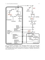
Fig. 1. Screening for dystrophin deletions using the multiplex PCR method. Track
1, deletion of exons 48-50; Track 2, deletion of exons 50-53; Track 3, deletion of
exon 53; Tracks 4 and 6, no deletion; Track 5, deletion of exon 52; and Track 7,
deletion of exon 45.
1. Prepare a master mix of all the components, except the DNA. Aliquot 8 yL into a
thin-walled 0.5-mL Eppendorf tube.
2. Add 2 pL of DNA solution (10-25 ng/pL) (see Note 3).
3. Add one drop of light paraffin oil, and place on a PCR machine with a preheated
block at 94’C (see Note 4).
4. PCR cycling conditions: Initial denaturation: 94°C for 3 min, followed by 30 cycles
of 94°C for 1 min, 60°C for 1 min, and 72°C for 2,3, or 4 min. (The synthesis time
is extended by 1 min every 10 rounds.) Final synthesis: 72°C for 5 min.
5. Add 2.5 pL of 5X TBE loading buffer.
6. Load 6 pL of reaction on a 2% agarose gel (1% Nusieve/l% BCL agarose), and
carry out electrophoresis at 100 mA for approx 1 h with ethidium bromide
(0.5 mg/mL) in both the gel and the TBE running buffer (see Note 5).
7. Once separation of the bands is complete, photograph the gel on a UV trans-
illuminator (Fig. 1) (see Notes 6 and 7).
3.1.2. SSCP/Heteroduplex Analysis (see Note 8)
3.1.2.1. PCR
CONDITIONS
The final concentrations of the reaction components are: 67 miVTris, pH 8.3,
16.6 mb4NH4S04, 3.7 mJ4MgC12, 85 pg/mL BSA, 0.5 fleach primer, 3 mM
dNTPs, 20-50 ng of genomic DNA, and
0.5
U of
Taq
polymerase in a total
volume of 10 pL.