The role of chitosan in protection of soybean from sudden death syndrome caused by fusarium solani f sp glycines
Bạn đang xem bản rút gọn của tài liệu. Xem và tải ngay bản đầy đủ của tài liệu tại đây (281.98 KB, 6 trang )
Bioresource Technology 98 (2007) 1353–1358
0960-8524/$ - see front matter © 2006 Elsevier Ltd. All rights reserved.
doi:10.1016/j.biortech.2006.05.029
The role of chitosan in protection of soybean from sudden death
syndrome caused by Fusarium solani f. sp. glycines
Benjaphorn Prapagdee
a,¤
, Kanignun Kotchadat
a
, Acharaporn Kumsopa
a
,
Niphon Visarathanonth
b
a
Faculty of Environment and Resource Studies, Mahidol University, Salaya, Nakhon Pathom 73170, Thailand
b
Department of Plant Pathology, Faculty of Agriculture, Kasetsart University, Bang Khen, Bangkok 10220, Thailand
Received 12 September 2005; received in revised form 18 May 2006; accepted 21 May 2006
Available online 7 July 2006
Abstract
The in vitro antifungal properties of chitosan and its role in protection of soybean from a sudden death syndrome (SDS) were evalu-
ated. Chitosan inhibited the radial and submerged growth of F. solani f. sp. glycines with a marked eVect at concentrations up to 1 mg/ml
indicating antifungal property and at 3 mg/ml was able to delay SDS symptoms expression on soybean leaves for over three days after
fungal inoculation when applied preventively. Chitosan was able to induce the level of chitinase activity in soybean resulting in the retar-
dation of SDS development in soybean leaves. However, the SDS symptoms gradually appeared and were associated with the reduction
of chitinase activity level after Wve days of infection period. These results suggested the role of chitosan in partially protecting soybeans
from F. solani f. sp. glycines infection.
© 2006 Elsevier Ltd. All rights reserved.
Keywords: Fusarium solani; Chitosan; Sudden death syndrome; Soybean
1. Introduction
Sudden death syndrome (SDS), caused by the soil-borne
fungus F. solani f. sp. glycines, is an economically harmful
disease of soybean (Rupe, 1989). SDS causes rapid defolia-
tion of soybean, resulting in reducing both the quality and
quantity of soybean product (Roy et al., 1989; Rupe, 1989).
The development of SDS is favored by cool and wet rhizo-
spheric conditions through the growing season (Scherm
and Yang, 1996). There is no total elimination of this dis-
ease because F. solani f. sp. glycines as mycelium and chla-
mydospores can survive in the soil and tolerate to the
unfavorable conditions (Rupe and Gbur, 1995). The use of
chemical substances for controlling Fusarium pathogen,
mainly methyl bromide as a broad spectrum disinfectant,
has been found to be eVective (Allen et al., 2004). However,
the excessive application of chemical fungicides led to
increase in fungicide resistance in pathogens and a contin-
ued presence of the pathogens in other areas of the Weld
(Bourbos et al., 1997) as well as contamination of the envi-
ronment. Additionally, the fungicides contaminated in the
environment tend to accumulate in agricultural products
and human body via the food chain.
Chitosan (poly--(1,4)-
D-glucosamine), a transformed
oligosaccharide, is obtained by alkaline deacetylation of
chitin, one of the most abundant natural biopolymers, that
is extracted from the exoskeleton of crustaceans such as
shrimps and crabs, as well as the cell walls of some fungi
(Sandford, 1989; Roller and Covill, 1999; Domard and
Domard, 2002). Thus, chitosan has attracted tremendous
attention as a potentially important biological resource due
to its biological properties including biocompatibility, non-
toxicity and biodegradability (Kurita, 1998). It has been
widely applied in the Welds of agriculture, environment,
*
Corresponding author. Tel.: +662 441 5000x187; fax: +662 441 9509
10.
E-mail address: (B. Prapagdee).
1354 B. Prapagdee et al. / Bioresource Technology 98 (2007) 1353–1358
pharmaceuticals, medicines and industrial food processing
(Sandford, 1989; Shahidi et al., 1999; Liu et al., 2001).
The interest in the antimicrobial properties of chitosan
has focused on its possible role in plant protection. Chito-
san has been found to interfere with the growth of several
plant pathogenic fungi e.g., Fusarium solani, F. oxysporum,
Puccinia arachidis, Botrytis cinerea, Colletotrichum gloeo-
sporioides (Shimosaka et al., 1993; Bell et al., 1998; Sathiya-
bama and Balasubramanian, 1998; Ben-Shalom et al., 2003;
Bautista-Baños et al., 2003). Chitosan caused morphologi-
cal changes, structural alterations and molecular disorgani-
zation of the fungal cells reXecting its fungistatic or
fungicidal potential (Hadwiger et al., 1986; Benhamou,
1996). The potential of chitosan to protect fungal diseases
of various horticultural plants has been studied in various
investigations (Benhamou et al., 1994; Lafontaine and
Benhamou, 1996; Ben-Shalom et al., 2003; Bautista-Baños
et al., 2003). Chitosan has also been found to activate
several biological processes of plant defense responses such
as enzymatic activities. Plant defense-related enzymes were
known to participate in early defense mechanisms and
to prevent pathogen infections (Ben-Shalom et al., 2003;
Bautista-Baños et al., 2006).
This work describes the potential of chitosan as an anti-
fungal agent on the growth of F. solani f. sp. glycines. Con-
sequently, chitosan was evaluated as an eVective biological
substance for the soybean protection from SDS symptoms
expression.
2. Methods
2.1. Materials
Chitosan from crab shell was obtained from Seafresh
Chitosan (Lab) Co. Ltd., Thailand. The degree of deacety-
lation of chitosan was 85% and the molecular weight was
2 £ 10
5
daltons. The viscosity of 1% chitosan solution in 1%
acetic acid and moisture content were 149 centipoise and
8.97%, respectively. The puriWed chitosan was prepared as
described by Benhamou (1992). Soybean (Glycine max (L.)
Merr.) seeds (SJ5 cultivar) were obtained from Department
of Agriculture, Ministry of Agriculture and Cooperatives,
Thailand. All cultural media were purchased from Difco
Laboratories, USA. Chemicals were obtained from Sigma–
Adlrich (USA).
2.2. Fungal culture and growth
F. solani f. sp. glycines was maintained on Potato Dex-
trose Agar (PDA) medium. It was aerobically cultivated in
Potato Dextrose Broth (PDB) at 28 °C with continuous
shaking at 150 rpm.
Antifungal assay of chitosan was conducted for both the
radial and submerged growth determination of F. solani f.
sp. glycines. PuriWed chitosan was dissolved in 0.25 N HCl
under continuous stirring, and the pH was adjusted to 5.6
with 2 N NaOH and then sterilized as previously described
(Bell et al., 1998). For the radial growth determination, the
sterile chitosan solution was added into PDA at concentra-
tions of 1, 3 or 5 mg/ml. Each PDA plate was seeded with
6-mm-diameter mycelial plugs of F. solani f. sp. glycines and
incubated at 28 °C in the dark. The fungal growth was
measured daily for seven days (Bell et al., 1998). Growth
inhibition was expressed as the percentage of inhibition of
radial growth relative to the control.
For the submerged growth determination, the sterile
chitosan solution was added into PDB to obtain the same
chitosan concentrations of the radial growth determination.
Spore suspension of F. solani f. sp. glycines was inoculated
in chitosan-supplemented PDB to give a Wnal volume of
1 £ 10
4
spores/ml and incubated for one day. The fungal
growth was monitored daily by dry weight determination
for 10 days (Yonni et al., 2004).
2.3. Evaluation of the role of chitosan in protection of
soybean from SDS development
The use of chitosan as a natural antifungal agent against
SDS in soybeans was investigated as described previously
(Sathiyabama and Balasubramanian, 1998) with some
modiWcation. Soybean seeds (SJ5 cultivar) were grown with
autoclaved soil and usually watered until being at V1
growth stage (14-day-old). The experiment used a Com-
pletely Randomized Design (CRD) which was divided into
six treatments with Wve replications. Both chitosan and F.
solani f. sp. glycines were not applied in T
1
as negative con-
trol. The surface of soybean leaf was sprayed with 1 mg/ml
of benomyl as chemical antifungal agent for T
3
. Soybean
leaves of other treatments including T
2
, T
4
, T
5
and T
6
were
sprayed with 100 l of chitosan solution at concentrations
of 0, 1, 3 and 5 mg/ml on the abaxial surface, respectively.
After 24 h, all treatments, except T
1
, were inoculated with
100 l of spore suspension (1 £ 10
3
spores/ml) of F. solani f.
sp. glycines on the abaxial surface. All inoculated soybean
plants were covered with water-sprayed polyethylene bags
for 24 h. The visible symptoms appearance of all soybean
plants was observed daily for 9 days. Finally at the 14-day,
all soybean plants were harvested for growth determination
of root length, stem height and dry weight.
2.4. Chitinase activity assay in soybean leaves
After fungal inoculation, chitosan-untreated and 3 mg/
ml of chitosan-treated leaves were collected for chitinase
activity assay at 1, 2, 3, 4, 5, 6, 8, 10, 12 and 14-day. The
intercellular Xuid of soybean leaves was prepared by grind-
ing leaf tissues and collecting by centrifugation for total
protein and chitinase activity assay. The total protein con-
centration was determined for the cleared intercellular Xuid
prior to their use in enzyme activity assays. Total protein
was determined by Coomassie Blue Protein Assay (BioRad,
USA) according to the sensitive method of Bradford
(1976). The chitinase activity assay was quantitative detec-
tion by measuring the amount of reducing sugars (N-acetyl-
B. Prapagdee et al. / Bioresource Technology 98 (2007) 1353–1358 1355
D-glucosamine, GlcNAc) liberated during the hydrolysis of
chitin solution as previously described (Shimosaka et al.,
1993). One unit of chitinase enzyme was deWned as the
amount of enzyme catalyzing the turnover of 1 mol of
GlcNAc per minute under the assay conditions. All experi-
ments were independently repeated at least three times and
representative data are shown.
2.5. Statistical analysis
The means and standard deviation of radial growth, sub-
merged growth and chitinase activity were calculated. Data
from soybean plant growth were statically analyzed by
using the analysis of variance (ANOVA) and DUNCAN
multiple range tests if a signiWcant diVerence was detected
(p < 0.05). SPSS, version 10.0 was used for statistical ana-
lysis.
3. Results and discussion
3.1. EVects of chitosan as a natural antifungal agent
on inhibition of the radial and submerged growth of
F. solani f. sp. glycines
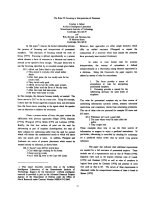
There was no halo formation of F. solani f. sp. glycines
cultivated on 0 and 1 mg/ml of chitosan but the growth on 3
and 5 mg/ml chitosan-amended plates was restricted rela-
tive to that of the control (Fig. 1) and the percentages of
radial growth inhibition were 38.2 and 54.6, respectively.
Furthermore, they also formed a halo around the colony on
the agar surface (data not shown). The halo-forming prop-
erty was used for testing the chitosanolytic activity in the
screening of Fusarium species, especially F. splendens and
F. solani. F. solani f. sp. phaseoli formed halo around the
colony on the 2.5 mg/ml of chitosan-containing agar plates
(Shimosaka et al., 1993).
Allan and Hadwiger (1979) suggested that the presence
of chitosan within the cell walls of some fungi rendered
those strains more resistant to the antifungal property of
externally-amended chitosan. Roller and Covill (1999),
however, found that chitosan reduced the growth rate of
Mucor racemosus at 1 mg/ml and at 5 mg/ml completely
prevented the growth of Byssochlamys spp. Benhamou
(1992) found that chitosan at 3 to 6 mg/ml inhibited the
radial growth of F. oxysporum f. sp. radicis-lycopersici, the
causative agent of tomato crown and root rot. The decrease
in growth inhibition was obtained with chitosan at concen-
trations less than 3 mg/ml. Based on the results from the
in vitro studies, inhibition of the radial growth of F. solani f.
sp. glycines was possibly due to the antifungal property of
chitosan. Several mechanisms for the antifungal action of
chitosan have been proposed. Two models had been pro-
posed to explain the antifungal activity of chitosan. Firstly,
the activity of chitosan was related to its ability to directly
interfere with the membrane function (Stössel and Leuba,
1984). Secondly, the interaction of chitosan with fungal
DNA and mRNA is the basis of its antifungal eVect (Had-
wiger et al., 1986).
Studies on the eVect of chitosan on submerged growth of
F. solani f. sp. glycines using dry weight measurements over
a period 10 days at 28 °C showed complete inhibition of the
growth of F. solani
f. sp. glycines at all concentrations of
chitosan (Fig. 2). However, an abnormal mycelial morphol-
ogy including hyphal swelling and cytoplasm aggregation
of F. solani f. sp. glycines was observed with 3 and 5 mg/ml
of chitosan. But none of these abnormal shapes were exhib-
ited in 1 mg/ml of chitosan-treated cells (data not shown).
Chitosan at concentrations ranging from 1 to 6 mg/ml
induced morphological changes in F. oxysporum f. sp. radi-
cis-lycopersici (Benhamou, 1992). These alterations could
Fig. 1. The radial growth of F. solani f. sp. glycines on chitosan-supple-
mented PDA plate. F. solani f. sp. glycines was cultivated on PDA plates
amended with 0, 1, 3 and 5 mg/ml of chitosan at 30 °C at 7-day of incuba-
tion period. The diameters of fungal colonies that grew on 0 (ᮀ), 1 (᭡), 3
(᭺) and 5 (᭹) mg/ml of chitosan-supplemented PDA plates were mea-
sured daily for 7 days of incubation period. Values presented are means
and standard deviation of triplicate assays.
Fig. 2. EVect of chitosan on the submerged growth of F. solani f. sp. gly-
cines F. solani f. sp. glycines was cultivated in PDB amended with 0 (ᮀ), 1
(᭡), 3 (᭺) and 5 (᭹) mg/ml of chitosan to give Wnal volume of 1 £ 10
4
spores/ml and incubated at 30 °C with continuous shaking at 150 rpm.
The fungal growth was monitored by dry-weight determination at 0, 12-h,
1, 2, 3, 4, 5, 6, 7, 8, 9 and 10-day of incubation period. Growth was
expressed as mg of cell dry weight per ml of cell sample. The values pre-
sented are the mean and standard deviation of three independent experi-
ments.
1356 B. Prapagdee et al. / Bioresource Technology 98 (2007) 1353–1358
be related with damages in the cell membrane structural
integrity due to chitosan presence, leading to the release of
some macromolecules caused by an increment of mem-
brane permeability (Stössel and Leuba, 1984).
3.2. Preventive application of chitosan on SDS symptoms
expression in soybeans
The visible foliar symptoms of soybean SDS appeared
only one day after fungal inoculation in chitosan-untreated
leaves (T
2
). A number of small brown blotches developed
on leaves and rapidly became necrotic within three days
after fungal inoculation. Some necrotic blotches became
larger and changed to pale brown. Then, the symptoms
developed daily with the increase in dead tissue until the
leaves turned to yellow and Wnally dropped oV, leaving the
petioles attached to the stem. No signiWcant retardation of
SDS development was observed at 1 mg/ml of chitosan (T
4
)
and even 1 mg/ml of benomyl-treated leaves (T
3
). Their
foliar symptoms still appeared similar to that of chitosan-
untreated leaves. The third day after inoculation, the foliar
symptoms obviously appeared in 5 mg/ml of chitosan-
treated leaves (T
6
). Although T
6
showed a slightly retardant
eVect on the expression of SDS symptom, the number of
necrotic blotches was greater than that of 3 mg/ml of chito-
san-treated leaves (T
5
).
The foliar symptoms on T
5
were clearly visible Wve days
after inoculation. Furthermore, the number of necrotic
blotches formed on 3 mg/ml of chitosan-treated leaves was
reduced relative to chitosan-untreated leaves. The symp-
tom appearance also increased slightly with time; however,
the symptom severity was less than that of chitosan-
untreated leaves. The results clearly indicated that an eVec-
tive dose of chitosan at 3 mg/ml could retard SDS symptom
expression on soybean leaves over three days after fungal
inoculation.
In a fungal-plant interaction, chitosan could activate the
defense response mechanisms in plant cells and completely
inhibit all RNA synthesis of some fungi and Wnally reduce
cell viability as well as suppress the fungal growth (Had-
wiger et al., 1986). Chitosan might enter the plant cells
through wounds on the leaf surface (Sathiyabama and
Balasubramanian, 1998). Chitosan in plant cells could be
localized in the nucleus of plant leaves and actually interact
with the cellular DNA leading to biochemical reactions in
the plant cells (Hadwiger et al., 1981; Hadwiger et al., 1986).
Thus, chitosan could induce resistance in pea against F.
solani f. sp. pisi by accumulating defense response proteins
(Kendra et al., 1989). Additionally, Sathiyabama and Bala-
subramanian (1998) found that chitosan at 1 mg/ml could
reduce uredospores of P. arachidis. However, chitosan
could not absolutely protect the soybean from SDS because
the foliar symptoms still appeared later. This was possibly
due to either the severity of F. solani f. sp. glycines invasion
or a reduction of the defense response components in soy-
beans.
3.3. EVect of chitosan on the growth of soybean plant
After 14 days of fungal inoculation, soybean plants of all
treatments were harvested for growth determination of
root length, stem height and dry weight. No signiWcant
diVerences (p < 0.05) in means of root length and stem
height of soybean plants were found in all treatments
(Table 1). In contrast, the signiWcant diVerence (p <0.05) in
mean was found on dry weight of soybean plants. There
was maximum increase per gram of dry weight in 3 mg/ml
of chitosan-treated leaves (0.634g) (T
5
) as compared to chito-
san-untreated leaves (T
2
). As a result, chitosan at 3mg/ml
could provide the higher soybean growth than other chito-
san-treated leaves and 1 mg/ml of benomyl-treated leaves
(T
3
) due to its role in protecting soybeans against SDS
symptom development.
3.4. The level of chitinase activity in infected soybean leaves
To investigate the level of chitinase activity in infected
soybean leaves, chitosan-untreated and 3 mg/ml of chito-
san-treated leaves were collected for chitinase activity assay
after fungal inoculation. The level of chitinase activity in
3 mg/ml of chitosan-treated leaves was drastically increased
from 12.4 to 17.9 U/mg protein after three days of fungal
inoculation (Fig. 3). The low level of chitinase activity was
probably responsible for the earlier observed SDS symp-
tom expression in chitosan-untreated leaves. In addition to
chitosan-treated leaves, there were almost no macroscopic
foliar symptoms of SDS on leaves during the high level
of chitinase activity period. Then, chitinase activity in
chitosan-treated and chitosan-untreated leaves sharply
decreased from 6 to 14 days after fungal inoculation. The
symptoms seemed to gradually appear and be associated
with the decrease of chitinase activity level after 5 days of
T
a
bl
e
1
EVects of chitosan on the growth of soybean plants
A
The in vivo experiment was divided into 6 treatments.
T
1
D Negative control (without F. solani f. sp. glycines)
T
2
D Positive control (inoculated with F. solani f. sp. glycines)
T
3
D Treated with 1 mg/ml of benomyl and F. solani f. sp. glycines
T
4
D Treated with 1 mg/ml of chitosan and F. solani f. sp. glycines
T
5
D Treated with 3 mg/ml of chitosan and F. solani f. sp. glycines
T
6
D Treated with 5 mg/ml of chitosan and F. solani f. sp. glycines.
B
Means were not signiWcantly diVerent (p < 0.05) according to the ana-
lysis of variance.
C
Means followed by the same letter within column were not signiW-
cantly diVerent (p < 0.05) according to Duncan’s multiple range test.
Treatment
A
Means § SD
Root length
B
(cm) Stem height
B
(cm) Dry weight
C
(g)
T
1
25.0 § 2.2 64.4 § 7.5 0.992 § 0.109
d
T
2
22.9 § 4.7 72.4 § 11.4 0.442 § 0.082
ab
T
3
25.6 § 6.2 77.8 § 16.3 0.528 § 0.084
bc
T
4
24.0 § 2.7 71.6 § 16.3 0.498 § 0.080
ab
T
5
22.6 § 4.2 76.7 § 18.3 0.634 § 0.087
c
T
6
21.3 § 5.1 72.0 § 16.4 0.446 § 0.083
ab
B. Prapagdee et al. / Bioresource Technology 98 (2007) 1353–1358 1357
fungal inoculation. The results could imply that the appli-
cation of chitosan might sensitize the soybean plant
responses in protecting themselves from the phytopatho-
genic fungal invasion by elaboration of chitinase activity.
Higher plants have the ability to initiate various defense
mechanisms, when they are infected either by phytopatho-
gens or after treatment with biotic and abiotic elicitors.
Chitosan had been shown to act as a potent oligosaccharide
elicitor which can induce defense response mechanisms
in several plants, mostly dicots. Chitinase, a hydrolytic
enzyme, was one of the pathogenesis-related proteins which
might be implicated in plant defense system against patho-
genic fungi (Shibuya and Minami, 2001). Chitinase and
-1,3-glucanase are defense response proteins that are pro-
duced by F. solani f. sp. pisi when cells were induced with
chitosan (Kendra et al., 1989). Furthermore, chitinase and
-1,3-glucanase are eVective in inhibiting the in vitro growth
of several fungi (Mauch et al., 1988). Celery, Apium graveo-
lens, treated with chitosan showed an increase in chitinase
activity of 20-fold compared to that of chitosan-untreated
plants and exhibited a delay in symptom expression caused
by F. oxysporum (Krebs and Grumet, 1993). Similarly,
chitosan stimulated chitinase production in cucumber plant
and protected this plant from root rot disease caused by
Pythium aphanidermatum (Ghauoth et al., 1994).
The evidence suggested that chitosan could induce active
defense responses just as chitinase enzyme in soybean
induces the resistance against F. solani f. sp. glycines. Chito-
san, a potent elicitor, could induce resistance components
as endogenous salicyclic acid, intercellular chitinase and -
1,3-glucanase activity in Arachis hypogaea against leaf rust
caused by P. arachidis (Sathiyabama and Balasubrama-
nian, 1998).
4. Conclusions
Chitosan played an important role in the growth sup-
pression of F. solani f. sp. glycines and the protection of soy-
bean plant against SDS. The radial and submerged growth
of F. solani f. sp. glycines were reduced by chitosan concen-
tration up to 1 mg/ml. The eVective dose of chitosan (3 mg/
ml) although could retard the SDS symptom expression in
soybean leaves over three days after fungal inoculation, it
could not absolutely protect the soybean from disease inci-
dence however; the foliar symptoms still appeared later.
Chitinase activity in soybean could increase the resistance
in soybean against F. solani f. sp. glycines because this
enzyme was able to degrade the fungal cell walls inhibiting
the fungal growth and symptom expression.
Acknowledgements
The authors thank the Department of Agriculture, Min-
istry of Agriculture and Cooperatives, Thailand for provid-
ing a strain of F. solani f. sp. glycines and Dr. Edward A.
Grand for a critical reading of the manuscript. This
research work was partially supported by the grant from
the Post-Graduate Education, Training and Research Pro-
gram in Environmental Science, Technology and Manage-
ment under Higher Education Development Project of the
Commission on Higher Education, Ministry of Education,
Thailand.
References
Allan, C.R., Hadwiger, L.A., 1979. The fungicidal eVect of chitosan on
fungi of varying cell wall component. Exp. Mycol. 3, 285–287.
Allen, T.W., Enebak, S.A., Carey, W.A., 2004. Evaluation of fungicides for
control of species of Fusarium on longleaf pine seed. Crop Prot. 23,
979–982.
Bautista-Baños, S., Hernández-López, M., Bosquez-Molina, E., Wilson,
C.L., 2003. EVects of chitosan and plant extracts on growth of Colleto-
trichum gloeosporioides, anthracnose levels and quality of papaya fruit.
Crop Prot. 22, 1087–1092.
Bautista-Baños, S., Hernández-Lauzardo, A.N., Velázquez-del Valle,
M.G., Hernández-López, M., Ait Barka, E., Bosquez-Molina, E., Wil-
son, C.L., 2006. Chitosan as a potential natural compound to control
pre and postharvest diseases of horticultural commodities. Crop Prot.
25, 108–118.
Bell, A.A., Hubbard, J.C., Liu, L., 1998. EVects of chitin and chitosan
on the incidence and severity of Fusarium yellows of celery. Plant Dis.
82, 322–328.
Benhamou, N., 1992. Ultrastructural and cytochemical aspects of chitosan
on Fusarium oxysporum f. sp. radicis-lycopersici, agent of tomato crown
and root rot. Phytopathology 82, 1185–1193.
Benhamou, N., 1996. Elicitor-induced plant defense pathways. Trends
Plant Sci. 1, 233–240.
Benhamou, N., Lafontaine, P.J., Nicole, M., 1994. Induction of systemic
resistance to Fusarium crown and root rot in tomato plants by seed
treatment with chitosan. Phytopathology 84, 1432–1444.
Ben-Shalom, N., Ardi, R., Pinto, R., Aki, C., Fallik, E., 2003. Controlling
gray mold caused by Botrytis cinerea in cucumber plants by means of
chitosan. Crop Prot. 22, 285–290.
Fig. 3. The level of chitinase activity in fungal infected soybean leaves
Both chitosan-untreated and 3.0 mg/ml of chitosan-treated soybean leaves
were inoculated with 100 l of fungal spore suspension (1 £ 10
3
spores/ml)
on the abaxial surface. Soybean leaves were harvested daily until 14 days
for chitinase activity assay. The extraction of intercellular Xuid from
chitosan-untreated leaves (᭺) and 3 mg/ml of chitosan-treated leaves (᭹)
and chitinase activity assay were performed as previously describes
(Shimosaka et al., 1993). Values presented are means and standard devia-
tion of triplicate experiments.
1358 B. Prapagdee et al. / Bioresource Technology 98 (2007) 1353–1358
Bourbos, V.A., Skoudridakis, M.T., Darakist, G.A., Koulizakis, M., 1997.
Calcium cyanamide and soil solarization for the control of Fusarium
solani f. sp. cucurbitae in greenhouse cucumber. Crop Prot. 16, 383–386.
Bradford, M.M., 1976. A rapid and sensitive method for the quantitation
of microgram quantities of protein utilizing the principle of protein-
dye binding. Anal. Biochem. 72, 248–254.
Domard, A., Domard, M., 2002. Chitosan: structure – properties relation-
ship and biomedical applications. In: Dumitriu, S. (Ed.), Polymeric
Biomaterials. Marcel Dekker, New York, pp. 187–212.
Ghauoth, A.E., Arul, J., Grenier, J., Benhamou, N., Asselin, A., Belanger,
G., 1994. EVects of chitosan on cucumber plants: Suppression of Pyth-
ium aphanidermatum and induction of defense reactions. Phytopathol-
ogy 84, 313–320.
Hadwiger, L.A., Beckman, J.M., Adams, M.J., 1981. Localization of fungal
components in the pea-Fusarium interaction detected immunochemi-
cally with anti-chitosan and antifungal cell wall antisera. Plant Physiol.
67, 170–175.
Hadwiger, L.A., Kendra, D.F., Fristensky, B.W., Wagoner, N., 1986. Chito-
san both activates genes in plants and inhibits RNA synthesis in fungi.
In: Muzzarelli, R.A., Jeuniaux, C., Gooday, G.W. (Eds.), Chitin in
Nature and Technology. Plenum Press, New York, pp. 209–214.
Kendra, D.F., Christian, D., Hadwiger, L.A., 1989. Chitosan oligomers
from Fusarium solani/pea interactions, chitinase/-glucanase digestion
of sporeling and from fungal wall chitin actively inhibition fungal
growth and enhance disease resistance. Physiol. Mol. Plant Pathol. 35,
215–230.
Krebs, S.L., Grumet, R., 1993. Characterization of celery hydrolytic
enzymes induced in response to infection by Fusarium oxysporum.
Physiol. Mol. Plant Pathol. 43, 193–208.
Kurita, K., 1998. Chemistry and application of chitin and chitosan. Polym.
Degrad. Stabil. 59, 117–120.
Lafontaine, P.J., Benhamou, N., 1996. Chitosan treatment: an emerging
strategy for enhancing resistance of greenhouse tomato plants to infec-
tion by Fusarium oxysporum f. sp. radicis-lycopersici. Biocontrol Sci.
Techn. 6, 11–124.
Liu, X.D., Nishi, N., Tokura, S., Sakari, N., 2001. Chitosan coated cotton
Wber: preparation and physical properties. Carbohyd. Polym. 44, 233–238.
Mauch, F., Mauch-MaNi, B., Boller, T., 1988. Antifungal hydrolase in pea
tissue II inhibition of fungal growth by combinations of chitinase and
-(1,3)-glucanase. Plant Physiol. 88, 936–942.
Roller, S., Covill, N., 1999. The antifungal properties of chitosan in labora-
tory media and apple juice. Int. J Food Microbiol. 47, 67–77.
Roy, K.W., Lawrence, G.W., Hodges, H.H., McLean, K.S., Killebrew, J.F.,
1989. Sudden death syndrome of soybean: Fusarium solani as incitant
and relation of Heterodera glycines to disease severity. Phytopathology
79, 191–197.
Rupe, J.C., 1989. Frequency and pathogenicity of Fusarium solani recovered
from soybeans with sudden death syndrome. Plant Dis. 73, 581–584.
Rupe, J.C., Gbur Jr., E.E., 1995. EVect of plant age maturity group and the
environment on disease progress of sudden death syndrome of soy-
bean. Plant Dis. 79, 139–143.
Sandford, P.A., 1989. Chitin: commercial uses and potential applications.
In: Skjak-Break, G., Anthosen, T., Sandford, P. (Eds.), Chitin and
Chitosan: Sources, Chemistry, Biochemistry, Physical Properties and
Applications. Elsevier Applied Science, London, pp. 51–69.
Sathiyabama, M., Balasubramanian, R., 1998. Chitosan induces resistance
components in Arachis hypogaea against leaf rust caused by Puccinia
arachidis Speg. Crop Prot. 17, 307–313.
Scherm, H., Yang, X.B., 1996. Development of sudden death syndrome of
soybean in relation to soil temperature and soil water matric potential.
Phytopathology 86, 642–649.
Shahidi, F., Arachchi, J.K.V., Jeon, Y.J., 1999. Food applications of chitin
and chitosans. Trends Food Sci. Tech. 10, 37–51.
Shibuya, N., Minami, E., 2001. Oligosaccharide signaling for defence
responses in plant. Physiol. Mol. Plant Pathol. 59, 223–233.
Shimosaka, M., Nogawa, M., Ohno, Y., Okazaki, M., 1993. Chitosanase
from the plant pathogenic fungus, Fusarium solani f. sp. phaseoli-puriW
-
cation and some properties. Biosci. Biotech. Biochem. 57, 231–235.
Stössel, P., Leuba, J.L., 1984. EVect of chitosan, chitin and some amino-
sugars on growth of various soilborne phytopathogenic fungi. Phyto-
pathology 111, 82–90.
Yonni, F., Moreira, M.T., Fasoli, H., Grandi, L., Cabral, D., 2004. Simple
and easy method for the determination of fungal growth and decolou-
rative capacity in solid media. Int. Biodeterio. Biodegrad. 54, 283–287.