influence of bias enhanced nucleation on thermal conductance through chemical vapor deposited diamond films
Bạn đang xem bản rút gọn của tài liệu. Xem và tải ngay bản đầy đủ của tài liệu tại đây (1.22 MB, 8 trang )
46 IEEE TRANSACTIONS ON COMPONENTS AND PACKAGING TECHNOLOGIES, VOL. 31, NO. 1, MARCH 2008
Influence of Bias-Enhanced Nucleation on Thermal
Conductance Through Chemical Vapor
Deposited Diamond Films
Baratunde A. Cola, Ratnakar Karru, Changrui Cheng, Xianfan Xu, and Timothy S. Fisher
Abstract—This work describes an experimental study of the
cross-plane thermal conductance of plasma-enhanced chemical
vapor deposited (PECVD) diamond films grown as a result of
bias-enhanced nucleation (BEN). The diamond films are grown on
silicon wafers using a two-step process in which a nucleation layer
of amorphous or diamond like (DLC) carbon is first deposited on
the silicon under the influence of a voltage bias. Then, conditions
are adjusted to allow for polycrystalline diamond (PD) growth.
The nucleation layer is essential for seeding diamond growth
on smooth substrates and for optimizing PD properties such as
grain size, orientation, transparency, adhesion, and roughness. A
photoacoustic (PA) technique is employed to measure the thermal
conductivities of and the thermal interface resistances between the
layers in the diamond film structure. The influence of nucleation
layers that are 70, 240, 400, and 650 nm thick on the thermal
conductance of the diamond film structure is characterized. The
thermal conductivity of the nucleation layer exhibits a thickness
dependence for relatively thin layers. For each sample, the thermal
conductivity of the PD is higher than 500 W
m K (measure-
ment sensitivity limit). A resistive network for the diamond film
structure is developed. The resistance at the silicon/nucleation
interface is less than 10
m K W (measurement sensitivity
limit), which is of the order of theoretical predictions. The
minimum diamond film structure resistance occurs when the nu-
cleation layer is thinnest. When the nucleation layer is sufficiently
thick, it begins to exhibit bulk behavior, and the resistance at the
nucleation/PD interface dominates the thermal resistance of the
diamond film structure.
Index Terms—Coatings, diamond, microelectronics, photoa-
coustic, plasma-enhanced chemical vapor deposited (PECVD),
thermal interface resistance, thin films.
NOMENCLATURE
m .
Intermediate coefficient.
Modulation frequency, s .
Intensity of laser light, W m .
Imaginary unity.
Manuscript received November 22, 2006; revised April 27, 2007. This work
was supported by the NASA Institute for Nanoelectronics and Computing
(INaC) and Purdue University. This work was recommended for publication
by Associate Editor B. Sammakia upon evaluation of the reviewers comments.
B. A. Cola, R. Karru, X. Xu, and T. S. Fisher are with Purdue University,
West Lafayette, IN 47907 USA (e-mail: tsfi).
C. Cheng is with Butler International, Inc., West Lafayette, IN 47907 USA.
Color versions of one or more of the figures in this paper are available online
at .
Digital Object Identifier 10.1109/TCAPT.2007.906725
Thermal conductivity, W m K .
Thermal conductivity uncertainty, W m K .
Thickness, m.
Thermal interface resistance, m K W .
Thermal interface resistance uncertainty,
m
K W .
Time, s.
Greek symbols
Thermal diffusivity, m s .
Phase shift.
Complex temperature, K.
Reflectivity.
,m .
Modulated angular frequency, 2 s .
Subscripts
Nucleation layer.
Nucleation and polycrystalline diamond interface.
Polycrystalline diamond layer.
Silicon and nucleation interface.
Diamond film structure.
I. I
NTRODUCTION
B
ECAUSE of the steady increase in device density of elec-
tronic circuits and components, driven by improvements
in fabrication technologies, effective and efficient thermal
management is required to alleviate problems that lead to poor
reliability and longevity. Because of its extreme hardness,
mechanical stability, chemical inertness, dielectric strength,
and high thermal conductivity, diamond can be an excellent
packaging material [1]–[7]. The thermal conductance of di-
amond films has been measured in several studies [8]–[13].
The high in-plane and cross-plane thermal conductivities of
diamond make it particularly effective to spread heat away from
hot spots to a heat sink. However, as heat dissipation increases,
the thermal resistance of the nucleation layer and its associated
interfaces will consume a larger portion of the thermal budget.
Despite its obvious advantages as a thermal enhancement mate-
rial, polycrystalline diamond films have not been widely used in
microelectronic components, largely because of difficulties in
1521-3331/$25.00 © 2007 IEEE
COLA et al.: INFLUENCE OF BIAS-ENHANCED NUCLEATION ON THERMAL CONDUCTANCE 47
heterogeneous material integration and cost. Here, we consider
the thermal characteristics of a promising material integration
approach involving bias-enhanced nucleation (BEN) [14] of
plasma-enhanced chemical vapor deposited (PECVD) diamond
films.
To ensure the suitability of diamond films in a cooling design,
thermal conductance must be measured, preferably by a nonin-
trusive technique, and the effect of nucleation on thermal per-
formance should be characterized. In this work, a photoacoustic
(PA) technique [15]–[17] is used to measure the thermal conduc-
tivities of and the thermal interface resistances between nucle-
ation and polycrystalline diamond (PD) layers. The thermal con-
ductivities are used in conjunction with measured thicknesses to
calculate the thermal resistance of each layer; these resistances
are summed in series with the interface resistances to determine
the thermal resistance of the diamond film structure.
PECVD has become a popular method to synthesize diamond
films for microelectronic applications because it offers low
sample contamination, relatively low growth temperatures, and
an ability to control the film characteristics [18]. During the
synthesis of such films using PECVD and BEN, the formation
of an amorphous carbon or diamond like carbon (DLC) layer
(nucleation stage) precedes the growth of well faceted diamond
grains (growth stage) [7], [19]. In this study, we examine the
thermal behavior of the products of each stage of the growth
process separately as well as together, with attention given
to the nucleation conditions and time allowed for nucleation.
By examining thermal conductance in this piecewise manner,
we seek to elucidate the factors that contribute to a PECVD
synthesized diamond film’s overall thermal resistance, thereby
allowing more thorough understanding of its thermal perfor-
mance.
II. E
XPERIMENTAL
METHODS
A. Film Synthesis and Sample Preparation
The films used in this study were synthesized in a SEKI
AX5200S microwave plasma CVD system. A molybdenum
holder concentrates the plasma over silicon substrates
(0.580 mm thick). Substrate pretreatment, such as ultrasonic
scratching with diamond powder, is usually required to deposit
diamond films on silicon surfaces [19]. However, such a treat-
ment is undesirable in applications where surface roughness
needs to be small. BEN is a well established alternative to attain
high nucleation densities on smooth surfaces such as silicon
[19]–[23]. BEN eliminates the need for additional cleaning
steps because it occurs in the deposition chamber under the
same vacuum environment used for diamond film growth. For
this study, a constant DC bias of
250 V was applied during
the nucleation stage [14]. This bias level was found to produce
successful nucleation on silicon in our system [7].
Methane and hydrogen were used as the source gases, and
nucleation was carried out in 300 W plasma for 15, 30, 45, and
60 min. While diamond film synthesis usually occurs at much
higher plasma powers (more than 1 kW), 300 W was used for
nucleation in this study because plasma arching was observed at
both high plasma power and high bias voltage. After nucleation,
the bias was turned off for the growth stage to allow the plasma
power to increase. 1200 W plasma power was used during the
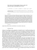
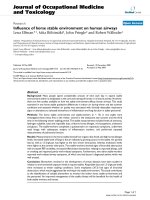
Fig. 1. (a) Schematic of a typical diamond film grown by PECVD using BEN.
The silicon wafer is 580
m thick. (b) FESEM of diamond film for 45-min
nucleation time. (c) Higher resolution (6.5 X) FESEM that shows a close up of
the nucleation layer.
growth stage to promote the growth of thick films. A chamber
pressure above 55 torr is required to safely support a plasma
power above 1000 W in our system. The process parameters are
summarized in Table I, and a typical film structure is shown in
Fig. 1. For each nucleation duration, two different samples were
fabricated: a sample consisting of a silicon substrate and a nucle-
ation layer (herein referred to as the “nucleation sample”), and a
sample consisting of a silicon substrate, a nucleation layer, and
aPDfilm (herein referred to as the “PD growth sample”). Typ-
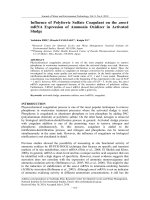
ical Raman spectra of the nucleation and PD growth samples are
shown in Fig. 2. The low signal to noise ratio and the presence
of a G-band mode in the PD growth samples’ spectra are due to
the simultaneous excitation of the PD and nucleation layers. The
Raman spectra supports field-emission scanning electron micro-
scope (FESEM) observations of an amorphous-like carbon film
in the nucleation layer and diamond growth in the PD layer. As
determined from FESEM, the average particle size in the nu-
cleation layers ranged from 5 to 25 nm; the average increased
with increased nucleation time. Consequently, the average sur-
face roughness of the nucleation layer increased with nucleation
time as well. The average PD grain size ranged from 1 to 3
m
and slightly increased as the nucleation time increased.
B. Photoacoustic (PA) Technique
The PA technique is one of many proven techniques to mea-
sure thermal conductivity of thin films, and it has recently been
used to measure the thermal resistance of separable interfaces
[17]. The PA technique provides high accuracy [16], yet in com-
parison to other techniques to measure thermal conductance
across thin films and planar interfaces, it is relatively simple to
implement. Reference [15] provides a detailed description of the
technique.
Theory: The sample used for PA measurement can have
a backing layer (0) and
successive layers (1, 2, )on
which the
-coordinate originates from the surface of layer
and points outward. The multilayered material is heated by a
modulated laser beam with an intensity of 1/2
1 ,
and absorption of the laser beam is allowed in any layer, and
in more than one layer. The backing material (0) and a gas
medium
1 in contact with the surface layer are
considered to be thermally thick. The transient temperature
field in the multilayer sample and gas can be derived by solving
48 IEEE TRANSACTIONS ON COMPONENTS AND PACKAGING TECHNOLOGIES, VOL. 31, NO. 1, MARCH 2008
Fig. 2. (a) Raman spectrum for the nucleation layer. The merging of the D-band
and G-band suggests the presence of an amorphous carbon state. (b) Raman
spectrum for the PD layer. The D-band peak near 1320 cm
can indicate a
preference for diamond. The peak near 500 cm
is from silicon.
TABLE I
P
ROCESS PARAMETERS FOR THE GROWTH OF PECVD DIAMOND FILMS
a set of 1-D heat conduction equations [15], [24]. Details of the
derivation process have been described in [15]. The solution of
the complex temperature distribution
in the gas can be
expressed as
(1)
where
is complex, and is a function of the thermal prop-
erties of the multilayered sample. The general formulation of
is long and readily available in the literature; thus the
reader is referred to [15]–[17] for its full formulation.
The temperature in the gas layer is related to the phase
shift and the amplitude of the pressure or PA signal using
a thermal piston analogy where the heated gas near the
sample surface pushes the rest of the gas up like a piston
[15]. The phase shift of the PA signal is calculated as
4, and the amplitude of the PA signal is
calculated as
, where
and are the ambient temperature and pressure, respec-
tively.
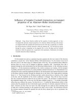
Experimental Details: The experimental setup is shown
schematically in Fig. 3. A fiber laser is used as the heating
source. An acoustic-optical modulator (AOM) driven by a
function generator modulates the laser power with a sinusoidal
function. For this study, the modulation frequency ranges from
2 to 20 kHz, and the output power of the laser is approximately
350 mW at the modulation mode. The laser beam is reflected
and focused and then directed onto the sample mounted at
the bottom of the PA cell. To promote complete laser power
absorption at the sample surface, 80 nm of titanium is deposited
on the samples. The PA cell is the same as the one used in the
studies of [17]. The acoustic signal is sensed by a microphone
embedded in the side wall of the cell. The signal is transferred
to the lock-in amplifier, where the amplitude and phase are
measured. The phase shift of the acoustic signal is used to
determine thermal properties because it is more stable than the
amplitude signal in the current experimental setup and thus
provides higher measurement precision.
In order to account for delay in the PA response due to the
time needed for the acoustic wave to travel from the sample sur-
face to the microphone, and due to acoustic resonance in the
cell, a silicon wafer (0.580 mm thick) is used as a reference or
calibration sample. 80 nm of titanium is deposited on the sil-
icon reference and test samples at the same time to allow for
similar surface reflectivity and laser absorption. Within the fre-
quency range of this study, the reference sample is thick enough
to be considered a bulk material (much thicker than the thermal
penetration depth,
); therefore the phase shift
is
90 . The calibrated phase shift of the sample, , is calcu-
lated as
90, where is the measured
phase shift for the sample, and
is the measured
phase shift for the reference. The experimental setup is cali-
brated before each measurement and at each frequency. After
the signal stabilizes, phase-shift data are recorded every 8 s and
averaged every 5 min. In order to determine the drift of the sig-
nals with time, the references are also tested after each sample
measurement.
The measured and calibrated phase shift of the acoustic signal
is used in conjunction with the general PA model of [15] to es-
timate the thermal interface resistance between the silicon sub-
strate and nucleation layer,
, and between the nucleation
layer and the PD layer,
, and the thermal conductivities
of the nucleation layer,
, and the PD layer, . In order to
estimate theses four quantities, the nucleation samples are mea-
sured and the two unknowns,
and , are obtained. Then
the PD growth samples are measured with
and input
as known values, and the remaining two unknowns,
and
, are estimated. The sample measurement procedure is il-
lustrated in Fig. 4. For each measurement set, the unknowns are
solved by fitting the PA model to the experimental data using
a least-squares algorithm where trial values of the unknown
thermal properties are used to calculate the phase shift of the PA
signal at each experimental frequency. The sum of the square
of the difference between calculated and experimental values
of phase shift is calculated. The trial
and values for which
COLA et al.: INFLUENCE OF BIAS-ENHANCED NUCLEATION ON THERMAL CONDUCTANCE 49
Fig. 3. Schematic diagram of the photoacoustic apparatus.
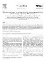
Fig. 4. In the first measurement set the nucleation sample is measured, and and are determined. These measured values are then used in the second
measurement set (PD growth sample) to determine
and . The sample layer labeling used in the PA model is presented for each measurement set as
well.
the least square is obtained are taken as the property values. The
piecewise examination of the diamond film structures allows for
each measurement set to have only two unknowns (
and ) that
are uncoupled in the governing equations, thereby allowing the
least-squares fits to be unique.
Experimental uncertainty is primarily determined by the un-
certainty in thickness measurements, and for the PD growth
samples, the uncertainty in the
and measurements as
well. The effects of uncertainties associated with other ‘known’
material properties used in the PA model, uncertainty associated
with laser energy drift, and uncertainty associated with phase
shift measurements were negligible in comparison. Uncertainty
in the measured thermal properties is determined by finding the
range of the property values that result from changing the nucle-
ation and PD layer thicknesses, and (for the PD growth samples)
and within their uncertainty range in the PA model.
The PA signal is primarily influenced by the sample layers or
interfaces that are the most resistive to heat flow. Consequently,
there are limits on the magnitudes of property values that can be
sensed with the technique. These limitations are determined by
experimental error and/or by varying the desired property in the
PA model to conditions where further changes in the property
50 IEEE TRANSACTIONS ON COMPONENTS AND PACKAGING TECHNOLOGIES, VOL. 31, NO. 1, MARCH 2008
Fig. 5. Thicknesses of the nucleation layer and PD as a function of nucleation
time. PD growth is shown to be independent of nucleation time, and is the same
thickness for each case. The measurement error, as given in Table II, is less than
the size of the data point markers.
produce a negligible change in the calculated phase shift. Ex-
perimental uncertainty is sufficiently large in this study that the
sensitivity limits are determined by it alone. If the thermal con-
ductivity uncertainty for a layer is
, then the smallest measur-
able thermal interface resistance for that layer is approximately
. Similarly, if the uncertainty of the interface resistance
is
, then the largest measurable thermal conductivity for that
layer is approximately l
.
III. R
ESULTS AND DISCUSSION
The thermal resistances of PECVD-via-BEN diamond films
were measured on samples with nucleation times of 15, 30, 45,
and 60 min. First, only nucleation was performed, and the layer
formed in each case was analyzed. Then, on new samples, nu-
cleation followed by 10 h of PD growth forms the diamond
film structure. The thicknesses of the nucleation layer and the
diamond film structure were measured from FESEM images,
and the resulting data are presented in Table II. The PD layer
thickness is determined by identifying the boundary between
nucleation and PD in the diamond film structure FESEM image.
For each case, the location of this boundary coincided with the
measured thickness of the nucleation layer (without PD), thus
corroborating this result. The amount of PD grown is approxi-
mately the same for each nucleation time, as shown in Fig. 5.
Fig. 6 shows that the nucleation layer retains its pre-PD growth
thickness after the PD is grown, verifying an important assump-
tion in our analysis. A thermal resistance network for the dia-
mond film structure is presented in Fig. 7. The thermal resis-
tance of the nucleation and PD layers are given as
and , respectively.
A summary of resistances for the diamond film structure is
shown in Table III, and the data trend is illustrated in Fig. 8.
As discussed in detail later, the large jump in resistance from
a nucleation layer thickness of 400 to 650 nm is due to poor
bonding of the PD to the 650 nm-thick nucleation layer.
is
the first resistance encountered by heat flowing from silicon to a
diamond film as shown in Fig. 7. This resistance is the result of
acoustic mismatch and imperfect interfacial contact, and theo-
retical modeling by Zeng and Chen suggest that it is very small,
on the order of 10
m K W [25]. For the samples in this
study,
is less than the smallest resistance that can be sensed
Fig. 6. FESEM of nucleation layer without (a), and with (b) PD growth for a
nucleation time of 45 min. In each case, the nucleation layer is approximately
400 nm thick. The top layer in (a) is titanium, which is deposited for the purpose
of laser energy absorption.
Fig. 7. Thermal circuit for diamond films synthesized by PECVD using BEN.
is the thermal resistance of the entire diamond film structure.
TABLE II
T
HICKNESS AS A
FUNCTION OF
NUCLEATION TIME FOR THE
NUCLEATION
LAYER AND DIAMOND FILM STRUCTURE.U
NCERTAINTY IN
THE
THICKNESS MEASUREMENTS IS ESTIMATED FROM THE
PRECISION OF THE SCALE BARS IN FESEM IMAGES
with our experimental technique. These sensitivity minima are
all on the order of 10
m K W ; therefore, we conclude that
the nucleation layer is in good contact with the silicon substrate,
and
is predicted relatively well by the modeling of Zeng
and Chen. The next resistive path in the diamond film network
is
. Each value is calculated from the measurements
for the different nucleation samples. A thickness dependence
of the nucleation layer’s thermal conductivity is apparent for
relatively thin layers, causing a nonlinear relationship between
and as illustrated in Fig. 8. As the layer becomes suffi-
ciently thick,
converges to a consistent value, and begins
to display a linear relationship with respect to
as expected.
This trend can be explained by the change in the structure of
the nucleation layer with increased thickness. The nucleation
layer is more discontinuous and has smaller particle sizes (e.g.,
more grain boundaries and voids) near its interface with the sil-
icon substrate than in the section of the layer that accumulates
with increasing thickness. Thus, as the nucleation layer becomes
COLA et al.: INFLUENCE OF BIAS-ENHANCED NUCLEATION ON THERMAL CONDUCTANCE 51
TABLE III
R
ESISTANCE
COMPONENTS OF
DIAMOND
FILMS SYNTHESIZED BY
PECVD U
SING
BEN. UNCERTAINTY IN THE
RESISTANCE
VALUES IS THE
RESULT OF
UNCERTAINTY IN THE
THICKNESS AND
MEASUREMENTS. AND ARE
BELOW THE
MEASUREMENT
SENSITIVITY
Fig. 8. Dominate resistance components, and , of the diamond
films’ thermal resistance,
, as a function of nucleation layer thickness. The
measurement error, as given in Table III, is less than the size of the data point
markers.
thicker, the more continuous section with larger particles (away
from the interface) grows thicker and has a greater influence
on the measured
, and eventually dominates thermal trans-
port across this layer. The measured
values are larger than
the room-temperature thermal conductivity value of amorphous
carbon, 1.60 W
m K [26]; therefore, it is reasonable to as-
sume that the carbon in the nucleation layer is DLC, which con-
tains higher crystal order than amorphous carbon.
As presented in Table III and illustrated in Fig. 8,
most significantly affects the thermal performance of the
diamond film structure.
decreases with decreasing nu-
cleation layer thickness because of reduced growth or thermal
stresses at the nucleation/PD interface. We postulate that when
the nucleation layer becomes sufficiently thick, the thermal
stresses generated at its interface with the PD weakens the
bond between the nucleation layer and the PD and may cause
the section of the nucleation layer near the PD interface to
break into clusters, creating voids. This adverse effect may
be exacerbated by the fact that the surface roughness of the
nucleation layer increases as the nucleation layer thickens.
This weakening of the nucleation/PD interfacial bond impedes
thermal transport across the interface. To further demonstrate
this point, the PD growth samples were cut in half after testing
and their cross-sections were imaged with a field-emission
scanning electron microscope. As illustrated in Fig. 9, the
thickest nucleation layer (60 min nucleation time) resulted in
the formation of large voids in the nucleation/PD interface;
such voids will impede thermal transport across the interface.
Fig. 9. (a) Nucleation/PD interface after 45 min of nucleation. The nucleation
layer and the PD layer appear well connected. (b) Nucleation/PD interface after
60 min of nucleation. Stress-induced voids are present at the nucleation/PD in-
terface.
Additionally, for 60 min of nucleation, the PD layer’s adhesion
was very poor as evidenced by easy peeling of the diamond
layer from the substrate (due most likely to increased stress
concentration at the discrete contact points).
is the final resistive component of the diamond film
structure. Each
value is determined from the measured
for the different PD growth samples. For each sample,
is measured to be above the largest thermal conductivity
that can be sensed with our experimental technique (approxi-
mately 500 W
m K ). Because each PD layer is approxi-
mately 5.8
m thick and has an average grain size between 1 and
3
m, is expected to be near 10 W m K [8], which is
consistent with the results of this study. For each sample (since
is constant for each nucleation layer sample), is below
approximately 10
m K W and has a negligible effect on
the overall thermal resistance of the diamond film structure,
.
The minimum conduction resistance of the diamond film
structure occurs when the nucleation layer is thinnest because
of reduced thermal stress between the nucleation and PD layer.
We also note that the quality, hence thermal conductivity, of
PD is expected to increase with increasing nucleation layer
thickness or “seeding grain” size (the nucleation layer surface
asperities may be view as the seeding grains) due to the for-
mation of larger, more continuous PD columns that promote a
decrease in phonon-grain boundary scattering [1], [8]. How-
ever, as shown in this work, increased PD thermal conductivity,
via increased nucleation layer thickness, would be entirely
offset by increases in both
and to produce a larger
overall thermal resistance
.
IV. C
ONCLUSION
The thermal resistances of PECVD diamond films grown
from BEN layers have been measured for nucleation times of
15, 30, 45, and 60 min. An experimental technique, using PA
52 IEEE TRANSACTIONS ON COMPONENTS AND PACKAGING TECHNOLOGIES, VOL. 31, NO. 1, MARCH 2008
measurements, has been employed to measure the resistive
components of the diamond film structure. The resistance at
the silicon/nucleation boundary and the intrinsic resistance
of the PD layer were measured to have a negligible effect on
the diamond film structures’ resistance. For each nucleation
sample, the dominant resistances in the thermal network are
the intrinsic resistance of the nucleation layer and the nucle-
ation/PD interface resistance.
In general, this study shows that the thermal resistance of
PECVD diamond films grown from BEN strongly depends on
the structure of the nucleation layer and the quality of the nu-
cleation/PD interface. The thermal conductivity of the nucle-
ation layer is measured to be higher than that of amorphous
carbon and exhibits a thickness dependence for relatively thin
layers, while it converges to a consistent value when the layer
is sufficiently thick. Under the conditions of this study, smaller
nucleation times and consequentially thinner nucleation layer
thicknesses result in lower diamond film structure resistances.
As the nucleation layer thickens, the nucleation/PD interface re-
sistance, which dominates the overall resistance of the diamond
film, increases because of poor interfacial bonding.
In this study, the voltage bias, plasma conditions, and gas flow
ratios are the same for each case. Further work is recommended
to investigate the effects of these parameters on the structure
of the nucleation layer and its boundaries. The only substrate
used in this study is silicon. Diamond films grown by PECVD
using BEN on other relevant substrates remain to be explored.
Finally, the PD layers grown in this study are all approximately
5.8
m thick, the effect of PD layers of different thicknesses on
the nucleation/PD interface resistance is suggested for further
study.
A
CKNOWLEDGMENT
The authors wish thank A. Franklin, Dr. M. R. Maschmann,
and Dr. P. B. Amama for their help with FESEM and Raman
characterization.
R
EFERENCES
[1] K. E. Goodson and Y. S. Ju, “Heat conduction in novel electronic
films,” Annu. Rev. Mater. Sci., vol. 29, pp. 261–293, 1999.
[2] R. F. Davis, Diamond Films and Coatings. Norwich, NY: William
Andrew Publishing, 1993.
[3] M. N. R. Ashfold, P. W. May, C. A. Rego, and N. M. Everitt, “Thin film
diamond by chemical vapour deposition methods,” Chem. Soc. Rev.,
vol. 23, pp. 21–31, 1994.
[4] K. E. Spear and J. P. Dismukes, Synthetic Diamond: Emerging CVD
Science and Technology. Hoboken, NJ: Wiley, 1994.
[5] X. Zhu, D. M. Aslam, Y. Tang, B. H. Stark, and K. Najafi, “The fabri-
cation of all-diamond packaging panels with built-in interconnects for
wireless integrated microsystems,” J. Microelectromech. Syst., vol. 13,
no. 3, pp. 396–405, 2004.
[6] X. Zhu and D. M. Aslam, “CVD diamond thin film technology
for MEMs packaging,” Diamond Rel. Mater., vol. 15, no. 2–3, pp.
254–258, 2006.
[7] J. Chee, R. Karru, T. S. Fisher, and D. Peroulis, “DC-65 GHz charac-
terization of nanocrystalline diamond leaky film for reliable RF MEMS
switches,” in Proc. Eur. Microw. Conf., 2005, vol. 3, pp. 1527–1530.
[8] J. E. Graebner, S. Jin, G. W. Kammlott, J. A. Herb, and C. F. Gardinier,
“Large anisotropic thermal conductivity in synthetic diamond films,”
Nature, vol. 359, pp. 401–, 1992.
[9] K. E. Goodson, O. W. Käding, M. Rösler, and R. Zachai, “Experi-
mental investigation of thermal conduction normal to diamond-silicon
boundaries,” J. Appl. Phys., vol. 77, no. 4, pp. 1385–1392, 1995.
[10] J. E. Graebner, “Measurements of thermal conductivity and thermal
diffusivity of CVD diamond,” Int. J. Thermophys., vol. 19, no. 2, pp.
511–523, 1998.
[11] H. Verhoeven, H. Reiß, H J. Füßer, and R. Zachai, “Thermal resistance
of thin diamond films deposited at low temperatures,” Appl. Phys. Lett.,
vol. 69, no. 11, pp. 1562–1564, 1996.
[12] J. Hartmann, P. Voigt, and M. Reichling, “Measuring local thermal
conductivity in polycrystalline diamond with a high resolution pho-
tothermal microscope,” J. Appl. Phys., vol. 81, no. 7, pp. 2966–2972,
1997.
[13] E. Jansen and E. Obermeier, “Thermal conductivity measurements
of thin films based on micromechanical devices,” J. Micromech.
Microeng., vol. 6, pp. 118–121, 1996.
[14] J. Robertson, J. Gerber, S. Sattel, M. Weiler, K. Jung, and H. Ehrhardt,
“Mechanism of bias-enhanced nucleation on Si,” Appl. Phys. Lett., vol.
66, no. 24, pp. 3287–3289, 1995.
[15] H. Hu, X. Wang, and X. Xu, “Generalized theory of the photoacoustic
effect in a multilayer material,” J. Appl. Phys., vol. 86, no. 7, pp.
3953–3958, 1999.
[16] X. Wang, H. Hu, and X. Xu, “Photoacoustic measurement of thermal
conductivity of thin films and bulk materials,” ASME J. Heat Transf.,
vol. 123, pp. 138–144, 2001.
[17] B. A. Cola, J. Xu, C. Cheng, H. Hu, X. Xu, and T. S. Fisher,
“Photoa-
coustic characterization of carbon nanotube array interfaces,” J. Appl.
Phys., vol. 101, no. 054313, 2007.
[18] E. Kohn, M. Adamschik, P. Schmid, A. Denisenko, A. Aleksov, and W.
Ebert, “Prospects of diamond devices,” J. Phys. D: Appl. Phys., vol. 34,
pp. R77–R85, 2001.
[19] S T. Lee, Z. Lin, and X. Jiang, “CVD diamond films: Nucleation and
growth,” Mater. Sci. Eng., vol. 25, pp. 123–154, 1999.
[20] B. R. Stoner, G H. M. Ma, S. D. Wolter, and J. T. Glass, “Characteri-
zation of bias-enhanced nucleation of diamond on silicon by in vacuo
surface analysis and transmission electron microscopy,” Phys. Rev. B,
vol. 45, no. 19, pp. 11067–11084, 1992.
[21] M. Schreck, K H. Thürer, and B. Stritzker, “Limitations of the process
window for the bias-enhanced nucleation of heteroepitaxial diamond
films on silicon in the time domain,” J. Appl. Phys., vol. 81, no. 7, pp.
3092–3095, 1997.
[22] S. D. Wolter, F. Okuzumi, J. T. Prater, and Z. Sitar, “AC vs. DC bias-
enhanced nucleation of highly oriented diamond on silicon (100),” J.
Electrochem. Soc., vol. 149, no. 2, pp. G114–G117, 2002.
[23] A. N. Jones, W. Ahmed, C. A. Rego, M. J. Jackson, and R. Hall, “Nu-
cleation studies of pulsed bias-enhanced CVD of diamond on bioma-
terials,” J. Mater. Eng. Perform., vol. 15, no. 2, pp. 192–194, 2006.
[24] R. S. Quimby and W. M. Yen, “On the adequacy of one-dimensional
treatments of the photoacoustic effect,” J. Appl. Phys., vol. 51, no. 2,
pp. 1252–1253, 1980.
[25] T. Zeng and G. Chen, “Phonon heat conduction in thin films: Impact
of thermal boundary resistance and internal heat generation,” ASME J.
Heat Transf., vol. 123, pp. 340–347, 2001.
[26] F. P. Incropera and D. P. DeWitt, Heat and Mass Transfer, 5th ed.
Hoboken, NJ: Wiley, 2002.
Baratunde A. Cola received the B.E. and M.S.
degrees in mechanical engineering from Vanderbilt
University, Nashville, TN, in 2002 and 2004, respec-
tively, and is currently pursuing the Ph.D. degree in
mechanical engineering at Purdue University, West
Lafayette, IN.
His current research interests include nanomate-
rial synthesis, applications of carbon nanotubes, and
cooling of microelectronics.
Ratnakar Karru received the B.Tech. degree from
the Indian Institute of Technology, Kharagpur, in
2001 and the M.S. degree in mechanical engineering
from Purdue University, West Lafayette, IN, in 2003
where he is currently pursuing the M.S. degree in
electrical engineering.
His research involves synthesis and characteriza-
tion of diamond thin films, focusing on their applica-
tions in RF MEMS capacitive switches.
COLA et al.: INFLUENCE OF BIAS-ENHANCED NUCLEATION ON THERMAL CONDUCTANCE 53
Changrui Cheng received the Ph.D. degree from the
School of Mechanical Engineering, Purdue Univer-
sity, West Lafayette, IN, in 2006.
Currently, he works in Butler International, Inc.,
West Lafayette, as an Engineering Analyst. His re-
search focuses on molecular dynamics simulation of
laser micro-machining and computational modeling
in thermal-fluid.
Xianfan Xu received the M.S. and Ph.D. degrees in
mechanical engineering from the University of Cali-
fornia at Berkeley in 1991 and 1994, respectively.
He is a Professor of Mechanical Engineering with
Purdue University, West Lafayette, IN. His currentre-
search is laser based materials processing and diag-
nostics.
Timothy S. Fisher received the B.S. and Ph.D.
degrees in mechanical engineering from Cornell
University, Ithaca, NY, in 1991 and 1998, re-
spectively, and the M.S. degree from Vanderbilt
University, Nashville, TN, in 2002.
He joined the School of Mechanical Engineering
and Birck Nanotechnology Center, Purdue Univer-
sity, West Lafayette, IN, in 2002 after several years
at Vanderbilt University. Prior to his graduate studies,
he was employed from 1991 to 1993 as a Design En-
gineer in Motorola’s Automotive and Industrial Elec-
tronics Group. His research has included efforts in simulation and measurement
of nanoscale heat transfer, coupled electro-thermal effects in semiconductor de-
vices, nanoscale direct energy conversion, molecular electronics, microfluidic
devices, hydrogen storage, and computational methods ranging from atomistic
to continuum scales. His current efforts include theoretical, computational, and
experimental studies focused toward integration of nanoscale materials with
bulk materials for enhancement of electrical, thermal, and mass transport prop-
erties. Applications of his work cover a broad range of areas, including nano-
electronics, thermal interface materials, thermal-electrical energy conversion,
biosensors, and hydrogen storage. This work has also produced related studies
of controlled synthesis of nanomaterials, particularly carbon nanotubes.
Dr. Fisher is a member of Tau Beta Pi and Pi Tau Sigma. He serves on the
IEEE TC-9 Committee on Thermal Phenomena in Electronics, the ASME K-6
committee on Heat Transfer in Energy Systems, ASME K-16 Committee on
Thermal Management of Electronics.