analysis of cuo nanoparticles synthesized by sol gel
Bạn đang xem bản rút gọn của tài liệu. Xem và tải ngay bản đầy đủ của tài liệu tại đây (708.17 KB, 4 trang )
Nanoscience and Nanotechnology 2012, 2(3): 71-74
DOI: 10.5923/j.nn.20120203.05
Structure, Microstructure and Optical Absorption
Analysis of CuO Nanoparticles Synthesized by Sol-Gel
Route
P. Mallick
*
, S. Sahu
Department of Physics, North Orissa University, Baripada, 757003, India
Abstract We report the synthesis of CuO nanoparticles using different solvents by a low cost sol-gel route. Evolution of
structure, microstructure and optical absorption analysis of these nanoparticles were studied using X-ray diffraction (XRD)
and UV-Visible spectrophotometer. XRD analysis indicated that the crystallite size and strain are higher for the CuO
nanoparticle synthesized using propanol as solvent. Optical absorption analysis indicated the red shift of indirect band gap
and the blue shift of direct band gap. In the present case, red shift is associated with the formation of surface defects whereas
the blue shift is due to the quantum confinement effect seen for nanoparticle systems.
Keywords Transition Metal Oxide, Sol-Gel Route, Nanoparticle, Cuo
1. Introduction
In recent years, nanoscale metal oxides have attracted a
great deal of research interest because of both fundamental
and technological point of view. Among all the metal oxides,
cupric oxide (CuO) has attracted considerable attention be-
cause of its peculiar properties. CuO has been used as a basic
material in cuprate High-T
C
superconductors as the super-
conductivity in these classes of systems is associated with
Cu-O bondings[1,2]. Apart from this, CuO has investigated
as potential material for nanofluid in heat transfer applica-
tions[3], catalysts for the water-gas shift reaction[4], steam
reforming[5], CO oxidation of automobile exhaust gases[6],
photocathodes for photoelectrochemical water splitting ap-
plication[7] etc. For technological applications the detailed
understanding of size, morphology controlled emergence of
different properties are important.
The synthesis procedure plays crucial role in controlling
the size, shape of the nanostructure and hence detecting
different properties of the material. CuO nanoparticles have
been prepared by wet-chemistry route[8], sonochemical
preparation[9], alkoxidebased preparation[10], hydrothermal
process[11], solid-state reaction in the presence of a surfac-
tant[12] etc.
In the present study, we have synthesized CuO nanopar-
ticles using different solvent by a low cost sol-gel process.
The aim of the present paper is to study the effect of different
* Corresponding author:
(P. Mallick)
Published online at
Copyright © 2012 Scientific & Academic Publishing. All Rights Reserved
solvent on the structure, microstructure and optical band gap
of CuO synthesized by sol-gel route.
2. Experimental Methods
For the synthesis of CuO nanoparticles in sol-gel process,
5 gm of Cu(NO
3
)
2
H
2
O is dissolved into 20 ml of ethanol. In
order to see the effect of solvent, we use propanol instead of
ethanol for the synthesis of another CuO nanoparticles.
Cu(NO
3
)
2
.H
2
O dissolved in two different solvents separately
stirred for 1 hour to obtained for the homogenious solutions.
These solutions kept for 1 day for gel formation. Then the
gels were dried at 200℃ and calcined at 300℃ for 1 hour in
each step. Then the obtained powders pressed into pellets.
Finally these pellets were annealed at 500℃ for 1 hour.
The structural and optical properties of the CuO nanopar-
ticles were studied by using Bruker X-ray diffractometer (D8
Advance) and UV-Visible spectrophotometer (Simadzu,
UV-2450) respectively.
3. Results and Discussion
3.1. Structure and Microstructure of CuO Nanoparticles
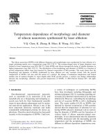
Figure 1 shows the XRD pattern of CuO nanoparticles
synthesized by sol-gel route using ethanol and propanol as
solvent. In both solvent cases all the obtained peaks in the
XRD pattern are well matched with the monoclinic phase of
CuO bulk crystals and well consistent with the JCPDS card
(card no: 048-1548). No impurity peak related to any other
phases of Cu like Cu(OH)
2
, Cu
2
O or Cu are seen in the ob-
72 P. Mallick et al.: Structure, Microstructure and Optical Absorption Analysis of
CuO Nanoparticles Synthesized by Sol-Gel Route
served XRD pattern. Our XRD results thus confirm synthesis
of pure and well crystalline CuO nanoparticles without any
impurity. The obtained results are well consistent with the
previously reported literature[13,14]. The XRD peaks
broaden and shift to higher angles for the CuO sample pre-
pared when propanol used as solvent. The peak shift could be
due to strain generation in the materials medium during
synthesis. Since two different batches CuO nanoparticles
were synthesized using ethanol and propanol as solvent. The
solvent might be influencing the microstructure of resultant
CuO nanoparticle.
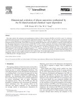
In order to understand the peak shift for CuO nanoparticle
synthesized by both solvent cases, we carried out William-
son–Hall (W–H) analysis[15] of the FWHM (β) of various
Bragg peaks appeared in the XRD pattern (Fig. 1). The W–H
plot of
cos
βθ
λ
versus
2sin
θ
λ
gives the value of microstrain
from the slope and particle size from the ordinate intersec-
tion. For pure particle size broadening this plot is expected to
be a horizontal line parallel to the sin θ axis, whereas in the
presence of strain, it has a non-zero slope. Figure 2 shows the
W-H plot for CuO nanoparticle synthesized with two dif-
ferent solvents. The obtained values of crystallite size and
microstrain for CuO nanoparticles synthesized under dif-
ferent solvent are given in Table 1. As indicated from the
Table 1, the crystallite size and strain is higher for the CuO
nanoparticle synthesized using propanol as solvent. The
shifting of XRD peaks to higher angle may be a consequence
of stain effect.
Figure 1. X-ray diffraction pattern of CuO nanoparticles synthesized by
sol-gel route with different solvent as mentioned
Figure 2. Williamson–Hall (W–H) plot for the CuO nanoparticles syn-
thesized by sol-gel route with different solvent as mentioned
Table 1. Evolution of crystallite size and strain of CuO nanoparticles
synthesized with different solvent as mentioned
Solvent
Crystallite Size (nm)
Strain (%)
Ethanol 28.57 0.22
Propanol
36.76
0.24
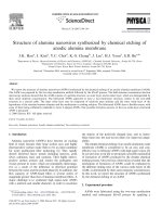
3.2. UV-Visible Characterization of CuO Nanoparticle
The variation of absorption coefficient,
α
of CuO
nanoparticles as a function of wavelength is shown in Fig. 3.
It is clearly seen from the figure that the absorption coeffi-
cient tends to decrease exponentially as the wavelength
increases. This behaviour is typical for many semiconductors
and can occur for a variety of reasons, such as internal elec-
tric fields within the crystal, deformation of lattice due to
strain caused by imperfection and inelastic scattering of
charge carriers by phonons[16-18]. The absorbance of CuO
sample synthesized with propanol solvent shows faster ex-
ponential decrease indicating more strain generation in this
case. The behaviour of absorbance shown in Fig. 3 is thus
agreed with the strain analysis using W-H plot discussed
above.
Figure 3. Variation of absorption coefficient of NiO nanoparticles as a
function of wavelength
The optical band gap of CuO nanoparticles were extracted
according to the following relation[19]:
()
n
g
Bh E
h
ν
α
ν
−
=
(1)
where
ν
h
is the incident photon energy,
α
is the ab-
sorption coefficient,
B
is a materials dependent constant
and
g
E
is the optical band gap. The value of
n
depends
on the nature of transition. Depending on whether the tran-
sition is direct allowed, direct forbidden, indirect allowed or
indirect forbidden, n takes the value 1/2, 3/2, 2 or 3 respec-
tively[20]. The usual method of determining
g
E
involves
plotting
1
()
n
h
αν
vs. photon energy,
ν
h
. Figure 4 and 5
show the variation of
1
()
n
h
αν
vs.
ν
h
for CuO nanoparti-
cles with n values of 1/2 and 2 respectively. The values of
direct and indirect band gap for CuO nanoparticles synthe-
sized with different solvent are shown in Table 2. The indi-
rect band gap of CuO nanoparticles synthesized using both
Nanoscience and Nanotechnology 2012, 2(3): 71-74 73
the solvents show similar values and the values red shifted ~
0.24 to 0.27 eV as compared to bulk value (1.45 eV)[21].
The increasing red shift with decreasing particle size sug-
gests that the defects responsible for the intra-gap states are
primarily of surface defects[22-24]. Our results thus indi-
cated that CuO nanoparticles prepared using ethanol as sol-
vent show more surface defects as compared to the CuO
nanoparticles prepared using propanol as solvent. Both the
CuO samples show higher direct band gap as compared to
bulk value (3.25 eV[24,25]). The blue shift in the direct band
edges as seen in present case is due to the quantum con-
finement effect[24,26].
Figure 4. Variation of
2
()h
αν
vs. photon energy,
h
ν
for CuO nano-
particles prepared sol-gel route with different solvent as mentioned
Figure 5. Variation of
1
2
()h
αν
vs. photon energy,
h
ν
for CuO nano-
particles prepared sol-gel route with different solvent as mentioned
Table 2. Evolution of direct and indirect optical band gap of CuO
nanoparticles synthesized with different solvent as mentioned
Solvent
Optical band gap (eV)
Direct Indirect
Ethanol 3.57 1.18
Propanol
3.57
1.21
4. Conclusions
CuO nanoparticles were synthesized by a low cost sol-gel
method. Effect of solvent on the structure, microstructure
and optical absorption properties of CuO nanoparticles were
studied. XRD analysis indicated that the crystallite size and
strain are higher for the CuO nanoparticle synthesized using
propanol as solvent. UV-Visible analysis also indicated the
higher strain generation for CuO nanoparticle synthesized
using propanol as solvent. Optical absorption analysis indi-
cated that the both the CuO samples show red shift of indi-
rect band gap due to the formation of surface defects. CuO
nanoparticles on the other hand show higher direct band gap
as compared to bulk value indicating blue shift of band gap
due to the quantum confinement effect.
REFERENCES
[1] F. Parmigiani and G. Samoggia, “Experimental Evidence of a
Fluctuating Charge State in Cupric Oxide”, Europhys. Lett. 7,
543(1988).
[2] X.G. Zheng, C.N. Xu, Y. Tomokiyo, E. Tanaka, H. Yamada
and Y. Soejima, “Observation of Charge Stripes in Cupric
Oxide”, Phys. Rev. Lett. 85, 5170 (2000).
[3] M H. Chang, H S. Liu and C.Y. Tai, “Preparation of copper
oxide nanoparticles and its application in nanofluid”, Powder
Technol. 207, 378 (2011).
[4] Y. She, Q. Zheng, L. Li, Y. Zhan, C. Chen, Y. Zheng and
X.Lin, “Rare earth oxide modified CuO/CeO
2
catalysts for
the water–gas shift reaction”, Int. J. Hydrogen Energy 34,
8929 (2009).
[5] P.P.C. Udani, P.V.D.S. Gunawardana, H.C. Lee, D.H. Kim,
“Steam reforming and oxidative steam reforming of metha-
nol over CuO–CeO
2
catalysts”, Int. J. Hydrogen Energy 34,
7648 (2009).
[6] J.L. Cao, G.S. Shao, Y. Wang, Y. Liu and Z.Y. Yuan, “CuO
catalysts supported on attapulgite clay for low-temperature
CO oxidation”, Catal. Commun. 9, 2555 (2008).
[7] C Y. Chiang, K. Aroh, N. Franson, V.R. Satsangi, S. Dass
and S. Ehrman, “Copper oxide nanoparticle made by flame
spray pyrolysis for photoelectrochemical water splitting –
Part II. Photoelectrochemical study”, Int. J. Hydrogen Ener-
gy 36, 15519 (2011).
[8] X.P. Gao, J.L. Bao and G.L. Pan, “Preparation and Electro-
chemical Performance of Polycrystalline and Single Crystal-
line CuO Nanorods as Anode Materials for Li Ion Battery”, J.
Phys. Chem. B 108, 5547 (2004).
[9] R. Vijaya Kumar, R. Elgamiel, Y. Diamant, and A. Gedanken,
“Sonochemical Preparation and Characterization of Nano-
crystalline Copper Oxide Embedded in Poly(vinyl alcohol)
and Its Effect on Crystal Growth of Copper Oxide”, Langmuir
17, 1406 (2001).
[10] C.L. Carnes, J. Stipp and K.J. Klabunde, “Synthesis, Cha-
racterization, and Adsorption Studies of Nanocrystalline
Copper Oxide and Nickel Oxide”, Langmuir 18, 1352 (2002).
[11] Y. Zhang, S. Wang, X. Li, L. Chen, Y. Qian and Z. Zhang,
“CuO shuttle-like nanocrystals synthesized by oriented at-
tachment”, J. Cryst. Growth 291, 196 (2006).
74 P. Mallick et al.: Structure, Microstructure and Optical Absorption Analysis of
CuO Nanoparticles Synthesized by Sol-Gel Route
[12] W. Wang, Y. Zhan and G. Wang, “One-step, solid-state
reaction to the synthesis of copper oxide nanorods in the
presence of a suitable surfactant”, Chem. Commun. 727
(2001).
[13] J.I. Langford and D. Louer, “High-resolution powder dif-
fraction studies of copper(II) oxide”, J. Appl. Crystallogr. 24,
149 (1991).
[14] M. Abaker, A. Umar, S. Baskoutas, S.H. Kim and S.W.
Hwang, “Structural and optical properties of CuO layered
hexagonal discs synthesized by a low-temperature hydro-
thermal process”, J. Phys. D: Appl. Phys. 44, 155405 (2011).
[15] G.K. Williamson and W.H. Hall, “X-ray line broadening
from filed aluminium and wolfram”, Acta Metall. 1, 22
(1953).
[16] T.S. Moss, G.J. Burrell and B. Ellis, Semiconductor Op-
to-Electronics, Butterworth & Co. Ltd, 1973.
[17] H.M. Honsi, S.A. Fayek, S.M. El-Sayed, M. Roushdy, M.A.
Soliman, “Optical properties and DC electrical conductivity
of Ge
28−x
Se
72
Sb
x
thin films”, Vacuum 81, 54 (2006)
[18] A. Sawaby, M.S. Selim, S.Y. Marzouk, M.A. Mostafa and A.
Hosny, “Structure, optical and electrochromic properties of
NiO thin films”, Physica B 405, 3412 (2010).
[19] N.F. Mott, E.A. Davies, Electronic Processes in
Non-Crystalline Materials (Clarendon Press, Oxford,1979).
[20] A.N. Banerjee and K.K. Chattopadhyay, in D. Depla and S.
Maheiu (Eds.), Reactive Sputter Deposition, Springer-Verlag
Berlin Heidelberg, 2008, p.465.
[21] B.A. Gizhevskii, Y.P. Sukhorukov, A.S. Moskvin, N.N.
Loshkareva, E.V. Mostovshchikova, A.E. Ermakov, E.A.
Kozlov, M.A. Uimin, V.S. Gaviko, “Anomalies in the optical
properties of nanocrystalline copper oxides CuO and Cu2O
near the fundamental absorption edge”, JETP 102, 297(2006).
[22] Y.P. Sukhorukov, B.A. Gizhevskii, E.V. Mostovshchikova,
A.Y. Yermakov, S.N. Tugushev and E.A. Kozlov, “Nano-
crystalline copper oxide for selective solar energy absorbers”,
Tech. Phys. Lett. 32, 132 (2006).
[23] S.G. Ovchinnikov, B.A. Gizhevskii, Y.P. Sukhorukov, A.E.
Ermakov, M.A. Uimin, E.A. Kozlov, Y. Kotov and A.A.V.
Bagazeev, “Specific features of the electronic structure and
optical spectra of nanoparticles with strong electron correla-
tions”, Phys. Solid State 49, 1116 (2007).
[24] S. Rehman, A. Mumtaz and S.K. Hasanain, “Size effects on
the magnetic and optical properties of CuO nanoparticles”, J.
Nanopart. Res. 13, 2497 (2011).
[25] F.P. Koffyberg and F.A. Benko, “A photoelectrochemical
determination of the position of the conduction and valence
band edges of p‐type C uO ”, J. A ppl. Phys. 53, 1173 (1982).
[26] S. Neeleshwar, C.L. Chen, C.B. Tsai, Y.Y. Chen, C.C. Chen,
S.G. Shyu and M.S. Seehra, “Size-dependent properties of
CdSe quantum dots”, Phys. Rev. B 71, 201307(R) (2005).