controllable synthesis of zno architectures by a surfactant-free hydrothermal process
Bạn đang xem bản rút gọn của tài liệu. Xem và tải ngay bản đầy đủ của tài liệu tại đây (677.14 KB, 3 trang )
Controllable synthesis of ZnO architectures by a surfactant-free hydrothermal process
Guixiang Du
a,
⁎
, Lidong Zhang
b
, Yan Feng
a
, Yanyan Xu
a
, YuXiu Sun
a
, Bin Ding
a
, Qian Wang
a
a
College of Chemistry, Tianjin Key Laboratory of Structure and Performance for functional Molecule, Tianjin Normal University, Tianjin 300387, China
b
School of Energy and Chemical Technology, Tianjin Bohai Vocational Technical College,Tianjin 300402, China
abstractarticle info
Article history:
Received 26 October 2011
Accepted 3 January 2012
Available online 10 January 2012
Keywords:
ZnO
Semiconductors
Microstructure
Surfactant-free
Hydrothermal
Various ZnO architectures like novel flowerlike structures radially assembled by rods, nails or towers and
novel radialized bundled tubular structures with diverse diameter in entire length were controllably synthe-
sized with different amine precursors by a simple surfactant-free hydrothermal process. It suggests that di-
verse amine sources, which possibly have different hydroxyl ion releasing rate contributing to different
reaction rates, probably play an importance role in controlling the assembly of different ZnO architectures,
besides temperature and time. X-ray powder diffraction (XRD) results prove the ZnO belonging to wurtzite
structure and room temperature photoluminescence (PL) demonstrates a high quality of the products.
© 2012 Elsevier B.V. All rights reserved.
1. Introduction
Zinc oxide (ZnO), an important wide-band gap semiconductor
material has been viewed as one of the promising nanomaterials
due to the applications in gas sensors, photocatalysts and solar cells
[1–3]. Since the properties of the material depend closely on the
mic rostructure, much effort has been fo cused on con trol ling
sizes, morphology and microst ructure of ZnO, and diverse ZnO
structures can be achieved by vap or-phase or solution processes
[4–10]. Complex procedures and equipment are involved in
vapor-phase processes , and therefore, more effective, simpler
and low cost solution ways are feasible to controllably synthesize
the ZnO architectures in a large-scale by changing reaction time,
surfactants and substrates [9,10]. However, it has been ra rely
rep orted that different precursors have been employed to control
themorphologiesandstructures.Wethinkthatdiverseamine
precursors, which possibly possess different hydroxyl ion releas-
ing rate, contributing to different reaction rates of zinc ion an d
hydroxyl ion, maybe control the assembly of different ZnO
architectures.
In this letter, we have s uccessfully achieved various ZnO architectures
including novel r adialized flowers a ssembled b y r ods, towers and n ails
with on e sharp tip, and novel tubular Z nO bundles compose d o f many
single tubes with different diameters from bottom to top by four amine
precursors by a s urfactant-free hydrothermal process. It suggests t hat dif-
ferent amine precursors play an important role in controlling of ZnO
structures possibly by tuning the reaction rate.
2. Experimental
In a typical synthesis, 0.0075 mol of glycol was added into 15 ml of
aqueous solution of Zn(NO
3
)
2
·6H
2
O (0.05 M) and ammonia (0.05 M),
the mixture was stirred for minutes and was transferred into 25 mL
Teflon-lined stainless steel autoclaves, sealed and maintained at certain
temperature and time. After the reaction, the samples was filtered out,
washed several times with distilled water and alcohol, and then dried
at 60 °C under air a tmosphere. Hexamethylenetetramine (HMTA),
ethanol amine (EA) and ethylenediamine (ED) were also used in
the parallel experiments.
XRD analyses were conducted on a Bruker D8A X-ray diffractometer
with a Cu Kα radiation (λ =0.15418 nm). Field emission scanning
electron microscopy (FE-SEM) and transmission electron microscopic
(FE-TEM) images were performed on a FEI Nova Nano SEM 230 micro-
scope and on a FEI Tecnai G
2
F20 microscopic, respectively. The room
temperature PL spectrum was recorded with a HORIBA JY FL-3 spectro-
photometer excited by a He-Cd laser with a wavelength of 325 nm.
3. Results and discussion
XRD results of all the samples have nearly same peaks. A represen-
tative XRD spectrum of the ZnO samples is shown in Fig. 1. All diffract ion
peaks can be indexed within the experimental error as a wurt zite struc-
ture of ZnO (a=0.324982 nm and c=0.520661 nm). No peaks associat-
ed with other crystalline forms are detected.
We first performed the experiments from four amine sources in
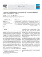
the presence of glycol at 90 °C for 20 h. Fig. 2a and the insert present
well-defined hexagonal ZnO rods with smooth ends obtained from
ammonia. When HMTA was used, the hexagonal ZnO bundles radially
assembled by tower-like structures (Fig. 2b) were gained, and it is
Materials Letters 73 (2012) 86–88
⁎ Corresponding author. Tel.: +86 22 23766515; fax: + 86 22 23766515.
E-mail address: (G. Du).
0167-577X/$ – see front matter © 2012 Elsevier B.V. All rights reserved.
doi:10.1016/j.matlet.2012.01.013
Contents lists available at SciVerse ScienceDirect
Materials Letters
journal homepage: www.elsevier.com/locate/matlet
clear that there are some steps on the side surface of a single tower.
Fig. 2b′ shows a typical TEM image of a ZnO tower with diameter of
300 nm, and the corresponding HRTEM image (upper right inset of
Fig. 2b) and SEAD pattern (lower right inset of Fig. 2b′) of the tower
were recorded. The (0002) lattice plane of hexagonal ZnO, with a lattice
spacing of about 0.52 nm, can be clearly identified in the lattice-
resolved HRTEM image. It indicates that the ZnO tower is single crystal-
line in nature and preferentially grows along the [0001] direction. The
wurtzite structure of tower was further confirmed by the SAED pattern.
Flowerlike ZnO bundles radially assembled by well-defined hexagonal
rods with even diameters were obtained from EA (Fig. 2c). But when
ED wa s replaced, novel flowerlike architectures (Fig. 2d), radially
ass embled by ZnO nails with a single sharp tip, quite different to
those in previous reports [9,10] and those of Fig. 2bandcformed.
It suggests that diverse amine sources, possibly having different
hydroxyl ion releasing rate, contributing to different re action
rates, probably cause the formation of different ZnO architectures.
Cer tainly, hereinto, glycol maybe plays a certain role in controlling
the morpholo gies in the process, and the correlativ e study is
underway.
When reactions were performed at 100 °C for 10 h by the four
amine sources, various novel bundled tubes and flowers (Fig. 3)
were produced. Novel hexagonal tubular ZnO with obvious different
diameter through entire length, presenting one closed sharp tip and
growing into bundles from the open ends in various directions,
were obtained from ammonia (Fig. 3a). The typical diameters of the
tubes at the open end and tips range between 500–600 nm an d between
100–150 nm, respectively, and the typical lengths are in the range of
10–12 um. To our best of knowledge, large-scale fabrication of ZnO tu-
bular radial bunches with changed diameter in each tube has been rarely
reported in a surfactant-free hydrothermal pr ocess. When HMTA was
employed, the hexagonal ZnO tubular bunches radially assembled by
closely p acked n a notubes with open en ds exposed were formed
(Fig. 3b), which is similar to the report by Yu et al. [11]. Fig. 3c reveals
the flowerlike ZnO with unsmooth surface obtained from EA, which is
different fr om traditionally rod-based flowerlike ZnO. The typical flower
shownintherightofFig. 3c indicates that it is composed of several
sharp-tip petals, in which each petal consists of some short, non-
smooth rods, similar to the underdeveloping ones. The ends of these
rods attach to each other and form the tip-like petal (the insert of the
right flower of Fig. 3c). In addition, instead of the petal-based flowe rs,
some unusual rod- based ZnO without obvious petals (typically
displayed in the left of Fig. 3c) extending radially from c enter to
form flowerlike structures are clearly seen, in which the end
faces of these rods seem to be “dissolved” and the hexagonal contour
is rough and blurry (the insert corresponding to the left flower of
Fig. 3c). It is easy to imagine that the rod-based ZnO flowers without ob-
vious petals could be gradually “dissolved”, and they are maybe the
forerunners of the petal-based flowers. Fig. 3d illustrates the rods or
20 30 40 50 60 70 80
(202)
(004)
(200)
(201)
(112)
(103)
(110)
(102)
(101)
(002)
(100)
Intensity(a.u.)
2
θ
(degree)
Fig. 1. A representative XRD pattern of the ZnO architectures obtained from HMTA.
c
d
4 um
a
500 nm
b
2 um
[0001]
b
’
[0001]
2 nm
0.52 nm
Fig. 2. ZnO architectures obtained from ammonia (a), HMTA (b), EA (c), ED (d) at 90 °C for 20 h. The insets of Fig. 2b are the SAED pattern and the HRTEM image of a nanotower,
respectively.
87G. Du et al. / Materials Letters 73 (2012) 86–88
tower structures obtained from ED, in which many of them were
com posed of discrete subunits, orientated and connected along
c-a xis. All the above results suggest that it is an effective way to
controllably synthesize ZnO by changing reactant precursors with
dif ferent ion releasing rate, besides chan ging the temperature
and time.
The optical properties of ZnO flowerlike bundles from HMTA were
observed by PL (Fig. 4). Generally, the UV emission at about 391 nm is
band-edge emission resulting from the recombination of free excitons
[11], while the green–yellow emission can be attributed to the recombi-
nation of photo-generated hole with electrons in singly occupied oxygen
vacancies. The green–yellow emission in our sample can be neglectable
compared with the intensive sharp UV emission. Therefore, the resul ts of
PL indicate that our growth method can produce a low concentration of
oxygen vacancies and high optical quality of single-crystal ZnO [8,12].
4. Conclusions
In summary, various ZnO architectures, especially novel radialized
flowerlike structures assembled by nails with one sharp tip and novel
tubular ZnO bundles composed of many single tubes with different
diameters from bottom to top were controllably synthesized with dif-
ferent amine precursors by a simple surfactant-free hydrothermal
process. It suggests that different amine precursors play an important
role in controlling the formation of various ZnO architectures possibly
by tuning the reaction rate, besides temperature and time. It not only
gives an effective way to controllably synthesize various ZnO archi-
tectures but also provides valuable information for the controlled
synthesis of other functional nanomaterials.
Acknowledgements
This work was s upported by Na tional Natu ral ScientificFoundationof
China (Nos. 51102180 and 21001081), Tianjin Science and Technology
Fund Project for High Education ( Nos. 20110311 and 20100504) and
the talent fund projects in Tianjin Normal University (No. 5RL078).
References
[1] Hochbaum AI, Yang PD. Chem Rev 2010;110:527–46.
[2] Lao JY, Huang JY, Wang DZ, Ren ZF. Nano Lett 2003;3:235–8.
[3] Zhang QF, Dandeneau CS, Zhou XY, Cao GZ. Adv Mater 2009;21:4087–108.
[4] He JH, Ho CH, Wang CW, Ding Y, Chen LJ, Wang ZL. Cryst Growth Des 2009;9:17–9.
[5] Qiu JS, Sun TJ. Mater Lett 2008;62:1528–31.
[6] Fan DH, Shen WZ, Zheng MJ, Zhu YF, Lu JJ. J Phys Chem C 2007;111:9116–21.
[7] Zhang XJ, Zhang XH, Zou K, Lee CS, Lee ST. J Am Chem Soc 2007;129:3527–32.
[8] Fang Z, Tang KB, Shen GZ, Chen D, Kong R, Lei SJ. Mater Lett 2006;60:2530–3.
[9] Zhang J, Sun LD, Yin JL, Su HL, Liao CS, Yan CH. Chem Mater 2002;14:4172–7.
[10] Xu F, Lu YN, Xie Y, Liu YF. J Phys Chem C 2009;113:1052–9.
[11] Yu QJ, Fu WY, Yu CL, Yang HB, Wei RH, Li MH. J Phys Chem C 2007;111:17521–6.
[12] Liu JP, Huang XT, Li YY, Duan JX, Ai HH, Ren L. Mater Sci Eng B 2006;127:85–90.
350 400 450 500 550 600 650
PL intensity(a.u.)
wavelength(nm)
Fig. 4. Room temperature PL spectrum of the ZnO architectures obtained from HMTA.
d
c
a
b
500 nm
Fig. 3. ZnO architectures obtained from ammonia (a), HMTA (b), EA (c), ED (d) at 100 °C for 10 h.
88 G. Du et al. / Materials Letters 73 (2012) 86–88